Filters
Clonality
Type
Reactivity
Gene Name
Isotype
Host
Application
Clone
92 results for " neuronal" - showing 50-92


 FCM (Flow Cytometry)
(Flow cytometric analysis of PC-12 cells with nNOS antibody at 1/50 dilution (red) compared with an unlabelled control (cells without incubation with primary antibody; black). Alexa Fluor 488-conjugated goat anti rabbit IgG was used as the secondary antibody.)
FCM (Flow Cytometry)
(Flow cytometric analysis of PC-12 cells with nNOS antibody at 1/50 dilution (red) compared with an unlabelled control (cells without incubation with primary antibody; black). Alexa Fluor 488-conjugated goat anti rabbit IgG was used as the secondary antibody.)
nNOS, Monoclonal Antibody (Cat# AAA30104)
Full Name
nNOS Antibody
Gene Names
NOS1; NOS; bNOS; nNOS; IHPS1; N-NOS; NC-NOS
Reactivity
Human, Mouse, Rat
Applications
Western Blot, Immunocytochemistry, Immunofluorescence, Immunohistochemistry, Immunoprecipitation, Flow Cytometry
Purity
ProA affinity purified
Pricing
 Standard Curve (Sample)
Neuronal nuclear Antigen, ELISA Kit (Cat# AAA27288)
Full Name
Human Neuronal nuclear Antigen ELISA Kit
Reactivity
Human
Pricing


 IF (Immunofluorescence)
(Figure 6. IF analysis of SCG10/STMN2 using anti- SCG10/STMN2 antibody (AAA19299).SCG10/STMN2 was detected in immunocytochemical section of U20S cells. Enzyme antigen retrieval was performed using IHC enzyme antigen retrieval reagent for 15 mins. The cells were blocked with 10% goat serum. And then incubated with 5μg/mL rabbit anti- SCG10/STMN2 Antibody (AAA19299) overnight at 4 degree C. DyLight®488 Conjugated Goat Anti-Rabbit IgG was used as secondary antibody at 1:100 dilution and incubated for 30 minutes at 37 degree C. The section was counterstained with DAPI. Visualize using a fluorescence microscope and filter sets appropriate for the label used.)
IF (Immunofluorescence)
(Figure 6. IF analysis of SCG10/STMN2 using anti- SCG10/STMN2 antibody (AAA19299).SCG10/STMN2 was detected in immunocytochemical section of U20S cells. Enzyme antigen retrieval was performed using IHC enzyme antigen retrieval reagent for 15 mins. The cells were blocked with 10% goat serum. And then incubated with 5μg/mL rabbit anti- SCG10/STMN2 Antibody (AAA19299) overnight at 4 degree C. DyLight®488 Conjugated Goat Anti-Rabbit IgG was used as secondary antibody at 1:100 dilution and incubated for 30 minutes at 37 degree C. The section was counterstained with DAPI. Visualize using a fluorescence microscope and filter sets appropriate for the label used.)
SCG10/STMN2, Polyclonal Antibody (Cat# AAA19299)
Full Name
Anti-SCG10/STMN2 Antibody
Gene Names
STMN2; SCG10; SCGN10
Reactivity
Human, Mouse, Rat
Applications
WB, IHC-P, ICC, IF, FC/FACS/FCM, EIA
Purity
Immunogen affinity purified.
Pricing
 Standard Curve (Sample)
enolase 2 (gamma, neuronal), ELISA Kit (Cat# AAA15495)
Full Name
Rat Neuron-specific enolase, NSE ELISA Kit
Gene Names
Eno2; NSE; RNEN3
Reactivity
Rat
Pricing
 Standard Curve (Sample)
Band 4.1 like protein 1 (EPB41L1), ELISA Kit (Cat# AAA27320)
Full Name
Human Band 4.1 like protein 1 (EPB41L1) ELISA Kit
Gene Names
EPB41L1; 4.1N; MRD11
Reactivity
Human
Pricing
 Standard Curve (Sample)
IL-16, ELISA Kit (Cat# AAA21893)
Full Name
Rat IL-16 (Interleukin 16) ELISA Kit
Gene Names
IL16; LCF; NIL16; PRIL16; prIL-16
Reactivity
Rat
Pricing
 Standard Curve (Sample)
Nicotinic acetylcholine receptor, ELISA Kit (Cat# AAA16118)
Full Name
Human Nicotinic acetylcholine receptor ELISA Kit
Gene Names
CHRNA4; EBN; BFNC; EBN1; NACHR; NACRA4; NACHRA4
Reactivity
Human
Pricing
 Standard Curve (Sample)
Interleukin 16, IL-16, ELISA Kit (Cat# AAA15290)
Full Name
Rat Interleukin 16, IL-16 ELISA Kit
Gene Names
IL16; LCF; NIL16; PRIL16; prIL-16
Reactivity
Rat
Pricing
 Standard Curve (Sample)
Neuron-Specific Enolase, ELISA Kit (Cat# AAA17376)
Full Name
Sheep Neuron-Specific Enolase ELISA Kit
Gene Names
ENO2; NSE
Reactivity
Sheep
Pricing


 WB (Western Blot)
(LRRN1 monoclonal antibody (M05), clone 3D11. Western Blot analysis of LRRN1 expression in PC-12.)
WB (Western Blot)
(LRRN1 monoclonal antibody (M05), clone 3D11. Western Blot analysis of LRRN1 expression in PC-12.)
LRRN1, Monoclonal Antibody (Cat# AAA26018)
Full Name
LRRN1 (Leucine Rich Repeat Neuronal 1, FIGLER3, KIAA1497, NLRR-1, LRCH4) (AP)
Reactivity
Human, Mouse, Rat
Applications
WB
Purity
Purified
Pricing


 FCM (Flow Cytometry)
(Flow cytometric analysis of SH-SY5Y cells with Cacng4 antibody at 1/100 dilution (fuchsia) compared with an unlabelled control (cells without incubation with primary antibody; yellow). Alexa Fluor 488-conjugated goat anti-rabbit IgG was used as the secondary antibody.)
FCM (Flow Cytometry)
(Flow cytometric analysis of SH-SY5Y cells with Cacng4 antibody at 1/100 dilution (fuchsia) compared with an unlabelled control (cells without incubation with primary antibody; yellow). Alexa Fluor 488-conjugated goat anti-rabbit IgG was used as the secondary antibody.)
CACNG4, Polyclonal Antibody (Cat# AAA29944)
Full Name
CACNG4 Antibody
Reactivity
Human, Rat
Applications
WB, ICC, IHC, FC/FACS
Purity
Peptide affinity purified.
Pricing
 Standard Curve (Sample)
Neuron-Specific Enolase, ELISA Kit (Cat# AAA17349)
Full Name
Mouse Neuron-Specific Enolase ELISA Kit
Gene Names
ENO2; NSE
Reactivity
Mouse
Pricing
 Standard Curve (Sample)
Nicotinic acetylcholine receptor, ELISA Kit (Cat# AAA16000)
Full Name
Mouse Nicotinic acetylcholine receptor ELISA Kit
Gene Names
CHRNA4; EBN; BFNC; EBN1; NACHR; NACRA4; NACHRA4
Reactivity
Mouse
Pricing
 Standard Curve (Sample)
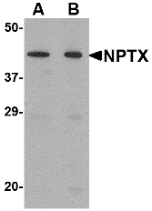
neuronal pentraxin I, ELISA Kit (Cat# AAA15654)
Full Name
Human Neuronal Pentraxin-1, NPTX-1 ELISA Kit
Gene Names
NPTX1; NP1
Reactivity
Human
Pricing
 Standard Curve (Sample)
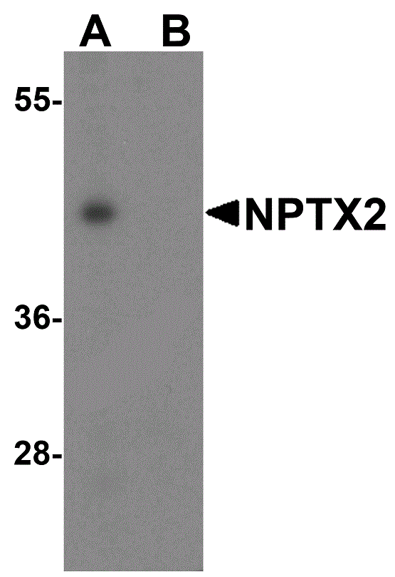
NOS1/nNOS, ELISA Kit (Cat# AAA21639)
Full Name
Human NOS1/nNOS (Nitric Oxide Synthase 1, Neuronal) ELISA Kit
Gene Names
NOS1; NOS; bNOS; nNOS; IHPS1; N-NOS; NC-NOS
Reactivity
Human
Pricing


 FCM (Flow Cytometry)
(Figure 12. Flow Cytometry analysis of U937 cells using anti-DYNLL1/PIN antibody (AAA19276).Overlay histogram showing U937 cells stained with AAA19276 (Blue line). The cells were blocked with 10% normal goat serum. And then incubated with rabbit anti-DYNLL1/PIN Antibody (AAA19276, 1μg/1x106 cells) for 30 min at 20 degree C. DyLight®488 conjugated goat anti-rabbit IgG (5-10μg/1x106 cells) was used as secondary antibody for 30 minutes at 20 degree C. Isotype control antibody (Green line) was rabbit IgG (1μg/1x106) used under the same conditions. Unlabelled sample (Red line) was also used as a control.)
FCM (Flow Cytometry)
(Figure 12. Flow Cytometry analysis of U937 cells using anti-DYNLL1/PIN antibody (AAA19276).Overlay histogram showing U937 cells stained with AAA19276 (Blue line). The cells were blocked with 10% normal goat serum. And then incubated with rabbit anti-DYNLL1/PIN Antibody (AAA19276, 1μg/1x106 cells) for 30 min at 20 degree C. DyLight®488 conjugated goat anti-rabbit IgG (5-10μg/1x106 cells) was used as secondary antibody for 30 minutes at 20 degree C. Isotype control antibody (Green line) was rabbit IgG (1μg/1x106) used under the same conditions. Unlabelled sample (Red line) was also used as a control.)
DYNLL1/PIN, Polyclonal Antibody (Cat# AAA19276)
Full Name
Anti-DYNLL1/PIN Antibody
Gene Names
DYNLL1; LC8; PIN; DLC1; DLC8; LC8a; DNCL1; hdlc1; DNCLC1
Reactivity
Human, Mouse, Rat
Applications
WB, IHC-P, ICC, IF, FC/FACS/FCM, EIA
Purity
Immunogen affinity purified.
Pricing
 Standard Curve (Sample)
Neuronal nuclear Antigen, ELISA Kit (Cat# AAA27066)
Full Name
Rat Neuronal nuclear Antigen ELISA Kit
Reactivity
Rat
Pricing
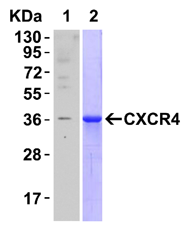
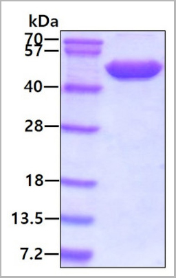
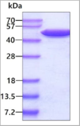 SDS-PAGE
(3ug by SDS-PAGE under reducing condition and visualized by coomassie blue stain.)
SDS-PAGE
(3ug by SDS-PAGE under reducing condition and visualized by coomassie blue stain.)
Alpha-enolase, Active Protein (Cat# AAA11794)
Full Name
Alpha-enolase, 1-434aa, Human, E Coli (Bioactivity Validated)
Gene Names
ENO1; NNE; PPH; MPB1; ENO1L1; HEL-S-17
Applications
Enzyme Activity, SDS-PAGE
Purity
> 95% by SDS-PAGE
Pricing
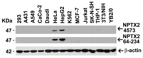
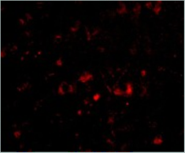
 IF (Immunofluorescence)
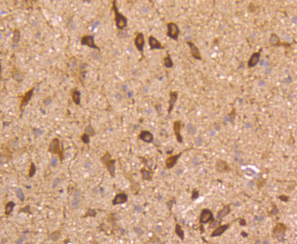
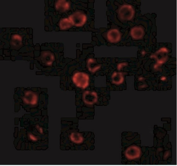
(Immunofluorescent analysis of 4% paraformaldehyde-fixed mouse brain issue labeling NPTX2 with AAA10933 at 20ug/mL, followed by goat anti-rabbit IgG secondaryantibody at 1/500 dilution (red) and DAPI staining (blue).)
IF (Immunofluorescence)
(Immunofluorescent analysis of 4% paraformaldehyde-fixed mouse brain issue labeling NPTX2 with AAA10933 at 20ug/mL, followed by goat anti-rabbit IgG secondaryantibody at 1/500 dilution (red) and DAPI staining (blue).)
NPTX2, Polyclonal Antibody (Cat# AAA10933)
Full Name
NPTX2 Antibody
Gene Names
NPTX2; NP2; NARP; NP-II
Reactivity
Human, Mouse, Rat
Applications
Western Blot, Immunohistochemistry, Immunofluorescence
Purity
NPTX2 Antibody is affinity chromatography purified via peptide column.
Pricing
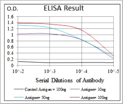 ELISA
(Control Antigen (100 ng); Purple line: Antigen(10ng); Blue line: Antigen (50 ng); Red line: Antigen (100ng).)
ELISA
(Control Antigen (100 ng); Purple line: Antigen(10ng); Blue line: Antigen (50 ng); Red line: Antigen (100ng).)
NRCAM, Monoclonal Antibody (Cat# AAA27925)
Full Name
Mouse Monoclonal Antibody to NRCAM
Reactivity
Human
Applications
Western Blot, Immunocytochemistry, Flow Cytometry
Pricing
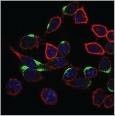
 IF (Immunofluorescence)
(AAA31175 staining Hela cells by IF/ICC. The samples were fixed with PFA and permeabilized in 0.1% Triton X-100,then blocked in 10% serum for 45 minutes at 25°C. Samples were then incubated with primary Ab(AAA31175 1:200) and mouse anti-beta tubulin Ab( 1:200) for 1 hour at 37°C. An AlexaFluor594 conjugated goat anti-rabbit IgG(H+L) Ab(Red) and an AlexaFluor488 conjugated goat anti-mouse IgG(H+L) Ab(Green) were used as the secondary antibody.The nuclear counter stain is DAPI(blue).)
IF (Immunofluorescence)
(AAA31175 staining Hela cells by IF/ICC. The samples were fixed with PFA and permeabilized in 0.1% Triton X-100,then blocked in 10% serum for 45 minutes at 25°C. Samples were then incubated with primary Ab(AAA31175 1:200) and mouse anti-beta tubulin Ab( 1:200) for 1 hour at 37°C. An AlexaFluor594 conjugated goat anti-rabbit IgG(H+L) Ab(Red) and an AlexaFluor488 conjugated goat anti-mouse IgG(H+L) Ab(Green) were used as the secondary antibody.The nuclear counter stain is DAPI(blue).)
Nicotinic Acetylcholine Receptor alpha 7, Polyclonal Antibody (Cat# AAA31175)
Full Name
Nicotinic Acetylcholine Receptor alpha 7 Antibody
Gene Names
CHRNA7; NACHRA7; CHRNA7-2
Reactivity
Human, Mouse, Rat
Applications
Western Blot, Immunofluorescence, Immunocytochemistry
Purity
The antiserum was purified by peptide affinity chromatography using SulfoLink™ Coupling Resin (Thermo Fisher Scientific).
Pricing

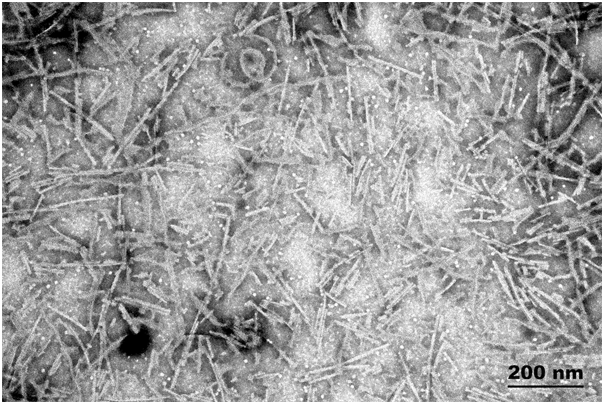
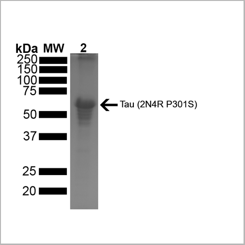 SDS-PAGE
(SDS-PAGE of ~67 kDa Human Tau Protein 2N4R P301S Preformed Fibrils. Lane 1: MW Ladder. Lane 2: Tau Protein Preformed Fibrils)
SDS-PAGE
(SDS-PAGE of ~67 kDa Human Tau Protein 2N4R P301S Preformed Fibrils. Lane 1: MW Ladder. Lane 2: Tau Protein Preformed Fibrils)
Tau441, Active Protein (Cat# AAA27663)
Full Name
Active Human Recombinant Tau441 (2N4R), P301S Mutant Protein Preformed Fibrils
Gene Names
MAPT; TAU; MSTD; PPND; DDPAC; MAPTL; MTBT1; MTBT2; FTDP-17; PPP1R103
Applications
Western Blot
Purity
>95%
Ion-Exchange Purified
Ion-Exchange Purified
Pricing
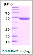
 SDS-PAGE
(3ug by SDS-PAGE under reducing condition and visualized by coomassie blue stain)
SDS-PAGE
(3ug by SDS-PAGE under reducing condition and visualized by coomassie blue stain)
Neuron-Specific Enolase (NSE), Active Protein (Cat# AAA11754)
Full Name
Neuron-Specific Enolase (NSE), 1-434aa, Human, E Coli (Bioactivity Validated)
Gene Names
ENO2; NSE; HEL-S-279
Applications
Enzyme Activity, SDS-PAGE
Purity
> 95% by SDS-PAGE
Pricing
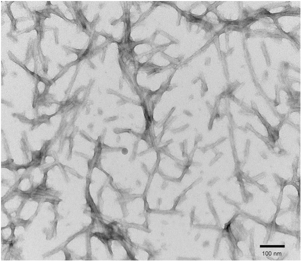
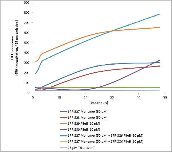 Application Data
(Thioflavin T is a fluorescent dye that binds to beta sheet-rich structures such as those in tau fibrils. Upon binding, the emission spectrum of the dye experiences a red-shift, and increased fluorescence intensity. Thioflavin T emission curves show increased fluorescence (correlated to tau aggregation) in tau K18 P301L monomers over time. Thioflavin T ex = 450 nm, em = 485 nm.)
Application Data
(Thioflavin T is a fluorescent dye that binds to beta sheet-rich structures such as those in tau fibrils. Upon binding, the emission spectrum of the dye experiences a red-shift, and increased fluorescence intensity. Thioflavin T emission curves show increased fluorescence (correlated to tau aggregation) in tau K18 P301L monomers over time. Thioflavin T ex = 450 nm, em = 485 nm.)
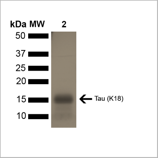
Tau, Active Protein (Cat# AAA27662)
Full Name
Active Human Recombinant Tau (K18), P301L Mutant Protein Monomer
Gene Names
MAPT; TAU; MSTD; PPND; DDPAC; MAPTL; MTBT1; MTBT2; FTDP-17; PPP1R103
Applications
Western Blot
Purity
Purity: >95%
Purification: Ion-exchange Purified
Purification: Ion-exchange Purified
Pricing


 Application Data
(Published customer image: Mouse anti V5 tag antibody, clone SV5-Pk1 used for the detection of V5 tagged WEEV_nsP3 protein by western blotting and immunofluorescenceImage caption: WEEV nsP3 interaction with host IKKbeta. A) U87MGs were transfected in a 6-well plate with 5 ug of pUC19 and WEEV_nsP3_HA for 24 hours. Cell lysates were resolved using SDS-PAGE and subsequently immunoblotted with V5 antibody and beta-actin served as a loading control. B) U87MGs were transfected with WEEV_nsP3_V5; cells were fixed after 24 hours and stained with antibodies against the endogenous IKKbeta and the V5 tag. Cells were incubated with appropriate secondary Alexa Fluor antibodies and the nuclei stained with DAPI. Co-localization of IKKbeta with WEEV_nsP3_V5 (yellow) was observed as shown by the arrows. B) Panels E -H serve as an example of transfected cells in a given field of view that show co-localization of IKKbeta and WEEV_nsP3_V5 24 hours post transfection. Panels I-L represent magnified images of other cells showing co-localization of IKKbeta and WEEV_nsP3_V5. Panel M is a magnified image of panel L. The co-localization was confirmed by Z-stack analysis. Co-localization was calculated to be approximately in 61% of cells (163 cells were counted of which 44% demonstrated expression of nsP3. Of those cells that expressed nsP3, 61% showed co-localization of both proteins). Images were taken using Nikon Eclipse TE2000-U at 60x magnification and are representative of 2 independent experiments.From: Amaya M, Voss K, Sampey G, Senina S, de la Fuente C, et al. (2014) The Role of IKKbeta in Venezuelan Equine Encephalitis Virus Infection. PLoS ONE 9(2): e86745.)
Application Data
(Published customer image: Mouse anti V5 tag antibody, clone SV5-Pk1 used for the detection of V5 tagged WEEV_nsP3 protein by western blotting and immunofluorescenceImage caption: WEEV nsP3 interaction with host IKKbeta. A) U87MGs were transfected in a 6-well plate with 5 ug of pUC19 and WEEV_nsP3_HA for 24 hours. Cell lysates were resolved using SDS-PAGE and subsequently immunoblotted with V5 antibody and beta-actin served as a loading control. B) U87MGs were transfected with WEEV_nsP3_V5; cells were fixed after 24 hours and stained with antibodies against the endogenous IKKbeta and the V5 tag. Cells were incubated with appropriate secondary Alexa Fluor antibodies and the nuclei stained with DAPI. Co-localization of IKKbeta with WEEV_nsP3_V5 (yellow) was observed as shown by the arrows. B) Panels E -H serve as an example of transfected cells in a given field of view that show co-localization of IKKbeta and WEEV_nsP3_V5 24 hours post transfection. Panels I-L represent magnified images of other cells showing co-localization of IKKbeta and WEEV_nsP3_V5. Panel M is a magnified image of panel L. The co-localization was confirmed by Z-stack analysis. Co-localization was calculated to be approximately in 61% of cells (163 cells were counted of which 44% demonstrated expression of nsP3. Of those cells that expressed nsP3, 61% showed co-localization of both proteins). Images were taken using Nikon Eclipse TE2000-U at 60x magnification and are representative of 2 independent experiments.From: Amaya M, Voss K, Sampey G, Senina S, de la Fuente C, et al. (2014) The Role of IKKbeta in Venezuelan Equine Encephalitis Virus Infection. PLoS ONE 9(2): e86745.)
V5-TAG, Monoclonal Antibody (Cat# AAA12081)
Full Name
MOUSE ANTI V5-TAG:HRP
Applications
Western Blot
Pricing
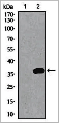
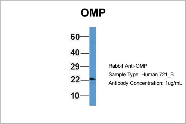
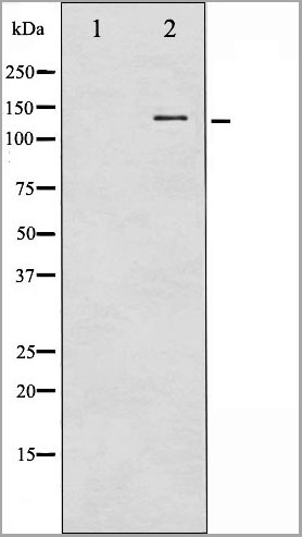
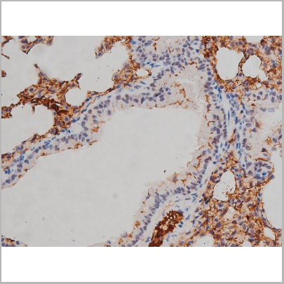
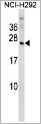 WB (Western Blot)
(OMP Antibody (N-term) western blot analysis in NCI-H292 cell line lysates (35ug/lane).This demonstrates the OMP antibody detected the OMP protein (arrow).)
WB (Western Blot)
(OMP Antibody (N-term) western blot analysis in NCI-H292 cell line lysates (35ug/lane).This demonstrates the OMP antibody detected the OMP protein (arrow).)
OMP, Polyclonal Antibody (Cat# AAA28786)
Full Name
OMP Antibody (N-term)
Reactivity
Human
Applications
Western Blot
Purity
This antibody is purified through a protein A column, followed by peptide affinity purification.
Pricing
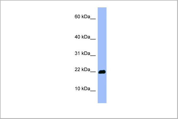
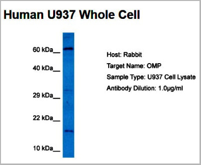 WB (Western Blot)
(Host: RabbitTarget Name: OMPSample Tissue: Human U937 Whole CellAntibody Dilution: 5ug/ml)
WB (Western Blot)
(Host: RabbitTarget Name: OMPSample Tissue: Human U937 Whole CellAntibody Dilution: 5ug/ml)
OMP, Polyclonal Antibody (Cat# AAA23571)
Full Name
OMP antibody - middle region
Reactivity
Tested Reactivity: Human, Rat
Predicted Species Reactivity: Goat, Human, Mouse, Rat
Predicted Species Reactivity: Goat, Human, Mouse, Rat
Applications
Western Blot
Purity
Affinity Purified
Pricing
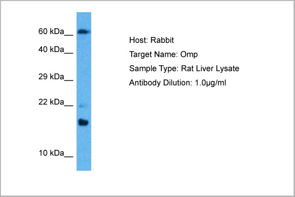
 WB (Western Blot)
(Western blot analysis of extracts of various celllines, using Phospho-Src (Tyr529) Antibody.)
WB (Western Blot)
(Western blot analysis of extracts of various celllines, using Phospho-Src (Tyr529) Antibody.)
Src, Polyclonal Antibody (Cat# AAA31009)
Full Name
Phospho-Src (Tyr530) Antibody
Gene Names
SRC; ASV; SRC1; THC6; c-SRC; p60-Src
Reactivity
Human, Mouse, Rat
Applications
Western Blot, Immunohistochemistry, Immunofluorescence, Immunocytochemistry
Purity
From purified rabbit serum by affinity purification via sequential chromatography on phospho-and non-phospho-peptide affinity columns.
Pricing


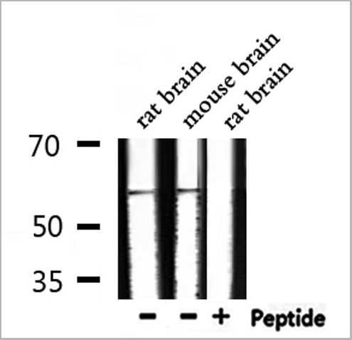
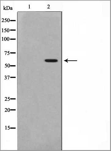
 WB (Western Blot)
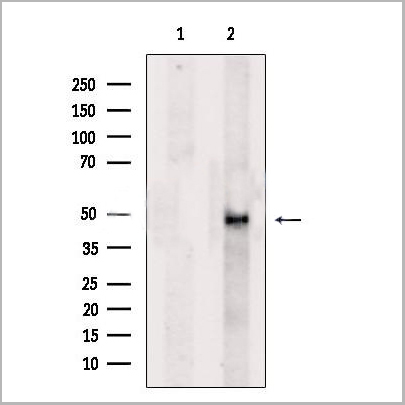
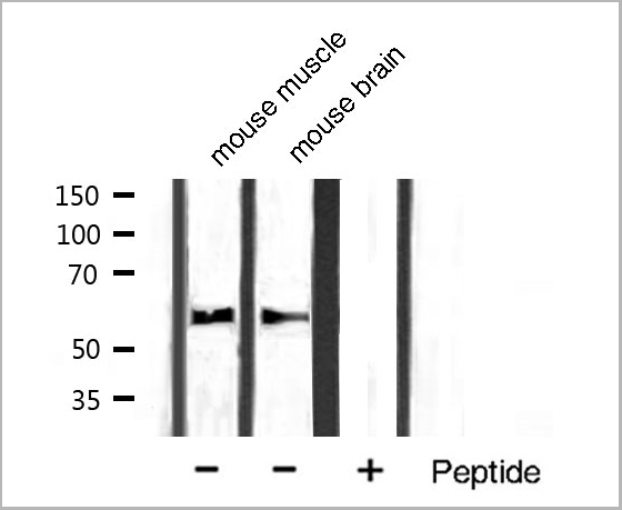
(Western blot analysis of n-NOS phosphorylation expression in A549 whole cell lysates, The lane on the left is treated with the antigen-specific peptide.)
WB (Western Blot)
(Western blot analysis of n-NOS phosphorylation expression in A549 whole cell lysates, The lane on the left is treated with the antigen-specific peptide.)
n-NOS, Polyclonal Antibody (Cat# AAA31028)
Full Name
Phospho-n-NOS (Ser852) Antibody
Gene Names
NOS1; NOS; bNOS; nNOS; IHPS1; N-NOS; NC-NOS
Reactivity
Human, Mouse, Rat
Applications
Western Blot, Immunohistochemistry, Immunofluorescence, Immunocytochemistry
Purity
From purified rabbit serum by affinity purification via sequential chromatography on phospho-and non-phospho-peptide affinity columns.
Pricing
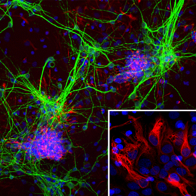 IS (Immunostaining)
(Immunostaining of cultured E20 rat cortical neurons and glia stained with anti-nestin antibody (AAA14229, red, 1:500) and anti-MAP2 antibody (, green, 1:500). The blue is Hoechst staining for nuclear DNA. The nestin antibody labels developing astrocytes and neuronal stem cells in a clearly filamentous fashion, while the MAP2 antibody stains dendrites and perikarya of mature neurons.)
IS (Immunostaining)
(Immunostaining of cultured E20 rat cortical neurons and glia stained with anti-nestin antibody (AAA14229, red, 1:500) and anti-MAP2 antibody (, green, 1:500). The blue is Hoechst staining for nuclear DNA. The nestin antibody labels developing astrocytes and neuronal stem cells in a clearly filamentous fashion, while the MAP2 antibody stains dendrites and perikarya of mature neurons.)
Nestin, Monoclonal Antibody (Cat# AAA14229)
Full Name
Anti-Nestin
Gene Names
NES; Nbla00170
Applications
Western Blot, Immunofluorescence
Purity
Protein G purified culture supernatant
Pricing

 WB (Western Blot)
(Western blot analysis of extracts of various tissue sample, using Src Antibody.)
WB (Western Blot)
(Western blot analysis of extracts of various tissue sample, using Src Antibody.)
Src, Polyclonal Antibody (Cat# AAA31086)
Full Name
Src Antibody
Gene Names
SRC; ASV; SRC1; THC6; c-SRC; p60-Src
Reactivity
Human, Mouse, Rat, Monkey
Applications
Western Blot, Immunohistochemistry, Immunofluorescence, Immunocytochemistry
Purity
Peptide affinity purification
Pricing


 Application Data
(Published customer image: Mouse anti V5 tag antibody, clone SV5-Pk1 used for the detection of V5 tagged WEEV_nsP3 protein by western blotting and immunofluorescenceImage caption: WEEV nsP3 interaction with host IKKbeta. A) U87MGs were transfected in a 6-well plate with 5 ug of pUC19 and WEEV_nsP3_HA for 24 hours. Cell lysates were resolved using SDS-PAGE and subsequently immunoblotted with V5 antibody and beta-actin served as a loading control. B) U87MGs were transfected with WEEV_nsP3_V5; cells were fixed after 24 hours and stained with antibodies against the endogenous IKKbeta and the V5 tag. Cells were incubated with appropriate secondary Alexa Fluor antibodies and the nuclei stained with DAPI. Co-localization of IKKbeta with WEEV_nsP3_V5 (yellow) was observed as shown by the arrows. B) Panels E -H serve as an example of transfected cells in a given field of view that show co-localization of IKKbeta and WEEV_nsP3_V5 24 hours post transfection. Panels I-L represent magnified images of other cells showing co-localization of IKKbeta and WEEV_nsP3_V5. Panel M is a magnified image of panel L. The co-localization was confirmed by Z-stack analysis. Co-localization was calculated to be approximately in 61% of cells (163 cells were counted of which 44% demonstrated expression of nsP3. Of those cells that expressed nsP3, 61% showed co-localization of both proteins). Images were taken using Nikon Eclipse TE2000-U at 60x magnification and are representative of 2 independent experiments.From: Amaya M, Voss K, Sampey G, Senina S, de la Fuente C, et al. (2014) The Role of IKKbeta in Venezuelan Equine Encephalitis Virus Infection. PLoS ONE 9(2): e86745.)
Application Data
(Published customer image: Mouse anti V5 tag antibody, clone SV5-Pk1 used for the detection of V5 tagged WEEV_nsP3 protein by western blotting and immunofluorescenceImage caption: WEEV nsP3 interaction with host IKKbeta. A) U87MGs were transfected in a 6-well plate with 5 ug of pUC19 and WEEV_nsP3_HA for 24 hours. Cell lysates were resolved using SDS-PAGE and subsequently immunoblotted with V5 antibody and beta-actin served as a loading control. B) U87MGs were transfected with WEEV_nsP3_V5; cells were fixed after 24 hours and stained with antibodies against the endogenous IKKbeta and the V5 tag. Cells were incubated with appropriate secondary Alexa Fluor antibodies and the nuclei stained with DAPI. Co-localization of IKKbeta with WEEV_nsP3_V5 (yellow) was observed as shown by the arrows. B) Panels E -H serve as an example of transfected cells in a given field of view that show co-localization of IKKbeta and WEEV_nsP3_V5 24 hours post transfection. Panels I-L represent magnified images of other cells showing co-localization of IKKbeta and WEEV_nsP3_V5. Panel M is a magnified image of panel L. The co-localization was confirmed by Z-stack analysis. Co-localization was calculated to be approximately in 61% of cells (163 cells were counted of which 44% demonstrated expression of nsP3. Of those cells that expressed nsP3, 61% showed co-localization of both proteins). Images were taken using Nikon Eclipse TE2000-U at 60x magnification and are representative of 2 independent experiments.From: Amaya M, Voss K, Sampey G, Senina S, de la Fuente C, et al. (2014) The Role of IKKbeta in Venezuelan Equine Encephalitis Virus Infection. PLoS ONE 9(2): e86745.)
V5-TAG, Monoclonal Antibody (Cat# AAA11864)
Full Name
MOUSE ANTI V5-TAG:FITC
Applications
Immunofluorescence
Pricing


 Application Data
(Published customer image: Mouse anti V5 tag antibody, clone SV5-Pk1 used for the detection of V5 tagged WEEV_nsP3 protein by western blotting and immunofluorescenceImage caption: WEEV nsP3 interaction with host IKKbeta. A) U87MGs were transfected in a 6-well plate with 5 ug of pUC19 and WEEV_nsP3_HA for 24 hours. Cell lysates were resolved using SDS-PAGE and subsequently immunoblotted with V5 antibody and beta-actin served as a loading control. B) U87MGs were transfected with WEEV_nsP3_V5; cells were fixed after 24 hours and stained with antibodies against the endogenous IKKbeta and the V5 tag. Cells were incubated with appropriate secondary Alexa Fluor antibodies and the nuclei stained with DAPI. Co-localization of IKKbeta with WEEV_nsP3_V5 (yellow) was observed as shown by the arrows. B) Panels E -H serve as an example of transfected cells in a given field of view that show co-localization of IKKbeta and WEEV_nsP3_V5 24 hours post transfection. Panels I-L represent magnified images of other cells showing co-localization of IKKbeta and WEEV_nsP3_V5. Panel M is a magnified image of panel L. The co-localization was confirmed by Z-stack analysis. Co-localization was calculated to be approximately in 61% of cells (163 cells were counted of which 44% demonstrated expression of nsP3. Of those cells that expressed nsP3, 61% showed co-localization of both proteins). Images were taken using Nikon Eclipse TE2000-U at 60x magnification and are representative of 2 independent experiments.From: Amaya M, Voss K, Sampey G, Senina S, de la Fuente C, et al. (2014) The Role of IKKbeta in Venezuelan Equine Encephalitis Virus Infection. PLoS ONE 9(2): e86745.)
Application Data
(Published customer image: Mouse anti V5 tag antibody, clone SV5-Pk1 used for the detection of V5 tagged WEEV_nsP3 protein by western blotting and immunofluorescenceImage caption: WEEV nsP3 interaction with host IKKbeta. A) U87MGs were transfected in a 6-well plate with 5 ug of pUC19 and WEEV_nsP3_HA for 24 hours. Cell lysates were resolved using SDS-PAGE and subsequently immunoblotted with V5 antibody and beta-actin served as a loading control. B) U87MGs were transfected with WEEV_nsP3_V5; cells were fixed after 24 hours and stained with antibodies against the endogenous IKKbeta and the V5 tag. Cells were incubated with appropriate secondary Alexa Fluor antibodies and the nuclei stained with DAPI. Co-localization of IKKbeta with WEEV_nsP3_V5 (yellow) was observed as shown by the arrows. B) Panels E -H serve as an example of transfected cells in a given field of view that show co-localization of IKKbeta and WEEV_nsP3_V5 24 hours post transfection. Panels I-L represent magnified images of other cells showing co-localization of IKKbeta and WEEV_nsP3_V5. Panel M is a magnified image of panel L. The co-localization was confirmed by Z-stack analysis. Co-localization was calculated to be approximately in 61% of cells (163 cells were counted of which 44% demonstrated expression of nsP3. Of those cells that expressed nsP3, 61% showed co-localization of both proteins). Images were taken using Nikon Eclipse TE2000-U at 60x magnification and are representative of 2 independent experiments.From: Amaya M, Voss K, Sampey G, Senina S, de la Fuente C, et al. (2014) The Role of IKKbeta in Venezuelan Equine Encephalitis Virus Infection. PLoS ONE 9(2): e86745.)
V5-TAG, Monoclonal Antibody (Cat# AAA11930)
Full Name
MOUSE ANTI V5-TAG
Applications
Immunohistochemistry, Flow Cytometry, Immunofluorescence, Immunoprecipitation, Western Blot, Radioimmunoassay
Pricing


 Application Data
(Published customer image: Mouse anti V5 tag antibody, clone SV5-Pk1 used for the detection of V5 tagged WEEV_nsP3 protein by western blotting and immunofluorescenceImage caption: WEEV nsP3 interaction with host IKKbeta. A) U87MGs were transfected in a 6-well plate with 5 ug of pUC19 and WEEV_nsP3_HA for 24 hours. Cell lysates were resolved using SDS-PAGE and subsequently immunoblotted with V5 antibody and beta-actin served as a loading control. B) U87MGs were transfected with WEEV_nsP3_V5; cells were fixed after 24 hours and stained with antibodies against the endogenous IKKbeta and the V5 tag. Cells were incubated with appropriate secondary Alexa Fluor antibodies and the nuclei stained with DAPI. Co-localization of IKKbeta with WEEV_nsP3_V5 (yellow) was observed as shown by the arrows. B) Panels E -H serve as an example of transfected cells in a given field of view that show co-localization of IKKbeta and WEEV_nsP3_V5 24 hours post transfection. Panels I-L represent magnified images of other cells showing co-localization of IKKbeta and WEEV_nsP3_V5. Panel M is a magnified image of panel L. The co-localization was confirmed by Z-stack analysis. Co-localization was calculated to be approximately in 61% of cells (163 cells were counted of which 44% demonstrated expression of nsP3. Of those cells that expressed nsP3, 61% showed co-localization of both proteins). Images were taken using Nikon Eclipse TE2000-U at 60x magnification and are representative of 2 independent experiments.From: Amaya M, Voss K, Sampey G, Senina S, de la Fuente C, et al. (2014) The Role of IKKbeta in Venezuelan Equine Encephalitis Virus Infection. PLoS ONE 9(2): e86745.)
Application Data
(Published customer image: Mouse anti V5 tag antibody, clone SV5-Pk1 used for the detection of V5 tagged WEEV_nsP3 protein by western blotting and immunofluorescenceImage caption: WEEV nsP3 interaction with host IKKbeta. A) U87MGs were transfected in a 6-well plate with 5 ug of pUC19 and WEEV_nsP3_HA for 24 hours. Cell lysates were resolved using SDS-PAGE and subsequently immunoblotted with V5 antibody and beta-actin served as a loading control. B) U87MGs were transfected with WEEV_nsP3_V5; cells were fixed after 24 hours and stained with antibodies against the endogenous IKKbeta and the V5 tag. Cells were incubated with appropriate secondary Alexa Fluor antibodies and the nuclei stained with DAPI. Co-localization of IKKbeta with WEEV_nsP3_V5 (yellow) was observed as shown by the arrows. B) Panels E -H serve as an example of transfected cells in a given field of view that show co-localization of IKKbeta and WEEV_nsP3_V5 24 hours post transfection. Panels I-L represent magnified images of other cells showing co-localization of IKKbeta and WEEV_nsP3_V5. Panel M is a magnified image of panel L. The co-localization was confirmed by Z-stack analysis. Co-localization was calculated to be approximately in 61% of cells (163 cells were counted of which 44% demonstrated expression of nsP3. Of those cells that expressed nsP3, 61% showed co-localization of both proteins). Images were taken using Nikon Eclipse TE2000-U at 60x magnification and are representative of 2 independent experiments.From: Amaya M, Voss K, Sampey G, Senina S, de la Fuente C, et al. (2014) The Role of IKKbeta in Venezuelan Equine Encephalitis Virus Infection. PLoS ONE 9(2): e86745.)
V5-TAG, Monoclonal Antibody (Cat# AAA12211)
Full Name
MOUSE ANTI V5-TAG
Applications
Immunohistochemistry, Flow Cytometry, Immunofluorescence, Immunoprecipitation, Western Blot, Radioimmunoassay
Pricing
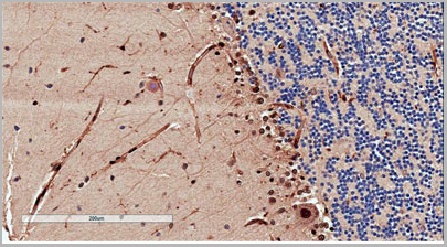
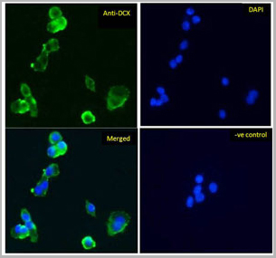
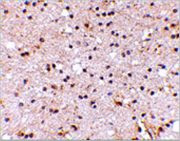
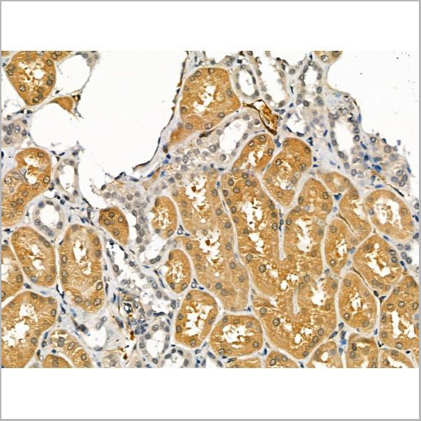
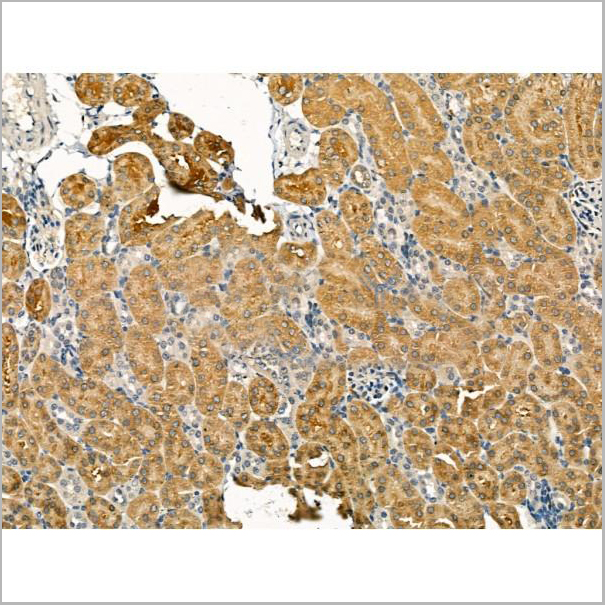
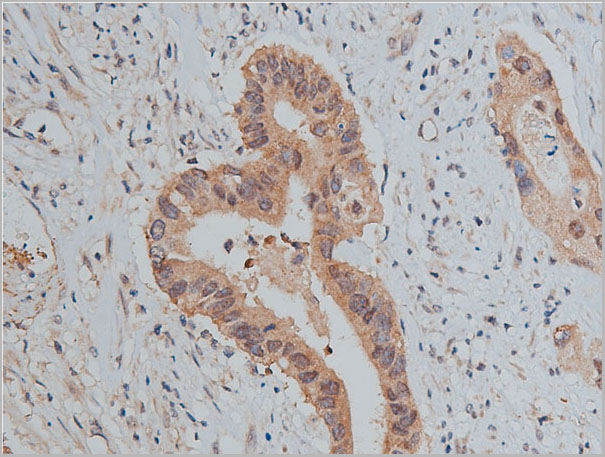
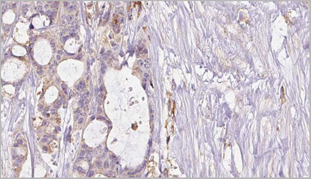
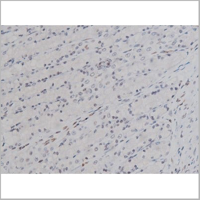
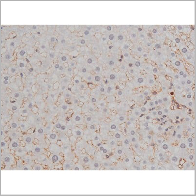
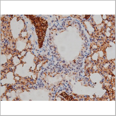
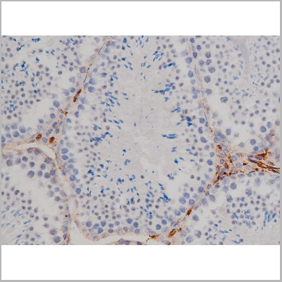
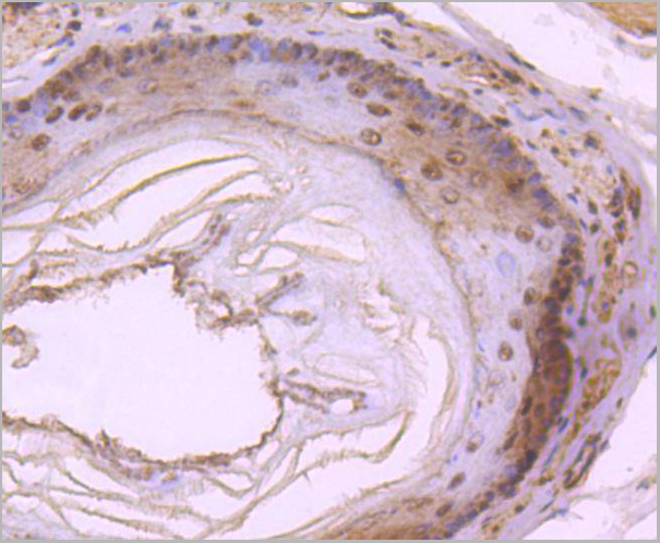
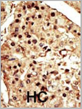
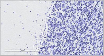 IHC (Immunohistochemistry)
(Negative Control showing staining of paraffin embedded Human Cerebellum, with no primary antibody.)
IHC (Immunohistochemistry)
(Negative Control showing staining of paraffin embedded Human Cerebellum, with no primary antibody.)
Doublecortin/DCX, Polyclonal Antibody (Cat# AAA13668)
Full Name
Goat anti-Doublecortin/DCX (aa232-242) Antibody
Gene Names
DCX; DC; DBCN; LISX; SCLH; XLIS
Reactivity
Species Reactivity: Mouse, Human
Expected from sequence similarity: Human, Mouse, Rat, Dog, Pig, Cow
Expected from sequence similarity: Human, Mouse, Rat, Dog, Pig, Cow
Applications
Peptide ELISA, Western Blot, Immunohistochemistry, Immunofluorescence
Purity
Purified from goat serum by ammonium sulphate precipitation followed by antigen affinity chromatography using the immunizing peptide.
Pricing
SLIT2N, Recombinant Protein (Cat# AAA14389)
Full Name
SLIT2N protein
Purity
>98% by SDS-PAGE
Pricing


 Application Data
(Published customer image: Mouse anti V5 tag antibody, clone SV5-Pk1 used for the detection of V5 tagged WEEV_nsP3 protein by western blotting and immunofluorescenceImage caption: WEEV nsP3 interaction with host IKKbeta. A) U87MGs were transfected in a 6-well plate with 5 ug of pUC19 and WEEV_nsP3_HA for 24 hours. Cell lysates were resolved using SDS-PAGE and subsequently immunoblotted with V5 antibody and beta-actin served as a loading control. B) U87MGs were transfected with WEEV_nsP3_V5; cells were fixed after 24 hours and stained with antibodies against the endogenous IKKbeta and the V5 tag. Cells were incubated with appropriate secondary Alexa Fluor antibodies and the nuclei stained with DAPI. Co-localization of IKKbeta with WEEV_nsP3_V5 (yellow) was observed as shown by the arrows. B) Panels E -H serve as an example of transfected cells in a given field of view that show co-localization of IKKbeta and WEEV_nsP3_V5 24 hours post transfection. Panels I-L represent magnified images of other cells showing co-localization of IKKbeta and WEEV_nsP3_V5. Panel M is a magnified image of panel L. The co-localization was confirmed by Z-stack analysis. Co-localization was calculated to be approximately in 61% of cells (163 cells were counted of which 44% demonstrated expression of nsP3. Of those cells that expressed nsP3, 61% showed co-localization of both proteins). Images were taken using Nikon Eclipse TE2000-U at 60x magnification and are representative of 2 independent experiments.From: Amaya M, Voss K, Sampey G, Senina S, de la Fuente C, et al. (2014) The Role of IKKbeta in Venezuelan Equine Encephalitis Virus Infection. PLoS ONE 9(2): e86745.)
Application Data
(Published customer image: Mouse anti V5 tag antibody, clone SV5-Pk1 used for the detection of V5 tagged WEEV_nsP3 protein by western blotting and immunofluorescenceImage caption: WEEV nsP3 interaction with host IKKbeta. A) U87MGs were transfected in a 6-well plate with 5 ug of pUC19 and WEEV_nsP3_HA for 24 hours. Cell lysates were resolved using SDS-PAGE and subsequently immunoblotted with V5 antibody and beta-actin served as a loading control. B) U87MGs were transfected with WEEV_nsP3_V5; cells were fixed after 24 hours and stained with antibodies against the endogenous IKKbeta and the V5 tag. Cells were incubated with appropriate secondary Alexa Fluor antibodies and the nuclei stained with DAPI. Co-localization of IKKbeta with WEEV_nsP3_V5 (yellow) was observed as shown by the arrows. B) Panels E -H serve as an example of transfected cells in a given field of view that show co-localization of IKKbeta and WEEV_nsP3_V5 24 hours post transfection. Panels I-L represent magnified images of other cells showing co-localization of IKKbeta and WEEV_nsP3_V5. Panel M is a magnified image of panel L. The co-localization was confirmed by Z-stack analysis. Co-localization was calculated to be approximately in 61% of cells (163 cells were counted of which 44% demonstrated expression of nsP3. Of those cells that expressed nsP3, 61% showed co-localization of both proteins). Images were taken using Nikon Eclipse TE2000-U at 60x magnification and are representative of 2 independent experiments.From: Amaya M, Voss K, Sampey G, Senina S, de la Fuente C, et al. (2014) The Role of IKKbeta in Venezuelan Equine Encephalitis Virus Infection. PLoS ONE 9(2): e86745.)
V5-TAG, Monoclonal Antibody (Cat# AAA11850)
Full Name
MOUSE ANTI V5-TAG:Biotin
Applications
Immunohistochemistry, Western Blot
Pricing
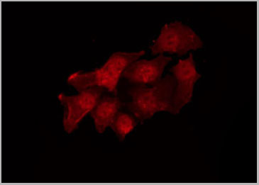
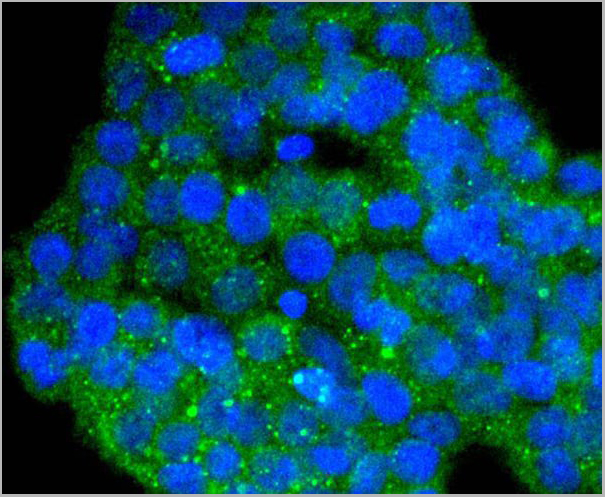
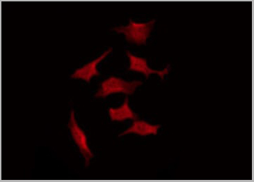 IF (Immunofluorescence)
(AAA30990 staining K562 by IF/ICC. The sample were fixed with PFA and permeabilized in 0.1% Triton X-100, then blocked in 10% serum for 45 minutes at 25 degree C. The primary antibody was diluted at 1/200 and incubated with the sample for 1 hour at 37 degree C. An Alexa Fluor 594 conjugated goat anti-rabbit IgG (H+L) Ab, diluted at 1/600, was used as the secondary antibody.)
IF (Immunofluorescence)
(AAA30990 staining K562 by IF/ICC. The sample were fixed with PFA and permeabilized in 0.1% Triton X-100, then blocked in 10% serum for 45 minutes at 25 degree C. The primary antibody was diluted at 1/200 and incubated with the sample for 1 hour at 37 degree C. An Alexa Fluor 594 conjugated goat anti-rabbit IgG (H+L) Ab, diluted at 1/600, was used as the secondary antibody.)
Lyn, Polyclonal Antibody (Cat# AAA30990)
Full Name
Phospho-Lyn (Tyr508) Antibody
Gene Names
LYN; JTK8; p53Lyn; p56Lyn
Reactivity
Human, Mouse, Rat
Applications
Western Blot, Immunohistochemistry, Immunofluorescence, Immunocytochemistry
Purity
From purified rabbit serum by affinity purification via sequential chromatography on phospho-and non-phospho-peptide affinity columns.
Pricing
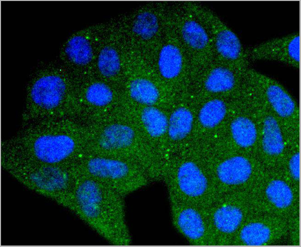
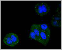

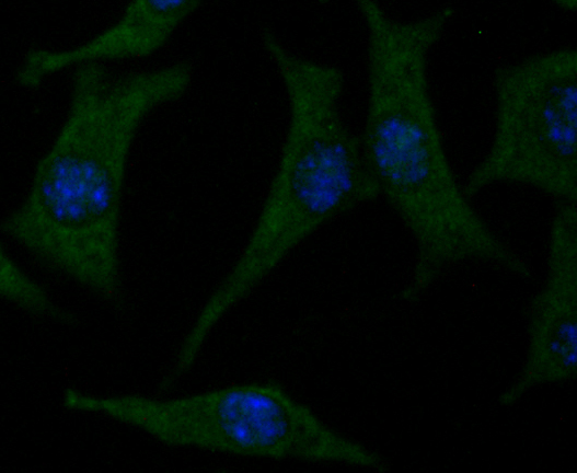
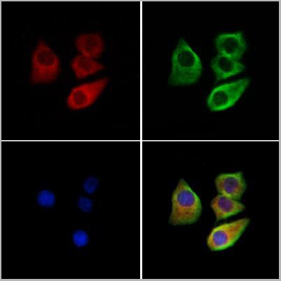
 ICC (Immunocytochemistry)
(ICC staining DYNLL1 in 293T cells (green). The nuclear counter stain is DAPI (blue). Cells were fixed in paraformaldehyde, permeabilised with 0.25% Triton X100/PBS.)
ICC (Immunocytochemistry)
(ICC staining DYNLL1 in 293T cells (green). The nuclear counter stain is DAPI (blue). Cells were fixed in paraformaldehyde, permeabilised with 0.25% Triton X100/PBS.)
DYNLL1, Monoclonal Antibody (Cat# AAA30208)
Full Name
DYNLL1 Antibody
Gene Names
DYNLL1; LC8; PIN; DLC1; DLC8; LC8a; DNCL1; hdlc1; DNCLC1
Reactivity
Human, Mouse, Rat
Applications
Western Blot, Immunocytochemistry, Immunofluorescence, Immunohistochemistry, Immunoprecipitation
Purity
ProA affinity purified
Pricing
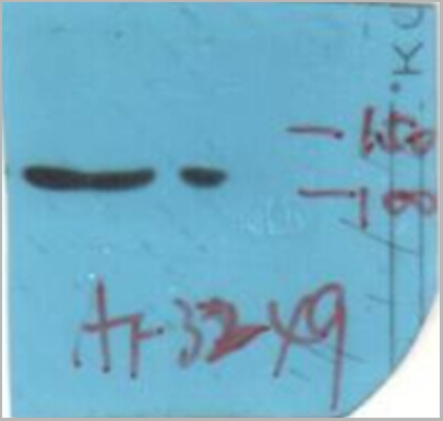
 WB (Western Blot)
(CHAK1 Antibody (AAA28787) western blot analysis in 293 cell line lysates (35ug/lane).This demonstrates the CHAK1 antibody detected the CHAK1 protein (arrow).)
WB (Western Blot)
(CHAK1 Antibody (AAA28787) western blot analysis in 293 cell line lysates (35ug/lane).This demonstrates the CHAK1 antibody detected the CHAK1 protein (arrow).)
TRPM7 (CHAK1), Polyclonal Antibody (Cat# AAA28787)
Full Name
TRPM7 (CHAK1) Antibody (C-term)
Gene Names
TRPM7; CHAK; CHAK1; ALSPDC; LTRPC7; LTrpC-7; TRP-PLIK
Reactivity
Human, Mouse
Predicted: Rat
Predicted: Rat
Applications
Western Blot, Immunohistochemistry
Purity
Purified Rabbit Polyclonal Antibody (Pab)
Pricing
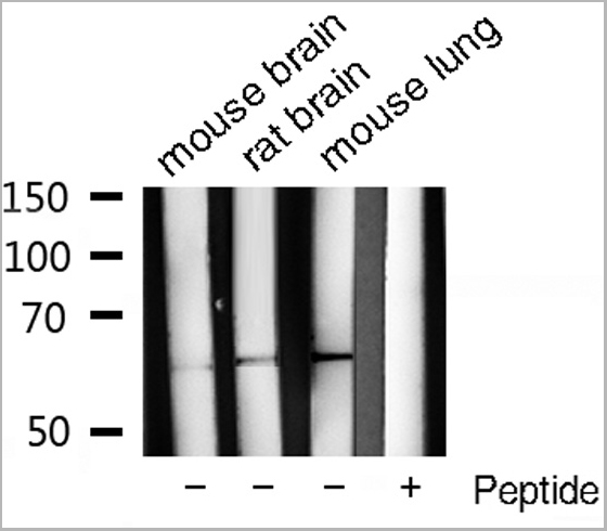
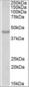
 WB (Western Blot)
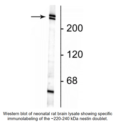
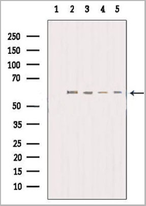
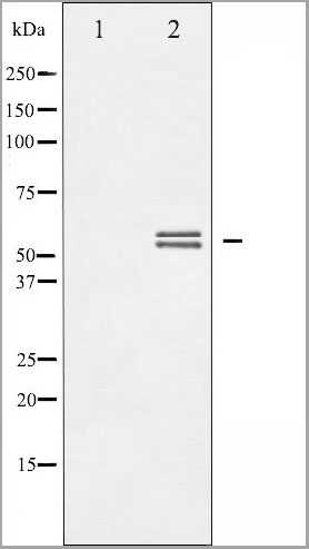
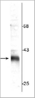
(Western blot of rat synaptic membrane (SPM) showing specific immunolabeling of the ~36 kDa stargazin protein.)
WB (Western Blot)
(Western blot of rat synaptic membrane (SPM) showing specific immunolabeling of the ~36 kDa stargazin protein.)
Stargazin, Polyclonal Antibody (Cat# AAA14233)
Full Name
Anti-Stargazin
Gene Names
Cacng2; stg; wag; waggler; AW060990; stargazer; stargazin; B230105C07Rik; B930041E13Rik
Reactivity
Tested: Rat
Expected: Mouse
Expected: Mouse
Applications
Western Blot
Purity
Prepared from pooled rabbit serum by affinity purification via chromatography on an affinity column to which the C-terminal peptide used as immunogen was coupled.
Pricing
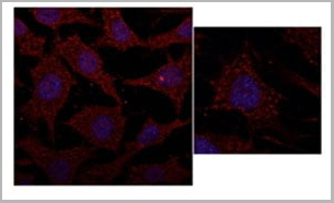
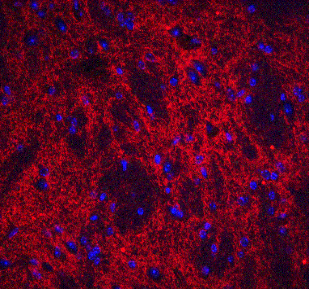
 IF (Immunofluorescence)
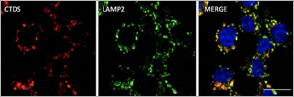
(Immunofluorescence - anti-CTSD Ab at 1/100 dilution in RAW264.7 cells; cells were fixed with PFA and permeabilized with 0.05% saponin)
IF (Immunofluorescence)
(Immunofluorescence - anti-CTSD Ab at 1/100 dilution in RAW264.7 cells; cells were fixed with PFA and permeabilized with 0.05% saponin)
Cathepsin D, Polyclonal Antibody (Cat# AAA13859)
Full Name
Cathepsin D Polyclonal Antibody
Gene Names
CTSD; CPSD; CLN10; HEL-S-130P
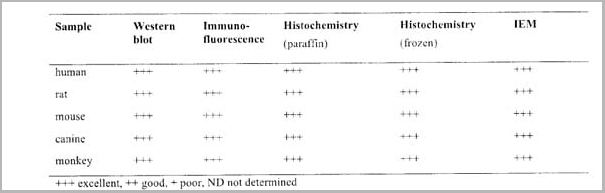
Reactivity
Reacts against human, rat, mouse, canine and monkey proteins.
Applications
Western Blot, Immunofluorescence, Immunohistochemistry, Immunohistochemistry
Pricing