Filters
Clonality
Type
Reactivity
Gene Name
Isotype
Host
Application
Clone
733 results for " l type" - showing 50-100


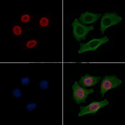
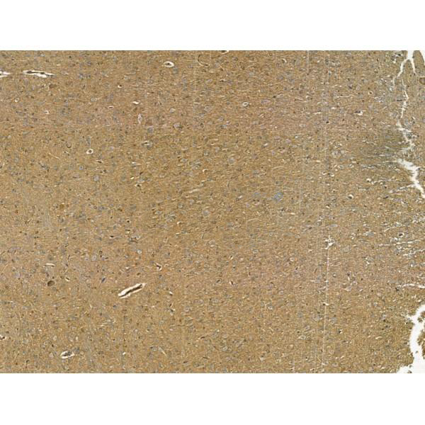
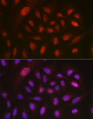
 IF (Immunofluorescence)
(Immunofluorescence analysis of HeLa cells using APP antibody at dilution of 1:100. Blue: DAPI for nuclear staining.)
IF (Immunofluorescence)
(Immunofluorescence analysis of HeLa cells using APP antibody at dilution of 1:100. Blue: DAPI for nuclear staining.)
APP, Polyclonal Antibody (Cat# AAA28326)
Full Name
APP Rabbit pAb
Gene Names
APP; AAA; AD1; PN2; ABPP; APPI; CVAP; ABETA; PN-II; CTFgamma
Reactivity
Human, Mouse, Rat
Applications
Western Blot, Immunohistochemistry, Immunofluorescence
Purity
Affinity purification
Pricing


 Application Data
(Staining of human peripheral blood lymphocytes with MOUSE ANTI HUMAN CD45RA:FITC (MCA88F))
Application Data
(Staining of human peripheral blood lymphocytes with MOUSE ANTI HUMAN CD45RA:FITC (MCA88F))
CD45RA, Monoclonal Antibody (Cat# AAA26770)
Full Name
CD45RA (CD45 Antigen, B220, GP180, Leukocyte Common Antigen, LCA, L-CA, LY5, LY-5, Protein Tyrosine Phosphatase Receptor Type C Polypeptide, PTPRC, T200, T200 Glycoprotein) (PE)
Reactivity
Human, Monkey
Applications
Flow Cytometry, Immunohistochemistry
Purity
Purified by protein G affinity chromatography from tissue culture supernatant.
Pricing
 Standard Curve (Sample)


 FCM (Flow Cytometry)
(Flow cytometric analysis of HepG2 cells with Cathepsin L/V/K/H antibody at 1/50 dilution (red) compared with an unlabelled control (cells without incubation with primary antibody; black). Alexa Fluor 488-conjugated goat anti rabbit IgG was used as the secondary antibody.)
FCM (Flow Cytometry)
(Flow cytometric analysis of HepG2 cells with Cathepsin L/V/K/H antibody at 1/50 dilution (red) compared with an unlabelled control (cells without incubation with primary antibody; black). Alexa Fluor 488-conjugated goat anti rabbit IgG was used as the secondary antibody.)
Cathepsin LVKH, Monoclonal Antibody (Cat# AAA30319)
Full Name
Cathepsin LVKH Antibody
Gene Names
CTSL1; MEP; CATL; CTSL
Reactivity
Human, Mouse, Rat
Applications
Western Blot, Immunocytochemistry, Immunofluorescence, Immunohistochemistry, Flow Cytometry
Purity
ProA affinity purified
Pricing


 ChIP (Chromatin Immunoprecipitation)
(Chromatin immunoprecipitation analysis of extracts of HeLa cells, using Acetyl-Histone H2B-K12 antibody and rabbit IgG.The amount of immunoprecipitated DNA was checked by quantitative PCR. Histogram was constructed by the ratios of the immunoprecipitated DNA to the input.)
ChIP (Chromatin Immunoprecipitation)
(Chromatin immunoprecipitation analysis of extracts of HeLa cells, using Acetyl-Histone H2B-K12 antibody and rabbit IgG.The amount of immunoprecipitated DNA was checked by quantitative PCR. Histogram was constructed by the ratios of the immunoprecipitated DNA to the input.)
Acetyl-Histone H2B-K12, Polyclonal Antibody (Cat# AAA28316)
Full Name
Acetyl-Histone H2B-K12 Rabbit pAb
Gene Names
HIST2H2BE; H2B; H2BQ; GL105; H2B.1; H2BFQ; H2BGL105
Reactivity
Human, Mouse, Rat
Applications
Western Blot, Immunohistochemistry, Immunofluorescence
Purity
Affinity purification
Pricing


 Application Data
(Staining of mouse spleen cells with Rat anti Mouse CD45 (Ly-5))
Application Data
(Staining of mouse spleen cells with Rat anti Mouse CD45 (Ly-5))
CD45, Monoclonal Antibody (Cat# AAA11856)
Full Name
RAT ANTI MOUSE CD45:FITC
Gene Names
Ptprc; loc; B220; Cd45; L-CA; Ly-5; T200; CD45R; Lyt-4
Applications
Flow Cytometry
Pricing


 IHC (Immunohistchemistry)
(Immunohistochemistry of paraffin-embedded mouse spleen using PTPN11 antibody at dilution of 1:100 (40x lens).)
IHC (Immunohistchemistry)
(Immunohistochemistry of paraffin-embedded mouse spleen using PTPN11 antibody at dilution of 1:100 (40x lens).)
PTPN11, Polyclonal Antibody (Cat# AAA10709)
Full Name
PTPN11 Polyclonal Antibody
Gene Names
PTPN11; CFC; NS1; SHP2; BPTP3; PTP2C; PTP-1D; SH-PTP2; SH-PTP3
Reactivity
Human, Mouse, Rat
Applications
Western Blot, Immunohistochemistry
Purity
Affinity Purification
Pricing


 WB (Western Blot)
(Anti-SOST Antibody (N-term)at 1:2000 dilution + human kidney lysatesLysates/proteins at 20 ug per lane.SecondaryGoat Anti-Rabbit IgG, (H+L), Peroxidase conjugated at 1/10000 dilutionPredicted band size : 24 kDaBlocking/Dilution buffer: 5% NFDM/TBST.)
WB (Western Blot)
(Anti-SOST Antibody (N-term)at 1:2000 dilution + human kidney lysatesLysates/proteins at 20 ug per lane.SecondaryGoat Anti-Rabbit IgG, (H+L), Peroxidase conjugated at 1/10000 dilutionPredicted band size : 24 kDaBlocking/Dilution buffer: 5% NFDM/TBST.)
SOST, Polyclonal Antibody (Cat# AAA28704)
Full Name
SOST Antibody (N-term)
Gene Names
SOST; CDD; VBCH; SOST1
Reactivity
Human, mouse (Predicted Reactivity: Bovine)
Applications
Western Blot, Immunohistochemistry
Purity
Purified Rabbit Polyclonal Antibody (Pab)
Pricing


 Application Data
(At 25 degree C. Samples were then incubated with primary Ab(At 37 degree C. An AlexaFluor594 conjugated goat anti-rabbit IgG(H+L) Ab(Red) and an AlexaFluor488 conjugated goat anti-mouse IgG(H+L) Ab(Green) were used as the secondary antibody.The nuclear counter stain is DAPI(blue).)
Application Data
(At 25 degree C. Samples were then incubated with primary Ab(At 37 degree C. An AlexaFluor594 conjugated goat anti-rabbit IgG(H+L) Ab(Red) and an AlexaFluor488 conjugated goat anti-mouse IgG(H+L) Ab(Green) were used as the secondary antibody.The nuclear counter stain is DAPI(blue).)
H2B, Polyclonal Antibody (Cat# AAA31349)
Full Name
Acetyl-H2B (Lys16) Antibody
Reactivity
Human, Mouse, Rat
Applications
Western Blot, Immunohistochemistry, Immunofluorescence, Immunocytochemistry, Peptide ELISA
Purity
The antiserum was purified by peptide affinity chromatography using SulfoLink Coupling Resin
Pricing


 Application Data
(Publised customer image:Mouse anti Human CD163 antibody, clone EDHu-1 used for the identification of perivascular macrophages in human brain by immunofluorescence.Image caption:Images demonstrating immunohistological stainings of amylin and double immunofluorescence staining against NG2/amylin, laminin/amylin and CD163/amylin in the hippocampus of the patient with AD and T2D.Amylin cell inclusions are indicated with arrows in (a) and shown in a higher magnification in (b). Pericytes with round cell bodies and NG2-positive coverage of the microvessel surface (green in c), without amylin cell inclusions (red in d), displayed round DAPI-positive cell nuclei (blue in e). The images in (c), (d) and (e) are merged in (f). Cells with more diffuse and weak NG2 staining (indicated by the arrowhead, green in g) and cytosolic amylin cell inclusions (red in h) showed altered cell nuclei (indicated with arrow, blue in i).The adjacent unaffected NG2-positive cell is indicated with an arrowhead in (i). The images in (g), (h) and (i) are merged in (j). Cells enclosed by laminin (green in K) contained amylin grains (red in l) and fragmented DAPI-positive cell nuclei (indicated with an arrow blue in m). The images in (k), (l) and (m) are merged in (n).Loss of NG2 coverage (green in o) was associated with polarized amylin cell inclusion (red in p) and fragmented DAPI-positive cell nuclei (indicated with an arrow, blue in q). The images in (o), (p) and (q) are merged in (r). Staining against macrophage marker CD163 (green in s) did not co-localize with amylin cell inclusions (red in t). The cell nucleus was stained with DAPI (blue in U). The images in (s), (t) and (u) are merged in (v). Scale bars: (a) 50 mum, (b), (c) to (v) 5 mum.From: Schultz, N. et al. (2016).Amylin alters human brain pericyte viability and NG2 expression.J Cereb Blood Flow & Metab. Jun 28 [Epub ahead of print]This is from an open access article distributed under the terms of the Creative Commons Attribution License.)
Application Data
(Publised customer image:Mouse anti Human CD163 antibody, clone EDHu-1 used for the identification of perivascular macrophages in human brain by immunofluorescence.Image caption:Images demonstrating immunohistological stainings of amylin and double immunofluorescence staining against NG2/amylin, laminin/amylin and CD163/amylin in the hippocampus of the patient with AD and T2D.Amylin cell inclusions are indicated with arrows in (a) and shown in a higher magnification in (b). Pericytes with round cell bodies and NG2-positive coverage of the microvessel surface (green in c), without amylin cell inclusions (red in d), displayed round DAPI-positive cell nuclei (blue in e). The images in (c), (d) and (e) are merged in (f). Cells with more diffuse and weak NG2 staining (indicated by the arrowhead, green in g) and cytosolic amylin cell inclusions (red in h) showed altered cell nuclei (indicated with arrow, blue in i).The adjacent unaffected NG2-positive cell is indicated with an arrowhead in (i). The images in (g), (h) and (i) are merged in (j). Cells enclosed by laminin (green in K) contained amylin grains (red in l) and fragmented DAPI-positive cell nuclei (indicated with an arrow blue in m). The images in (k), (l) and (m) are merged in (n).Loss of NG2 coverage (green in o) was associated with polarized amylin cell inclusion (red in p) and fragmented DAPI-positive cell nuclei (indicated with an arrow, blue in q). The images in (o), (p) and (q) are merged in (r). Staining against macrophage marker CD163 (green in s) did not co-localize with amylin cell inclusions (red in t). The cell nucleus was stained with DAPI (blue in U). The images in (s), (t) and (u) are merged in (v). Scale bars: (a) 50 mum, (b), (c) to (v) 5 mum.From: Schultz, N. et al. (2016).Amylin alters human brain pericyte viability and NG2 expression.J Cereb Blood Flow & Metab. Jun 28 [Epub ahead of print]This is from an open access article distributed under the terms of the Creative Commons Attribution License.)
CD163, Monoclonal Antibody (Cat# AAA12256)
Full Name
Mouse Anti Human CD163: RPE
Gene Names
CD163; M130; MM130; SCARI1
Reactivity
Human
Applications
Flow Cytometry
Pricing


 Application Data
(Published customer image Infiltration of GFP+ BM-cells in infarct and peri-infarct regions. (A-B) Dot plots of viable macrophages/granulocytes (CD11b+CD45high, top right quadrants) and microglia (CD11b+CD45dim, bottom right quadrants) in cortex from BM-chimeric unmanipulated mice and mice exposed to pMCAO. (C) Bar graph showing mean numbers of CD11b+CD45dim microglia and CD11b+CD45high macrophages/granulocytes in BM-chimeric mice 24 hours after pMCAO, subdivided based on expression of GFP (n = 5). Approximately 92% of of the CD45high population were GFP+. (D) Estimation and comparison of mean numbers of CD11b+CD45dim microglia in non-chimeric (n = 10) versus BM-chimeric mice (n = 5) 24 hours after of pMCAO shows significantly fewer CD11b+CD45dim microglial cells in irradiated mice. (E) Overview, showing distribution of infiltrating GFP+ BM-derived cells into infarct (IF) and peri-infarct (P-IF) regions 24 hours after pMCAO. (E-G) By 24 hours, GFP+ single cells (F) and vessel-associated aggregates of GFP+ cells (arrows in G) were observed in infarct and peri-infarct regions. Some of the vessel-associated cells were round, leukocyte-like cells (arrows) while others were elongated cells lining the vasculature (arrow heads in G and in insert). (H) Bar graph showing mean numbers of single GFP+ cells and vessel-associated aggregates of GFP+ cells in ipsi- and contralateral cortex 24 hours after surgery (n = 10). (I-P) Immunohistochemical staining of CD45.1 (I, K), CD45.2 (J, L), IgG2a (M, O) and CD45 (N, P) in ischemic tissue in BM-chimeric (I, J, M, N) and non-chimeric mice (K, L, O, P) 24 hours after pMCAO. N.D, none detected. Scale bars: 200 um (A), 10 um (B, C). 50 um (I-P) *P < 0.05, **P < 0.01, and ***P < 0.001.From: Clausen BH, Lambertsen KL, Babcock AA, Holm TH, Dagnaes-Hansen F, Finsen B. Interleukin-1beta and tumor necrosis factor-alpha are expressed by different subsets of microglia and macrophages after ischemic stroke in mice. J Neuroinflammation. 2008 Oct 23;5:46.)
Application Data
(Published customer image Infiltration of GFP+ BM-cells in infarct and peri-infarct regions. (A-B) Dot plots of viable macrophages/granulocytes (CD11b+CD45high, top right quadrants) and microglia (CD11b+CD45dim, bottom right quadrants) in cortex from BM-chimeric unmanipulated mice and mice exposed to pMCAO. (C) Bar graph showing mean numbers of CD11b+CD45dim microglia and CD11b+CD45high macrophages/granulocytes in BM-chimeric mice 24 hours after pMCAO, subdivided based on expression of GFP (n = 5). Approximately 92% of of the CD45high population were GFP+. (D) Estimation and comparison of mean numbers of CD11b+CD45dim microglia in non-chimeric (n = 10) versus BM-chimeric mice (n = 5) 24 hours after of pMCAO shows significantly fewer CD11b+CD45dim microglial cells in irradiated mice. (E) Overview, showing distribution of infiltrating GFP+ BM-derived cells into infarct (IF) and peri-infarct (P-IF) regions 24 hours after pMCAO. (E-G) By 24 hours, GFP+ single cells (F) and vessel-associated aggregates of GFP+ cells (arrows in G) were observed in infarct and peri-infarct regions. Some of the vessel-associated cells were round, leukocyte-like cells (arrows) while others were elongated cells lining the vasculature (arrow heads in G and in insert). (H) Bar graph showing mean numbers of single GFP+ cells and vessel-associated aggregates of GFP+ cells in ipsi- and contralateral cortex 24 hours after surgery (n = 10). (I-P) Immunohistochemical staining of CD45.1 (I, K), CD45.2 (J, L), IgG2a (M, O) and CD45 (N, P) in ischemic tissue in BM-chimeric (I, J, M, N) and non-chimeric mice (K, L, O, P) 24 hours after pMCAO. N.D, none detected. Scale bars: 200 um (A), 10 um (B, C). 50 um (I-P) *P < 0.05, **P < 0.01, and ***P < 0.001.From: Clausen BH, Lambertsen KL, Babcock AA, Holm TH, Dagnaes-Hansen F, Finsen B. Interleukin-1beta and tumor necrosis factor-alpha are expressed by different subsets of microglia and macrophages after ischemic stroke in mice. J Neuroinflammation. 2008 Oct 23;5:46.)
CD11b, Monoclonal Antibody (Cat# AAA12182)
Full Name
RAT ANTI MOUSE CD11b:FITC
Gene Names
Itgam; CR3; CR3A; MAC1; Cd11b; Ly-40; Mac-1; Mac-1a; CD11b/CD18; F730045J24Rik
Applications
Flow Cytometry
Pricing


 WB (Western Blot)
(Anti-CD63 Antibody (C-term)at 1:2000 dilution + human plasma lysatesLysates/proteins at 20 ug per lane.SecondaryGoat Anti-Rabbit IgG, (H+L), Peroxidase conjugated at 1/10000 dilutionPredicted band size : 25 kDaBlocking/Dilution buffer: 5% NFDM/TBST.)
WB (Western Blot)
(Anti-CD63 Antibody (C-term)at 1:2000 dilution + human plasma lysatesLysates/proteins at 20 ug per lane.SecondaryGoat Anti-Rabbit IgG, (H+L), Peroxidase conjugated at 1/10000 dilutionPredicted band size : 25 kDaBlocking/Dilution buffer: 5% NFDM/TBST.)
CD63, Polyclonal Antibody (Cat# AAA28725)
Full Name
CD63 Antibody (C-term)
Gene Names
CD63; MLA1; ME491; LAMP-3; OMA81H; TSPAN30
Reactivity
Human
Applications
Western Blot, Immunohistochemistry, Flow Cytometry
Purity
Peptide Affinity Purified Rabbit Polyclonal Antibody (Pab)
Pricing


 Application Data
(At 37 degree C. The primary antibody was diluted 1/200 and incubated with the sample for 1 hour at 37 degree C. A Alexa Fluor 594 conjugated goat polyclonal to rabbit IgG (H+L), diluted 1/600 was used as secondary antibody.)
Application Data
(At 37 degree C. The primary antibody was diluted 1/200 and incubated with the sample for 1 hour at 37 degree C. A Alexa Fluor 594 conjugated goat polyclonal to rabbit IgG (H+L), diluted 1/600 was used as secondary antibody.)
N Cadherin, Polyclonal Antibody (Cat# AAA31337)
Full Name
N Cadherin Antibody
Gene Names
CDH2; CDHN; NCAD; CD325; CDw325
Reactivity
Human, Mouse, Rat
Predicted Reactivity: Pig (100%), Zebrafish (100%), Bovine (100%), Horse (100%), Sheep (100%), Rabbit (100%), Dog (100%), Chicken (100%), Xenopus (100%)
Predicted Reactivity: Pig (100%), Zebrafish (100%), Bovine (100%), Horse (100%), Sheep (100%), Rabbit (100%), Dog (100%), Chicken (100%), Xenopus (100%)
Applications
Western Blot, Immunohistochemistry, Immunofluorescence, Immunocytochemistry, Peptide ELISA, Immunohistochemistry, Immunohistochemistry
Purity
The antiserum was purified by peptide affinity chromatography using SulfoLink Coupling Resin
Pricing


 Application Data
(Staining of CD209 transfected K562 cells with MOUSE ANTI HUMAN CD209: FITC)
Application Data
(Staining of CD209 transfected K562 cells with MOUSE ANTI HUMAN CD209: FITC)
DC-SIGN, Monoclonal Antibody (Cat# AAA26722)
Full Name
DC-SIGN (Dendritic Cell-specific ICAM3 grabbing Non-integrin, C Type Lectin Domain Family 4 Member L, CLEC4L, CD209, CD209 Antigen, CDSIGN, Dendritic Cell-specific ICAM-3-grabbing Non-integrin 1, DC SIGN1, DC-SIGN1, HIV GP120 Binding Protein, MGC129965) (
Reactivity
Human
Applications
Immunofluorescence
Purity
Purified by protein G affinity chromatography from tissue culture supernatant.
Pricing


 Application Data
(At 25 degree C. The primary antibody was diluted at 1/200 and incubated with the sample for 1 hour at 37 degree C. An Alexa Fluor 594 conjugated goat anti-rabbit IgG (H+L) Ab, diluted at 1/600, was used as the secondary antibody.)
Application Data
(At 25 degree C. The primary antibody was diluted at 1/200 and incubated with the sample for 1 hour at 37 degree C. An Alexa Fluor 594 conjugated goat anti-rabbit IgG (H+L) Ab, diluted at 1/600, was used as the secondary antibody.)
CD22, Polyclonal Antibody (Cat# AAA31418)
Full Name
Phospho-CD22 (Tyr842) Antibody
Gene Names
CD22; SIGLEC2; SIGLEC-2
Reactivity
Human, Mouse, Rat
Predicted Reactivity: Horse (83%)
Predicted Reactivity: Horse (83%)
Applications
Western Blot, Immunohistochemistry, Immunofluorescence, Immunocytochemistry, Peptide ELISA
Purity
The antibody is from purified rabbit serum by affinity purification via sequential chromatography on phospho-peptide and non-phospho-peptide affinity columns.
Pricing


 WB (Western Blot)
(Flow cytometric analysis of SK-BR-3 cells using MICA Antibody (Center) (green) compared to an isotype control of rabbit IgG(blue). AAA28710 was diluted at 1:25 dilution. An Alexa Fluor 488 goat anti-rabbit lgG at 1:400 dilution was used as the secondary antibody.)
WB (Western Blot)
(Flow cytometric analysis of SK-BR-3 cells using MICA Antibody (Center) (green) compared to an isotype control of rabbit IgG(blue). AAA28710 was diluted at 1:25 dilution. An Alexa Fluor 488 goat anti-rabbit lgG at 1:400 dilution was used as the secondary antibody.)
MICA, Polyclonal Antibody (Cat# AAA28710)
Full Name
MICA Antibody (Center)
Gene Names
MICA; MIC-A; PERB11.1
Reactivity
Human
Applications
Western Blot, Immunohistochemistry, Immunofluorescence, Flow Cytometry
Purity
Peptide Affinity Purified Rabbit Polyclonal Antibody (Pab)
Pricing


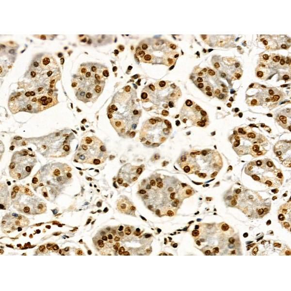
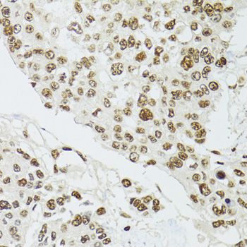
 IHC (Immunohistchemistry)
(Immunohistochemistry of paraffin-embedded human lung cancer using KRT20 Antibody at dilution of 1:100 (40x lens).)
IHC (Immunohistchemistry)
(Immunohistochemistry of paraffin-embedded human lung cancer using KRT20 Antibody at dilution of 1:100 (40x lens).)
KRT20, Polyclonal Antibody (Cat# AAA10645)
Full Name
KRT20 Polyclonal Antibody
Gene Names
KRT20; K20; CD20; CK20; CK-20; KRT21
Reactivity
Human, Mouse, Rat
Applications
Western Blot, Immunohistochemistry, Immunofluorescence
Purity
Affinity Purification
Pricing


 Application Data
(Staining of human peripheral blood granulocytes with Mouse anti Human CD45: Pacific Blue)
Application Data
(Staining of human peripheral blood granulocytes with Mouse anti Human CD45: Pacific Blue)
CD45, Monoclonal Antibody (Cat# AAA12202)
Full Name
MOUSE ANTI HUMAN CD45:FITC
Gene Names
PTPRC; LCA; LY5; B220; CD45; L-CA; T200; CD45R; GP180
Applications
Flow Cytometry
Pricing


 WB (Western Blot)
(Western blot analysis of HLA-B (arrow) using rabbit polyclonal HLA-B Antibody (N-term). 293 cell lysates (2 ug/lane) either nontransfected (Lane 1) or transiently transfected (Lane 2) with the HLA-B gene.)
WB (Western Blot)
(Western blot analysis of HLA-B (arrow) using rabbit polyclonal HLA-B Antibody (N-term). 293 cell lysates (2 ug/lane) either nontransfected (Lane 1) or transiently transfected (Lane 2) with the HLA-B gene.)
HLA-B, Polyclonal Antibody (Cat# AAA28743)
Full Name
HLA-B Antibody (N-term)
Reactivity
Human
Applications
Western Blot, Immunohistochemistry, Flow Cytometry
Purity
Peptide Affinity Purified Rabbit Polyclonal Antibody (Pab)
Pricing


 Application Data
(At 25 degree C. Samples were then incubated with primary Ab(At 37 degree C. An AlexaFluor594 conjugated goat anti-rabbit IgG(H+L) Ab(Red) and an AlexaFluor488 conjugated goat anti-mouse IgG(H+L) Ab(Green) were used as the secondary antibody.The nuclear counter stain is DAPI (blue).)
Application Data
(At 25 degree C. Samples were then incubated with primary Ab(At 37 degree C. An AlexaFluor594 conjugated goat anti-rabbit IgG(H+L) Ab(Red) and an AlexaFluor488 conjugated goat anti-mouse IgG(H+L) Ab(Green) were used as the secondary antibody.The nuclear counter stain is DAPI (blue).)
TOP2A, Polyclonal Antibody (Cat# AAA31306)
Full Name
Phospho-TOP2A (Thr1343) Antibody
Gene Names
TOP2A; TOP2; TP2A
Reactivity
Human, Mouse
Applications
Immunohistochemistry, Immunofluorescence, Immunocytochemistry, Peptide ELISA
Purity
The antibody is from purified rabbit serum by affinity purification via sequential chromatography on phospho-peptide and non-phospho-peptide affinity columns.
Pricing


 Application Data
(Published customer image Infiltration of GFP+ BM-cells in infarct and peri-infarct regions. (A-B) Dot plots of viable macrophages/granulocytes (CD11b+CD45high, top right quadrants) and microglia (CD11b+CD45dim, bottom right quadrants) in cortex from BM-chimeric unmanipulated mice and mice exposed to pMCAO. (C) Bar graph showing mean numbers of CD11b+CD45dim microglia and CD11b+CD45high macrophages/granulocytes in BM-chimeric mice 24 hours after pMCAO, subdivided based on expression of GFP (n = 5). Approximately 92% of of the CD45high population were GFP+. (D) Estimation and comparison of mean numbers of CD11b+CD45dim microglia in non-chimeric (n = 10) versus BM-chimeric mice (n = 5) 24 hours after of pMCAO shows significantly fewer CD11b+CD45dim microglial cells in irradiated mice. (E) Overview, showing distribution of infiltrating GFP+ BM-derived cells into infarct (IF) and peri-infarct (P-IF) regions 24 hours after pMCAO. (E-G) By 24 hours, GFP+ single cells (F) and vessel-associated aggregates of GFP+ cells (arrows in G) were observed in infarct and peri-infarct regions. Some of the vessel-associated cells were round, leukocyte-like cells (arrows) while others were elongated cells lining the vasculature (arrow heads in G and in insert). (H) Bar graph showing mean numbers of single GFP+ cells and vessel-associated aggregates of GFP+ cells in ipsi- and contralateral cortex 24 hours after surgery (n = 10). (I-P) Immunohistochemical staining of CD45.1 (I, K), CD45.2 (J, L), IgG2a (M, O) and CD45 (N, P) in ischemic tissue in BM-chimeric (I, J, M, N) and non-chimeric mice (K, L, O, P) 24 hours after pMCAO. N.D, none detected. Scale bars: 200 um (A), 10 um (B, C). 50 um (I-P) *P < 0.05, **P < 0.01, and ***P < 0.001.From: Clausen BH, Lambertsen KL, Babcock AA, Holm TH, Dagnaes-Hansen F, Finsen B. Interleukin-1beta and tumor necrosis factor-alpha are expressed by different subsets of microglia and macrophages after ischemic stroke in mice. J Neuroinflammation. 2008 Oct 23;5:46.)
Application Data
(Published customer image Infiltration of GFP+ BM-cells in infarct and peri-infarct regions. (A-B) Dot plots of viable macrophages/granulocytes (CD11b+CD45high, top right quadrants) and microglia (CD11b+CD45dim, bottom right quadrants) in cortex from BM-chimeric unmanipulated mice and mice exposed to pMCAO. (C) Bar graph showing mean numbers of CD11b+CD45dim microglia and CD11b+CD45high macrophages/granulocytes in BM-chimeric mice 24 hours after pMCAO, subdivided based on expression of GFP (n = 5). Approximately 92% of of the CD45high population were GFP+. (D) Estimation and comparison of mean numbers of CD11b+CD45dim microglia in non-chimeric (n = 10) versus BM-chimeric mice (n = 5) 24 hours after of pMCAO shows significantly fewer CD11b+CD45dim microglial cells in irradiated mice. (E) Overview, showing distribution of infiltrating GFP+ BM-derived cells into infarct (IF) and peri-infarct (P-IF) regions 24 hours after pMCAO. (E-G) By 24 hours, GFP+ single cells (F) and vessel-associated aggregates of GFP+ cells (arrows in G) were observed in infarct and peri-infarct regions. Some of the vessel-associated cells were round, leukocyte-like cells (arrows) while others were elongated cells lining the vasculature (arrow heads in G and in insert). (H) Bar graph showing mean numbers of single GFP+ cells and vessel-associated aggregates of GFP+ cells in ipsi- and contralateral cortex 24 hours after surgery (n = 10). (I-P) Immunohistochemical staining of CD45.1 (I, K), CD45.2 (J, L), IgG2a (M, O) and CD45 (N, P) in ischemic tissue in BM-chimeric (I, J, M, N) and non-chimeric mice (K, L, O, P) 24 hours after pMCAO. N.D, none detected. Scale bars: 200 um (A), 10 um (B, C). 50 um (I-P) *P < 0.05, **P < 0.01, and ***P < 0.001.From: Clausen BH, Lambertsen KL, Babcock AA, Holm TH, Dagnaes-Hansen F, Finsen B. Interleukin-1beta and tumor necrosis factor-alpha are expressed by different subsets of microglia and macrophages after ischemic stroke in mice. J Neuroinflammation. 2008 Oct 23;5:46.)
CD11b, Monoclonal Antibody (Cat# AAA12184)
Full Name
RAT ANTI MOUSE CD11b
Gene Names
Itgam; CR3; CR3A; MAC1; Cd11b; Ly-40; Mac-1; Mac-1a; CD11b/CD18; F730045J24Rik
Applications
Immunohistochemistry, Flow Cytometry, Immunofluorescence, Immunoprecipitation
Pricing


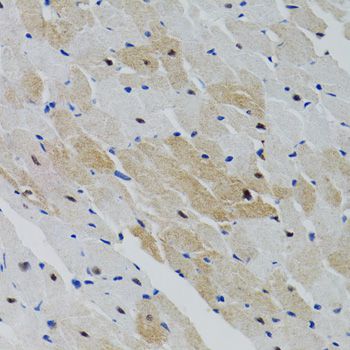
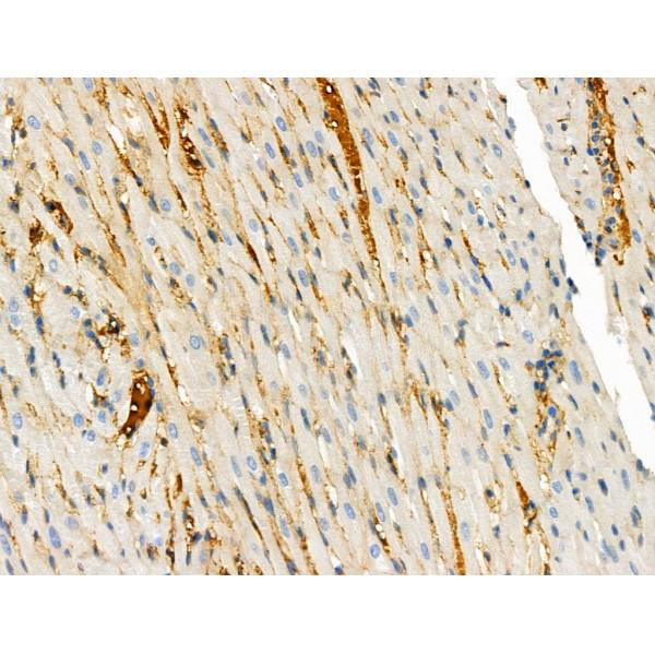
 IHC (Immunohistchemistry)
(Immunohistochemistry of paraffin-embedded mouse heart using KRT5 Antibody at dilution of 1:100 (40x lens).)
IHC (Immunohistchemistry)
(Immunohistochemistry of paraffin-embedded mouse heart using KRT5 Antibody at dilution of 1:100 (40x lens).)
KRT5, Polyclonal Antibody (Cat# AAA10706)
Full Name
KRT5 Polyclonal Antibody
Gene Names
KRT5; K5; CK5; DDD; DDD1; EBS2; KRT5A
Reactivity
Human, Mouse
Applications
Western Blot, Immunofluorescence
Purity
Affinity Purification
Pricing


 IF (Immunofluorescence)
(Immunofluorescence analysis of L929 cells using MMP9 Rabbit pAb (AAA10629) at dilution of 1:100.Secondary antibody: Cy3-conjugated Goat anti-Rabbit IgG (H+L) at 1:500 dilution. Blue: DAPI for nuclear staining.)
IF (Immunofluorescence)
(Immunofluorescence analysis of L929 cells using MMP9 Rabbit pAb (AAA10629) at dilution of 1:100.Secondary antibody: Cy3-conjugated Goat anti-Rabbit IgG (H+L) at 1:500 dilution. Blue: DAPI for nuclear staining.)
MMP9, Polyclonal Antibody (Cat# AAA10629)
Full Name
MMP9 Polyclonal Antibody
Gene Names
MMP9; GELB; CLG4B; MMP-9; MANDP2
Applications
Western Blot, Immunohistochemistry, Immunofluorescence, Immunocytochemistry
Purity
Affinity purification
Pricing


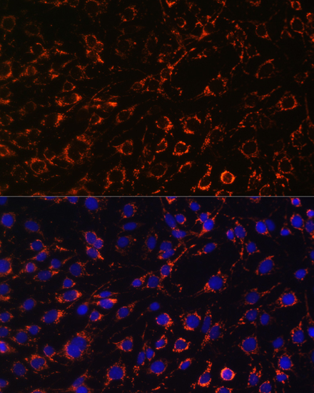


 IF (Immunofluorescence)
(Confocal immunofluorescence analysis of U2OS cells using TOM20 Polyclonal Antibody at dilution of 1:100. Blue: DAPI for nuclear staining.)
IF (Immunofluorescence)
(Confocal immunofluorescence analysis of U2OS cells using TOM20 Polyclonal Antibody at dilution of 1:100. Blue: DAPI for nuclear staining.)
TOM20, Polyclonal Antibody (Cat# AAA28341)
Full Name
TOM20 Rabbit pAb
Gene Names
TOMM20; MAS20; MOM19; TOM20
Reactivity
Human, Mouse, Rat
Applications
Western Blot, Immunohistochemistry, Immunofluorescence
Purity
Affinity purification
Pricing


 Application Data
(At 25 degree C. Samples were then incubated with primary Ab(At 37 degree C. An AlexaFluor594 conjugated goat anti-rabbit IgG(H+L) Ab(Red) and an AlexaFluor488 conjugated goat anti-mouse IgG(H+L) Ab(Green) were used as the secondary antibody.The nuclear counter stain is DAPI (blue).)
Application Data
(At 25 degree C. Samples were then incubated with primary Ab(At 37 degree C. An AlexaFluor594 conjugated goat anti-rabbit IgG(H+L) Ab(Red) and an AlexaFluor488 conjugated goat anti-mouse IgG(H+L) Ab(Green) were used as the secondary antibody.The nuclear counter stain is DAPI (blue).)
MSH2, Polyclonal Antibody (Cat# AAA31299)
Full Name
Phospho-MSH2 (Tyr238) Antibody
Gene Names
MSH2; FCC1; COCA1; HNPCC; LCFS2; HNPCC1
Reactivity
Human, Mouse, Rat
Applications
Immunohistochemistry, Immunofluorescence, Immunocytochemistry, Peptide ELISA
Purity
The antibody is from purified rabbit serum by affinity purification via sequential chromatography on phospho-peptide and non-phospho-peptide affinity columns.
Pricing


 Application Data
(At 25 degree C. The primary antibody was diluted at 1/200 and incubated with the sample for 1 hour at 37 degree C. An Alexa Fluor 594 conjugated goat anti-rabbit IgG (H+L) antibody(Red), diluted at 1/600, was used as secondary antibody.)
Application Data
(At 25 degree C. The primary antibody was diluted at 1/200 and incubated with the sample for 1 hour at 37 degree C. An Alexa Fluor 594 conjugated goat anti-rabbit IgG (H+L) antibody(Red), diluted at 1/600, was used as secondary antibody.)
Ret, Polyclonal Antibody (Cat# AAA31389)
Full Name
Phospho-Ret (Tyr1096) Antibody
Gene Names
RET; PTC; MTC1; HSCR1; MEN2A; MEN2B; RET51; CDHF12; CDHR16; RET-ELE1
Reactivity
Human, Mouse, Rat
Predicted Reactivity: Pig (100%), Bovine (100%), Sheep (100%), Rabbit (100%), Dog (100%)
Predicted Reactivity: Pig (100%), Bovine (100%), Sheep (100%), Rabbit (100%), Dog (100%)
Applications
Western Blot, Immunohistochemistry, Immunofluorescence, Immunocytochemistry, Peptide ELISA
Purity
The antibody is from purified rabbit serum by affinity purification via sequential chromatography on phospho-peptide and non-phospho-peptide affinity columns.
Pricing


 ChIP (Chromatin Immunoprecipitation)
(Chromatin immunoprecipitation analysis of extracts of MCF7 cells, using SIRT1 antibody and rabbit IgG.The amount of immunoprecipitated DNA was checked by quantitative PCR. Histogram was constructed by the ratios of the immunoprecipitated DNA to the input.)
ChIP (Chromatin Immunoprecipitation)
(Chromatin immunoprecipitation analysis of extracts of MCF7 cells, using SIRT1 antibody and rabbit IgG.The amount of immunoprecipitated DNA was checked by quantitative PCR. Histogram was constructed by the ratios of the immunoprecipitated DNA to the input.)
SIRT1, Polyclonal Antibody (Cat# AAA28348)
Full Name
[KO Validated] SIRT1 Rabbit pAb
Gene Names
SIRT1; SIR2L1
Reactivity
Human, Mouse, Rat
Applications
Western Blot, Immunohistochemistry, Immunofluorescence, Immunoprecipitation
Purity
Affinity purification
Pricing


 IHC (Immunohistchemistry)
(Figure 9. IHC analysis of BAK using anti-BAK antibody (AAA11654).BAK was detected in frozen section of rat cardiac muscle tissue. Heat mediated antigen retrieval was performed in citrate buffer (pH6, epitope retrieval solution) for 20 mins. The tissue section was blocked with 10% goat serum. The tissue section was then incubated with 1ug/ml rabbit anti-BAK Antibody (AAA11654) overnight at 4 degree C. Biotinylated goat anti-rabbit IgG was used as secondary antibody and incubated for 30 minutes at 37 degree C. The tissue section was developed using Strepavidin-Biotin-Complex (SABC) with DAB as the chromogen.)
IHC (Immunohistchemistry)
(Figure 9. IHC analysis of BAK using anti-BAK antibody (AAA11654).BAK was detected in frozen section of rat cardiac muscle tissue. Heat mediated antigen retrieval was performed in citrate buffer (pH6, epitope retrieval solution) for 20 mins. The tissue section was blocked with 10% goat serum. The tissue section was then incubated with 1ug/ml rabbit anti-BAK Antibody (AAA11654) overnight at 4 degree C. Biotinylated goat anti-rabbit IgG was used as secondary antibody and incubated for 30 minutes at 37 degree C. The tissue section was developed using Strepavidin-Biotin-Complex (SABC) with DAB as the chromogen.)
BAK, Polyclonal Antibody (Cat# AAA11654)
Full Name
Anti-BAK Antibody
Gene Names
BAK1; BAK; CDN1; BCL2L7; BAK-LIKE
Reactivity
Human, Mouse, Rat
Applications
Western Blot, Immunohistochemistry
Purity
Immunogen Affinity Purified
Pricing


 Application Data
(At 25 degree C. Samples were then incubated with primary Ab(At 37 degree C. An AlexaFluor594 conjugated goat anti-rabbit IgG(H+L) Ab(Red) and an AlexaFluor488 conjugated goat anti-mouse IgG(H+L) Ab(Green) were used as the secondary antibody.The nuclear counter stain is DAPI (blue).)
Application Data
(At 25 degree C. Samples were then incubated with primary Ab(At 37 degree C. An AlexaFluor594 conjugated goat anti-rabbit IgG(H+L) Ab(Red) and an AlexaFluor488 conjugated goat anti-mouse IgG(H+L) Ab(Green) were used as the secondary antibody.The nuclear counter stain is DAPI (blue).)
SIRT2, Polyclonal Antibody (Cat# AAA31286)
Full Name
Phospho-SIRT2 (Ser368) Antibody
Gene Names
SIRT2; SIR2; SIR2L; SIR2L2
Reactivity
Human, Mouse, Rat
Applications
Immunohistochemistry, Immunofluorescence, Immunocytochemistry, Peptide ELISA
Purity
The antibody is from purified rabbit serum by affinity purification via sequential chromatography on phospho-peptide and non-phospho-peptide affinity columns.
Pricing


 IP (Immunoprecipitation)
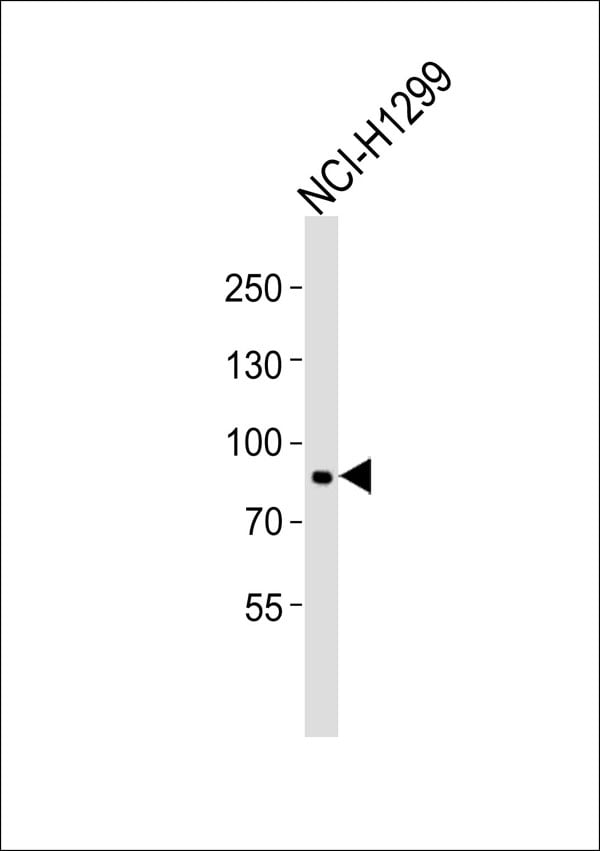
(HSPA1A was immunoprecipitated using:Lane A:0.5 mg Hela Whole Cell Lysate2 uL anti-HSPA1A rabbit monoclonal antibody and 15 ul of 50 % Protein G agarose.Primary antibody:Anti-HSPA1A rabbit monoclonal antibody,at 1:200 dilution Secondary antibody:Dylight 800-labeled antibody to rabbit IgG (H+L), at 1:5000 dilution Developed using the odssey technique.Performed under reducing conditions.Predicted band size: 70 kDaObserved band size: 70 kDa)
IP (Immunoprecipitation)
(HSPA1A was immunoprecipitated using:Lane A:0.5 mg Hela Whole Cell Lysate2 uL anti-HSPA1A rabbit monoclonal antibody and 15 ul of 50 % Protein G agarose.Primary antibody:Anti-HSPA1A rabbit monoclonal antibody,at 1:200 dilution Secondary antibody:Dylight 800-labeled antibody to rabbit IgG (H+L), at 1:5000 dilution Developed using the odssey technique.Performed under reducing conditions.Predicted band size: 70 kDaObserved band size: 70 kDa)
HSP70, Monoclonal Antibody (Cat# AAA27747)
Full Name
Recombinant Anti-HSP70 Antibody, Rabbit Monoclonal
Gene Names
HSPA1B; HSP72; HSPA1; HSX70; HSP70-1; HSP70-2; HSP70.1; HSP70.2; HSP70-1B
Reactivity
Human
Applications
Western Blot, Immunohistochemistry, Flow Cytometry, Immunocytochemistry, Immunofluorescence, Immunoprecipitation
Purity
Protein A
Pricing


 IF (Immunofluorescence)
(Confocal immunofluorescent analysis of NOS2A Antibody (Center) with hela cell followed by Alexa Fluor 488-conjugated goat anti-rabbit lgG (green). DAPI was used to stain the cell nuclear (blue).)
IF (Immunofluorescence)
(Confocal immunofluorescent analysis of NOS2A Antibody (Center) with hela cell followed by Alexa Fluor 488-conjugated goat anti-rabbit lgG (green). DAPI was used to stain the cell nuclear (blue).)
NOS2A, Polyclonal Antibody (Cat# AAA28780)
Full Name
NOS2A Antibody (Center)
Gene Names
NOS2; NOS; INOS; NOS2A; HEP-NOS
Reactivity
Human
Applications
Western Blot, Immunohistochemistry, Flow Cytometry, Immunofluorescence
Purity
Peptide Affinity Purified Rabbit Polyclonal Antibody (Pab)
Pricing


 IF (Immunofluorescence)
(Immunofluorescence analysis of A549 cells using CAMK1D antibody.)
IF (Immunofluorescence)
(Immunofluorescence analysis of A549 cells using CAMK1D antibody.)
CAMK1D, Polyclonal Antibody (Cat# AAA28183)
Full Name
CAMK1D
Gene Names
CAMK1D; CKLiK; CaM-K1; CaMKID
Reactivity
Human, Mouse, Rat
Applications
Western Blot, Immunohistochemistry
Pricing


 IF (Immunofluorescence)
(Immunofluorescence analysis of U-2 OS cells using WNT10B antibody at dilution of 1:100. Blue: DAPI for nuclear staining.)
IF (Immunofluorescence)
(Immunofluorescence analysis of U-2 OS cells using WNT10B antibody at dilution of 1:100. Blue: DAPI for nuclear staining.)
WNT10B, Polyclonal Antibody (Cat# AAA28334)
Full Name
WNT10B Rabbit pAb
Gene Names
WNT10B; SHFM6; WNT-12
Reactivity
Human, Mouse, Rat
Applications
Western Blot, Immunohistochemistry, Immunofluorescence
Purity
Affinity purification
Pricing


 IF (Immunofluorescence)
(Western blot analysis of lysate from RPMI 8226 cell line, using CD38 Antibody (C-term). AAA28702 was diluted at 1:1000. A goat anti-rabbit IgG H&L(HRP) at 1:5000 dilution was used as the secondary antibody. Lysate at 35ug.)
IF (Immunofluorescence)
(Western blot analysis of lysate from RPMI 8226 cell line, using CD38 Antibody (C-term). AAA28702 was diluted at 1:1000. A goat anti-rabbit IgG H&L(HRP) at 1:5000 dilution was used as the secondary antibody. Lysate at 35ug.)
CD38, Polyclonal Antibody (Cat# AAA28702)
Full Name
CD38 Antibody (C-term)
Gene Names
CD38; T10; ADPRC 1
Reactivity
Human (Predicted Reactivity: Monkey)
Applications
Western Blot, Immunohistochemistry, Immunofluorescence, Flow Cytometry
Purity
Purified Rabbit Polyclonal Antibody (Pab)
Pricing
 Standard Curve (Sample)
Regenerating Islet Derived Protein 1 Alpha (REG1a), ELISA Kit (Cat# AAA11179)
Full Name
Human Regenerating Islet Derived Protein 1 Alpha (REG1a) ELISA Kit
Gene Names
REG1A; P19; PSP; PTP; REG; ICRF; PSPS; PSPS1
Reactivity
Human
Pricing


 Application Data
(At 25 degree C. The primary antibody was diluted at 1/200 and incubated with the sample for 1 hour at 37 degree C. An Alexa Fluor 594 conjugated goat anti-rabbit IgG (H+L) Ab, diluted at 1/600, was used as the secondary antibody.)
Application Data
(At 25 degree C. The primary antibody was diluted at 1/200 and incubated with the sample for 1 hour at 37 degree C. An Alexa Fluor 594 conjugated goat anti-rabbit IgG (H+L) Ab, diluted at 1/600, was used as the secondary antibody.)
LAT, Polyclonal Antibody (Cat# AAA31413)
Full Name
Phospho-LAT (Tyr161) Antibody
Gene Names
LAT; LAT1; pp36
Reactivity
Human, Mouse, Rat
Predicted Reactivity: Pig (91%), Zebrafish (83%), Bovine (100%), Horse (91%), Sheep (100%), Rabbit (80%), Dog (82%)
Predicted Reactivity: Pig (91%), Zebrafish (83%), Bovine (100%), Horse (91%), Sheep (100%), Rabbit (80%), Dog (82%)
Applications
Western Blot, Immunohistochemistry, Immunofluorescence, Immunocytochemistry, Peptide ELISA
Purity
The antibody is from purified rabbit serum by affinity purification via sequential chromatography on phospho-peptide and non-phospho-peptide affinity columns.
Pricing


 IF (Immunofluorescence)
(Immunofluorescence analysis of U-2 OS cells using DYNLL1 Rabbit pAb (AAA28275) at dilution of 1:100 (40x lens). Blue: DAPI for nuclear staining.)
IF (Immunofluorescence)
(Immunofluorescence analysis of U-2 OS cells using DYNLL1 Rabbit pAb (AAA28275) at dilution of 1:100 (40x lens). Blue: DAPI for nuclear staining.)
DYNLL1, Polyclonal Antibody (Cat# AAA28275)
Full Name
DYNLL1 Polyclonal Antibody
Gene Names
DYNLL1; LC8; PIN; DLC1; DLC8; LC8a; DNCL1; hdlc1; DNCLC1
Applications
Western Blot, Immunohistochemistry, Immunofluorescence
Purity
Affinity Purification
Pricing


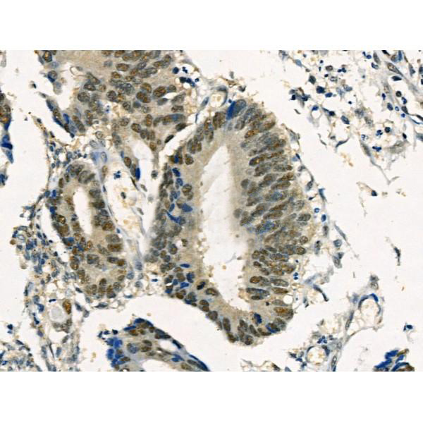
 IHC (Immunohistchemistry)
(Immunohistochemistry of paraffin-embedded human rectum using CDX2 antibody at dilution of 1:100 (40x lens).)
IHC (Immunohistchemistry)
(Immunohistochemistry of paraffin-embedded human rectum using CDX2 antibody at dilution of 1:100 (40x lens).)
CDX2, Polyclonal Antibody (Cat# AAA10710)
Full Name
CDX2 Polyclonal Antibody
Gene Names
CDX2; CDX3; CDX-3; CDX2/AS
Reactivity
Human, Mouse, Rat
Applications
Western Blot, Immunohistochemistry
Purity
Affinity Purification
Pricing


 ICC (Immunocytochemistry)
(ICC staining Histone H4 (acetyl K5) in SH-SY-5Y cells (green). Cells were fixed in paraformaldehyde, permeabilised with 0.25% Triton X100/PBS.)
ICC (Immunocytochemistry)
(ICC staining Histone H4 (acetyl K5) in SH-SY-5Y cells (green). Cells were fixed in paraformaldehyde, permeabilised with 0.25% Triton X100/PBS.)
Histone H4, Monoclonal Antibody (Cat# AAA30536)
Full Name
Histone H4 (Acetyl K5) Antibody
Gene Names
HIST2H4B; H4/o
Reactivity
Human, Mouse, Rat
Applications
Western Blot, Immunocytochemistry, Immunofluorescence, Immunohistochemistry, Immunoprecipitation
Purity
ProA affinity purified
Pricing
 Standard Curve (Sample)
Cortisol (COR), ELISA Kit (Cat# AAA11130)
Full Name
Porcine Cortisol (COR) ELISA Kit
Reactivity
Porcine
Pricing


 Application Data
(Staining of CD209 transfected K562 cells with Mouse anti Human CD209: Biotin)
Application Data
(Staining of CD209 transfected K562 cells with Mouse anti Human CD209: Biotin)
CD209, Monoclonal Antibody (Cat# AAA12042)
Full Name
MOUSE ANTI HUMAN CD209:RPE
Gene Names
CD209; CDSIGN; CLEC4L; DC-SIGN; DC-SIGN1
Applications
Flow Cytometry
Pricing


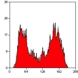
 FCM (Flow Cytometry)
(Flow cytometric analysis of Jurkat cells with MCL1 antibody at 1/50 dilution (blue) compared with an unlabelled control (cells without incubation with primary antibody; red). Alexa Fluor 488-conjugated goat anti rabbit IgG was used as the secondary antibody.)
FCM (Flow Cytometry)
(Flow cytometric analysis of Jurkat cells with MCL1 antibody at 1/50 dilution (blue) compared with an unlabelled control (cells without incubation with primary antibody; red). Alexa Fluor 488-conjugated goat anti rabbit IgG was used as the secondary antibody.)
MCL1, Monoclonal Antibody (Cat# AAA30035)
Full Name
MCL1 Antibody
Gene Names
MCL1; TM; EAT; MCL1L; MCL1S; Mcl-1; BCL2L3; MCL1-ES; bcl2-L-3; mcl1/EAT
Reactivity
Human, Mouse, Rat
Applications
Western Blot, Immunocytochemistry, Immunofluorescence, Immunohistochemistry, Immunoprecipitation, Flow Cytometry
Purity
ProA affinity purified
Pricing


 FCM (Flow Cytometry)
(Flow cytometric analysis of Hela cells with Bcl-XL antibody at 1/50 dilution (red) compared with an unlabelled control (cells without incubation with primary antibody; black). Alexa Fluor 488-conjugated goat anti rabbit IgG was used as the secondary antibody.)
FCM (Flow Cytometry)
(Flow cytometric analysis of Hela cells with Bcl-XL antibody at 1/50 dilution (red) compared with an unlabelled control (cells without incubation with primary antibody; black). Alexa Fluor 488-conjugated goat anti rabbit IgG was used as the secondary antibody.)
BCL-XL, Monoclonal Antibody (Cat# AAA30008)
Full Name
BCL-XL Antibody
Gene Names
BCL2L1; BCLX; BCL2L; BCLXL; BCLXS; Bcl-X; bcl-xL; bcl-xS; PPP1R52; BCL-XL/S
Reactivity
Human, Mouse, Rat
Applications
Western Blot, Immunocytochemistry, Immunofluorescence, Immunohistochemistry, Immunoprecipitation, Flow Cytometry
Purity
ProA affinity purified
Pricing


 IF (Immunofluorescence)
(Immunofluorescence analysis of A549 cells using BRCA1 antibody.)
IF (Immunofluorescence)
(Immunofluorescence analysis of A549 cells using BRCA1 antibody.)
BRCA1, Polyclonal Antibody (Cat# AAA10639)
Full Name
BRCA1 Polyclonal Antibody
Gene Names
BRCA1; IRIS; PSCP; BRCAI; BRCC1; FANCS; PNCA4; RNF53; BROVCA1; PPP1R53
Reactivity
Human, Mouse
Applications
Western Blot, Immunohistochemistry, Immunofluorescence
Purity
Affinity Purification
Pricing


 Application Data
(Staining of CD209 transfected K562 cells with Mouse anti Human CD209: Biotin)
Application Data
(Staining of CD209 transfected K562 cells with Mouse anti Human CD209: Biotin)
CD209, Monoclonal Antibody (Cat# AAA12140)
Full Name
MOUSE ANTI HUMAN CD209:FITC
Gene Names
CD209; CDSIGN; CLEC4L; DC-SIGN; DC-SIGN1
Applications
Flow Cytometry
Pricing


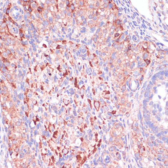
 IHC (Immunohistchemistry)
(Formalin-fixed and paraffin-embedded human breast carcinoma tissue reacted with MMP9 antibody (C-term) , which was peroxidase-conjugated to the secondary antibody, followed by DAB staining. This data demonstrates the use of this antibody for immunohistochemistry; clinical relevance has not been evaluated.)
IHC (Immunohistchemistry)
(Formalin-fixed and paraffin-embedded human breast carcinoma tissue reacted with MMP9 antibody (C-term) , which was peroxidase-conjugated to the secondary antibody, followed by DAB staining. This data demonstrates the use of this antibody for immunohistochemistry; clinical relevance has not been evaluated.)
MMP9, Polyclonal Antibody (Cat# AAA28681)
Full Name
MMP9 Antibody (C-term)
Gene Names
MMP9; GELB; CLG4B; MMP-9; MANDP2
Reactivity
Human, mouse
Applications
Western Blot, Immunohistochemistry
Purity
Purified Rabbit Polyclonal Antibody (Pab)
Pricing


 Application Data
(At 25 degree C. Samples were then incubated with primary Ab(At 37 degree C. An AlexaFluor594 conjugated goat anti-rabbit IgG(H+L) Ab(Red) and an AlexaFluor488 conjugated goat anti-mouse IgG(H+L) Ab(Green) were used as the secondary antibody.The nuclear counter stain is DAPI(blue).)
Application Data
(At 25 degree C. Samples were then incubated with primary Ab(At 37 degree C. An AlexaFluor594 conjugated goat anti-rabbit IgG(H+L) Ab(Red) and an AlexaFluor488 conjugated goat anti-mouse IgG(H+L) Ab(Green) were used as the secondary antibody.The nuclear counter stain is DAPI(blue).)
H2B, Polyclonal Antibody (Cat# AAA31348)
Full Name
Acetyl-H2B (Lys5) Antibody
Reactivity
Human, Mouse, Rat
Applications
Western Blot, Immunohistochemistry, Immunofluorescence, Immunocytochemistry, Peptide ELISA
Purity
The antiserum was purified by peptide affinity chromatography using SulfoLink Coupling Resin
Pricing
 Standard Curve (Sample)
Luteinizing Hormone-Releasing Hormone, LHRH, ELISA Kit (Cat# AAA11097)
Full Name
Mouse Luteinizing Hormone-Releasing Hormone, LHRH ELISA Kit
Gene Names
Gnrh1; Gnrh; Lhrh; SH-4; Gnrha; Rgnrhg1
Reactivity
Mouse
Pricing
 Standard Curve (Sample)
C terminal Telopeptides of type l collagen (ICTP), ELISA Kit (Cat# AAA16395)
Full Name
Rabbit C terminal Telopeptides of type l collagen (ICTP) ELISA Kit
Reactivity
Rabbit
Pricing


 Application Data
(At 25 degree C. Samples were then incubated with primary Ab(At 37 degree C. An AlexaFluor594 conjugated goat anti-rabbit IgG(H+L) Ab(Red) and an AlexaFluor488 conjugated goat anti-mouse IgG(H+L) Ab(Green) were used as the secondary antibody.The nuclear counter stain is DAPI (blue).)
Application Data
(At 25 degree C. Samples were then incubated with primary Ab(At 37 degree C. An AlexaFluor594 conjugated goat anti-rabbit IgG(H+L) Ab(Red) and an AlexaFluor488 conjugated goat anti-mouse IgG(H+L) Ab(Green) were used as the secondary antibody.The nuclear counter stain is DAPI (blue).)
SNAIL, Polyclonal Antibody (Cat# AAA31310)
Full Name
Phospho-SNAIL (Ser11) Antibody
Gene Names
SNAI1; SNA; SNAH; SNAIL; SLUGH2; SNAIL1; dJ710H13.1
Reactivity
Human, Mouse, Rat
Applications
Western Blot, Immunohistochemistry, Immunofluorescence, Immunocytochemistry, Peptide ELISA
Purity
The antibody is from purified rabbit serum by affinity purification via sequential chromatography on phospho-peptide and non-phospho-peptide affinity columns.
Pricing