Filters
Clonality
Type
Reactivity
Gene Name
Isotype
Host
Application
Clone
455 results for " T cells" - showing 50-100


 Application Data
(Immunofluorescesence staining of rat lymph node cryosection with Mouse anti Rat CD8beta, clone 341 , red in A and Mouse anti Rat anti Mouse CD4 antibody, clone W3/25 , green in B. C is the merged picture C with nuclei counterstained blue using DAPI. Low power)
Application Data
(Immunofluorescesence staining of rat lymph node cryosection with Mouse anti Rat CD8beta, clone 341 , red in A and Mouse anti Rat anti Mouse CD4 antibody, clone W3/25 , green in B. C is the merged picture C with nuclei counterstained blue using DAPI. Low power)
CD8 BETA, Monoclonal Antibody (Cat# AAA12017)
Full Name
MOUSE ANTI RAT CD8 BETA
Gene Names
Cd8b; Cd8b1
Applications
Flow Cytometry, Immunoprecipitation
Pricing


 Application Data
(Staining of mouse peritoneal macrophages with Rat anti Mouse CD11b)
Application Data
(Staining of mouse peritoneal macrophages with Rat anti Mouse CD11b)
CD11b, Monoclonal Antibody (Cat# AAA12057)
Full Name
RAT ANTI MOUSE CD11b:RPE
Gene Names
Itgam; CR3; CR3A; MAC1; Cd11b; Ly-40; Mac-1; Mac-1a; CD11b/CD18; F730045J24Rik
Applications
Flow Cytometry
Pricing


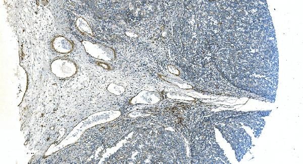
 Application Data
(Published customer image: Histological staining of control and HBOT wounds at post-wounding days 7 and 29. A -D) H&E staining. E -H) CD34 immunohistochemistry. I+J) CD68 immunohistochemistry.From: uk B, Tong M, Fijneman EMG, van Neck JW (2014) Hyperbaric Oxygen Therapy to Treat Diabetes Impaired Wound Healing in Rats. PLoS ONE 9(10): e108533.)
Application Data
(Published customer image: Histological staining of control and HBOT wounds at post-wounding days 7 and 29. A -D) H&E staining. E -H) CD34 immunohistochemistry. I+J) CD68 immunohistochemistry.From: uk B, Tong M, Fijneman EMG, van Neck JW (2014) Hyperbaric Oxygen Therapy to Treat Diabetes Impaired Wound Healing in Rats. PLoS ONE 9(10): e108533.)
CD68, Monoclonal Antibody (Cat# AAA12149)
Full Name
MOUSE ANTI RAT CD68
Applications
Immunohistochemistry, Flow Cytometry, Immunofluorescence, Immunoprecipitation, Immunohistochemistry
Pricing


 IHC (Immunohistchemistry)
(Immunohistochemistry of paraffin-embedded mouse spleen using IL2 antibody at dilution of 1:100 (40x lens).)
IHC (Immunohistchemistry)
(Immunohistochemistry of paraffin-embedded mouse spleen using IL2 antibody at dilution of 1:100 (40x lens).)
IL2, Polyclonal Antibody (Cat# AAA28327)
Full Name
IL2 Rabbit pAb
Gene Names
IL2; IL-2; TCGF; lymphokine
Reactivity
Human, Mouse, Rat
Applications
Western Blot, Immunohistochemistry
Purity
Affinity purification
Pricing


 ELISA
(Titration curve analysis of CTLA-4 mAbs to detect recombinant CTLA-4 in ELISA with antibodies at decreasing concentrations.)
ELISA
(Titration curve analysis of CTLA-4 mAbs to detect recombinant CTLA-4 in ELISA with antibodies at decreasing concentrations.)
CTLA-4, Monoclonal Antibody (Cat# AAA10982)
Full Name
CTLA-4 Antibody [1E6]
Gene Names
CTLA4; CD; GSE; GRD4; ALPS5; CD152; CTLA-4; IDDM12; CELIAC3
Reactivity
Human
Applications
Western Blot, Immunohistochemistry, Immunocytochemistry, Immunofluorescence
Purity
Protein A purified IgG1.
Pricing


 Application Data
(Staining of mouse splenocytes with Rat anti Mouse CD40: RPE)
Application Data
(Staining of mouse splenocytes with Rat anti Mouse CD40: RPE)
CD40, Monoclonal Antibody (Cat# AAA12023)
Full Name
RAT ANTI MOUSE CD40:RPE
Gene Names
Cd40; IGM; p50; Bp50; GP39; IMD3; TRAP; HIGM1; T-BAM; Tnfrsf5; AI326936
Applications
Flow Cytometry
Pricing


 Application Data
(Immunoperoxidase staining of mouse lymph node cryosection stained with Rat antii Mouse CD8alpha antibody, clone KT15 followed by horseradish peroxidase conjugatedGoat anti Rat IgG s a detection reagent. High power)
Application Data
(Immunoperoxidase staining of mouse lymph node cryosection stained with Rat antii Mouse CD8alpha antibody, clone KT15 followed by horseradish peroxidase conjugatedGoat anti Rat IgG s a detection reagent. High power)
CD8 ALPHA, Monoclonal Antibody (Cat# AAA12000)
Full Name
RAT ANTI MOUSE CD8 ALPHA
Gene Names
Cd8a; Ly-2; Ly-B; Ly-35; Lyt-2; BB154331
Applications
Immunohistochemistry, Flow Cytometry, Immunofluorescence
Pricing


 Application Data
(Published customer image: Increased accumulation of repair-associated macrophages surrounding collaterals in ischemic hind limbs is PAR2-dependent. (A) Stainings of CD206-positive macrophages (green) and SMA-positive vessels (red) in non-ischemic (control) and ischemic (ligated) hind limbs of WT, PAR1-/- and PAR2-/- mice are shown. Nuclei were visualized with DAPI (blue). Arrows indicate single macrophages in the non-ischemic adductor. Quantification of the average number of repair-associated macrophages per vessel is indicated on the right. (B) Correlation between the number of CD206-positive macrophages in the ischemic tissues and the expression of CD11b and (C) CD115 on monocytes. ** p)
Application Data
(Published customer image: Increased accumulation of repair-associated macrophages surrounding collaterals in ischemic hind limbs is PAR2-dependent. (A) Stainings of CD206-positive macrophages (green) and SMA-positive vessels (red) in non-ischemic (control) and ischemic (ligated) hind limbs of WT, PAR1-/- and PAR2-/- mice are shown. Nuclei were visualized with DAPI (blue). Arrows indicate single macrophages in the non-ischemic adductor. Quantification of the average number of repair-associated macrophages per vessel is indicated on the right. (B) Correlation between the number of CD206-positive macrophages in the ischemic tissues and the expression of CD11b and (C) CD115 on monocytes. ** p)
CD206, Monoclonal Antibody (Cat# AAA12120)
Full Name
RAT ANTI MOUSE CD206:FITC
Gene Names
Mrc1; MR; CD206; AW259686
Applications
Flow Cytometry
Pricing


 Application Data
(Staining of human peripheral blood lymphocytes with RAT ANTI HUMAN CD28:RPE)
Application Data
(Staining of human peripheral blood lymphocytes with RAT ANTI HUMAN CD28:RPE)
CD28, Monoclonal Antibody (Cat# AAA26793)
Full Name
CD28 (CD28 Antigen, CD28 Molecule, MGC138290, T cell Antigen CD28, T cell Specific Surface Glycoprotein, T cell Specific Surface Glycoprotein CD28, Tp44) (MaxLight 490)
Reactivity
Human
Applications
Flow Cytometry, Immunohistochemistry
Purity
Purified by Protein G Affinity Chromatography
Pricing


 Application Data
(Staining of mouse spleen with Rat anti Mouse CD4:RPE)
Application Data
(Staining of mouse spleen with Rat anti Mouse CD4:RPE)
CD4, Monoclonal Antibody (Cat# AAA12227)
Full Name
RAT ANTI MOUSE CD4
Gene Names
Cd4; L3T4; Ly-4
Applications
Immunohistochemistry, Flow Cytometry, Immunofluorescence, Immunoprecipitation, Western Blot
Pricing


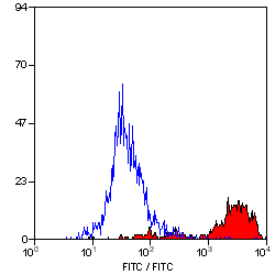
 FCM (Flow Cytometry)
(Flow cytometric analysis of Jurkat cells with NFAT1 antibody at 1/50 dilution (red) compared with an unlabelled control (cells without incubation with primary antibody; black). Alexa Fluor 488-conjugated goat anti rabbit IgG was used as the secondary antibody.)
FCM (Flow Cytometry)
(Flow cytometric analysis of Jurkat cells with NFAT1 antibody at 1/50 dilution (red) compared with an unlabelled control (cells without incubation with primary antibody; black). Alexa Fluor 488-conjugated goat anti rabbit IgG was used as the secondary antibody.)
NFAT1, Monoclonal Antibody (Cat# AAA30340)
Full Name
NFAT1 Antibody
Gene Names
NFATC2; NFAT1; NFATP
Reactivity
Human
Applications
Western Blot, Immunocytochemistry, Immunofluorescence, Immunohistochemistry, Flow Cytometry
Purity
ProA affinity purified
Pricing


 Application Data
(Staining of KG1 lymphocytes with Mouse anti Human CD59:FITC)
Application Data
(Staining of KG1 lymphocytes with Mouse anti Human CD59:FITC)
CD59, Monoclonal Antibody (Cat# AAA11916)
Full Name
MOUSE ANTI HUMAN CD59
Gene Names
CD59; 1F5; EJ16; EJ30; EL32; G344; MIN1; MIN2; MIN3; MIRL; HRF20; MACIF; MEM43; MIC11; MSK21; 16.3A5; HRF-20; MAC-IP; p18-20
Applications
Immunohistochemistry, Fluorescence Microscopy, Flow Cytometry, Immunofluorescence, Immunoprecipitation, Immunohistochemistry, Western Blot
Pricing


 Application Data
(Published Customer Image:Mouse CD31 antibody, clone ER-MP12 used for the demonstration of vasculature in mouse brain by immunofluorescence.Image caption:Inhibition of 2-AG hydrolysis reduces LPS-induced BBB permeability. a, b Fibrinogen levels in b plasma and the a ratio of brain to plasma fibrinogen were assessed by ELISA. n?=?5/7 mice per group. c, d Fluorescent immunostaining in the striatum for fibrinogen (red) and vascular marker (CD31; green) demonstrated leakage of fibrinogen into the brain with vehicle treatment, whereas vascular integrity was preserved when (e, f) MAGL was inhibited. g Extravascular fibrinogen was semi-quantitated in fluorescently labeled sections of the striatum. Bar graphs were plotted with mean?+/-SEM and data analyzed using one-way analysis of variance (ANOVA) with Tukey post-hoc comparisons. n = 5/7 mice per group. Significance is shown as *p?)
Application Data
(Published Customer Image:Mouse CD31 antibody, clone ER-MP12 used for the demonstration of vasculature in mouse brain by immunofluorescence.Image caption:Inhibition of 2-AG hydrolysis reduces LPS-induced BBB permeability. a, b Fibrinogen levels in b plasma and the a ratio of brain to plasma fibrinogen were assessed by ELISA. n?=?5/7 mice per group. c, d Fluorescent immunostaining in the striatum for fibrinogen (red) and vascular marker (CD31; green) demonstrated leakage of fibrinogen into the brain with vehicle treatment, whereas vascular integrity was preserved when (e, f) MAGL was inhibited. g Extravascular fibrinogen was semi-quantitated in fluorescently labeled sections of the striatum. Bar graphs were plotted with mean?+/-SEM and data analyzed using one-way analysis of variance (ANOVA) with Tukey post-hoc comparisons. n = 5/7 mice per group. Significance is shown as *p?)
CD31, Monoclonal Antibody (Cat# AAA12258)
Full Name
Rat Anti Mouse CD31: FITC
Gene Names
Pecam1; Cd31; Pecam; C85791; PECAM-1
Reactivity
Mouse
Applications
Flow Cytometry
Purity
Purified IgG prepared by affinity chromatography on Protein G from tissue culture supernatant
Pricing


 IF (Immunofluorescence)
(Immunofluorescence analysis of HeLa cells using CCT4 antibody. Blue: DAPI for nuclear staining.)
IF (Immunofluorescence)
(Immunofluorescence analysis of HeLa cells using CCT4 antibody. Blue: DAPI for nuclear staining.)
CCT4, Polyclonal Antibody (Cat# AAA10783)
Full Name
CCT4 Polyclonal Antibody
Gene Names
CCT4; SRB; Cctd; CCT-DELTA
Reactivity
Human, Mouse, Rat
Applications
Western Blot, Immunohistochemistry, Immunofluorescence
Purity
Affinity Purification
Pricing


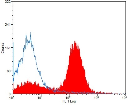
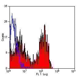
 FCM (Flow Cytometry)
(Flow cytometric analysis of Jurkat cells with CD137 antibody at 1/100 dilution (red) compared with an unlabelled control (cells without incubation with primary antibody; black).)
FCM (Flow Cytometry)
(Flow cytometric analysis of Jurkat cells with CD137 antibody at 1/100 dilution (red) compared with an unlabelled control (cells without incubation with primary antibody; black).)
CD137, Monoclonal Antibody (Cat# AAA29865)
Full Name
CD137 Antibody
Gene Names
TNFRSF9; ILA; 4-1BB; CD137; CDw137
Reactivity
Human, Mouse, Rat
Applications
Western Blot, Immunohistochemistry, Immunocytochemistry, Flow Cytometry
Purity
ProA affinity purified
Pricing


 Application Data
(Staining of mouse peritoneal macrophages with Rat anti Mouse CD11b)
Application Data
(Staining of mouse peritoneal macrophages with Rat anti Mouse CD11b)
CD11b, Monoclonal Antibody (Cat# AAA11889)
Full Name
RAT ANTI MOUSE CD11b:FITC
Gene Names
Itgam; CR3; CR3A; MAC1; Cd11b; Ly-40; Mac-1; Mac-1a; CD11b/CD18; F730045J24Rik
Applications
Flow Cytometry
Pricing


 FCM (Flow Cytometry)
(Flow cytometric analysis of K562 cells with ILF3 antibody at 1/100 dilution (purple) compared with an unlabelled control (cells without incubation with primary antibody; yellow). Alexa Fluor 488-conjugated goat anti-rabbit IgG was used as the secondary antibody.)
FCM (Flow Cytometry)
(Flow cytometric analysis of K562 cells with ILF3 antibody at 1/100 dilution (purple) compared with an unlabelled control (cells without incubation with primary antibody; yellow). Alexa Fluor 488-conjugated goat anti-rabbit IgG was used as the secondary antibody.)
ILF3, Monoclonal Antibody (Cat# AAA30498)
Full Name
ILF3 Antibody
Gene Names
ILF3; CBTF; DRBF; MMP4; MPP4; NF90; NFAR; NF110; NF90a; NF90b; NFAR2; TCP80; DRBP76; NF110b; NFAR-1; TCP110; MPHOSPH4; NF-AT-90
Reactivity
Human, Mouse, Rat
Applications
Western Blot, Immunoprecipitation, Immunocytochemistry, Immunofluorescence, Immunohistochemistry, Flow Cytometry
Purity
ProA affinity purified
Pricing


 WB (Western Blot)
(HSPA1B monoclonal antibody Western Blot analysis of HSPA1B expression in Raw 264.7.)
WB (Western Blot)
(HSPA1B monoclonal antibody Western Blot analysis of HSPA1B expression in Raw 264.7.)
HSPA1A, Monoclonal Antibody (Cat# AAA25423)
Full Name
HSPA1A (Heat Shock 70kD Protein 1A/1B, Heat Shock 70kD Protein 1/2, HSP70.1/HSP70.2, HSP70-1/HSP70-2, HSPA1, HSPA1B) (HRP)
Gene Names
HSPA1A; HSP72; HSPA1; HSP70I; HSP70-1; HSP70.1; HSP70-1A; HEL-S-103
Reactivity
Human, Mouse, Rat
Applications
Immunohistochemistry, Western Blot
Purity
Purified by Protein A Affinity Chromatography.
Pricing


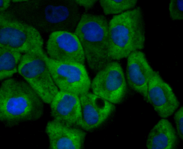
 IF (Immunofluorescence)
(Figure 11. IF analysis of H Cadherin/CDH13 using anti- H Cadherin/CDH13 antibody (AAA19252).H Cadherin/CDH13 was detected in immunocytochemical section of SIHA cells. Enzyme antigen retrieval was performed using IHC enzyme antigen retrieval reagent for 15 mins. The cells were blocked with 10% goat serum. And then incubated with 5μg/mL rabbit anti- H Cadherin/CDH13 Antibody (AAA19252) overnight at 4 degree C. DyLight®488 Conjugated Goat Anti-Rabbit IgG was used as secondary antibody at 1:100 dilution and incubated for 30 minutes at 37 degree C. The section was counterstained with DAPI. Visualize using a fluorescence microscope and filter sets appropriate for the label used.)
IF (Immunofluorescence)
(Figure 11. IF analysis of H Cadherin/CDH13 using anti- H Cadherin/CDH13 antibody (AAA19252).H Cadherin/CDH13 was detected in immunocytochemical section of SIHA cells. Enzyme antigen retrieval was performed using IHC enzyme antigen retrieval reagent for 15 mins. The cells were blocked with 10% goat serum. And then incubated with 5μg/mL rabbit anti- H Cadherin/CDH13 Antibody (AAA19252) overnight at 4 degree C. DyLight®488 Conjugated Goat Anti-Rabbit IgG was used as secondary antibody at 1:100 dilution and incubated for 30 minutes at 37 degree C. The section was counterstained with DAPI. Visualize using a fluorescence microscope and filter sets appropriate for the label used.)
H Cadherin/CDH13, Polyclonal Antibody (Cat# AAA19252)
Full Name
Anti-H Cadherin/CDH13 Antibody
Gene Names
CDH13; CDHH; P105
Reactivity
Human, Rat
Applications
Western Blot, Immunohistochemistry, Immunocytochemistry, Immunofluorescence, Flow Cytometry, Direct ELISA
Purity
Immunogen affinity purified.
Pricing


 Application Data
(Publised customer image:Mouse anti Human CD163 antibody, clone EDHu-1 used for the identification of perivascular macrophages in human brain by immunofluorescence.Image caption:Images demonstrating immunohistological stainings of amylin and double immunofluorescence staining against NG2/amylin, laminin/amylin and CD163/amylin in the hippocampus of the patient with AD and T2D.Amylin cell inclusions are indicated with arrows in (a) and shown in a higher magnification in (b). Pericytes with round cell bodies and NG2-positive coverage of the microvessel surface (green in c), without amylin cell inclusions (red in d), displayed round DAPI-positive cell nuclei (blue in e). The images in (c), (d) and (e) are merged in (f). Cells with more diffuse and weak NG2 staining (indicated by the arrowhead, green in g) and cytosolic amylin cell inclusions (red in h) showed altered cell nuclei (indicated with arrow, blue in i).The adjacent unaffected NG2-positive cell is indicated with an arrowhead in (i). The images in (g), (h) and (i) are merged in (j). Cells enclosed by laminin (green in K) contained amylin grains (red in l) and fragmented DAPI-positive cell nuclei (indicated with an arrow blue in m). The images in (k), (l) and (m) are merged in (n).Loss of NG2 coverage (green in o) was associated with polarized amylin cell inclusion (red in p) and fragmented DAPI-positive cell nuclei (indicated with an arrow, blue in q). The images in (o), (p) and (q) are merged in (r). Staining against macrophage marker CD163 (green in s) did not co-localize with amylin cell inclusions (red in t). The cell nucleus was stained with DAPI (blue in U). The images in (s), (t) and (u) are merged in (v). Scale bars: (a) 50 mum, (b), (c) to (v) 5 mum.From: Schultz, N. et al. (2016).Amylin alters human brain pericyte viability and NG2 expression.J Cereb Blood Flow & Metab. Jun 28 [Epub ahead of print]This is from an open access article distributed under the terms of the Creative Commons Attribution License.)
Application Data
(Publised customer image:Mouse anti Human CD163 antibody, clone EDHu-1 used for the identification of perivascular macrophages in human brain by immunofluorescence.Image caption:Images demonstrating immunohistological stainings of amylin and double immunofluorescence staining against NG2/amylin, laminin/amylin and CD163/amylin in the hippocampus of the patient with AD and T2D.Amylin cell inclusions are indicated with arrows in (a) and shown in a higher magnification in (b). Pericytes with round cell bodies and NG2-positive coverage of the microvessel surface (green in c), without amylin cell inclusions (red in d), displayed round DAPI-positive cell nuclei (blue in e). The images in (c), (d) and (e) are merged in (f). Cells with more diffuse and weak NG2 staining (indicated by the arrowhead, green in g) and cytosolic amylin cell inclusions (red in h) showed altered cell nuclei (indicated with arrow, blue in i).The adjacent unaffected NG2-positive cell is indicated with an arrowhead in (i). The images in (g), (h) and (i) are merged in (j). Cells enclosed by laminin (green in K) contained amylin grains (red in l) and fragmented DAPI-positive cell nuclei (indicated with an arrow blue in m). The images in (k), (l) and (m) are merged in (n).Loss of NG2 coverage (green in o) was associated with polarized amylin cell inclusion (red in p) and fragmented DAPI-positive cell nuclei (indicated with an arrow, blue in q). The images in (o), (p) and (q) are merged in (r). Staining against macrophage marker CD163 (green in s) did not co-localize with amylin cell inclusions (red in t). The cell nucleus was stained with DAPI (blue in U). The images in (s), (t) and (u) are merged in (v). Scale bars: (a) 50 mum, (b), (c) to (v) 5 mum.From: Schultz, N. et al. (2016).Amylin alters human brain pericyte viability and NG2 expression.J Cereb Blood Flow & Metab. Jun 28 [Epub ahead of print]This is from an open access article distributed under the terms of the Creative Commons Attribution License.)
CD163, Monoclonal Antibody (Cat# AAA12256)
Full Name
Mouse Anti Human CD163: RPE
Gene Names
CD163; M130; MM130; SCARI1
Reactivity
Human
Applications
Flow Cytometry
Pricing


 Application Data
(Published customer image Infiltration of GFP+ BM-cells in infarct and peri-infarct regions. (A-B) Dot plots of viable macrophages/granulocytes (CD11b+CD45high, top right quadrants) and microglia (CD11b+CD45dim, bottom right quadrants) in cortex from BM-chimeric unmanipulated mice and mice exposed to pMCAO. (C) Bar graph showing mean numbers of CD11b+CD45dim microglia and CD11b+CD45high macrophages/granulocytes in BM-chimeric mice 24 hours after pMCAO, subdivided based on expression of GFP (n = 5). Approximately 92% of of the CD45high population were GFP+. (D) Estimation and comparison of mean numbers of CD11b+CD45dim microglia in non-chimeric (n = 10) versus BM-chimeric mice (n = 5) 24 hours after of pMCAO shows significantly fewer CD11b+CD45dim microglial cells in irradiated mice. (E) Overview, showing distribution of infiltrating GFP+ BM-derived cells into infarct (IF) and peri-infarct (P-IF) regions 24 hours after pMCAO. (E-G) By 24 hours, GFP+ single cells (F) and vessel-associated aggregates of GFP+ cells (arrows in G) were observed in infarct and peri-infarct regions. Some of the vessel-associated cells were round, leukocyte-like cells (arrows) while others were elongated cells lining the vasculature (arrow heads in G and in insert). (H) Bar graph showing mean numbers of single GFP+ cells and vessel-associated aggregates of GFP+ cells in ipsi- and contralateral cortex 24 hours after surgery (n = 10). (I-P) Immunohistochemical staining of CD45.1 (I, K), CD45.2 (J, L), IgG2a (M, O) and CD45 (N, P) in ischemic tissue in BM-chimeric (I, J, M, N) and non-chimeric mice (K, L, O, P) 24 hours after pMCAO. N.D, none detected. Scale bars: 200 um (A), 10 um (B, C). 50 um (I-P) *P < 0.05, **P < 0.01, and ***P < 0.001.From: Clausen BH, Lambertsen KL, Babcock AA, Holm TH, Dagnaes-Hansen F, Finsen B. Interleukin-1beta and tumor necrosis factor-alpha are expressed by different subsets of microglia and macrophages after ischemic stroke in mice. J Neuroinflammation. 2008 Oct 23;5:46.)
Application Data
(Published customer image Infiltration of GFP+ BM-cells in infarct and peri-infarct regions. (A-B) Dot plots of viable macrophages/granulocytes (CD11b+CD45high, top right quadrants) and microglia (CD11b+CD45dim, bottom right quadrants) in cortex from BM-chimeric unmanipulated mice and mice exposed to pMCAO. (C) Bar graph showing mean numbers of CD11b+CD45dim microglia and CD11b+CD45high macrophages/granulocytes in BM-chimeric mice 24 hours after pMCAO, subdivided based on expression of GFP (n = 5). Approximately 92% of of the CD45high population were GFP+. (D) Estimation and comparison of mean numbers of CD11b+CD45dim microglia in non-chimeric (n = 10) versus BM-chimeric mice (n = 5) 24 hours after of pMCAO shows significantly fewer CD11b+CD45dim microglial cells in irradiated mice. (E) Overview, showing distribution of infiltrating GFP+ BM-derived cells into infarct (IF) and peri-infarct (P-IF) regions 24 hours after pMCAO. (E-G) By 24 hours, GFP+ single cells (F) and vessel-associated aggregates of GFP+ cells (arrows in G) were observed in infarct and peri-infarct regions. Some of the vessel-associated cells were round, leukocyte-like cells (arrows) while others were elongated cells lining the vasculature (arrow heads in G and in insert). (H) Bar graph showing mean numbers of single GFP+ cells and vessel-associated aggregates of GFP+ cells in ipsi- and contralateral cortex 24 hours after surgery (n = 10). (I-P) Immunohistochemical staining of CD45.1 (I, K), CD45.2 (J, L), IgG2a (M, O) and CD45 (N, P) in ischemic tissue in BM-chimeric (I, J, M, N) and non-chimeric mice (K, L, O, P) 24 hours after pMCAO. N.D, none detected. Scale bars: 200 um (A), 10 um (B, C). 50 um (I-P) *P < 0.05, **P < 0.01, and ***P < 0.001.From: Clausen BH, Lambertsen KL, Babcock AA, Holm TH, Dagnaes-Hansen F, Finsen B. Interleukin-1beta and tumor necrosis factor-alpha are expressed by different subsets of microglia and macrophages after ischemic stroke in mice. J Neuroinflammation. 2008 Oct 23;5:46.)
CD11b, Monoclonal Antibody (Cat# AAA12182)
Full Name
RAT ANTI MOUSE CD11b:FITC
Gene Names
Itgam; CR3; CR3A; MAC1; Cd11b; Ly-40; Mac-1; Mac-1a; CD11b/CD18; F730045J24Rik
Applications
Flow Cytometry
Pricing


 Application Data
(Staining of human peripheral blood lymphocytes with MOUSE ANTI HUMAN CD3:ALEXA 647)
Application Data
(Staining of human peripheral blood lymphocytes with MOUSE ANTI HUMAN CD3:ALEXA 647)
CD3, Monoclonal Antibody (Cat# AAA26718)
Full Name
CD3 (AP)
Reactivity
Human
Applications
Immunohistochemistry
Purity
Purified by Protein G Affinity Chromatography
Pricing


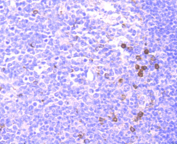
 IHC (Immunohistchemistry)
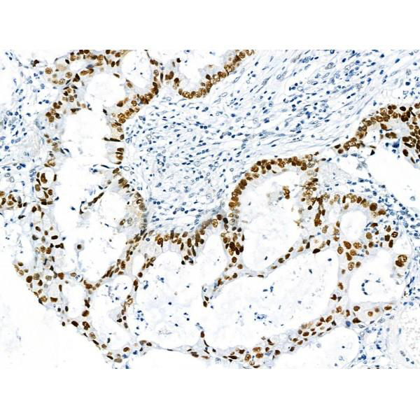
(Immunohistochemical analysis of paraffin-embedded colon cancer tissues using PBK mouse mAb with DAB staining.)
IHC (Immunohistchemistry)
(Immunohistochemical analysis of paraffin-embedded colon cancer tissues using PBK mouse mAb with DAB staining.)
PBK/TOPK, Monoclonal Antibody (Cat# AAA14113)
Full Name
Anti-PBK/TOPK Mouse mAb
Gene Names
PBK; SPK; CT84; TOPK; HEL164; Nori-3
Reactivity
Human, Rat
Applications
Western Blot
Pricing


 FCM (Flow Cytometry)
(Flow cytometric analysis of SH-SY-5Y cells with ILF2 antibody at 1/100 dilution (red) compared with an unlabelled control (cells without incubation with primary antibody; black). Alexa Fluor 488-conjugated goat anti rabbit IgG was used as the secondary antibody.)
FCM (Flow Cytometry)
(Flow cytometric analysis of SH-SY-5Y cells with ILF2 antibody at 1/100 dilution (red) compared with an unlabelled control (cells without incubation with primary antibody; black). Alexa Fluor 488-conjugated goat anti rabbit IgG was used as the secondary antibody.)
ILF2, Monoclonal Antibody (Cat# AAA30430)
Full Name
ILF2 Antibody
Gene Names
ILF2; NF45; PRO3063
Reactivity
Human, Mouse, Rat
Applications
Western Blot, Immunoprecipitation, Immunohistochemistry, Flow Cytometry
Purity
ProA affinity purified
Pricing


 Application Data
(Immunoperoxidase staining of mouse lymph node cryosection stained with Rat antii Mouse CD8alpha antibody, clone KT15 followed by horseradish peroxidase conjugatedGoat anti Rat IgG s a detection reagent. High power)
Application Data
(Immunoperoxidase staining of mouse lymph node cryosection stained with Rat antii Mouse CD8alpha antibody, clone KT15 followed by horseradish peroxidase conjugatedGoat anti Rat IgG s a detection reagent. High power)
CD8 ALPHA, Monoclonal Antibody (Cat# AAA12055)
Full Name
RAT ANTI MOUSE CD8 ALPHA:RPE
Gene Names
Cd8a; Ly-2; Ly-B; Ly-35; Lyt-2; BB154331
Applications
Flow Cytometry
Pricing


 Application Data
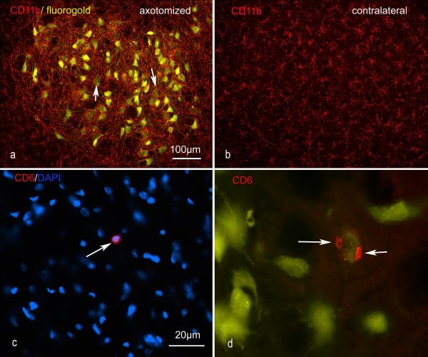
(Published customer image: Representative images of the inflammatory changes in the facial nucleus during axonal regeneration, one week following facial nerve transaction. a, b: CD11b immunoreactivity for microglia is increased in the axotomized facial nucleus, and microglia enwrap the facial motor neurons, e.g. at arrows. The regenerating neurons were retrogradely labelled with fluorogold. c, d: CD6- positive T-cells accumulated in the injured motor nucleus (arrows). They had little cytoplasm but dense nuclei (c) and were sometimes clustered around neurons retrogradely labelled with fluorogold (d). The scale bar in (a) also applies to (b) and that in (c) also applies to (d).From: Shokouhi et al. BMC Neuroscience 2010 11:13.)
Application Data
(Published customer image: Representative images of the inflammatory changes in the facial nucleus during axonal regeneration, one week following facial nerve transaction. a, b: CD11b immunoreactivity for microglia is increased in the axotomized facial nucleus, and microglia enwrap the facial motor neurons, e.g. at arrows. The regenerating neurons were retrogradely labelled with fluorogold. c, d: CD6- positive T-cells accumulated in the injured motor nucleus (arrows). They had little cytoplasm but dense nuclei (c) and were sometimes clustered around neurons retrogradely labelled with fluorogold (d). The scale bar in (a) also applies to (b) and that in (c) also applies to (d).From: Shokouhi et al. BMC Neuroscience 2010 11:13.)
CD11b, Monoclonal Antibody (Cat# AAA12146)
Full Name
MOUSE ANTI RAT CD11b:FITC
Gene Names
ITGAM; CD11B
Applications
Flow Cytometry
Pricing


 Application Data
(At 25 degree C. The primary antibody was diluted at 1/200 and incubated with the sample for 1 hour at 37 degree C. An Alexa Fluor 594 conjugated goat anti-rabbit IgG (H+L) Ab, diluted at 1/600, was used as the secondary antibody.)
Application Data
(At 25 degree C. The primary antibody was diluted at 1/200 and incubated with the sample for 1 hour at 37 degree C. An Alexa Fluor 594 conjugated goat anti-rabbit IgG (H+L) Ab, diluted at 1/600, was used as the secondary antibody.)
CD22, Polyclonal Antibody (Cat# AAA31418)
Full Name
Phospho-CD22 (Tyr842) Antibody
Gene Names
CD22; SIGLEC2; SIGLEC-2
Reactivity
Human, Mouse, Rat
Predicted Reactivity: Horse (83%)
Predicted Reactivity: Horse (83%)
Applications
Western Blot, Immunohistochemistry, Immunofluorescence, Immunocytochemistry, Peptide ELISA
Purity
The antibody is from purified rabbit serum by affinity purification via sequential chromatography on phospho-peptide and non-phospho-peptide affinity columns.
Pricing


 Application Data
(At 25 degree C. The primary antibody was diluted at 1/200 and incubated with the sample for 1 hour at 37 degree C. An Alexa Fluor 594 conjugated goat anti-rabbit IgG (H+L) Ab, diluted at 1/600, was used as the secondary antibody.)
Application Data
(At 25 degree C. The primary antibody was diluted at 1/200 and incubated with the sample for 1 hour at 37 degree C. An Alexa Fluor 594 conjugated goat anti-rabbit IgG (H+L) Ab, diluted at 1/600, was used as the secondary antibody.)
p53, Polyclonal Antibody (Cat# AAA31429)
Full Name
Phospho-p53 (Ser392) Antibody
Gene Names
TP53; P53; BCC7; LFS1; TRP53
Reactivity
Human, Mouse
Predicted Reactivity: Pig (88%), Bovine (88%), Sheep (88%), Rabbit (88%)
Predicted Reactivity: Pig (88%), Bovine (88%), Sheep (88%), Rabbit (88%)
Applications
Western Blot, Immunohistochemistry, Immunofluorescence, Immunocytochemistry, Peptide ELISA
Purity
The antibody is from purified rabbit serum by affinity purification via sequential chromatography on phospho-peptide and non-phospho-peptide affinity columns.
Pricing


 IHC (Immunohistochemistry)
(At 1/100 staining Human pancreatic cancer by IHC-P. The sample was formaldehyde fixed and a heat mediated antigen retrieval step in citrate buffer was performed. The sample was then blocked and incubated with the primary antibody at 4 degree C overnight. An HRP conjugated anti-Rabbit antibody was used as the secondary antibody.)
IHC (Immunohistochemistry)
(At 1/100 staining Human pancreatic cancer by IHC-P. The sample was formaldehyde fixed and a heat mediated antigen retrieval step in citrate buffer was performed. The sample was then blocked and incubated with the primary antibody at 4 degree C overnight. An HRP conjugated anti-Rabbit antibody was used as the secondary antibody.)
p53, Polyclonal Antibody (Cat# AAA31355)
Full Name
Acetyl-p53 (Lys381) Antibody
Gene Names
TP53; P53; BCC7; LFS1; TRP53
Reactivity
Human, Mouse, Rat
Predicted Reactivity: Rabbit (90%), Dog (100%)
Predicted Reactivity: Rabbit (90%), Dog (100%)
Applications
Western Blot, Immunohistochemistry, Peptide ELISA
Purity
The antiserum was purified by peptide affinity chromatography using SulfoLink Coupling Resin
Pricing


 Application Data
(Staining of human peripheral blood lymphocytes with MOUSE ANTI HUMAN CD3:ALEXA 647)
Application Data
(Staining of human peripheral blood lymphocytes with MOUSE ANTI HUMAN CD3:ALEXA 647)
CD3, Monoclonal Antibody (Cat# AAA26769)
Full Name
CD3 (PE)
Reactivity
Human
Applications
Flow Cytometry, Immunohistochemistry
Purity
Purified by Protein G Affinity Chromatography
Pricing


 ELISA
(Titration curve analysis of CTLA-4 mAbs to detect recombinant CTLA-4 in ELISA with antibodies at decreasing concentrations.)
ELISA
(Titration curve analysis of CTLA-4 mAbs to detect recombinant CTLA-4 in ELISA with antibodies at decreasing concentrations.)
CTLA-4, Monoclonal Antibody (Cat# AAA10984)
Full Name
CTLA-4 Antibody [8A1]
Gene Names
CTLA4; CD; GSE; GRD4; ALPS5; CD152; CTLA-4; IDDM12; CELIAC3
Reactivity
Human
Applications
Western Blot, Immunohistochemistry, Immunocytochemistry, Immunofluorescence
Purity
Protein A purified IgG1.
Pricing


 ELISA
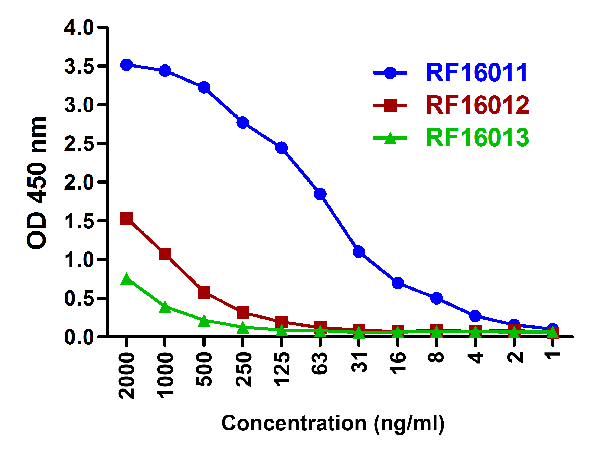
(Titration curve analysis of TIGIT mAbs to detect recombinant TIGIT in ELISA at decreasing concentrations.)
ELISA
(Titration curve analysis of TIGIT mAbs to detect recombinant TIGIT in ELISA at decreasing concentrations.)
TIGIT, Monoclonal Antibody (Cat# AAA11004)
Full Name
TIGIT Antibody [4A10]
Gene Names
TIGIT; VSIG9; VSTM3; WUCAM
Reactivity
Human
Applications
Western Blot, Immunohistochemistry, Immunocytochemistry, Immunofluorescence, Flow Cytometry
Purity
Protein A purified
Pricing


 IHC (Immunohistochemistry)
(At 1/100 staining Mouse kidney tissue by IHC-P. The sample was formaldehyde fixed and a heat mediated antigen retrieval step in citrate buffer was performed. The sample was then blocked and incubated with the primary antibody at 4 degree C overnight. An HRP conjugated anti-Rabbit antibody was used as the secondary antibody.)
IHC (Immunohistochemistry)
(At 1/100 staining Mouse kidney tissue by IHC-P. The sample was formaldehyde fixed and a heat mediated antigen retrieval step in citrate buffer was performed. The sample was then blocked and incubated with the primary antibody at 4 degree C overnight. An HRP conjugated anti-Rabbit antibody was used as the secondary antibody.)
p38 MAPK, Polyclonal Antibody (Cat# AAA31333)
Full Name
Phospho-p38 MAPK (Thr180/Tyr182) Antibody
Gene Names
MAPK14; RK; p38; CSBP; EXIP; Mxi2; CSBP1; CSBP2; CSPB1; PRKM14; PRKM15; SAPK2A; p38ALPHA
Reactivity
Human, Mouse, Rat
Predicted Reactivity: Pig (100%), Bovine (100%), Horse (100%), Sheep (100%), Rabbit (100%), Dog (100%), Xenopus (91%)
Predicted Reactivity: Pig (100%), Bovine (100%), Horse (100%), Sheep (100%), Rabbit (100%), Dog (100%), Xenopus (91%)
Applications
Western Blot, Immunohistochemistry, Immunofluorescence, Immunocytochemistry, Immunoprecipitation, Peptide ELISA
Purity
The antibody is from purified rabbit serum by affinity purification via sequential chromatography on phospho-peptide and non-phospho-peptide affinity columns.
Pricing


 IF (Immunofluorescence)
(Immunofluorescence of IL-17 in mouse thymus tissue with IL-17 Antibody at 20 μg/mL.)
IF (Immunofluorescence)
(Immunofluorescence of IL-17 in mouse thymus tissue with IL-17 Antibody at 20 μg/mL.)
IL-17, Polyclonal Antibody (Cat# AAA10926)
Full Name
IL-17 Antibody
Gene Names
IL17A; IL17; CTLA8; IL-17; IL-17A
Reactivity
Human, Mouse
Applications
Western Blot
Purity
IL-17 Antibody is affinity chromatography purified via peptide column.
Pricing


 Application Data
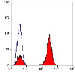
(Staining of mouse spleen cells with Rat anti Mouse CD3 Epsilon (T3): APC)
Application Data
(Staining of mouse spleen cells with Rat anti Mouse CD3 Epsilon (T3): APC)
CD3, Monoclonal Antibody (Cat# AAA12175)
Full Name
RAT ANTI MOUSE CD3
Gene Names
Cd3e; CD3; T3e; AI504783; CD3epsilon
Applications
Immunohistochemistry, Flow Cytometry
Pricing


 Application Data
(Staining of human peripheral blood granulocytes with Mouse anti Human CD45: Pacific Blue)
Application Data
(Staining of human peripheral blood granulocytes with Mouse anti Human CD45: Pacific Blue)
CD45, Monoclonal Antibody (Cat# AAA12202)
Full Name
MOUSE ANTI HUMAN CD45:FITC
Gene Names
PTPRC; LCA; LY5; B220; CD45; L-CA; T200; CD45R; GP180
Applications
Flow Cytometry
Pricing


 Application Data
(Immunoperoxidase staining of mouse lymph node cryosection stained with Rat antii Mouse CD8alpha antibody, clone KT15 followed by horseradish peroxidase conjugatedGoat anti Rat IgG s a detection reagent. High power)
Application Data
(Immunoperoxidase staining of mouse lymph node cryosection stained with Rat antii Mouse CD8alpha antibody, clone KT15 followed by horseradish peroxidase conjugatedGoat anti Rat IgG s a detection reagent. High power)
CD8 ALPHA, Monoclonal Antibody (Cat# AAA11885)
Full Name
RAT ANTI MOUSE CD8 ALPHA:FITC
Gene Names
Cd8a; Ly-2; Ly-B; Ly-35; Lyt-2; BB154331
Applications
Flow Cytometry
Pricing


 ELISA
(Titration curve analysis of VISTA antibody to detect recombinant VISTA in ELISA at decreasing concentrations.)
ELISA
(Titration curve analysis of VISTA antibody to detect recombinant VISTA in ELISA at decreasing concentrations.)
VISTA, Monoclonal Antibody (Cat# AAA11015)
Full Name
VISTA Antibody [9E4]
Gene Names
VSIR; B7H5; GI24; B7-H5; PD-1H; SISP1; VISTA; PP2135; C10orf54; DD1alpha
Reactivity
Human
Applications
Immunohistochemistry, Immunocytochemistry, Immunofluorescence, Flow Cytometry
Purity
Protein A purified
Pricing


 Application Data
(Published customer image Infiltration of GFP+ BM-cells in infarct and peri-infarct regions. (A-B) Dot plots of viable macrophages/granulocytes (CD11b+CD45high, top right quadrants) and microglia (CD11b+CD45dim, bottom right quadrants) in cortex from BM-chimeric unmanipulated mice and mice exposed to pMCAO. (C) Bar graph showing mean numbers of CD11b+CD45dim microglia and CD11b+CD45high macrophages/granulocytes in BM-chimeric mice 24 hours after pMCAO, subdivided based on expression of GFP (n = 5). Approximately 92% of of the CD45high population were GFP+. (D) Estimation and comparison of mean numbers of CD11b+CD45dim microglia in non-chimeric (n = 10) versus BM-chimeric mice (n = 5) 24 hours after of pMCAO shows significantly fewer CD11b+CD45dim microglial cells in irradiated mice. (E) Overview, showing distribution of infiltrating GFP+ BM-derived cells into infarct (IF) and peri-infarct (P-IF) regions 24 hours after pMCAO. (E-G) By 24 hours, GFP+ single cells (F) and vessel-associated aggregates of GFP+ cells (arrows in G) were observed in infarct and peri-infarct regions. Some of the vessel-associated cells were round, leukocyte-like cells (arrows) while others were elongated cells lining the vasculature (arrow heads in G and in insert). (H) Bar graph showing mean numbers of single GFP+ cells and vessel-associated aggregates of GFP+ cells in ipsi- and contralateral cortex 24 hours after surgery (n = 10). (I-P) Immunohistochemical staining of CD45.1 (I, K), CD45.2 (J, L), IgG2a (M, O) and CD45 (N, P) in ischemic tissue in BM-chimeric (I, J, M, N) and non-chimeric mice (K, L, O, P) 24 hours after pMCAO. N.D, none detected. Scale bars: 200 um (A), 10 um (B, C). 50 um (I-P) *P < 0.05, **P < 0.01, and ***P < 0.001.From: Clausen BH, Lambertsen KL, Babcock AA, Holm TH, Dagnaes-Hansen F, Finsen B. Interleukin-1beta and tumor necrosis factor-alpha are expressed by different subsets of microglia and macrophages after ischemic stroke in mice. J Neuroinflammation. 2008 Oct 23;5:46.)
Application Data
(Published customer image Infiltration of GFP+ BM-cells in infarct and peri-infarct regions. (A-B) Dot plots of viable macrophages/granulocytes (CD11b+CD45high, top right quadrants) and microglia (CD11b+CD45dim, bottom right quadrants) in cortex from BM-chimeric unmanipulated mice and mice exposed to pMCAO. (C) Bar graph showing mean numbers of CD11b+CD45dim microglia and CD11b+CD45high macrophages/granulocytes in BM-chimeric mice 24 hours after pMCAO, subdivided based on expression of GFP (n = 5). Approximately 92% of of the CD45high population were GFP+. (D) Estimation and comparison of mean numbers of CD11b+CD45dim microglia in non-chimeric (n = 10) versus BM-chimeric mice (n = 5) 24 hours after of pMCAO shows significantly fewer CD11b+CD45dim microglial cells in irradiated mice. (E) Overview, showing distribution of infiltrating GFP+ BM-derived cells into infarct (IF) and peri-infarct (P-IF) regions 24 hours after pMCAO. (E-G) By 24 hours, GFP+ single cells (F) and vessel-associated aggregates of GFP+ cells (arrows in G) were observed in infarct and peri-infarct regions. Some of the vessel-associated cells were round, leukocyte-like cells (arrows) while others were elongated cells lining the vasculature (arrow heads in G and in insert). (H) Bar graph showing mean numbers of single GFP+ cells and vessel-associated aggregates of GFP+ cells in ipsi- and contralateral cortex 24 hours after surgery (n = 10). (I-P) Immunohistochemical staining of CD45.1 (I, K), CD45.2 (J, L), IgG2a (M, O) and CD45 (N, P) in ischemic tissue in BM-chimeric (I, J, M, N) and non-chimeric mice (K, L, O, P) 24 hours after pMCAO. N.D, none detected. Scale bars: 200 um (A), 10 um (B, C). 50 um (I-P) *P < 0.05, **P < 0.01, and ***P < 0.001.From: Clausen BH, Lambertsen KL, Babcock AA, Holm TH, Dagnaes-Hansen F, Finsen B. Interleukin-1beta and tumor necrosis factor-alpha are expressed by different subsets of microglia and macrophages after ischemic stroke in mice. J Neuroinflammation. 2008 Oct 23;5:46.)
CD11b, Monoclonal Antibody (Cat# AAA12184)
Full Name
RAT ANTI MOUSE CD11b
Gene Names
Itgam; CR3; CR3A; MAC1; Cd11b; Ly-40; Mac-1; Mac-1a; CD11b/CD18; F730045J24Rik
Applications
Immunohistochemistry, Flow Cytometry, Immunofluorescence, Immunoprecipitation
Pricing


 ELISA
(Titration curve analysis of TIGIT mAbs to detect recombinant TIGIT in ELISA at decreasing concentrations.)
ELISA
(Titration curve analysis of TIGIT mAbs to detect recombinant TIGIT in ELISA at decreasing concentrations.)
TIGIT, Monoclonal Antibody (Cat# AAA11006)
Full Name
TIGIT Antibody [4A12]
Gene Names
TIGIT; VSIG9; VSTM3; WUCAM
Reactivity
Human
Applications
Western Blot, Immunohistochemistry, Immunocytochemistry, Immunofluorescence, Flow Cytometry
Purity
Protein A purified
Pricing


 Application Data
(Published customer image: Representative images of the inflammatory changes in the facial nucleus during axonal regeneration, one week following facial nerve transaction. a, b: CD11b immunoreactivity for microglia is increased in the axotomized facial nucleus, and microglia enwrap the facial motor neurons, e.g. at arrows. The regenerating neurons were retrogradely labelled with fluorogold. c, d: CD6- positive T-cells accumulated in the injured motor nucleus (arrows). They had little cytoplasm but dense nuclei (c) and were sometimes clustered around neurons retrogradely labelled with fluorogold (d). The scale bar in (a) also applies to (b) and that in (c) also applies to (d).From: Shokouhi et al. BMC Neuroscience 2010 11:13.)
Application Data
(Published customer image: Representative images of the inflammatory changes in the facial nucleus during axonal regeneration, one week following facial nerve transaction. a, b: CD11b immunoreactivity for microglia is increased in the axotomized facial nucleus, and microglia enwrap the facial motor neurons, e.g. at arrows. The regenerating neurons were retrogradely labelled with fluorogold. c, d: CD6- positive T-cells accumulated in the injured motor nucleus (arrows). They had little cytoplasm but dense nuclei (c) and were sometimes clustered around neurons retrogradely labelled with fluorogold (d). The scale bar in (a) also applies to (b) and that in (c) also applies to (d).From: Shokouhi et al. BMC Neuroscience 2010 11:13.)
CD11b, Monoclonal Antibody (Cat# AAA12045)
Full Name
MOUSE ANTI RAT CD11b:RPE
Gene Names
ITGAM; CD11B
Applications
Flow Cytometry
Pricing


 Application Data
(Published customer image: Representative images of the inflammatory changes in the facial nucleus during axonal regeneration, one week following facial nerve transaction. a, b: CD11b immunoreactivity for microglia is increased in the axotomized facial nucleus, and microglia enwrap the facial motor neurons, e.g. at arrows. The regenerating neurons were retrogradely labelled with fluorogold. c, d: CD6- positive T-cells accumulated in the injured motor nucleus (arrows). They had little cytoplasm but dense nuclei (c) and were sometimes clustered around neurons retrogradely labelled with fluorogold (d). The scale bar in (a) also applies to (b) and that in (c) also applies to (d).From: Shokouhi et al. BMC Neuroscience 2010 11:13.)
Application Data
(Published customer image: Representative images of the inflammatory changes in the facial nucleus during axonal regeneration, one week following facial nerve transaction. a, b: CD11b immunoreactivity for microglia is increased in the axotomized facial nucleus, and microglia enwrap the facial motor neurons, e.g. at arrows. The regenerating neurons were retrogradely labelled with fluorogold. c, d: CD6- positive T-cells accumulated in the injured motor nucleus (arrows). They had little cytoplasm but dense nuclei (c) and were sometimes clustered around neurons retrogradely labelled with fluorogold (d). The scale bar in (a) also applies to (b) and that in (c) also applies to (d).From: Shokouhi et al. BMC Neuroscience 2010 11:13.)
CD11b, Monoclonal Antibody (Cat# AAA11969)
Full Name
MOUSE ANTI RAT CD11b
Gene Names
ITGAM; CD11B
Applications
Immunohistochemistry, Flow Cytometry, Immunofluorescence, Immunoprecipitation
Pricing


 ChIP (Chromatin Immunoprecipitation)
(Chromatin immunoprecipitation analysis of extracts of HeLa cells, using Acetyl-Histone H3-K4 antibody and rabbit IgG.The amount of immunoprecipitated DNA was checked by quantitative PCR. Histogram was constructed by the ratios of the immunoprecipitated DNA to the input.)
ChIP (Chromatin Immunoprecipitation)
(Chromatin immunoprecipitation analysis of extracts of HeLa cells, using Acetyl-Histone H3-K4 antibody and rabbit IgG.The amount of immunoprecipitated DNA was checked by quantitative PCR. Histogram was constructed by the ratios of the immunoprecipitated DNA to the input.)
Acetyl-Histone H3-K4, Polyclonal Antibody (Cat# AAA28322)
Full Name
Acetyl-Histone H3-K4 Rabbit pAb
Gene Names
HIST3H3; H3t; H3.4; H3/g; H3FT
Reactivity
Human, Mouse, Rat, Other (Wide Range)
Applications
Western Blot, Immunofluorescence
Purity
Affinity purification
Pricing


 Application Data
(Immunoperoxidase staining of mouse lymph node cryosection stained with Rat antii Mouse CD8alpha antibody, clone KT15 followed by horseradish peroxidase conjugatedGoat anti Rat IgG s a detection reagent. High power)
Application Data
(Immunoperoxidase staining of mouse lymph node cryosection stained with Rat antii Mouse CD8alpha antibody, clone KT15 followed by horseradish peroxidase conjugatedGoat anti Rat IgG s a detection reagent. High power)
CD8 ALPHA, Monoclonal Antibody (Cat# AAA12218)
Full Name
RAT ANTI MOUSE CD8 ALPHA:APC
Gene Names
Cd8a; Ly-2; Ly-B; Ly-35; Lyt-2; BB154331
Applications
Flow Cytometry
Pricing

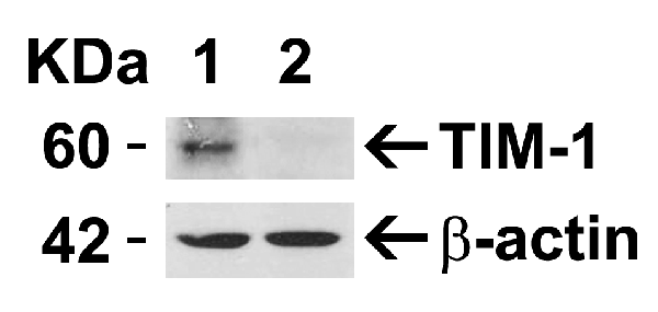


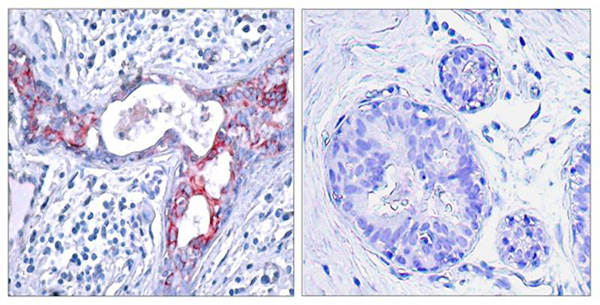
 IHC (Immunohistchemistry)
(Figure 5 Immunohistochemistry Validation of TIM-1Immunohistochemical analysis of paraffin-embedded human uterus tissue using anti-TIM-1 antibody (3809) at 10 μg/ml. Tissue was fixed with formaldehyde and blocked with 10% serum for 1 h at RT; antigen retrieval was by heat mediation with a citrate buffer (pH6). Samples were incubated with primary antibody overnight at 4˚ C. A goat anti-rabbit IgG H&L (HRP) at 1/250 was used as secondary. Counter stained with Hematoxylin.)
IHC (Immunohistchemistry)
(Figure 5 Immunohistochemistry Validation of TIM-1Immunohistochemical analysis of paraffin-embedded human uterus tissue using anti-TIM-1 antibody (3809) at 10 μg/ml. Tissue was fixed with formaldehyde and blocked with 10% serum for 1 h at RT; antigen retrieval was by heat mediation with a citrate buffer (pH6). Samples were incubated with primary antibody overnight at 4˚ C. A goat anti-rabbit IgG H&L (HRP) at 1/250 was used as secondary. Counter stained with Hematoxylin.)
TIM-1, Polyclonal Antibody (Cat# AAA10935)
Full Name
TIM-1 Antibody
Gene Names
HAVCR1; TIM; KIM1; TIM1; HAVCR; KIM-1; TIM-1; TIMD1; TIMD-1; HAVCR-1
Reactivity
Human, Mouse
Applications
Western Blot, Immunohistochemistry, Immunofluorescence
Purity
TIM-1 Antibody is affinity chromatography purified via peptide column.
Pricing


 IP (Immunoprecipitation)
(HSPA1A was immunoprecipitated using:Lane A:0.5 mg Hela Whole Cell Lysate2 uL anti-HSPA1A rabbit monoclonal antibody and 15 ul of 50 % Protein G agarose.Primary antibody:Anti-HSPA1A rabbit monoclonal antibody,at 1:200 dilution Secondary antibody:Dylight 800-labeled antibody to rabbit IgG (H+L), at 1:5000 dilution Developed using the odssey technique.Performed under reducing conditions.Predicted band size: 70 kDaObserved band size: 70 kDa)
IP (Immunoprecipitation)
(HSPA1A was immunoprecipitated using:Lane A:0.5 mg Hela Whole Cell Lysate2 uL anti-HSPA1A rabbit monoclonal antibody and 15 ul of 50 % Protein G agarose.Primary antibody:Anti-HSPA1A rabbit monoclonal antibody,at 1:200 dilution Secondary antibody:Dylight 800-labeled antibody to rabbit IgG (H+L), at 1:5000 dilution Developed using the odssey technique.Performed under reducing conditions.Predicted band size: 70 kDaObserved band size: 70 kDa)
HSP70, Monoclonal Antibody (Cat# AAA27747)
Full Name
Recombinant Anti-HSP70 Antibody, Rabbit Monoclonal
Gene Names
HSPA1B; HSP72; HSPA1; HSX70; HSP70-1; HSP70-2; HSP70.1; HSP70.2; HSP70-1B
Reactivity
Human
Applications
Western Blot, Immunohistochemistry, Flow Cytometry, Immunocytochemistry, Immunofluorescence, Immunoprecipitation
Purity
Protein A
Pricing


 WB (Western Blot)
(Western blot analysis of extracts from Hela cells, treated with EGF or calf intestinal phosphatase (CIP), using GSK3beta (Phospho-Ser9) Antibody.)
WB (Western Blot)
(Western blot analysis of extracts from Hela cells, treated with EGF or calf intestinal phosphatase (CIP), using GSK3beta (Phospho-Ser9) Antibody.)
GSK3beta, Polyclonal Antibody (Cat# AAA29709)
Full Name
GSK3beta (Phospho-Ser9) Antibody
Reactivity
Human, Mouse, Rat
Applications
Western Blot, Immunohistochemistry, Immunofluorescence
Purity
Antibodies were produced by immunizing rabbits with synthetic phosphopeptide and KLH conjugates. Antibodies were purified by affinity-chromatography using epitope-specific phosphopeptide. Non-phospho specific antibodies were removed by chromatogramphy usi
Pricing


 Application Data
(Staining of mouse peritoneal macrophages with Rat anti Mouse CD11b)
Application Data
(Staining of mouse peritoneal macrophages with Rat anti Mouse CD11b)
CD11b, Monoclonal Antibody (Cat# AAA12219)
Full Name
RAT ANTI MOUSE CD11b:Low Endotoxin
Gene Names
Itgam; CR3; CR3A; MAC1; Cd11b; Ly-40; Mac-1; Mac-1a; CD11b/CD18; F730045J24Rik
Applications
Immunohistochemistry, Flow Cytometry, Functional Assay, Immunofluorescence, Immunoprecipitation, Immunohistochemistry
Pricing


 WB (Western Blot)
(Sample: Recombinant PRF1, Human;Antibody: Rabbit Anti-Human PRF1 Ab)
WB (Western Blot)
(Sample: Recombinant PRF1, Human;Antibody: Rabbit Anti-Human PRF1 Ab)
Perforin 1 (PRF1), Active Protein (Cat# AAA21142)
Full Name
Active Perforin 1 (PRF1)
Gene Names
PRF1; P1; PFP; HPLH2
Reactivity
Homo sapiens (Human)
Applications
Cell culture; Activity Assays.
Purity
>80%
Pricing


 Application Data
(Staining of canine peripheral blood lymphocytes with Rat anti Canine CD4: Alexa Fluor 488)
Application Data
(Staining of canine peripheral blood lymphocytes with Rat anti Canine CD4: Alexa Fluor 488)
CD4, Monoclonal Antibody (Cat# AAA12064)
Full Name
RAT ANTI DOG CD4:FITC
Applications
Flow Cytometry
Pricing