Filters
Clonality
Type
Reactivity
Gene Name
Isotype
Host
Application
Clone
227 results for " Other Antibodies" - showing 50-100
Malaria Plasmodium Lactate Dehydrogenase (pLDH), Monoclonal Antibody (Cat# AAA13402)
Full Name
MAb TO MALARIA PLDH
Applications
ELISA
Purity
> or = 90% pure (SDS-PAGE). Protein A Chromatography
Pricing


 Application Data
(Published customer image: Increased accumulation of repair-associated macrophages surrounding collaterals in ischemic hind limbs is PAR2-dependent. (A) Stainings of CD206-positive macrophages (green) and SMA-positive vessels (red) in non-ischemic (control) and ischemic (ligated) hind limbs of WT, PAR1-/- and PAR2-/- mice are shown. Nuclei were visualized with DAPI (blue). Arrows indicate single macrophages in the non-ischemic adductor. Quantification of the average number of repair-associated macrophages per vessel is indicated on the right. (B) Correlation between the number of CD206-positive macrophages in the ischemic tissues and the expression of CD11b and (C) CD115 on monocytes. ** p)
Application Data
(Published customer image: Increased accumulation of repair-associated macrophages surrounding collaterals in ischemic hind limbs is PAR2-dependent. (A) Stainings of CD206-positive macrophages (green) and SMA-positive vessels (red) in non-ischemic (control) and ischemic (ligated) hind limbs of WT, PAR1-/- and PAR2-/- mice are shown. Nuclei were visualized with DAPI (blue). Arrows indicate single macrophages in the non-ischemic adductor. Quantification of the average number of repair-associated macrophages per vessel is indicated on the right. (B) Correlation between the number of CD206-positive macrophages in the ischemic tissues and the expression of CD11b and (C) CD115 on monocytes. ** p)
CD206, Monoclonal Antibody (Cat# AAA12121)
Full Name
RAT ANTI MOUSE CD206:FITC
Gene Names
Mrc1; MR; CD206; AW259686
Applications
Flow Cytometry
Pricing

 WB (Western Blot)
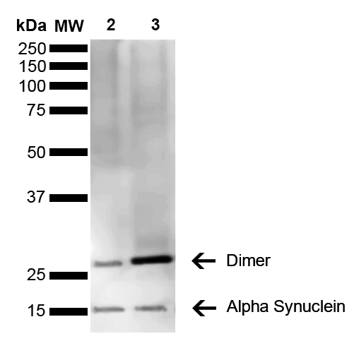
(Western Blot analysis of Mouse, Rat Brain showing detection of 14 kDa Alpha Synuclein protein using Mouse Anti-Alpha Synuclein Monoclonal Antibody, Clone 3C11. Lane 1: Molecular Weight Ladder (MW). Lane 2: Mouse brain cell lysate. Lane 3: Rat brain cell lysate. Load: 15 ug. Block: 5% Skim Milk in 1X TBST. Primary Antibody: Mouse Anti-Alpha Synuclein Monoclonal Antibody at 1:1000 for 2 hours at RT. Secondary Antibody: Goat Anti-Mouse HRP:IgG at 1:3000 for 1 hour at RT. Color Development: ECL solution (Super Signal West Pico) for 5 min in RT. Predicted/Observed Size: 14 kDa. Other Band(s): ~30 kDa (dimer).)
WB (Western Blot)
(Western Blot analysis of Mouse, Rat Brain showing detection of 14 kDa Alpha Synuclein protein using Mouse Anti-Alpha Synuclein Monoclonal Antibody, Clone 3C11. Lane 1: Molecular Weight Ladder (MW). Lane 2: Mouse brain cell lysate. Lane 3: Rat brain cell lysate. Load: 15 ug. Block: 5% Skim Milk in 1X TBST. Primary Antibody: Mouse Anti-Alpha Synuclein Monoclonal Antibody at 1:1000 for 2 hours at RT. Secondary Antibody: Goat Anti-Mouse HRP:IgG at 1:3000 for 1 hour at RT. Color Development: ECL solution (Super Signal West Pico) for 5 min in RT. Predicted/Observed Size: 14 kDa. Other Band(s): ~30 kDa (dimer).)
Alpha Synuclein, Monoclonal Antibody (Cat# AAA17810)
Full Name
Alpha Synuclein Antibody, Clone 3C11
Gene Names
SNCA; PD1; NACP; PARK1; PARK4
Reactivity
Human; Mouse; Rat
Applications
WB, DB, ICC, IF, EIA
Purity
Protein G Purified
Pricing


 IP (Immunoprecipitation)
(Immunoprecipitation analysis of 150ug extracts of MCF7 cells using 3ug HIST3H3 antibody. Western blot was performed from the immunoprecipitate using HIST3H3 antibody at a dilition of 1:500.)
IP (Immunoprecipitation)
(Immunoprecipitation analysis of 150ug extracts of MCF7 cells using 3ug HIST3H3 antibody. Western blot was performed from the immunoprecipitate using HIST3H3 antibody at a dilition of 1:500.)
HIST3H3, Polyclonal Antibody (Cat# AAA28284)
Full Name
HIST3H3 Polyclonal Antibody
Gene Names
HIST3H3; H3t; H3.4; H3/g; H3FT
Reactivity
Human, Mouse, Rat, Other (Wide Range)
Applications
Western Blot, Immunohistochemistry, Immunoprecipitation
Purity
Affinity Purification
Pricing

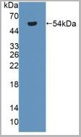
 WB (Western Blot)
(Western Blot detection against Immunogen using (AAA25718) (37kD).)
WB (Western Blot)
(Western Blot detection against Immunogen using (AAA25718) (37kD).)
HPSE, Monoclonal Antibody (Cat# AAA25718)
Full Name
HPSE (Heparanase, Endo-glucoronidase, Heparanase-1, Hpa1, HEP, HPA, HPA1, HPR1, HPSE1, HSE1) (PE)
Reactivity
Human
Applications
Western Blot
Purity
Purified by Protein A Affinity Chromatography.
Pricing


 Application Data
(C:FGFR2/isolectinB4 (C) and FGFR1/isolectinB4 (D) staining of apparent mesenchymal cells and the subpopulation of endothelial cells. Virtually all other dispersed apparent mesenchymal cells express FGFR1 and FGFR2 (merged image in E). F: FGFR2 (F) and FGFR1 (G) staining in clustered cells of epithelial origin (inferred by morphology here) demonstrating that epithelial cells express both FGFR1 and FGFR2 (merged image with DAPI staining in H).)
Application Data
(C:FGFR2/isolectinB4 (C) and FGFR1/isolectinB4 (D) staining of apparent mesenchymal cells and the subpopulation of endothelial cells. Virtually all other dispersed apparent mesenchymal cells express FGFR1 and FGFR2 (merged image in E). F: FGFR2 (F) and FGFR1 (G) staining in clustered cells of epithelial origin (inferred by morphology here) demonstrating that epithelial cells express both FGFR1 and FGFR2 (merged image with DAPI staining in H).)
FGFR2, Polyclonal Antibody (Cat# AAA14790)
Full Name
FGFR2, NT (FGFR2, BEK, KGFR, KSAM, Fibroblast growth factor receptor 2, K-sam, Keratinocyte growth factor receptor, CD332)
Gene Names
FGFR2; BEK; JWS; BBDS; CEK3; CFD1; ECT1; KGFR; TK14; TK25; BFR-1; CD332; K-SAM
Reactivity
Human, Monkey, Mouse, Rat
Applications
EL/EIA, WB, IHC, FC/FACS, IF
Purity
Affinity Purified
Purified by Protein A affinity chromatography.
Purified by Protein A affinity chromatography.
Pricing
 Standard Curve (Sample)
Antineutrophil cytoplasmic antibodies (ANCA), ELISA Kit (Cat# AAA12724)
Full Name
Rat Antineutrophil cytoplasmic antibodies (ANCA) ELISA Kit
Reactivity
Rat
Pricing

 Application Data
(Lane 1: 1x PBS was added at the sample hole (Negative);Lane 2: S-RBD protein ( 2.08mg/ml) with dilution at 1:100 (Positive);Lane 3: S-RBD protein ( 2.08mg/ml) with dilution at 1:1000 (Positive);Lane 4: S-RBD protein ( 2.08mg/ml) with dilution at 1:2000 (Positive);Lane 5: S-RBD protein ( 2.08mg/ml) with dilution at 1:4000 (Positive);Lane 6: S-RBD protein ( 2.08mg/ml) with dilution at 1:8000 (Positive);Lane 7: S-RBD protein ( 2.08mg/ml) with dilution at 1:16,000 (Positive);Lane 8: Lysis buffer (Negative);Lane 9: SARS-CoV-2 vaccinum which was purchased from other company (concentration is unknown).)
Application Data
(Lane 1: 1x PBS was added at the sample hole (Negative);Lane 2: S-RBD protein ( 2.08mg/ml) with dilution at 1:100 (Positive);Lane 3: S-RBD protein ( 2.08mg/ml) with dilution at 1:1000 (Positive);Lane 4: S-RBD protein ( 2.08mg/ml) with dilution at 1:2000 (Positive);Lane 5: S-RBD protein ( 2.08mg/ml) with dilution at 1:4000 (Positive);Lane 6: S-RBD protein ( 2.08mg/ml) with dilution at 1:8000 (Positive);Lane 7: S-RBD protein ( 2.08mg/ml) with dilution at 1:16,000 (Positive);Lane 8: Lysis buffer (Negative);Lane 9: SARS-CoV-2 vaccinum which was purchased from other company (concentration is unknown).)
COVID 19 Coronavirus, Reagent (Cat# AAA13552)
Full Name
SARS-CoV-2 Spike Antigen Rapid Test (Uncut pad)
Pricing


 Application Data
(C:FGFR2/isolectinB4 (C) and FGFR1/isolectinB4 (D) staining of apparent mesenchymal cells and the subpopulation of endothelial cells. Virtually all other dispersed apparent mesenchymal cells express FGFR1 and FGFR2 (merged image in E). F: FGFR2 (F) and FGFR1 (G) staining in clustered cells of epithelial origin (inferred by morphology here) demonstrating that epithelial cells express both FGFR1 and FGFR2 (merged image with DAPI staining in H).)
Application Data
(C:FGFR2/isolectinB4 (C) and FGFR1/isolectinB4 (D) staining of apparent mesenchymal cells and the subpopulation of endothelial cells. Virtually all other dispersed apparent mesenchymal cells express FGFR1 and FGFR2 (merged image in E). F: FGFR2 (F) and FGFR1 (G) staining in clustered cells of epithelial origin (inferred by morphology here) demonstrating that epithelial cells express both FGFR1 and FGFR2 (merged image with DAPI staining in H).)
FGFR2, Polyclonal Antibody (Cat# AAA26854)
Full Name
FGFR2, NT (FGFR2, BEK, KGFR, KSAM, Fibroblast growth factor receptor 2, K-sam, Keratinocyte growth factor receptor, CD332) (FITC)
Gene Names
FGFR2; BEK; JWS; BBDS; CEK3; CFD1; ECT1; KGFR; TK14; TK25; BFR-1; CD332; K-SAM
Reactivity
Human, Monkey, Mouse, Rat
Applications
WB, IHC, IF, FC/FACS
Purity
Purified by Protein G Affinity Chromatography.
Pricing


 Application Data
(C:FGFR2/isolectinB4 (C) and FGFR1/isolectinB4 (D) staining of apparent mesenchymal cells and the subpopulation of endothelial cells. Virtually all other dispersed apparent mesenchymal cells express FGFR1 and FGFR2 (merged image in E). F: FGFR2 (F) and FGFR1 (G) staining in clustered cells of epithelial origin (inferred by morphology here) demonstrating that epithelial cells express both FGFR1 and FGFR2 (merged image with DAPI staining in H).)
Application Data
(C:FGFR2/isolectinB4 (C) and FGFR1/isolectinB4 (D) staining of apparent mesenchymal cells and the subpopulation of endothelial cells. Virtually all other dispersed apparent mesenchymal cells express FGFR1 and FGFR2 (merged image in E). F: FGFR2 (F) and FGFR1 (G) staining in clustered cells of epithelial origin (inferred by morphology here) demonstrating that epithelial cells express both FGFR1 and FGFR2 (merged image with DAPI staining in H).)
FGFR2, Polyclonal Antibody (Cat# AAA26851)
Full Name
FGFR2, NT (FGFR2, BEK, KGFR, KSAM, Fibroblast growth factor receptor 2, K-sam, Keratinocyte growth factor receptor, CD332) (AP)
Gene Names
FGFR2; BEK; JWS; BBDS; CEK3; CFD1; ECT1; KGFR; TK14; TK25; BFR-1; CD332; K-SAM
Reactivity
Human, Monkey, Mouse, Rat
Applications
IF, EIA, IHC, WB
Purity
Purified by Protein G Affinity Chromatography.
Pricing


 Application Data
(Published customer image: Mouse anti Human CD49d antibody, clone HP2/1 used for binding efficiency determinationImage caption:Binding efficiencies (BE) of different a4beta7 molecules composed of distinct a4 mutants to monoclonal antibodies against a4, beta7 or the a4beta7 heterodimer. Binding efficiency is determined by the ratio between the mean fluorescence of antibody binding to each a4 molecule and of the binding in a mock-transfected cell culture (see Materials and Methods for details). Dark gray bars represent binding to the human (wild type) a4 clone, whereas light gray bars are those of binding to the different a4 mutants (as shown in the x-axis). a4 mutants which included substitutions at codon 201 are boxed. A, binding of anti-a4 2b4 antibody. B, binding of anti-a4 HP2/1 antibody. C, BE of different anti-a4 and beta7 antibodies to the human a4 and the quintuple a4 mutant (5 aa mut). Bars represent the range of standard errors deduced from triplicate experiments. p-values of Student's t tests are shown above each comparison. NS, non-significant (> 0.05).From:Darc M, Hait SH, Soares EA, Cicala C, Seuanez HN, et al. (2011) Polymorphisms in the a4 Integrin of Neotropical Primates: Insights for Binding of Natural Ligands and HIV-1 gp120 to the Human a4beta7. PLoS ONE 6(9): e24461.)
Application Data
(Published customer image: Mouse anti Human CD49d antibody, clone HP2/1 used for binding efficiency determinationImage caption:Binding efficiencies (BE) of different a4beta7 molecules composed of distinct a4 mutants to monoclonal antibodies against a4, beta7 or the a4beta7 heterodimer. Binding efficiency is determined by the ratio between the mean fluorescence of antibody binding to each a4 molecule and of the binding in a mock-transfected cell culture (see Materials and Methods for details). Dark gray bars represent binding to the human (wild type) a4 clone, whereas light gray bars are those of binding to the different a4 mutants (as shown in the x-axis). a4 mutants which included substitutions at codon 201 are boxed. A, binding of anti-a4 2b4 antibody. B, binding of anti-a4 HP2/1 antibody. C, BE of different anti-a4 and beta7 antibodies to the human a4 and the quintuple a4 mutant (5 aa mut). Bars represent the range of standard errors deduced from triplicate experiments. p-values of Student's t tests are shown above each comparison. NS, non-significant (> 0.05).From:Darc M, Hait SH, Soares EA, Cicala C, Seuanez HN, et al. (2011) Polymorphisms in the a4 Integrin of Neotropical Primates: Insights for Binding of Natural Ligands and HIV-1 gp120 to the Human a4beta7. PLoS ONE 6(9): e24461.)
CD49d, Monoclonal Antibody (Cat# AAA12002)
Full Name
MOUSE ANTI HUMAN CD49d
Gene Names
ITGA4; IA4; CD49D
Reactivity
Bovine, Cat, Cynomolgus monkey, Goat, Horse, Llama, Mink, Mustelid, Pig, Rabbit, Rat, Rhesus Monkey
Applications
Immunohistochemistry, Flow Cytometry, Functional Assay, Immunoprecipitation
Pricing
 Standard Curve (Sample)
CD59 glycoprotein, ELISA Kit (Cat# AAA17456)
Full Name
Human CD59 glycoprotein ELISA Kit
Gene Names
CD59; 1F5; EJ16; EJ30; EL32; G344; MIN1; MIN2; MIN3; MIRL; HRF20; MACIF; MEM43; MIC11; MSK21; 16.3A5; HRF-20; MAC-IP; p18-20
Reactivity
Human
Pricing
 Standard Curve (Sample)
Antineutrophil cytoplasmic antibodies (ANCA), ELISA Kit (Cat# AAA12865)
Full Name
Mouse Antineutrophil cytoplasmic antibodies (ANCA) ELISA Kit
Reactivity
Mouse
Pricing


 IF (Immunofluorescence)
(Immunofluorescent analysis of 4% paraformaldehyde-fixed, 0.1% Triton X-100 permeabilized U-2 OS (human osteosarcoma cell line) cells labeling DLL3 with (1:25), followed by Dylight 488- conjugated goat anti- rabbit IgG secondary antibody at (1:200) (green). Immunofluorescence image showing nucleus and weak cytoplasm staining on U-2 OScell line. Cytoplasmic actin is detected with Dylight 554 Phalloidin (1:100) (red).)
IF (Immunofluorescence)
(Immunofluorescent analysis of 4% paraformaldehyde-fixed, 0.1% Triton X-100 permeabilized U-2 OS (human osteosarcoma cell line) cells labeling DLL3 with (1:25), followed by Dylight 488- conjugated goat anti- rabbit IgG secondary antibody at (1:200) (green). Immunofluorescence image showing nucleus and weak cytoplasm staining on U-2 OScell line. Cytoplasmic actin is detected with Dylight 554 Phalloidin (1:100) (red).)
DLL3, Polyclonal Antibody (Cat# AAA26850)
Full Name
DLL3, CT (DLL3, Delta-like protein 3, Drosophila Delta homolog 3) (APC)
Gene Names
DLL3; SCDO1
Reactivity
Human
Applications
IF, IHC, WB
Purity
Purified by Protein A Affinity Chromatography.
Pricing

 Application Data
anti-Human IgM, Polyclonal Antibody (Cat# AAA14450)
Full Name
Affinity Purified Goat anti-Human IgM
Reactivity
Human
Purity
Antigen Affinity Purified
Pricing


 Application Data
(C:FGFR2/isolectinB4 (C) and FGFR1/isolectinB4 (D) staining of apparent mesenchymal cells and the subpopulation of endothelial cells. Virtually all other dispersed apparent mesenchymal cells express FGFR1 and FGFR2 (merged image in E). F: FGFR2 (F) and FGFR1 (G) staining in clustered cells of epithelial origin (inferred by morphology here) demonstrating that epithelial cells express both FGFR1 and FGFR2 (merged image with DAPI staining in H).)
Application Data
(C:FGFR2/isolectinB4 (C) and FGFR1/isolectinB4 (D) staining of apparent mesenchymal cells and the subpopulation of endothelial cells. Virtually all other dispersed apparent mesenchymal cells express FGFR1 and FGFR2 (merged image in E). F: FGFR2 (F) and FGFR1 (G) staining in clustered cells of epithelial origin (inferred by morphology here) demonstrating that epithelial cells express both FGFR1 and FGFR2 (merged image with DAPI staining in H).)
FGFR2, Polyclonal Antibody (Cat# AAA26856)
Full Name
FGFR2, NT (FGFR2, BEK, KGFR, KSAM, Fibroblast growth factor receptor 2, K-sam, Keratinocyte growth factor receptor, CD332) (PE)
Gene Names
FGFR2; BEK; JWS; BBDS; CEK3; CFD1; ECT1; KGFR; TK14; TK25; BFR-1; CD332; K-SAM
Reactivity
Human, Monkey, Mouse, Rat
Applications
WB, IHC, IF, FC/FACS
Purity
Purified by Protein G Affinity Chromatography.
Pricing


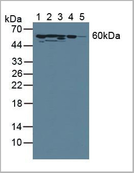
 WB (Western Blot)
(FG Pancreatic Carcinoma Cell Lines stably expressing vector along (FG-V) the b3 integrin subunit (FG-b3) or a b3 truncation mutant (FG-759x). Src Mab (AAA28639) was diluted 1:500 in 1% BSA/TBST and incubated Overnight at 4 degree C. After washing 3x 5 min. with TBST the blots were incubated with 1:5000 Goat anti-mouse or Goat anti-rabbit secondary antibody for 1 hr at Room temperature. The blots were again washed 3x 5 min. with TBST and developed using ECL reagent.Data and protocol kindly provided by Dr. Weis of Cheresh Lab, UCSD.)
WB (Western Blot)
(FG Pancreatic Carcinoma Cell Lines stably expressing vector along (FG-V) the b3 integrin subunit (FG-b3) or a b3 truncation mutant (FG-759x). Src Mab (AAA28639) was diluted 1:500 in 1% BSA/TBST and incubated Overnight at 4 degree C. After washing 3x 5 min. with TBST the blots were incubated with 1:5000 Goat anti-mouse or Goat anti-rabbit secondary antibody for 1 hr at Room temperature. The blots were again washed 3x 5 min. with TBST and developed using ECL reagent.Data and protocol kindly provided by Dr. Weis of Cheresh Lab, UCSD.)
SRC, Monoclonal Antibody (Cat# AAA28639)
Full Name
SRC Antibody
Gene Names
SRC; ASV; SRC1; c-SRC; p60-Src
Reactivity
Human, mouse
Applications
WB, EIA, IF
Purity
This antibody is purified through a protein G column, followed by dialysis against PBS.
Pricing


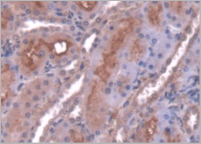
 IHC (Immunohistchemistry)
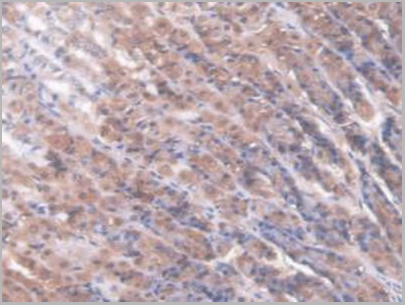
(Figure 6. IHC analysis of AMD1 using anti-AMD1 antibody (AAA19177).AMD1 was detected in paraffin-embedded section of human mammary cancer tissue. Heat mediated antigen retrieval was performed in citrate buffer (pH6, epitope retrieval solution) for 20 mins. The tissue section was blocked with 10% goat serum. The tissue section was then incubated with 1ug/ml rabbit anti-AMD1 Antibody (AAA19177) overnight at 4 degree C. Biotinylated goat anti-rabbit IgG was used as secondary antibody and incubated for 30 minutes at 37 degree C. The tissue section was developed using Strepavidin-Biotin-Complex (SABC) with DAB as the chromogen.)
IHC (Immunohistchemistry)
(Figure 6. IHC analysis of AMD1 using anti-AMD1 antibody (AAA19177).AMD1 was detected in paraffin-embedded section of human mammary cancer tissue. Heat mediated antigen retrieval was performed in citrate buffer (pH6, epitope retrieval solution) for 20 mins. The tissue section was blocked with 10% goat serum. The tissue section was then incubated with 1ug/ml rabbit anti-AMD1 Antibody (AAA19177) overnight at 4 degree C. Biotinylated goat anti-rabbit IgG was used as secondary antibody and incubated for 30 minutes at 37 degree C. The tissue section was developed using Strepavidin-Biotin-Complex (SABC) with DAB as the chromogen.)
AMD1/Adometdc, Polyclonal Antibody (Cat# AAA19177)
Full Name
Anti-AMD1/Adometdc Antibody
Gene Names
AMD1; AMD; SAMDC; ADOMETDC
Reactivity
Human, Mouse, Rat
No cross reactivity with other proteins.
No cross reactivity with other proteins.
Applications
IHC, WB
Purity
Immunogen affinity purified
Pricing


 WB (Western Blot)
(Western Blot Analysis of human U-87 cell lysate using Vimentin Rabbit Recombinant Monoclonal Antibody (VIM/1937R).)
WB (Western Blot)
(Western Blot Analysis of human U-87 cell lysate using Vimentin Rabbit Recombinant Monoclonal Antibody (VIM/1937R).)
Vimentin, Antibody (Cat# AAA23937)
Full Name
Vimentin (Mesenchymal Cell Marker)
Reactivity
Human
Applications
WB, IHC
Purity
Purified Ab with BSA and Azide at 200ug/ml OR Purified Ab WITHOUT BSA and Azide at 1.0mg/ml
Pricing
 Standard Curve (Sample)
Anti-Islet Cell Antibodies (ICA), ELISA Kit (Cat# AAA13025)
Full Name
Human Anti-Islet Cell Antibodies (ICA) ELISA Kit
Gene Names
1300017J02Rik; Ica; mICA
Reactivity
Human
Pricing


 Application Data
(C:FGFR2/isolectinB4 (C) and FGFR1/isolectinB4 (D) staining of apparent mesenchymal cells and the subpopulation of endothelial cells. Virtually all other dispersed apparent mesenchymal cells express FGFR1 and FGFR2 (merged image in E). F: FGFR2 (F) and FGFR1 (G) staining in clustered cells of epithelial origin (inferred by morphology here) demonstrating that epithelial cells express both FGFR1 and FGFR2 (merged image with DAPI staining in H).)
Application Data
(C:FGFR2/isolectinB4 (C) and FGFR1/isolectinB4 (D) staining of apparent mesenchymal cells and the subpopulation of endothelial cells. Virtually all other dispersed apparent mesenchymal cells express FGFR1 and FGFR2 (merged image in E). F: FGFR2 (F) and FGFR1 (G) staining in clustered cells of epithelial origin (inferred by morphology here) demonstrating that epithelial cells express both FGFR1 and FGFR2 (merged image with DAPI staining in H).)
FGFR2, Polyclonal Antibody (Cat# AAA26852)
Full Name
FGFR2, NT (FGFR2, BEK, KGFR, KSAM, Fibroblast growth factor receptor 2, K-sam, Keratinocyte growth factor receptor, CD332) (APC)
Gene Names
FGFR2; BEK; JWS; BBDS; CEK3; CFD1; ECT1; KGFR; TK14; TK25; BFR-1; CD332; K-SAM
Reactivity
Human, Monkey, Mouse, Rat
Applications
FC/FACS, IF, IHC, WB
Purity
Purified by Protein G Affinity Chromatography.
Pricing
HLA Class 1 Antigen A24,A11,A2403, Monoclonal Antibody (Cat# AAA14669)
Full Name
HLA Class 1 Antigen A24,A11,A2403 (X-Reactive)
Reactivity
Human
Applications
Flow Cytometry
Purity
Ascites
Pricing
Prostate Specific Antigen (PSA) Total, Monoclonal Antibody (Cat# AAA13377)
Full Name
MAb to PSA, Total
Gene Names
KLK3; APS; PSA; hK3; KLK2A1
Applications
Lateral Flow
Purity
>90% pure (SDS-PAGE). Protein A chromatography
Pricing
Goat anti Human IgG Fc, Secondary Antibody (Cat# AAA14316)
Full Name
Goat anti Human IgG Fc
Reactivity
Minimal cross-reaction to Bovine, Horse, and Mouse Serum Proteins.
Purity
Human IgG Fc antibody was purified by affinity chromatography.
Pricing
Insulin, Monoclonal Antibody (Cat# AAA13369)
Full Name
MAb to Insulin
Gene Names
INS; ILPR; IRDN; IDDM2; MODY10
Applications
Immunoassay, Radioimmunoassay
Purity
>90% pure (SDS-PAGE). Protein A chromatography
Pricing
HLA Class 1 Antigen B35,B57,B75,B77(X-Reactive), Monoclonal Antibody (Cat# AAA14681)
Full Name
HLA Class 1 Antigen B35,B57,B75,B77(X-Reactive)
Reactivity
Human
Applications
Cell Typing
Purity
Ascites
Pricing
Staphylococcus Enterotoxin B (SEB), Monoclonal Antibody (Cat# AAA13381)
Full Name
MAb to SEB
Applications
ELISA
Purity
>90% pure (SDS-PAGE). Protein A chromatography.
Product is 0.2um filtered.
Product is 0.2um filtered.
Pricing
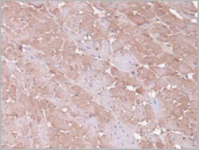
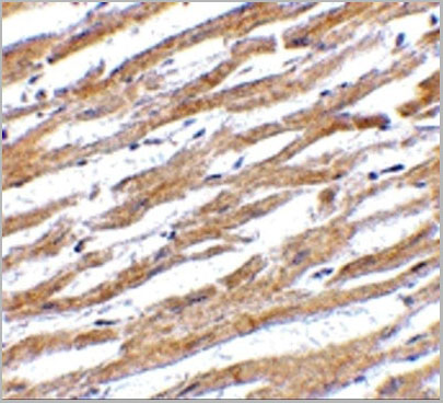
 IHC (Immunohistochemistry)
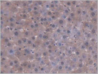
(DAB staining on IHC-P; Samples: Rat Small intestine Tissue; Primary Ab: 20ug/ml Rabbit Anti-Rat AT Antibody Second Ab: 2ug/mL HRP-Linked Caprine Anti-Rabbit IgG Polyclonal Antibody (Immunohistchemistry))
IHC (Immunohistochemistry)
(DAB staining on IHC-P; Samples: Rat Small intestine Tissue; Primary Ab: 20ug/ml Rabbit Anti-Rat AT Antibody Second Ab: 2ug/mL HRP-Linked Caprine Anti-Rabbit IgG Polyclonal Antibody (Immunohistchemistry))
Antithrombin, Polyclonal Antibody (Cat# AAA21014)
Full Name
Antithrombin (AT) Polyclonal Antibody
Reactivity
Rat
Applications
Westen Blot, Immunohistochemistry, Immunocytochemistry, Immunoprecipitation
Purity
Antigen-specific affinity chromatography followed by Protein A affinity chromatography
Pricing
Adenovirus, Hexon, Polyclonal Antibody (Cat# AAA14723)
Full Name
Adenovirus, Hexon
Applications
Immunohistochemistry, Immunofluorescence, Western Blot
Purity
Purified
Pricing
HLA Class 1 Antigen A11, Monoclonal Antibody (Cat# AAA14666)
Full Name
Anti-Human HLA Class 1 Antigen A11 (Biotin)
Applications
Flow Cytometry
Purity
Purified by Ion Exchange chromatography
Pricing
Wnt3 alpha, Polyclonal Antibody (Cat# AAA14338)
Full Name
Wnt3 alpha antibody
Gene Names
WNT3A; WNT3; WNT-3A
Reactivity
Human Wnt3
Applications
Western Blot
Pricing
Beta 2 Microglobulin, Monoclonal Antibody (Cat# AAA13382)
Full Name
MAb to Beta 2 Microglobulin
Gene Names
B2M; B2M
Reactivity
Human
Applications
Flow Cytometry, Immunoprecipitation, Radioimmunoassay, Western Blot
Purity
>95% pure (SDS-PAGE). Ion exchange chromatography and precipitation
Pricing
alpha-Gal Epitope, Monoclonal Antibody (Cat# AAA14679)
Full Name
alpha-Gal Epitope (Galalpha1-3Galbeta1-4GlcNAc-R)
Applications
Western Blot, Immunohistochemistry, Flow Cytometry
Purity
Supernatant
Pricing
Interleukin 6 Receptor, gp80, non-neutralizing, Monoclonal Antibody (Cat# AAA14678)
Full Name
Interleukin 6 Receptor, gp80, non-neutralizing (CD126, IL-6R)
Applications
Flow Cytometry
Purity
Purified
Pricing
Synaptosomal Protein 25kDa (SNAP-25) (a.a. 183-197) cleaved, Monoclonal Antibody (Cat# AAA13384)
Full Name
MAb to SNAP-25, cleaved
Applications
Immunohistochemistry
Purity
Product is sterile filtered.
Pricing
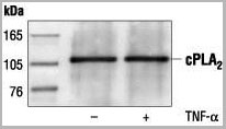
 WB (Western Blot)
(Western blot analysis of extracts from HeLa, C6 and NIH/3T3 cells, using AAA14717.)
WB (Western Blot)
(Western blot analysis of extracts from HeLa, C6 and NIH/3T3 cells, using AAA14717.)
c-Phospholipase A2, Polyclonal Antibody (Cat# AAA14717)
Full Name
c-Phospholipase A2 (Cytosolic PLA2, cPLA2)
Reactivity
Human
Applications
Western Blot, Immunoprecipitation
Purity
Purified by Protein A and peptide affinity chromatography
Pricing
Papillomavirus, Polyclonal Antibody (Cat# AAA13466)
Full Name
Papillomavirus
Reactivity
Crossreactive with HPV types 16 & 11 (other types not tested)
Applications
Immunohistochemistry
Pricing
IgM, Monoclonal Antibody (Cat# AAA13385)
Full Name
MAb to Human IgM
Reactivity
Human
Applications
Lateral Flow
Purity
Ion Exchange Chromatography
Pricing
Cortisol, Monoclonal Antibody (Cat# AAA13371)
Full Name
MAb to Cortisol
Purity
> 95% pure. Protein G Chromatography
Pricing
HLA Class 1 Antigen A11, Monoclonal Antibody (Cat# AAA14664)
Full Name
HLA Class 1 Antigen A11
Reactivity
Human
Applications
Flow Cytometry
Purity
Ascites
Pricing
Gram Positive Bacteria, Monoclonal Antibody (Cat# AAA13366)
Full Name
MAb to Gram Positive Bacteria
Applications
Immunofluorescence
Purity
>90% pure. Protein A chromatography
Pricing
Ferritin, Monoclonal Antibody (Cat# AAA13374)
Full Name
MAb to Human Ferritin
Reactivity
Human
Applications
Immunoassay
Purity
>=95% pure (SDS-PAGE). Protein G chromatography
Pricing
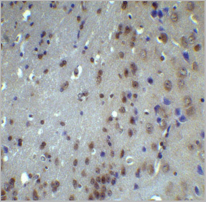
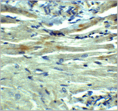
 IHC (Immunohistochemistry)
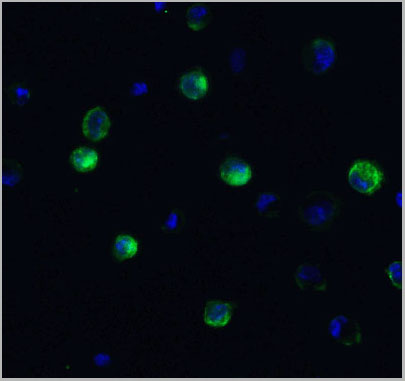
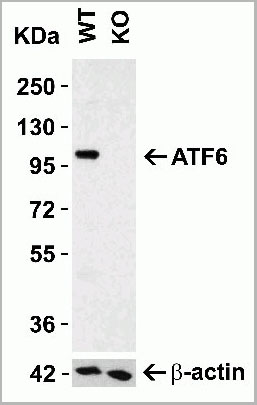
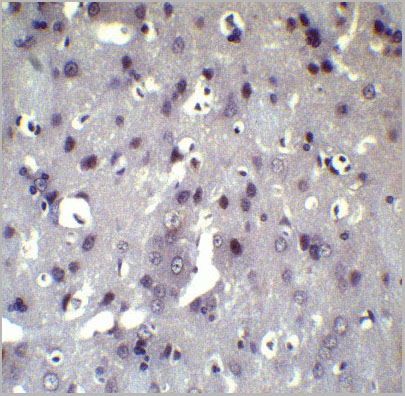
(Figure 8 Immunohistochemistry Validation of ATF6 in Rat Brain Tissue Immunohistochemical analysis of paraffin-embedded Rat brain tissue using anti-ATF6 antibody (AAA10937) at 5 ug/ml. Tissue was fixed with formaldehyde and blocked with 10% serum for 1 h at RT; antigen retrieval was by heat mediation with a citrate buffer (pH6). Samples were incubated with primary antibody overnight at 4°C. A goat anti-rabbit IgG H&L (HRP) at 1/250 was used as secondary. Counter stained with Hematoxylin.)
IHC (Immunohistochemistry)
(Figure 8 Immunohistochemistry Validation of ATF6 in Rat Brain Tissue Immunohistochemical analysis of paraffin-embedded Rat brain tissue using anti-ATF6 antibody (AAA10937) at 5 ug/ml. Tissue was fixed with formaldehyde and blocked with 10% serum for 1 h at RT; antigen retrieval was by heat mediation with a citrate buffer (pH6). Samples were incubated with primary antibody overnight at 4°C. A goat anti-rabbit IgG H&L (HRP) at 1/250 was used as secondary. Counter stained with Hematoxylin.)
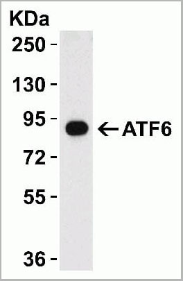
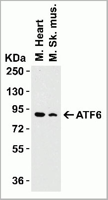
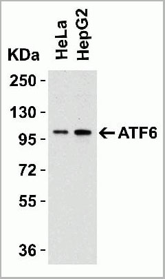
ATF6, Polyclonal Antibody (Cat# AAA10937)
Full Name
ATF6 Antibody
Gene Names
ATF6; ATF6A
Reactivity
Human, Mouse, Rat
Applications
Western Blot, Immunohistochemistry, Immunofluorescence
Purity
ATF6 Antibody is affinity chromatography purified via peptide column.
Pricing
C9 Tag, Polyclonal Antibody (Cat# AAA13688)
Full Name
Anti-C9 Tag Polyclonal Antibody, rabbit IgG
Applications
Western Blot
Purity
Protein A immunoaffinity chromatography
Pricing
Mast Cell Tryptase, alpha and beta isoforms, Monoclonal Antibody (Cat# AAA13400)
Full Name
MAb to Mast Cell Tryptase
Reactivity
Human
Applications
Western Blot, Immunohistochemistry
Purity
Protein G Chromatography
Pricing
Clostridium botulinum Type A Neurotoxin Complex, Antibody (Cat# AAA13408)
Full Name
Rabbit anti C. botulinum Neurotoxin Type A
Applications
Dot Blot, Western Blot
Purity
Protein G Sepharose Chromatography
Pricing
SDMA, Polyclonal Antibody (Cat# AAA14719)
Full Name
SDMA (Symmetric Dimethylarginine)
Applications
Western Blot, Immunohistochemistry, Immunocytochemistry, Radioimmunoassay
Purity
Serum
Pricing
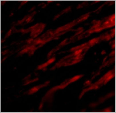
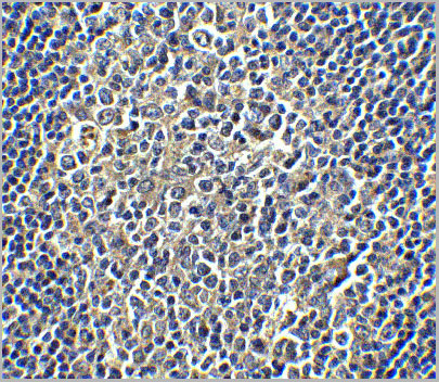
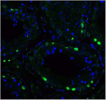
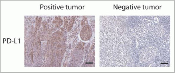 IF (Immunofluorescence)
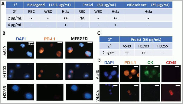
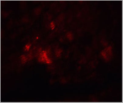
(Figure 12 Immunohistochemistry Validation of PD-L1in Human Tumors (Gadiot et al., 2011)Immunohistochemical analysis of patient tumors labeling PD-L1 with anti-PD-L1 antibodies (AAA10941). Several anti-PD-L1 antibodies were tested for staining, “Only 1 antibody gave no background staining and was competitively blocked by the addition of PD-L1Fc protein (AAA10941)”.)
IF (Immunofluorescence)
(Figure 12 Immunohistochemistry Validation of PD-L1in Human Tumors (Gadiot et al., 2011)Immunohistochemical analysis of patient tumors labeling PD-L1 with anti-PD-L1 antibodies (AAA10941). Several anti-PD-L1 antibodies were tested for staining, “Only 1 antibody gave no background staining and was competitively blocked by the addition of PD-L1Fc protein (AAA10941)”.)
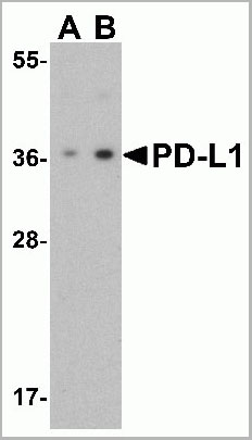
PDL-1, Polyclonal Antibody (Cat# AAA10941)
Full Name
PDL-1 Antibody
Gene Names
CD274; B7-H; B7H1; PDL1; PD-L1; PDCD1L1; PDCD1LG1
Applications
Western Blot, Immunohistochemistry, Immunofluorescence, Flow Cytometry
Purity
PD-L1 Antibody is affinity chromatography purified via peptide column.
Pricing
Dengue Virus NS1, Monoclonal Antibody (Cat# AAA13398)
Full Name
MAb to Dengue Virus NS1
Applications
Immunofluorescence, Lateral Flow
Purity
> 90% pure. Protein A Chromatography
Pricing
HLA Class 1 Antigen A1,A11,A26, Monoclonal Antibody (Cat# AAA14657)
Full Name
HLA Class 1 Antigen A1,A11,A26 (X-Reactive)
Applications
Cell Typing
Purity
Ascites
Pricing