Filters
Clonality
Type
Reactivity
Gene Name
Isotype
Host
Application
Clone
370 results for " F" - showing 50-100


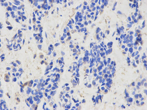
 ICC (Immunocytochemistry)
(Anti- TLR7 Picoband antibody, AAA11659, ICCICC: A549 Cell)
ICC (Immunocytochemistry)
(Anti- TLR7 Picoband antibody, AAA11659, ICCICC: A549 Cell)
TLR7, Polyclonal Antibody (Cat# AAA11659)
Full Name
Anti-TLR7 Antibody
Gene Names
TLR7; TLR7-like
Reactivity
Human, Mouse, Rat
Applications
Western Blot, Immunohistochemistry, Immunocytochemistry
Purity
Immunogen Affinity Purified
Pricing


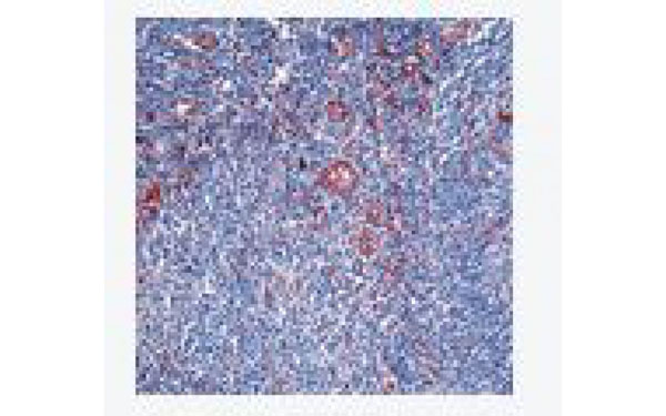
 IHC (Immunohistochemistry)
(Anti-Desmin Picoband antibody, AAA11607-7.JPGIHC(P): Rat Cardiac Muscle Tissue)
IHC (Immunohistochemistry)
(Anti-Desmin Picoband antibody, AAA11607-7.JPGIHC(P): Rat Cardiac Muscle Tissue)
Desmin, Polyclonal Antibody (Cat# AAA11607)
Full Name
Anti-Desmin Antibody
Gene Names
DES; CSM1; CSM2; LGMD2R
Reactivity
Human, Mouse, Rat
Applications
Western Blot, Immunohistochemistry, Immunohistochemistry
Purity
Immunogen affinity purified.
Pricing


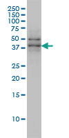
 WB (Western Blot)
(Figure 9 KD Validation in HeLa Cells (Horinaka et al., 2005)HeLa cells were transfected with DR4siRNA or LacZ control siRNA. At 24 h after transfection, the cells were treated with or without 20 μM luteolin for 24 h. Western blot analysis was carried out with anti-DR4 antibodies. DR4 expression was markedly reduced after DR4 knockdown.)
WB (Western Blot)
(Figure 9 KD Validation in HeLa Cells (Horinaka et al., 2005)HeLa cells were transfected with DR4siRNA or LacZ control siRNA. At 24 h after transfection, the cells were treated with or without 20 μM luteolin for 24 h. Western blot analysis was carried out with anti-DR4 antibodies. DR4 expression was markedly reduced after DR4 knockdown.)
DR4, Polyclonal Antibody (Cat# AAA10950)
Full Name
DR4 Antibody
Gene Names
TNFRSF10A; DR4; APO2; CD261; TRAILR1; TRAILR-1
Reactivity
Human
Applications
Western Blot
Purity
DR4 Antibody is affinity chromatography purified via peptide column.
Pricing


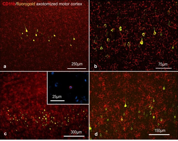
 Application Data
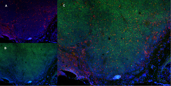
(Published customer image: Representative images of the inflammatory changes in the facial nucleus during axonal regeneration, one week following facial nerve transaction. a, b: CD11b immunoreactivity for microglia is increased in the axotomized facial nucleus, and microglia enwrap the facial motor neurons, e.g. at arrows. The regenerating neurons were retrogradely labelled with fluorogold. c, d: CD6- positive T-cells accumulated in the injured motor nucleus (arrows). They had little cytoplasm but dense nuclei (c) and were sometimes clustered around neurons retrogradely labelled with fluorogold (d). The scale bar in (a) also applies to (b) and that in (c) also applies to (d).From: Shokouhi et al. BMC Neuroscience 2010 11:13.)
Application Data
(Published customer image: Representative images of the inflammatory changes in the facial nucleus during axonal regeneration, one week following facial nerve transaction. a, b: CD11b immunoreactivity for microglia is increased in the axotomized facial nucleus, and microglia enwrap the facial motor neurons, e.g. at arrows. The regenerating neurons were retrogradely labelled with fluorogold. c, d: CD6- positive T-cells accumulated in the injured motor nucleus (arrows). They had little cytoplasm but dense nuclei (c) and were sometimes clustered around neurons retrogradely labelled with fluorogold (d). The scale bar in (a) also applies to (b) and that in (c) also applies to (d).From: Shokouhi et al. BMC Neuroscience 2010 11:13.)
CD11b, Monoclonal Antibody (Cat# AAA11969)
Full Name
MOUSE ANTI RAT CD11b
Gene Names
ITGAM; CD11B
Applications
Immunohistochemistry, Flow Cytometry, Immunofluorescence, Immunoprecipitation
Pricing


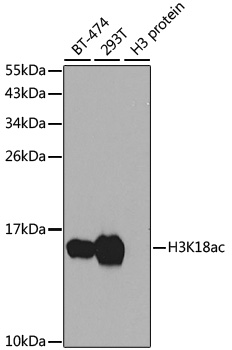
 IF (Immunofluorescence)
(Immunofluorescence analysis of NIH/3T3 cells using Acetyl-Histone H3-K18 antibody.)
IF (Immunofluorescence)
(Immunofluorescence analysis of NIH/3T3 cells using Acetyl-Histone H3-K18 antibody.)
Histone H3-K18, Polyclonal Antibody (Cat# AAA29843)
Full Name
Acetyl-Histone H3-K18 pAb
Gene Names
HIST1H3A; H3/A; H3FA
Reactivity
Human, Mouse, Rat, Other (Wide Range)
Applications
Western Blot, Immunohistochemistry, Immunofluorescence, Immunoprecipitation, Chromatin Immunoprecipitation, Chromatin Immunoprecipitation
Purity
Affinity purification
Pricing


 IP (Immunoprecipitation)
(HSPA1A was immunoprecipitated using:Lane A:0.5 mg Hela Whole Cell Lysate2 uL anti-HSPA1A rabbit monoclonal antibody and 15 ul of 50 % Protein G agarose.Primary antibody:Anti-HSPA1A rabbit monoclonal antibody,at 1:200 dilution Secondary antibody:Dylight 800-labeled antibody to rabbit IgG (H+L), at 1:5000 dilution Developed using the odssey technique.Performed under reducing conditions.Predicted band size: 70 kDaObserved band size: 70 kDa)
IP (Immunoprecipitation)
(HSPA1A was immunoprecipitated using:Lane A:0.5 mg Hela Whole Cell Lysate2 uL anti-HSPA1A rabbit monoclonal antibody and 15 ul of 50 % Protein G agarose.Primary antibody:Anti-HSPA1A rabbit monoclonal antibody,at 1:200 dilution Secondary antibody:Dylight 800-labeled antibody to rabbit IgG (H+L), at 1:5000 dilution Developed using the odssey technique.Performed under reducing conditions.Predicted band size: 70 kDaObserved band size: 70 kDa)
HSP70, Monoclonal Antibody (Cat# AAA27747)
Full Name
Recombinant Anti-HSP70 Antibody, Rabbit Monoclonal
Gene Names
HSPA1B; HSP72; HSPA1; HSX70; HSP70-1; HSP70-2; HSP70.1; HSP70.2; HSP70-1B
Reactivity
Human
Applications
Western Blot, Immunohistochemistry, Flow Cytometry, Immunocytochemistry, Immunofluorescence, Immunoprecipitation
Purity
Protein A
Pricing


 WB (Western Blot)
(Western blot analysis of extracts from Hela cells, treated with EGF or calf intestinal phosphatase (CIP), using GSK3beta (Phospho-Ser9) Antibody.)
WB (Western Blot)
(Western blot analysis of extracts from Hela cells, treated with EGF or calf intestinal phosphatase (CIP), using GSK3beta (Phospho-Ser9) Antibody.)
GSK3beta, Polyclonal Antibody (Cat# AAA29709)
Full Name
GSK3beta (Phospho-Ser9) Antibody
Reactivity
Human, Mouse, Rat
Applications
Western Blot, Immunohistochemistry, Immunofluorescence
Purity
Antibodies were produced by immunizing rabbits with synthetic phosphopeptide and KLH conjugates. Antibodies were purified by affinity-chromatography using epitope-specific phosphopeptide. Non-phospho specific antibodies were removed by chromatogramphy usi
Pricing


 Application Data
(Published customer image:Phycoerythrin conjugated Mouse anti Human CD83 antibody, clone HB15e used for the evaluation of CD83 expression on monocyte derived dendritic cells by flow cytometry.Phenotypic characterization of immunogenic and tolerogenic moDC populations by flow cytometry. Monocytes were negatively selected from PBMC using magnetic beads. Immature moDC were generated with IL-4 and GM-CSF for 6 days. 15d-PGJ2 (PGJ2 DC) and dexamethasone plus 1alpha,25-dihydroxyvitamin were added to generate tolerogenic moDC, respectively (PGJ2 DC and Dex/VD3 DC). To generate immunogenic moDC, immature moDC were stimulated for 24 h with LPS, polyI:C and a cytokine cocktail containing TNF-alpha, IL-1beta, IL-6 and PGE2, respectively. The phenotypes of the cells were analyzed by flow cytometry. Live cells were gated according to FSC/SSC. One representative experiment out of three is shown.From: Sprater F, Hovden A-O, Appel S (2012)Expression of ESE-3 Isoforms in Immunogenic and Tolerogenic Human Monocyte-Derived Dendritic Cells.PLoS ONE 7(11): e49577.)
Application Data
(Published customer image:Phycoerythrin conjugated Mouse anti Human CD83 antibody, clone HB15e used for the evaluation of CD83 expression on monocyte derived dendritic cells by flow cytometry.Phenotypic characterization of immunogenic and tolerogenic moDC populations by flow cytometry. Monocytes were negatively selected from PBMC using magnetic beads. Immature moDC were generated with IL-4 and GM-CSF for 6 days. 15d-PGJ2 (PGJ2 DC) and dexamethasone plus 1alpha,25-dihydroxyvitamin were added to generate tolerogenic moDC, respectively (PGJ2 DC and Dex/VD3 DC). To generate immunogenic moDC, immature moDC were stimulated for 24 h with LPS, polyI:C and a cytokine cocktail containing TNF-alpha, IL-1beta, IL-6 and PGE2, respectively. The phenotypes of the cells were analyzed by flow cytometry. Live cells were gated according to FSC/SSC. One representative experiment out of three is shown.From: Sprater F, Hovden A-O, Appel S (2012)Expression of ESE-3 Isoforms in Immunogenic and Tolerogenic Human Monocyte-Derived Dendritic Cells.PLoS ONE 7(11): e49577.)
CD83, Monoclonal Antibody (Cat# AAA12281)
Full Name
MOUSE ANTI HUMAN CD83: FITC
Reactivity
Human
Applications
Flow Cytometry
Pricing


 Application Data
(Published customer image: Histological staining of control and HBOT wounds at post-wounding days 7 and 29. A -D) H&E staining. E -H) CD34 immunohistochemistry. I+J) CD68 immunohistochemistry.From: uk B, Tong M, Fijneman EMG, van Neck JW (2014) Hyperbaric Oxygen Therapy to Treat Diabetes Impaired Wound Healing in Rats. PLoS ONE 9(10): e108533.)
Application Data
(Published customer image: Histological staining of control and HBOT wounds at post-wounding days 7 and 29. A -D) H&E staining. E -H) CD34 immunohistochemistry. I+J) CD68 immunohistochemistry.From: uk B, Tong M, Fijneman EMG, van Neck JW (2014) Hyperbaric Oxygen Therapy to Treat Diabetes Impaired Wound Healing in Rats. PLoS ONE 9(10): e108533.)
CD68, Monoclonal Antibody (Cat# AAA12147)
Full Name
MOUSE ANTI RAT CD68:Biotin
Applications
Flow Cytometry
Pricing


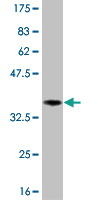
 WB (Western Blot)
(Western blot analysis of TAF7 over-expressed 293 cell line, cotransfected with TAF7 Validated Chimera RNAi (Lane 2) or non-transfected control (Lane 1). Blot probed with TAF7 monoclonal antibody. GAPDH (36.1kD) used as specificity and loading control.)
WB (Western Blot)
(Western blot analysis of TAF7 over-expressed 293 cell line, cotransfected with TAF7 Validated Chimera RNAi (Lane 2) or non-transfected control (Lane 1). Blot probed with TAF7 monoclonal antibody. GAPDH (36.1kD) used as specificity and loading control.)
TAF7, Monoclonal Antibody (Cat# AAA24383)
Full Name
TAF7 (TAF2F, TAFII55, Transcription Initiation Factor TFIID Subunit 7, RNA Polymerase II TBP-associated Factor Subunit F, Transcription Initiation Factor TFIID 55kD Subunit, TAF(II)55, TAFII-55) (AP)
Gene Names
TAF7; TAF2F; TAFII55
Reactivity
Human
Applications
Immunohistochemistry, Western Blot
Purity
Purified by Protein A Affinity Chromatography.
Pricing


 Application Data
(At 25 degree C. Samples were then incubated with primary Ab(At 37 degree C. An AlexaFluor594 conjugated goat anti-rabbit IgG(H+L) Ab(Red) and an AlexaFluor488 conjugated goat anti-mouse IgG(H+L) Ab(Green) were used as the secondary antibody.The nuclear counter stain is DAPI(blue).)
Application Data
(At 25 degree C. Samples were then incubated with primary Ab(At 37 degree C. An AlexaFluor594 conjugated goat anti-rabbit IgG(H+L) Ab(Red) and an AlexaFluor488 conjugated goat anti-mouse IgG(H+L) Ab(Green) were used as the secondary antibody.The nuclear counter stain is DAPI(blue).)
H2B, Polyclonal Antibody (Cat# AAA31348)
Full Name
Acetyl-H2B (Lys5) Antibody
Reactivity
Human, Mouse, Rat
Applications
Western Blot, Immunohistochemistry, Immunofluorescence, Immunocytochemistry, Peptide ELISA
Purity
The antiserum was purified by peptide affinity chromatography using SulfoLink Coupling Resin
Pricing


 ICC (Immunocytochemistry)
(ICC staining HBG1/2 in PC-12 cells (red). The nuclear counter stain is DAPI (blue). Cells were fixed in paraformaldehyde, permeabilised with 0.25% Triton X100/PBS.)
ICC (Immunocytochemistry)
(ICC staining HBG1/2 in PC-12 cells (red). The nuclear counter stain is DAPI (blue). Cells were fixed in paraformaldehyde, permeabilised with 0.25% Triton X100/PBS.)
Hemoglobin Subunit gamma 1 and 2, Monoclonal Antibody (Cat# AAA30321)
Full Name
Hemoglobin Subunit gamma 1 and 2 Antibody
Gene Names
HBG1; HBGA; HBGR; HBG-T2; HSGGL1; PRO2979
Reactivity
Human
Applications
Western Blot, Immunocytochemistry, Immunofluorescence, Immunohistochemistry
Purity
ProA affinity purified
Pricing


 ICC (Immunocytochemistry)
(Anti- SLC2A1 Picoband antibody, AAA11642, ICCICC: A549 Cell)
ICC (Immunocytochemistry)
(Anti- SLC2A1 Picoband antibody, AAA11642, ICCICC: A549 Cell)
SLC2A1, Polyclonal Antibody (Cat# AAA11642)
Full Name
Anti-SLC2A1 Antibody
Gene Names
SLC2A1; CSE; PED; DYT9; GLUT; DYT17; DYT18; EIG12; GLUT1; HTLVR; GLUT-1; SDCHCN; GLUT1DS
Reactivity
Human, Mouse, Rat
Applications
Western Blot, Immunohistochemistry, Immunocytochemistry
Purity
Immunogen Affinity Purified
Pricing
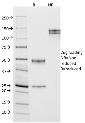
 SDS-PAGE
(Lane 1: 5 uL MW Marker. Lane 2: Reduced Mouse IgG Whole Molecule. Lane 3: Reduced Mouse F(c) Fragment. Lane 4: Reduced Mouse F(ab) Fragment. Lane 5: Mouse IgM Kappa Myeloma Protein. Load: 1 ug per lane. Predicted/Observed size: IgG at 50 and 25 kDa; F(c) at 25 kDa; F(ab) at 25 kDa; IgM K at 70 and 23 kDa. Observed F(c) Fragment migrates slightly higher.)
SDS-PAGE
(Lane 1: 5 uL MW Marker. Lane 2: Reduced Mouse IgG Whole Molecule. Lane 3: Reduced Mouse F(c) Fragment. Lane 4: Reduced Mouse F(ab) Fragment. Lane 5: Mouse IgM Kappa Myeloma Protein. Load: 1 ug per lane. Predicted/Observed size: IgG at 50 and 25 kDa; F(c) at 25 kDa; F(ab) at 25 kDa; IgM K at 70 and 23 kDa. Observed F(c) Fragment migrates slightly higher.)
Mouse IgG (Fc), Immunoglobulin (Cat# AAA14352)
Full Name
Mouse IgG (Fc)
Purity
Single arc observed by IEP.
Pricing


 IHC (Immunohistchemistry)
(At 1/100 staining Human ovarian cancer and adjacent normal tissues by IHC-P. The sample was formaldehyde fixed and a heat mediated antigen retrieval step in citrate buffer was performed. The sample was then blocked and incubated with the primary antibody at 4 degree C overnight. An HRP conjugated anti-Rabbit antibody was used as the secondary antibody.)
IHC (Immunohistchemistry)
(At 1/100 staining Human ovarian cancer and adjacent normal tissues by IHC-P. The sample was formaldehyde fixed and a heat mediated antigen retrieval step in citrate buffer was performed. The sample was then blocked and incubated with the primary antibody at 4 degree C overnight. An HRP conjugated anti-Rabbit antibody was used as the secondary antibody.)
alpha 1 Catenin, Polyclonal Antibody (Cat# AAA31319)
Full Name
Phospho-alpha 1 Catenin (Ser655/Thr658) Antibody
Gene Names
CTNNA1; CAP102
Reactivity
Human, Mouse, Rat
Applications
Western Blot, Immunohistochemistry, Peptide ELISA
Purity
The antibody is from purified rabbit serum by affinity purification via sequential chromatography on phospho-peptide and non-phospho-peptide affinity columns.
Pricing


 Application Data
(Published customer image: Leukocyte infiltration in COX-2-M/-M and COX-2+/+ mice. MPO enzymatic activity (panel A) was statistically similar in COX-2-M/-M and COX-2+/+ livers at 6 h and 24 h post-IRI. Ly-6G+ neutrophil (panel B) and granulocyte (panel C) infiltration were also comparable in COX-2-M/-M and COX-2+/+ livers after IRI. Mac-1+ (panel D) and CD68 (panel E) infiltrating macrophages were significantly reduced in COX-2-M/-M livers at 24 h post-reperfusion, but were statistically indistinguishable in COX-2-M/-M and COX-2+/+ livers at 6 h after IRI. No statistical differences in MMP-9 expression (panel F) could be demonstrated in livers of COX-2-M/-M and COX-2+/+ mice post-IRI. Representative immunostaining (panel G) of infiltrating Ly-6G+ (a,b,e,f) and Mac-1+ (c,d,g,h) leukocytes in livers of COX-2+/+ (a,c,e,g) and COX-2-M/-M (b,d,f,h) mice at 6 h (a to d) and 24 h (e to h) post IRI; (n = 5 -6/group; * indicates p)
Application Data
(Published customer image: Leukocyte infiltration in COX-2-M/-M and COX-2+/+ mice. MPO enzymatic activity (panel A) was statistically similar in COX-2-M/-M and COX-2+/+ livers at 6 h and 24 h post-IRI. Ly-6G+ neutrophil (panel B) and granulocyte (panel C) infiltration were also comparable in COX-2-M/-M and COX-2+/+ livers after IRI. Mac-1+ (panel D) and CD68 (panel E) infiltrating macrophages were significantly reduced in COX-2-M/-M livers at 24 h post-reperfusion, but were statistically indistinguishable in COX-2-M/-M and COX-2+/+ livers at 6 h after IRI. No statistical differences in MMP-9 expression (panel F) could be demonstrated in livers of COX-2-M/-M and COX-2+/+ mice post-IRI. Representative immunostaining (panel G) of infiltrating Ly-6G+ (a,b,e,f) and Mac-1+ (c,d,g,h) leukocytes in livers of COX-2+/+ (a,c,e,g) and COX-2-M/-M (b,d,f,h) mice at 6 h (a to d) and 24 h (e to h) post IRI; (n = 5 -6/group; * indicates p)
CD68, Monoclonal Antibody (Cat# AAA12105)
Full Name
RAT ANTI MOUSE CD68:FITC
Gene Names
Cd68; Lamp4; gp110; Scard1
Applications
Flow Cytometry
Pricing


 IHC (Immunohistochemistry)
(At 1/100 staining Human pancreatic cancer by IHC-P. The sample was formaldehyde fixed and a heat mediated antigen retrieval step in citrate buffer was performed. The sample was then blocked and incubated with the primary antibody at 4 degree C overnight. An HRP conjugated anti-Rabbit antibody was used as the secondary antibody.)
IHC (Immunohistochemistry)
(At 1/100 staining Human pancreatic cancer by IHC-P. The sample was formaldehyde fixed and a heat mediated antigen retrieval step in citrate buffer was performed. The sample was then blocked and incubated with the primary antibody at 4 degree C overnight. An HRP conjugated anti-Rabbit antibody was used as the secondary antibody.)
Histone H2B, Polyclonal Antibody (Cat# AAA31351)
Full Name
Phospho-Histone H2B (Tyr43) Antibody
Reactivity
Human, Mouse
Applications
Western Blot, Immunohistochemistry, Peptide ELISA
Purity
The antibody is from purified rabbit serum by affinity purification via sequential chromatography on phospho-peptide and non-phospho-peptide affinity columns.
Pricing


 Application Data
(Published customer image: Representative images of the inflammatory changes in the facial nucleus during axonal regeneration, one week following facial nerve transaction. a, b: CD11b immunoreactivity for microglia is increased in the axotomized facial nucleus, and microglia enwrap the facial motor neurons, e.g. at arrows. The regenerating neurons were retrogradely labelled with fluorogold. c, d: CD6- positive T-cells accumulated in the injured motor nucleus (arrows). They had little cytoplasm but dense nuclei (c) and were sometimes clustered around neurons retrogradely labelled with fluorogold (d). The scale bar in (a) also applies to (b) and that in (c) also applies to (d).From: Shokouhi et al. BMC Neuroscience 2010 11:13.)
Application Data
(Published customer image: Representative images of the inflammatory changes in the facial nucleus during axonal regeneration, one week following facial nerve transaction. a, b: CD11b immunoreactivity for microglia is increased in the axotomized facial nucleus, and microglia enwrap the facial motor neurons, e.g. at arrows. The regenerating neurons were retrogradely labelled with fluorogold. c, d: CD6- positive T-cells accumulated in the injured motor nucleus (arrows). They had little cytoplasm but dense nuclei (c) and were sometimes clustered around neurons retrogradely labelled with fluorogold (d). The scale bar in (a) also applies to (b) and that in (c) also applies to (d).From: Shokouhi et al. BMC Neuroscience 2010 11:13.)
CD11b, Monoclonal Antibody (Cat# AAA11971)
Full Name
MOUSE ANTI RAT CD11b
Gene Names
ITGAM; CD11B
Applications
Immunohistochemistry, Flow Cytometry, Immunofluorescence, Immunoprecipitation
Pricing


 FCM (Flow Cytometry)
(Flow cytometric analysis of A549 cells with alpha Actinin 4 antibody at 1/100 dilution (red) compared with an unlabelled control (cells without incubation with primary antibody; black).)
FCM (Flow Cytometry)
(Flow cytometric analysis of A549 cells with alpha Actinin 4 antibody at 1/100 dilution (red) compared with an unlabelled control (cells without incubation with primary antibody; black).)
alpha Actinin 4, Monoclonal Antibody (Cat# AAA30392)
Full Name
alpha Actinin 4 Antibody
Gene Names
ACTN4; FSGS; FSGS1; ACTININ-4
Reactivity
Human, Mouse, Rat
Applications
Western Blot, Immunocytochemistry, Immunofluorescence, Immunoprecipitation, Immunohistochemistry, Flow Cytometry
Purity
ProA affinity purified
Pricing


 WB (Western Blot)
(Anti-HA Tag mouse monoclonal antibody at 1:1000 dilutionLane A: HA-GST (Recombinant protein)(30ng)Lane B: GST-HA (Recombinant protein)(10ng)Lane C: HA-ARG1-myc transfected 293 cell lysate (2ug)Lane D: HA-mFABP4-myc transfected 293 cell lysate (2ug)Lane E: myc-mFABP4-HA transfected 293 cell lysate (0.5ug)Lane F: myc-ARG1-HA transfected 293 cell lysate (2ug)SecondaryGoat Anti-Mouse IgG H&L (Dylight800) at 1/15000 dilution.Developed using the Odyssey technique. Performed under reducing conditions.)
WB (Western Blot)
(Anti-HA Tag mouse monoclonal antibody at 1:1000 dilutionLane A: HA-GST (Recombinant protein)(30ng)Lane B: GST-HA (Recombinant protein)(10ng)Lane C: HA-ARG1-myc transfected 293 cell lysate (2ug)Lane D: HA-mFABP4-myc transfected 293 cell lysate (2ug)Lane E: myc-mFABP4-HA transfected 293 cell lysate (0.5ug)Lane F: myc-ARG1-HA transfected 293 cell lysate (2ug)SecondaryGoat Anti-Mouse IgG H&L (Dylight800) at 1/15000 dilution.Developed using the Odyssey technique. Performed under reducing conditions.)
HA, Monoclonal Antibody (Cat# AAA27712)
Full Name
Anti-HA tag Antibody, Mouse Monoclonal
Reactivity
HA tag
Applications
Western Blot, Flow Cytometry, Immunocytochemistry, Immunofluorescence, Immunoprecipitation
Purity
Protein A
Pricing


 IHC (Immunohistochemistry)
(At 1/100 staining Mouse lung tissue by IHC-P. The sample was formaldehyde fixed and a heat mediated antigen retrieval step in citrate buffer was performed. The sample was then blocked and incubated with the primary antibody at 4 degree C overnight. An HRP conjugated anti-Rabbit antibody was used as the secondary antibody.)
IHC (Immunohistochemistry)
(At 1/100 staining Mouse lung tissue by IHC-P. The sample was formaldehyde fixed and a heat mediated antigen retrieval step in citrate buffer was performed. The sample was then blocked and incubated with the primary antibody at 4 degree C overnight. An HRP conjugated anti-Rabbit antibody was used as the secondary antibody.)
Histone H2A, Polyclonal Antibody (Cat# AAA31347)
Full Name
Acetyl-Histone H2A (Lys5) Antibody
Gene Names
HIST1H2AB; H2A/m; H2AFM
Reactivity
Human, Mouse, Rat
Applications
Western Blot, Immunohistochemistry, Peptide ELISA
Purity
The antiserum was purified by peptide affinity chromatography using SulfoLink Coupling Resin
Pricing


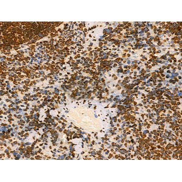
 IHC (Immunohistchemistry)
(Immunohistochemical analysis of paraffin-embedded rectum cancer tissues using ZEB1 mouse mAb with DAB staining.)
IHC (Immunohistchemistry)
(Immunohistochemical analysis of paraffin-embedded rectum cancer tissues using ZEB1 mouse mAb with DAB staining.)
ZEB1, Monoclonal Antibody (Cat# AAA14140)
Full Name
Anti-ZEB1 Mouse mAb
Gene Names
ZEB1; BZP; TCF8; AREB6; FECD6; NIL2A; PPCD3; ZFHEP; ZFHX1A; DELTAEF1
Reactivity
Human
Applications
Western Blot, Immunohistochemistry, Immunocytochemistry, Flow Cytometry
Pricing


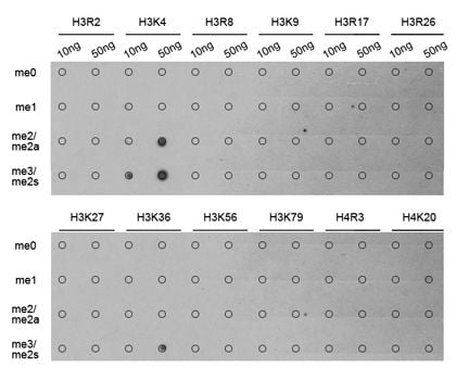

 ChIP (Chromatin Immunoprecipitation)
(Chromatin immunoprecipitation analysis extracts of 293 cell line, using TriMethyl-Histone H3-K4 antibody and rabbit IgG. The amount of immunoprecipitated DNA was checked by quantitative PCR. Histogram was constructed by the ratios of the immunoprecipitated DNA to the input.)
ChIP (Chromatin Immunoprecipitation)
(Chromatin immunoprecipitation analysis extracts of 293 cell line, using TriMethyl-Histone H3-K4 antibody and rabbit IgG. The amount of immunoprecipitated DNA was checked by quantitative PCR. Histogram was constructed by the ratios of the immunoprecipitated DNA to the input.)
H3K4me3, Antibody (Cat# AAA10662)
Full Name
Histone H3K4me3 Polyclonal Antibody
Gene Names
HIST3H3; H3t; H3.4; H3/g; H3FT
Reactivity
Human, Mouse, Rat, Other (Wide Range)
Applications
Western Blot, Immunohistochemistry, Immunofluorescence, Immunoprecipitation, Chromatin Immunoprecipitation, Chromatin Immunoprecipitation
Purity
Affinity Purification
Pricing


 ICC (Immunocytochemistry)
(ICC staining Histone H3 in Hela cells (green). The nuclear counter stain is DAPI (blue). Cells were fixed in paraformaldehyde, permeabilised with 0.25% Triton X100/PBS.)
ICC (Immunocytochemistry)
(ICC staining Histone H3 in Hela cells (green). The nuclear counter stain is DAPI (blue). Cells were fixed in paraformaldehyde, permeabilised with 0.25% Triton X100/PBS.)
Histone H3, Monoclonal Antibody (Cat# AAA30242)
Full Name
Histone H3 Antibody
Gene Names
HIST1H3A; H3/A; H3FA
Reactivity
Human, Mouse, Rat
Applications
Western Blot, Immunocytochemistry, Immunofluorescence, Immunohistochemistry, Chromatin Immunoprecipitation
Purity
ProA affinity purified
Pricing


 Application Data
(Analysis of Protein Array containing more than 19, 000 full-length human proteins using CD10 Mouse Monoclonal Antibody (MME/1870)Z- and S- Score: The Z-score represents the strength of a signal that a monoclonal antibody (MAb) (in combination with a fluorescently-tagged anti-IgG secondary antibody) produces when binding to a particular protein on the HuProtTM array. Z-scores are described in units of standard deviations (SD's) above the mean value of all signals generated on that array. If targets on HuProtTM are arranged in descending order of the Z-score, the S-score is the difference (also in units of SD's) between the Z-score. S-score therefore represents the relative target specificity of a MAb to its intended target. A MAb is considered to specific to its intended target, if the MAb has an S-score of at least 2.5. For example, if a MAb binds to protein X with a Z-score of 43 and to protein Y with a Z-score of 14, then the S-score for the binding of that MAb to protein X is equal to 29.)
Application Data
(Analysis of Protein Array containing more than 19, 000 full-length human proteins using CD10 Mouse Monoclonal Antibody (MME/1870)Z- and S- Score: The Z-score represents the strength of a signal that a monoclonal antibody (MAb) (in combination with a fluorescently-tagged anti-IgG secondary antibody) produces when binding to a particular protein on the HuProtTM array. Z-scores are described in units of standard deviations (SD's) above the mean value of all signals generated on that array. If targets on HuProtTM are arranged in descending order of the Z-score, the S-score is the difference (also in units of SD's) between the Z-score. S-score therefore represents the relative target specificity of a MAb to its intended target. A MAb is considered to specific to its intended target, if the MAb has an S-score of at least 2.5. For example, if a MAb binds to protein X with a Z-score of 43 and to protein Y with a Z-score of 14, then the S-score for the binding of that MAb to protein X is equal to 29.)
CD10, Monoclonal Antibody (Cat# AAA23901)
Full Name
CD10 (Membrane Metalloendopeptidase)
Gene Names
MME; NEP; SFE; CD10; CALLA; CMT2T; SCA43
Reactivity
Human. Others not tested.
Applications
Immunohistochemistry
Pricing


 ICC (Immunocytochemistry)
(ICC staining FBXL2 in SH-SY5Y cells (green). The nuclear counter stain is DAPI (blue). Cells were fixed in paraformaldehyde, permeabilised with 0.25% Triton X100/PBS.)
ICC (Immunocytochemistry)
(ICC staining FBXL2 in SH-SY5Y cells (green). The nuclear counter stain is DAPI (blue). Cells were fixed in paraformaldehyde, permeabilised with 0.25% Triton X100/PBS.)
FBXL2, Polyclonal Antibody (Cat# AAA29885)
Full Name
FBXL2 Antibody
Gene Names
FBXL2; FBL2; FBL3
Reactivity
Human, Mouse, Rat
Applications
Western Blot, Immunocytochemistry, Immunohistochemistry
Purity
ProA affinity purified
Pricing


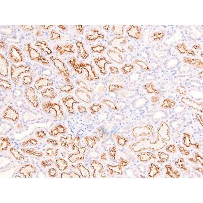 IHC (Immunohistochemistry)
(At 1/200 staining Human kidney tissue sections by IHC-P. The tissue was formaldehyde fixed and a heat mediated antigen retrieval step in citrate buffer was performed. The tissue was then blocked and incubated with the antibody for 1.5 hours at 22 degree C. An HRP conjugated goat anti-rabbit antibody was used as the secondary antibody.)
IHC (Immunohistochemistry)
(At 1/200 staining Human kidney tissue sections by IHC-P. The tissue was formaldehyde fixed and a heat mediated antigen retrieval step in citrate buffer was performed. The tissue was then blocked and incubated with the antibody for 1.5 hours at 22 degree C. An HRP conjugated goat anti-rabbit antibody was used as the secondary antibody.)
RhoA, Polyclonal Antibody (Cat# AAA31383)
Full Name
Phospho-RhoA (Ser188) Antibody
Gene Names
RHOA; ARHA; ARH12; RHO12; RHOH12
Reactivity
Human, Mouse, Rat
Predicted Reactivity: Pig (100%), Bovine (100%), Sheep (100%), Dog (100%)
Predicted Reactivity: Pig (100%), Bovine (100%), Sheep (100%), Dog (100%)
Applications
Western Blot, Immunohistochemistry, Peptide ELISA
Purity
The antibody is from purified rabbit serum by affinity purification via sequential chromatography on phospho-peptide and non-phospho-peptide affinity columns.
Pricing
 Standard Curve (Sample)
free haemoglobin, f-Hb, ELISA Kit (Cat# AAA14967)
Full Name
Human free haemoglobin, f-Hb ELISA Kit
Reactivity
Human
Pricing


 FCM (Flow Cytometry)
(Flow cytometric analysis of Jurkat cells with TCF7L2 antibody at 1/50 dilution (red) compared with an unlabelled control (cells without incubation with primary antibody; black). Alexa Fluor 488-conjugated goat anti rabbit IgG was used as the secondary antibody.)
FCM (Flow Cytometry)
(Flow cytometric analysis of Jurkat cells with TCF7L2 antibody at 1/50 dilution (red) compared with an unlabelled control (cells without incubation with primary antibody; black). Alexa Fluor 488-conjugated goat anti rabbit IgG was used as the secondary antibody.)
TCF7L2, Monoclonal Antibody (Cat# AAA30140)
Full Name
TCF7L2 Antibody
Gene Names
TCF7L2; TCF4; TCF-4
Reactivity
Human, Mouse, Rat
Applications
Western Blot, Immunocytochemistry, Immunofluorescence, Immunohistochemistry, Immunoprecipitation, Flow Cytometry
Purity
ProA affinity purified
Pricing


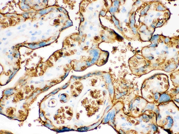
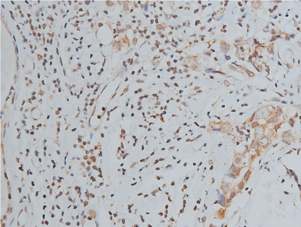
 IHC (Immunohistochemistry)
(Anti- Annexin A3 Picoband antibody, AAA11641, IHC(F)IHC(F): Human Placenta Tissue)
IHC (Immunohistochemistry)
(Anti- Annexin A3 Picoband antibody, AAA11641, IHC(F)IHC(F): Human Placenta Tissue)
Annexin A3, Polyclonal Antibody (Cat# AAA11641)
Full Name
Anti-Annexin A3 Antibody
Gene Names
ANXA3; ANX3
Reactivity
Human, Mouse, Rat
Applications
Western Blot, Immunohistochemistry
Purity
Immunogen Affinity Purified
Pricing


 Application Data
(Published customer image: Leukocyte infiltration in COX-2-M/-M and COX-2+/+ mice. MPO enzymatic activity (panel A) was statistically similar in COX-2-M/-M and COX-2+/+ livers at 6 h and 24 h post-IRI. Ly-6G+ neutrophil (panel B) and granulocyte (panel C) infiltration were also comparable in COX-2-M/-M and COX-2+/+ livers after IRI. Mac-1+ (panel D) and CD68 (panel E) infiltrating macrophages were significantly reduced in COX-2-M/-M livers at 24 h post-reperfusion, but were statistically indistinguishable in COX-2-M/-M and COX-2+/+ livers at 6 h after IRI. No statistical differences in MMP-9 expression (panel F) could be demonstrated in livers of COX-2-M/-M and COX-2+/+ mice post-IRI. Representative immunostaining (panel G) of infiltrating Ly-6G+ (a,b,e,f) and Mac-1+ (c,d,g,h) leukocytes in livers of COX-2+/+ (a,c,e,g) and COX-2-M/-M (b,d,f,h) mice at 6 h (a to d) and 24 h (e to h) post IRI; (n = 5 -6/group; * indicates p)
Application Data
(Published customer image: Leukocyte infiltration in COX-2-M/-M and COX-2+/+ mice. MPO enzymatic activity (panel A) was statistically similar in COX-2-M/-M and COX-2+/+ livers at 6 h and 24 h post-IRI. Ly-6G+ neutrophil (panel B) and granulocyte (panel C) infiltration were also comparable in COX-2-M/-M and COX-2+/+ livers after IRI. Mac-1+ (panel D) and CD68 (panel E) infiltrating macrophages were significantly reduced in COX-2-M/-M livers at 24 h post-reperfusion, but were statistically indistinguishable in COX-2-M/-M and COX-2+/+ livers at 6 h after IRI. No statistical differences in MMP-9 expression (panel F) could be demonstrated in livers of COX-2-M/-M and COX-2+/+ mice post-IRI. Representative immunostaining (panel G) of infiltrating Ly-6G+ (a,b,e,f) and Mac-1+ (c,d,g,h) leukocytes in livers of COX-2+/+ (a,c,e,g) and COX-2-M/-M (b,d,f,h) mice at 6 h (a to d) and 24 h (e to h) post IRI; (n = 5 -6/group; * indicates p)
CD68, Monoclonal Antibody (Cat# AAA12103)
Full Name
RAT ANTI MOUSE CD68:Biotin
Gene Names
Cd68; Lamp4; gp110; Scard1
Applications
Flow Cytometry
Pricing
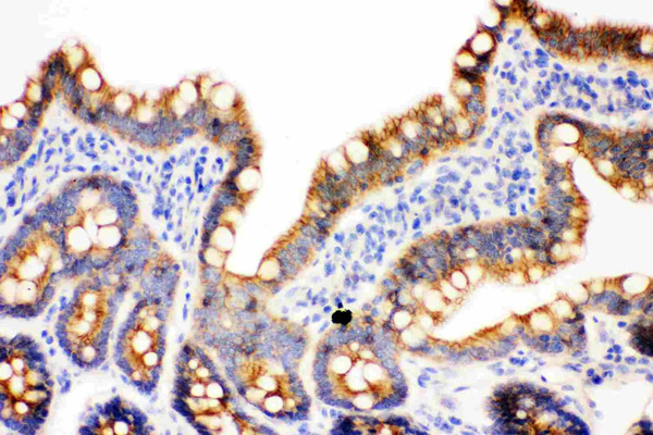


 IHC (Immunohistchemistry)
(Anti- gamma Catenin Picoband antibody, AAA11635, IHC(F)IHC(F): Rat Intestine Tissue)
IHC (Immunohistchemistry)
(Anti- gamma Catenin Picoband antibody, AAA11635, IHC(F)IHC(F): Rat Intestine Tissue)
Junction plakoglobin, Polyclonal Antibody (Cat# AAA11635)
Full Name
Anti-gamma Catenin Antibody
Gene Names
JUP; DP3; PDGB; PKGB; CTNNG; DPIII; ARVD12
Reactivity
Human, Mouse, Rat. No cross reactivity with other proteins.
Applications
Western Blot, Immunohistochemistry, Immunohistochemistry
Purity
Immunogen affinity purified.
Pricing


 Application Data
(Published customer imageSLAMF3 blockade in human hepatocytes is associated with lower susceptibility to HCV. (A) SLAMF3 was stained in primary human hepatocytes (PHHs) and cells from the Huh-7 human hepatoma cell line with a specific antibody (HLy9.1.25 clone; grey) and an isotype-matched control (empty). One of four independent experiments is shown. Huh-7 cells were transfected with scrambled control (sc) siRNA or three specific siRNAs (#1, #2 and #3) targeting SLAMF3, prior to infection with HCVcc; siRNA efficiency was checked by quantifying SLAMF3 mRNA (B) and the CD81 expression level (C) by flow cytometry analysis at 48 h post-transfection. Results are presented as the mean +/-SD (n = 3). Intracellular viral RNA was quantified at 72 h p.i. (D) and the infection was measured at 72 h p.i. by focus-forming units FFUs counting (E) (as inhibition percent; mean of three independent experiments; error bars: SD. **p)
Application Data
(Published customer imageSLAMF3 blockade in human hepatocytes is associated with lower susceptibility to HCV. (A) SLAMF3 was stained in primary human hepatocytes (PHHs) and cells from the Huh-7 human hepatoma cell line with a specific antibody (HLy9.1.25 clone; grey) and an isotype-matched control (empty). One of four independent experiments is shown. Huh-7 cells were transfected with scrambled control (sc) siRNA or three specific siRNAs (#1, #2 and #3) targeting SLAMF3, prior to infection with HCVcc; siRNA efficiency was checked by quantifying SLAMF3 mRNA (B) and the CD81 expression level (C) by flow cytometry analysis at 48 h post-transfection. Results are presented as the mean +/-SD (n = 3). Intracellular viral RNA was quantified at 72 h p.i. (D) and the infection was measured at 72 h p.i. by focus-forming units FFUs counting (E) (as inhibition percent; mean of three independent experiments; error bars: SD. **p)
CD229, Monoclonal Antibody (Cat# AAA11951)
Full Name
MOUSE ANTI HUMAN CD229
Gene Names
LY9; hly9; mLY9; CD229; SLAMF3
Applications
Flow Cytometry, Immunoprecipitation
Pricing


 Application Data
(Published customer image: Leukocyte infiltration in COX-2-M/-M and COX-2+/+ mice. MPO enzymatic activity (panel A) was statistically similar in COX-2-M/-M and COX-2+/+ livers at 6 h and 24 h post-IRI. Ly-6G+ neutrophil (panel B) and granulocyte (panel C) infiltration were also comparable in COX-2-M/-M and COX-2+/+ livers after IRI. Mac-1+ (panel D) and CD68 (panel E) infiltrating macrophages were significantly reduced in COX-2-M/-M livers at 24 h post-reperfusion, but were statistically indistinguishable in COX-2-M/-M and COX-2+/+ livers at 6 h after IRI. No statistical differences in MMP-9 expression (panel F) could be demonstrated in livers of COX-2-M/-M and COX-2+/+ mice post-IRI. Representative immunostaining (panel G) of infiltrating Ly-6G+ (a,b,e,f) and Mac-1+ (c,d,g,h) leukocytes in livers of COX-2+/+ (a,c,e,g) and COX-2-M/-M (b,d,f,h) mice at 6 h (a to d) and 24 h (e to h) post IRI; (n = 5 -6/group; * indicates p)
Application Data
(Published customer image: Leukocyte infiltration in COX-2-M/-M and COX-2+/+ mice. MPO enzymatic activity (panel A) was statistically similar in COX-2-M/-M and COX-2+/+ livers at 6 h and 24 h post-IRI. Ly-6G+ neutrophil (panel B) and granulocyte (panel C) infiltration were also comparable in COX-2-M/-M and COX-2+/+ livers after IRI. Mac-1+ (panel D) and CD68 (panel E) infiltrating macrophages were significantly reduced in COX-2-M/-M livers at 24 h post-reperfusion, but were statistically indistinguishable in COX-2-M/-M and COX-2+/+ livers at 6 h after IRI. No statistical differences in MMP-9 expression (panel F) could be demonstrated in livers of COX-2-M/-M and COX-2+/+ mice post-IRI. Representative immunostaining (panel G) of infiltrating Ly-6G+ (a,b,e,f) and Mac-1+ (c,d,g,h) leukocytes in livers of COX-2+/+ (a,c,e,g) and COX-2-M/-M (b,d,f,h) mice at 6 h (a to d) and 24 h (e to h) post IRI; (n = 5 -6/group; * indicates p)
CD68, Monoclonal Antibody (Cat# AAA12110)
Full Name
RAT ANTI MOUSE CD68
Gene Names
Cd68; Lamp4; gp110; Scard1
Applications
Immunohistochemistry, Flow Cytometry, Immunofluorescence, Immunoprecipitation, Immunohistochemistry, Western Blot
Pricing


 IF (Immunofluorescence)
(Immunofluorescence analysis of HepG2 cell using CAPZA2 antibody. Blue: DAPI for nuclear staining)
IF (Immunofluorescence)
(Immunofluorescence analysis of HepG2 cell using CAPZA2 antibody. Blue: DAPI for nuclear staining)
CAPZA2, Polyclonal Antibody (Cat# AAA29715)
Full Name
CAPZA2 Antibody
Gene Names
CAPZA2; CAPZ; CAPPA2
Reactivity
Human, Mouse, Rat
Applications
Western Blot, Immunohistochemistry, Immunofluorescence
Purity
Antibodies were purified by affinity purification using immunogen.
Pricing


 WB (Western Blot)
(Western blot analysis of ABCF2 over-expressed 293 cell line, cotransfected with ABCF2 Validated Chimera RNAi (Lane 2) or non-transfected control (Lane 1). Blot probed with ABCF2 monoclonal antibody GAPDH (36.1kD) used as specificity and loading control.)
WB (Western Blot)
(Western blot analysis of ABCF2 over-expressed 293 cell line, cotransfected with ABCF2 Validated Chimera RNAi (Lane 2) or non-transfected control (Lane 1). Blot probed with ABCF2 monoclonal antibody GAPDH (36.1kD) used as specificity and loading control.)
ABCF2, Monoclonal Antibody (Cat# AAA24704)
Full Name
ABCF2 (ATP-binding Cassette Sub-family F Member 2, Iron-inhibited ABC Transporter 2, HUSSY-18, DKFZp586K1823, EST133090, M-ABC1) (Biotin)
Gene Names
ABCF2; ABC28; HUSSY18; HUSSY-18; EST133090
Reactivity
Human
Applications
Immunohistochemistry, Immunoprecipitation, Western Blot
Purity
Purified by Protein A Affinity Chromatography.
Pricing


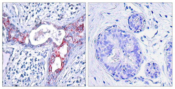
 Application Data
(Formalin fixed, paraffin embedded human breast cancer biopsy stained with Mouse anti Human estrogen receptor beta5 antibody followed by HRP polymer detection and DAB substrate development following heat mediated antigen retrieval using citrate buffer at pH6.2 (low power))
Application Data
(Formalin fixed, paraffin embedded human breast cancer biopsy stained with Mouse anti Human estrogen receptor beta5 antibody followed by HRP polymer detection and DAB substrate development following heat mediated antigen retrieval using citrate buffer at pH6.2 (low power))
ESTROGEN RECEPTOR BETA 5, Monoclonal Antibody (Cat# AAA12216)
Full Name
MOUSE ANTI HUMAN ESTROGEN RECEPTOR BETA 5
Gene Names
ESR2; Erb; ESRB; ESTRB; NR3A2; ER-BETA; ESR-BETA
Applications
Immunohistochemistry, Western Blot
Pricing


 Application Data
(Published customer image Infiltration of GFP+ BM-cells in infarct and peri-infarct regions. (A-B) Dot plots of viable macrophages/granulocytes (CD11b+CD45high, top right quadrants) and microglia (CD11b+CD45dim, bottom right quadrants) in cortex from BM-chimeric unmanipulated mice and mice exposed to pMCAO. (C) Bar graph showing mean numbers of CD11b+CD45dim microglia and CD11b+CD45high macrophages/granulocytes in BM-chimeric mice 24 hours after pMCAO, subdivided based on expression of GFP (n = 5). Approximately 92% of of the CD45high population were GFP+. (D) Estimation and comparison of mean numbers of CD11b+CD45dim microglia in non-chimeric (n = 10) versus BM-chimeric mice (n = 5) 24 hours after of pMCAO shows significantly fewer CD11b+CD45dim microglial cells in irradiated mice. (E) Overview, showing distribution of infiltrating GFP+ BM-derived cells into infarct (IF) and peri-infarct (P-IF) regions 24 hours after pMCAO. (E-G) By 24 hours, GFP+ single cells (F) and vessel-associated aggregates of GFP+ cells (arrows in G) were observed in infarct and peri-infarct regions. Some of the vessel-associated cells were round, leukocyte-like cells (arrows) while others were elongated cells lining the vasculature (arrow heads in G and in insert). (H) Bar graph showing mean numbers of single GFP+ cells and vessel-associated aggregates of GFP+ cells in ipsi- and contralateral cortex 24 hours after surgery (n = 10). (I-P) Immunohistochemical staining of CD45.1 (I, K), CD45.2 (J, L), IgG2a (M, O) and CD45 (N, P) in ischemic tissue in BM-chimeric (I, J, M, N) and non-chimeric mice (K, L, O, P) 24 hours after pMCAO. N.D, none detected. Scale bars: 200 um (A), 10 um (B, C). 50 um (I-P) *P < 0.05, **P < 0.01, and ***P < 0.001.From: Clausen BH, Lambertsen KL, Babcock AA, Holm TH, Dagnaes-Hansen F, Finsen B. Interleukin-1beta and tumor necrosis factor-alpha are expressed by different subsets of microglia and macrophages after ischemic stroke in mice. J Neuroinflammation. 2008 Oct 23;5:46.)
Application Data
(Published customer image Infiltration of GFP+ BM-cells in infarct and peri-infarct regions. (A-B) Dot plots of viable macrophages/granulocytes (CD11b+CD45high, top right quadrants) and microglia (CD11b+CD45dim, bottom right quadrants) in cortex from BM-chimeric unmanipulated mice and mice exposed to pMCAO. (C) Bar graph showing mean numbers of CD11b+CD45dim microglia and CD11b+CD45high macrophages/granulocytes in BM-chimeric mice 24 hours after pMCAO, subdivided based on expression of GFP (n = 5). Approximately 92% of of the CD45high population were GFP+. (D) Estimation and comparison of mean numbers of CD11b+CD45dim microglia in non-chimeric (n = 10) versus BM-chimeric mice (n = 5) 24 hours after of pMCAO shows significantly fewer CD11b+CD45dim microglial cells in irradiated mice. (E) Overview, showing distribution of infiltrating GFP+ BM-derived cells into infarct (IF) and peri-infarct (P-IF) regions 24 hours after pMCAO. (E-G) By 24 hours, GFP+ single cells (F) and vessel-associated aggregates of GFP+ cells (arrows in G) were observed in infarct and peri-infarct regions. Some of the vessel-associated cells were round, leukocyte-like cells (arrows) while others were elongated cells lining the vasculature (arrow heads in G and in insert). (H) Bar graph showing mean numbers of single GFP+ cells and vessel-associated aggregates of GFP+ cells in ipsi- and contralateral cortex 24 hours after surgery (n = 10). (I-P) Immunohistochemical staining of CD45.1 (I, K), CD45.2 (J, L), IgG2a (M, O) and CD45 (N, P) in ischemic tissue in BM-chimeric (I, J, M, N) and non-chimeric mice (K, L, O, P) 24 hours after pMCAO. N.D, none detected. Scale bars: 200 um (A), 10 um (B, C). 50 um (I-P) *P < 0.05, **P < 0.01, and ***P < 0.001.From: Clausen BH, Lambertsen KL, Babcock AA, Holm TH, Dagnaes-Hansen F, Finsen B. Interleukin-1beta and tumor necrosis factor-alpha are expressed by different subsets of microglia and macrophages after ischemic stroke in mice. J Neuroinflammation. 2008 Oct 23;5:46.)
CD11b, Monoclonal Antibody (Cat# AAA12185)
Full Name
RAT ANTI MOUSE CD11b
Gene Names
Itgam; CR3; CR3A; MAC1; Cd11b; Ly-40; Mac-1; Mac-1a; CD11b/CD18; F730045J24Rik
Applications
Immunohistochemistry, Flow Cytometry, Immunofluorescence, Immunoprecipitation
Pricing


 Application Data
(Published customer image Infiltration of GFP+ BM-cells in infarct and peri-infarct regions. (A-B) Dot plots of viable macrophages/granulocytes (CD11b+CD45high, top right quadrants) and microglia (CD11b+CD45dim, bottom right quadrants) in cortex from BM-chimeric unmanipulated mice and mice exposed to pMCAO. (C) Bar graph showing mean numbers of CD11b+CD45dim microglia and CD11b+CD45high macrophages/granulocytes in BM-chimeric mice 24 hours after pMCAO, subdivided based on expression of GFP (n = 5). Approximately 92% of of the CD45high population were GFP+. (D) Estimation and comparison of mean numbers of CD11b+CD45dim microglia in non-chimeric (n = 10) versus BM-chimeric mice (n = 5) 24 hours after of pMCAO shows significantly fewer CD11b+CD45dim microglial cells in irradiated mice. (E) Overview, showing distribution of infiltrating GFP+ BM-derived cells into infarct (IF) and peri-infarct (P-IF) regions 24 hours after pMCAO. (E-G) By 24 hours, GFP+ single cells (F) and vessel-associated aggregates of GFP+ cells (arrows in G) were observed in infarct and peri-infarct regions. Some of the vessel-associated cells were round, leukocyte-like cells (arrows) while others were elongated cells lining the vasculature (arrow heads in G and in insert). (H) Bar graph showing mean numbers of single GFP+ cells and vessel-associated aggregates of GFP+ cells in ipsi- and contralateral cortex 24 hours after surgery (n = 10). (I-P) Immunohistochemical staining of CD45.1 (I, K), CD45.2 (J, L), IgG2a (M, O) and CD45 (N, P) in ischemic tissue in BM-chimeric (I, J, M, N) and non-chimeric mice (K, L, O, P) 24 hours after pMCAO. N.D, none detected. Scale bars: 200 um (A), 10 um (B, C). 50 um (I-P) *P < 0.05, **P < 0.01, and ***P < 0.001.From: Clausen BH, Lambertsen KL, Babcock AA, Holm TH, Dagnaes-Hansen F, Finsen B. Interleukin-1beta and tumor necrosis factor-alpha are expressed by different subsets of microglia and macrophages after ischemic stroke in mice. J Neuroinflammation. 2008 Oct 23;5:46.)
Application Data
(Published customer image Infiltration of GFP+ BM-cells in infarct and peri-infarct regions. (A-B) Dot plots of viable macrophages/granulocytes (CD11b+CD45high, top right quadrants) and microglia (CD11b+CD45dim, bottom right quadrants) in cortex from BM-chimeric unmanipulated mice and mice exposed to pMCAO. (C) Bar graph showing mean numbers of CD11b+CD45dim microglia and CD11b+CD45high macrophages/granulocytes in BM-chimeric mice 24 hours after pMCAO, subdivided based on expression of GFP (n = 5). Approximately 92% of of the CD45high population were GFP+. (D) Estimation and comparison of mean numbers of CD11b+CD45dim microglia in non-chimeric (n = 10) versus BM-chimeric mice (n = 5) 24 hours after of pMCAO shows significantly fewer CD11b+CD45dim microglial cells in irradiated mice. (E) Overview, showing distribution of infiltrating GFP+ BM-derived cells into infarct (IF) and peri-infarct (P-IF) regions 24 hours after pMCAO. (E-G) By 24 hours, GFP+ single cells (F) and vessel-associated aggregates of GFP+ cells (arrows in G) were observed in infarct and peri-infarct regions. Some of the vessel-associated cells were round, leukocyte-like cells (arrows) while others were elongated cells lining the vasculature (arrow heads in G and in insert). (H) Bar graph showing mean numbers of single GFP+ cells and vessel-associated aggregates of GFP+ cells in ipsi- and contralateral cortex 24 hours after surgery (n = 10). (I-P) Immunohistochemical staining of CD45.1 (I, K), CD45.2 (J, L), IgG2a (M, O) and CD45 (N, P) in ischemic tissue in BM-chimeric (I, J, M, N) and non-chimeric mice (K, L, O, P) 24 hours after pMCAO. N.D, none detected. Scale bars: 200 um (A), 10 um (B, C). 50 um (I-P) *P < 0.05, **P < 0.01, and ***P < 0.001.From: Clausen BH, Lambertsen KL, Babcock AA, Holm TH, Dagnaes-Hansen F, Finsen B. Interleukin-1beta and tumor necrosis factor-alpha are expressed by different subsets of microglia and macrophages after ischemic stroke in mice. J Neuroinflammation. 2008 Oct 23;5:46.)
CD11b, Monoclonal Antibody (Cat# AAA12183)
Full Name
RAT ANTI MOUSE CD11b:FITC
Gene Names
Itgam; CR3; CR3A; MAC1; Cd11b; Ly-40; Mac-1; Mac-1a; CD11b/CD18; F730045J24Rik
Applications
Flow Cytometry
Pricing


 Application Data
(Immunoperoxidase staining of a human tonsil cryosection with Mouse anti Human CD163 antibody, clone EDHu-1 followed by the Histar detection system . Low power)
Application Data
(Immunoperoxidase staining of a human tonsil cryosection with Mouse anti Human CD163 antibody, clone EDHu-1 followed by the Histar detection system . Low power)
CD163, Monoclonal Antibody (Cat# AAA12100)
Full Name
MOUSE ANTI HUMAN CD163
Gene Names
CD163; M130; MM130
Applications
Immunohistochemistry, Flow Cytometry, Immunofluorescence, Immunoassay, Immunohistochemistry, Western Blot
Pricing


 IHC (Immunohistchemistry)
(Anti-GLUT4 Picoband antibody, AAA11601-6.JPGIHC(P): Mouse Skeletal Muscle Tissue)
IHC (Immunohistchemistry)
(Anti-GLUT4 Picoband antibody, AAA11601-6.JPGIHC(P): Mouse Skeletal Muscle Tissue)
GLUT4, Polyclonal Antibody (Cat# AAA11601)
Full Name
Anti-GLUT4 Antibody
Gene Names
SLC2A4; GLUT4
Reactivity
Human, Mouse, Rat
Applications
Western Blot, Immunohistochemistry, Immunohistochemistry
Purity
Immunogen affinity purified.
Pricing


 IHC (Immunohistochemistry)
(Immunohistochemistry of paraffin-embedded mouse heart using HIST1H2AG antibody at dilution of 1:100 (40x lens).)
IHC (Immunohistochemistry)
(Immunohistochemistry of paraffin-embedded mouse heart using HIST1H2AG antibody at dilution of 1:100 (40x lens).)
HIST1H2AG, Polyclonal Antibody (Cat# AAA28259)
Full Name
HIST1H2AG Polyclonal Antibody
Gene Names
HIST1H2AG; H2AG; H2A/p; H2AFP; H2A.1b; pH2A/f
Reactivity
Human, Mouse, Rat
Applications
Western Blot, Immunohistochemistry
Purity
Affinity Purification
Pricing
 IHC (Immunohistochemistry)
(Formalin-fixed, paraffin-embedded human tonsil stained with a-1-Antitrypsin (AAT) antibody using peroxidase-conjugate and AEC chromogen. Note cytoplasmic staining of endothelial cells.)
IHC (Immunohistochemistry)
(Formalin-fixed, paraffin-embedded human tonsil stained with a-1-Antitrypsin (AAT) antibody using peroxidase-conjugate and AEC chromogen. Note cytoplasmic staining of endothelial cells.)
alpha 1 Antitrypsin, Polyclonal Antibody (Cat# AAA14320)
Full Name
alpha 1 Antitrypsin antibody
Gene Names
SERPINA1; PI; A1A; AAT; PI1; A1AT; PRO2275; alpha1AT
Reactivity
Human
Applications
Immunohistochemistry
Pricing


 Application Data
(Published customer image: Representative images of the inflammatory changes in the facial nucleus during axonal regeneration, one week following facial nerve transaction. a, b: CD11b immunoreactivity for microglia is increased in the axotomized facial nucleus, and microglia enwrap the facial motor neurons, e.g. at arrows. The regenerating neurons were retrogradely labelled with fluorogold. c, d: CD6- positive T-cells accumulated in the injured motor nucleus (arrows). They had little cytoplasm but dense nuclei (c) and were sometimes clustered around neurons retrogradely labelled with fluorogold (d). The scale bar in (a) also applies to (b) and that in (c) also applies to (d).From: Shokouhi et al. BMC Neuroscience 2010 11:13.)
Application Data
(Published customer image: Representative images of the inflammatory changes in the facial nucleus during axonal regeneration, one week following facial nerve transaction. a, b: CD11b immunoreactivity for microglia is increased in the axotomized facial nucleus, and microglia enwrap the facial motor neurons, e.g. at arrows. The regenerating neurons were retrogradely labelled with fluorogold. c, d: CD6- positive T-cells accumulated in the injured motor nucleus (arrows). They had little cytoplasm but dense nuclei (c) and were sometimes clustered around neurons retrogradely labelled with fluorogold (d). The scale bar in (a) also applies to (b) and that in (c) also applies to (d).From: Shokouhi et al. BMC Neuroscience 2010 11:13.)
CD11b, Monoclonal Antibody (Cat# AAA11876)
Full Name
MOUSE ANTI RAT CD11b:FITC
Gene Names
ITGAM; CD11B
Applications
Flow Cytometry
Pricing


 Application Data
(Analysis of Protein Array containing more than 19, 000 full-length human proteins using Oct-2 Mouse Monoclonal Antibody (OCT2/2137) Z- and S- Score: The Z-score represents the strength of a signal that a monoclonal antibody (MAb) (in combination with a fluorescently-tagged anti-IgG secondary antibody) produces when binding to a particular protein on the HuProtTM array. Z-scores are described in units of standard deviations (SD's) above the mean value of all signals generated on that array. If targets on HuProtTM are arranged in descending order of the Z-score, the S-score is the difference (also in units of SD's) between the Z-score. S-score therefore represents the relative target specificity of a MAb to its intended target. A MAb is considered to specific to its intended target, if the MAb has an S-score of at least 2.5. For example, if a MAb binds to protein X with a Z-score of 43 and to protein Y with a Z-score of 14, then the S-score for the binding of that MAb to protein X is equal to 29.)
Application Data
(Analysis of Protein Array containing more than 19, 000 full-length human proteins using Oct-2 Mouse Monoclonal Antibody (OCT2/2137) Z- and S- Score: The Z-score represents the strength of a signal that a monoclonal antibody (MAb) (in combination with a fluorescently-tagged anti-IgG secondary antibody) produces when binding to a particular protein on the HuProtTM array. Z-scores are described in units of standard deviations (SD's) above the mean value of all signals generated on that array. If targets on HuProtTM are arranged in descending order of the Z-score, the S-score is the difference (also in units of SD's) between the Z-score. S-score therefore represents the relative target specificity of a MAb to its intended target. A MAb is considered to specific to its intended target, if the MAb has an S-score of at least 2.5. For example, if a MAb binds to protein X with a Z-score of 43 and to protein Y with a Z-score of 14, then the S-score for the binding of that MAb to protein X is equal to 29.)
OCT-2 (POU2F2), Monoclonal Antibody (Cat# AAA23902)
Full Name
OCT-2 (POU2F2) (B-Cell Marker)
Gene Names
POU2F2; OCT2; OTF2; Oct-2
Reactivity
Human. Others not known.
Applications
Western Blot, Immunohistochemistry
Pricing


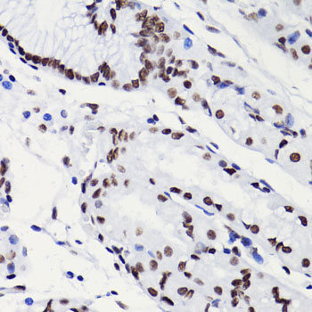
 IHC (Immunohistochemistry)
(AAA30929 at 1/200 staining human colon tissue sections by IHC-P. The tissue was formaldehyde fixed and a heat mediated antigen retrieval step in citrate buffer was performed. The tissue was then blocked and incubated with the antibody for 1.5 hours at 22 degree C. An HRP conjugated goat anti-rabbit antibody was used as the secondary.)
IHC (Immunohistochemistry)
(AAA30929 at 1/200 staining human colon tissue sections by IHC-P. The tissue was formaldehyde fixed and a heat mediated antigen retrieval step in citrate buffer was performed. The tissue was then blocked and incubated with the antibody for 1.5 hours at 22 degree C. An HRP conjugated goat anti-rabbit antibody was used as the secondary.)
Histone H3, Polyclonal Antibody (Cat# AAA30929)
Full Name
Histone H3 Antibody
Gene Names
HIST1H3A; H3/A; H3FA
Reactivity
Human, Mouse, Rat
Applications
Western Blot, Immunohistochemistry, Immunofluorescence, Immunocytochemistry
Purity
The antiserum was purified by peptide affinity chromatography using SulfoLink Coupling Resin.
Pricing


 Application Data
(At 25 degree C. Samples were then incubated with primary Ab(At 37 degree C. An AlexaFluor594 conjugated goat anti-rabbit IgG(H+L) Ab(Red) and an AlexaFluor488 conjugated goat anti-mouse IgG(H+L) Ab(Green) were used as the secondary antibody.The nuclear counter stain is DAPI (blue).)
Application Data
(At 25 degree C. Samples were then incubated with primary Ab(At 37 degree C. An AlexaFluor594 conjugated goat anti-rabbit IgG(H+L) Ab(Red) and an AlexaFluor488 conjugated goat anti-mouse IgG(H+L) Ab(Green) were used as the secondary antibody.The nuclear counter stain is DAPI (blue).)
Histone H3, Polyclonal Antibody (Cat# AAA31322)
Full Name
Phospho-Histone H3 (Thr3) Antibody
Gene Names
HIST1H3A; H3/A; H3FA
Reactivity
Human, Mouse, Rat
Applications
Immunohistochemistry, Immunofluorescence, Immunocytochemistry, Peptide ELISA
Purity
The antibody is from purified rabbit serum by affinity purification via sequential chromatography on phospho-peptide and non-phospho-peptide affinity columns.
Pricing


 WB (Western Blot)
(Western blot analysis of ABCF2 over-expressed 293 cell line, cotransfected with ABCF2 Validated Chimera RNAi (Lane 2) or non-transfected control (Lane 1). Blot probed with ABCF2 monoclonal antibody GAPDH (36.1kD) used as specificity and loading control.)
WB (Western Blot)
(Western blot analysis of ABCF2 over-expressed 293 cell line, cotransfected with ABCF2 Validated Chimera RNAi (Lane 2) or non-transfected control (Lane 1). Blot probed with ABCF2 monoclonal antibody GAPDH (36.1kD) used as specificity and loading control.)
ABCF2, Monoclonal Antibody (Cat# AAA24115)
Full Name
ABCF2 (ATP-binding Cassette Sub-family F Member 2, Iron-inhibited ABC Transporter 2, HUSSY-18, DKFZp586K1823, EST133090, M-ABC1) (AP)
Gene Names
ABCF2; ABC28; HUSSY18; HUSSY-18; EST133090
Reactivity
Human
Applications
Immunohistochemistry, Immunoprecipitation, Western Blot
Purity
Purified by Protein A Affinity Chromatography.
Pricing


 Application Data
(Proximity Ligation Analysis of protein-protein interactions between STAT1 and PDGFRB Mahlavu cells were stained with anti-STAT1 rabbit purified polyclonal (1:1200) and 131071 (1:50). Each red dot represents the detection of protein-protein interaction complex, and nuclei were counterstained with DAPI (blue).)
Application Data
(Proximity Ligation Analysis of protein-protein interactions between STAT1 and PDGFRB Mahlavu cells were stained with anti-STAT1 rabbit purified polyclonal (1:1200) and 131071 (1:50). Each red dot represents the detection of protein-protein interaction complex, and nuclei were counterstained with DAPI (blue).)
PDGFRB, Monoclonal Antibody (Cat# AAA25194)
Full Name
PDGFRB (PDGFR, PDGFR1, Platelet-derived Growth Factor Receptor beta, Beta Platelet-derived Growth Factor Receptor, Beta-type Platelet-derived Growth Factor Receptor, CD140 Antigen-like Family Member B, Platelet-derived Growth Factor Receptor 1, CD140b) (F
Gene Names
PDGFRB; IMF1; KOGS; IBGC4; JTK12; PDGFR; PENTT; CD140B; PDGFR1; PDGFR-1
Reactivity
Human
Applications
Immunoprecipitation, Western Blot
Purity
Purified by Protein A Affinity Chromatography.
Pricing


 WB (Western Blot)
(Anti-Beta-Tubulin mouse monoclonal antibody at 1:2000 dilution Lane A: HepG2 Whole Cell Lysate Lane B: Daudi Whole Cell Lysate Lane C: MOLT-4 Whole Cell Lysate Lane D: A549 Whole Cell Lysate Lane E: 293T Whole Cell Lysate Lane F: HelaS3 Whole Cell Lysate Lysates/proteins at 30 ug per lane. Secondary Goat Anti-Mouse IgG H&L (Dylight800) at 1/15000 dilution. Developed using the Odyssey technique. Performed under reducing conditions. Predicted band size:50 kDa Observed band size:54 kDa)
WB (Western Blot)
(Anti-Beta-Tubulin mouse monoclonal antibody at 1:2000 dilution Lane A: HepG2 Whole Cell Lysate Lane B: Daudi Whole Cell Lysate Lane C: MOLT-4 Whole Cell Lysate Lane D: A549 Whole Cell Lysate Lane E: 293T Whole Cell Lysate Lane F: HelaS3 Whole Cell Lysate Lysates/proteins at 30 ug per lane. Secondary Goat Anti-Mouse IgG H&L (Dylight800) at 1/15000 dilution. Developed using the Odyssey technique. Performed under reducing conditions. Predicted band size:50 kDa Observed band size:54 kDa)
Beta-Tubulin, Monoclonal Antibody (Cat# AAA27713)
Full Name
Beta-Tubulin Loading Control Antibody, Mouse MAb
Reactivity
Human
Applications
Western Blot, Immunohistochemistry, Immunocytochemistry, Immunofluorescence, Immunoprecipitation
Purity
Protein A
Pricing