Filters
Clonality
Type
Reactivity
Gene Name
Isotype
Host
Application
Clone
190 results for " Conjugated Polyclonal Antibodies" - showing 50-100


 IHC (Immunohistchemistry)
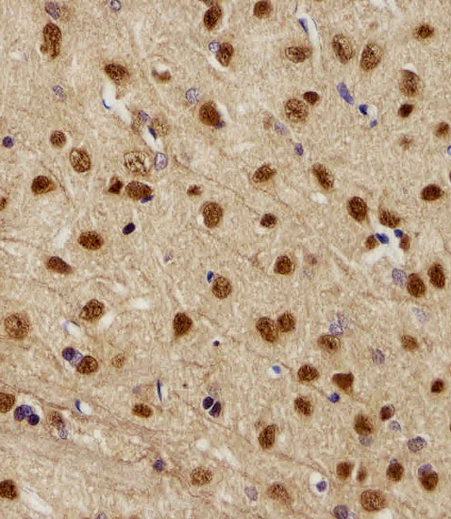
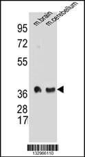
(Immunohistochemistry Analysis: Representative lot data. (Fig. 1 and 2) Paraffin-embedded mouse and human brain tissue was prepared using heat-induced epitope retrieval in citrate buffer, pH 6.0. Immunostaining was performed using a 1:100 dilution. Reactivity was detected using the IHC-Select Detection Kit. Staining pattern appears as cytoplasmic. (Fig. 3 and 4) Paraffin-embedded mouse and mouse olfactory lobe and cerebellum brain tissue was prepared using heat-induced epitope retrieval in citrate buffer, pH 6.0. Immunostaining was performed using a Chicken IgY Antibody 1:100 dilution of Cat. No. AB15894, anti-Tbr2. Reactivity was detected using the IHC-Select Detection Kit. Immunoreactivity seen here is mostly nuclear.)
IHC (Immunohistchemistry)
(Immunohistochemistry Analysis: Representative lot data. (Fig. 1 and 2) Paraffin-embedded mouse and human brain tissue was prepared using heat-induced epitope retrieval in citrate buffer, pH 6.0. Immunostaining was performed using a 1:100 dilution. Reactivity was detected using the IHC-Select Detection Kit. Staining pattern appears as cytoplasmic. (Fig. 3 and 4) Paraffin-embedded mouse and mouse olfactory lobe and cerebellum brain tissue was prepared using heat-induced epitope retrieval in citrate buffer, pH 6.0. Immunostaining was performed using a Chicken IgY Antibody 1:100 dilution of Cat. No. AB15894, anti-Tbr2. Reactivity was detected using the IHC-Select Detection Kit. Immunoreactivity seen here is mostly nuclear.)
EOMES, Polyclonal Antibody (Cat# AAA26889)
Full Name
EOMES (Eomesodermin Homolog, T-box Brain Protein 2, TBR2, T-brain-2, TBR-2)
Gene Names
Eomes; Tbr2; TBR-2; C77258
Reactivity
Mouse, Human, Rat
Applications
Immunohistochemistry, Western Blot
Purity
Purified by affinity chromatography.
Pricing


 IF (Immunofluorescence)
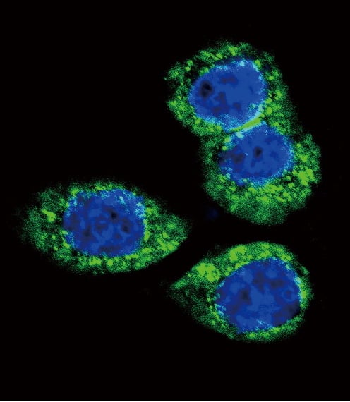
(Confocal immunofluorescent analysis of CTNNB1 Antibody (C-term) with T47D cell followed by Alexa Fluor 488-conjugated goat anti-rabbit lgG (green). Actin filaments have been labeled with Alexa Fluor 555 phalloidin (red). DAPI was used to stain the cell nuclear (blue).)
IF (Immunofluorescence)
(Confocal immunofluorescent analysis of CTNNB1 Antibody (C-term) with T47D cell followed by Alexa Fluor 488-conjugated goat anti-rabbit lgG (green). Actin filaments have been labeled with Alexa Fluor 555 phalloidin (red). DAPI was used to stain the cell nuclear (blue).)
CTNNB1, Polyclonal Antibody (Cat# AAA28720)
Full Name
CTNNB1 Antibody (C-term)
Gene Names
CTNNB1; CTNNB; MRD19; armadillo
Reactivity
Human (Predicted Reactivity: Bovine, Mouse, Rat)
Applications
Western Blot, Immunohistochemistry, Flow Cytometry, Immunofluorescence
Purity
Peptide Affinity Purified Rabbit Polyclonal Antibody (Pab)
Pricing


 WB (Western Blot)
(Western Blot Analysis: Representative lot data. Lysate from HEK-293 cells was resolved by electrophoresis, transferred to PVDF and probed with anti-PI3 Kinase, p110beta (0.05 ug/mL). Proteins were visualized using donkey anti-rabbit secondary antibody conjugated to HRP and chemiluminescence detection. Arrow indicates PI3 Kinase, p110beta (~110kD).)
WB (Western Blot)
(Western Blot Analysis: Representative lot data. Lysate from HEK-293 cells was resolved by electrophoresis, transferred to PVDF and probed with anti-PI3 Kinase, p110beta (0.05 ug/mL). Proteins were visualized using donkey anti-rabbit secondary antibody conjugated to HRP and chemiluminescence detection. Arrow indicates PI3 Kinase, p110beta (~110kD).)
Phosphoinositide 3 Kinase, p110 beta, Polyclonal Antibody (Cat# AAA26898)
Full Name
Phosphoinositide 3 Kinase, p110 beta (DKFZp779K1237, MGC133043, p110beta, Phosphatidylinositol 3 Kinase Catalytic beta Polypeptide, Phosphatidylinositol-4,5-bisphosphate 3-kinase Catalytic Subunit beta Isoform, Phosphoinositide 3 Kinase Catalytic beta Pol
Gene Names
PIK3CA; MCM; CWS5; MCAP; PI3K; CLOVE; MCMTC; p110-alpha
Reactivity
Human
Applications
Immunocytochemistry, Western Blot, Immunoprecipitation, Immunohistochemistry
Purity
Purified by Immunoaffinity chromatography.
Pricing


 IP (Immunoprecipitation)
(Immunoprecipitating GAPDH in Hela whole cell lysate Lane 1: Mouse control IgG instead of AAA27043 in Hela whole cell lysate. Lane 2: AAA27043 (5ul) + Hela whole cell lysate (500ug) Lane 3: Hela whole cell lysate (10ug) For western blotting, the blot was detected with AAA27043 at 1:5000, and a HRPconjugated Protein G antibody was used as the secondary antibody at 1:2000)
IP (Immunoprecipitation)
(Immunoprecipitating GAPDH in Hela whole cell lysate Lane 1: Mouse control IgG instead of AAA27043 in Hela whole cell lysate. Lane 2: AAA27043 (5ul) + Hela whole cell lysate (500ug) Lane 3: Hela whole cell lysate (10ug) For western blotting, the blot was detected with AAA27043 at 1:5000, and a HRPconjugated Protein G antibody was used as the secondary antibody at 1:2000)
GAPDH, Monoclonal Antibody (Cat# AAA27043)
Full Name
GAPDH Monoclonal Antibody
Reactivity
Human, Rat, Rabbit, Mouse
Applications
Western Blot, Immunohistochemistry, Immunoprecipitation, Immunofluorescence
Purity
>95%, Protein G purified
Pricing


 IF (Immunofluorescence)
(Fluorescent confocal image of Hela cell stained with HDAC2 Antibody (C-term) .Hela cells were fixed with 4% PFA (20 min), permeabilized with Triton X-100 (0.1%, 10 min), then incubated with HDAC2 primary antibody (1:25, 1 h at 37 degree C). For secondary antibody, Alexa Fluor 488 conjugated donkey anti-rabbit antibody (green) was used (1:400, 50 min at 37 degree C).Cytoplasmic actin was counterstained with Alexa Fluor 555 (red) conjugated Phalloidin (7units/ml, 1 h at 37 degree C). Nuclei were counterstained with DAPI (blue) (10 ug/ml, 10 min). hHDAC2 immunoreactivity is localized to Nucleus significantly.)
IF (Immunofluorescence)
(Fluorescent confocal image of Hela cell stained with HDAC2 Antibody (C-term) .Hela cells were fixed with 4% PFA (20 min), permeabilized with Triton X-100 (0.1%, 10 min), then incubated with HDAC2 primary antibody (1:25, 1 h at 37 degree C). For secondary antibody, Alexa Fluor 488 conjugated donkey anti-rabbit antibody (green) was used (1:400, 50 min at 37 degree C).Cytoplasmic actin was counterstained with Alexa Fluor 555 (red) conjugated Phalloidin (7units/ml, 1 h at 37 degree C). Nuclei were counterstained with DAPI (blue) (10 ug/ml, 10 min). hHDAC2 immunoreactivity is localized to Nucleus significantly.)
HDAC2, Polyclonal Antibody (Cat# AAA28801)
Full Name
HDAC2 Antibody (C-term)
Gene Names
HDAC2; HD2; RPD3; YAF1
Reactivity
Human
Applications
Western Blot, Immunohistochemistry, Immunofluorescence
Pricing


 IHC (Immunohistchemistry)
(Immunohistochemistry Analysis: Representative lot data. (Fig. 1 and 2) Paraffin-embedded mouse and human brain tissue was prepared using heat-induced epitope retrieval in citrate buffer, pH 6.0. Immunostaining was performed using a 1:100 dilution. Reactivity was detected using the IHC-Select Detection Kit. Staining pattern appears as cytoplasmic. (Fig. 3 and 4) Paraffin-embedded mouse and mouse olfactory lobe and cerebellum brain tissue was prepared using heat-induced epitope retrieval in citrate buffer, pH 6.0. Immunostaining was performed using a Chicken IgY Antibody 1:100 dilution of Cat. No. AB15894, anti-Tbr2. Reactivity was detected using the IHC-Select Detection Kit. Immunoreactivity seen here is mostly nuclear.)
IHC (Immunohistchemistry)
(Immunohistochemistry Analysis: Representative lot data. (Fig. 1 and 2) Paraffin-embedded mouse and human brain tissue was prepared using heat-induced epitope retrieval in citrate buffer, pH 6.0. Immunostaining was performed using a 1:100 dilution. Reactivity was detected using the IHC-Select Detection Kit. Staining pattern appears as cytoplasmic. (Fig. 3 and 4) Paraffin-embedded mouse and mouse olfactory lobe and cerebellum brain tissue was prepared using heat-induced epitope retrieval in citrate buffer, pH 6.0. Immunostaining was performed using a Chicken IgY Antibody 1:100 dilution of Cat. No. AB15894, anti-Tbr2. Reactivity was detected using the IHC-Select Detection Kit. Immunoreactivity seen here is mostly nuclear.)
EOMES, Polyclonal Antibody (Cat# AAA26890)
Full Name
EOMES (Eomesodermin Homolog, T-box Brain Protein 2, TBR2, T-brain-2, TBR-2)
Gene Names
Eomes; Tbr2; TBR-2; C77258
Reactivity
Mouse, Human, Rat
Applications
Immunohistochemistry, Western Blot
Purity
Purified by affinity chromatography.
Pricing


 IP (Immunoprecipitation)
(Immunoprecipitating GFP in 293F whole cell lysate transfected with GFPLane 1: Mouse control IgG2b instead of AAA28064 in 293F whole cell lysate transfected with GFPLane 2: AAA28064 (4ug) + 293F whole cell lysate transfected with GFP (500ug)Lane 3: 293F whole cell lysate transfected with GFP (5ug)For western blotting, the blot was detected with AAA28064 at 1:2000, and a HRP-conjugated Protein G antibody was used as the secondary antibody at 1:50000)
IP (Immunoprecipitation)
(Immunoprecipitating GFP in 293F whole cell lysate transfected with GFPLane 1: Mouse control IgG2b instead of AAA28064 in 293F whole cell lysate transfected with GFPLane 2: AAA28064 (4ug) + 293F whole cell lysate transfected with GFP (500ug)Lane 3: 293F whole cell lysate transfected with GFP (5ug)For western blotting, the blot was detected with AAA28064 at 1:2000, and a HRP-conjugated Protein G antibody was used as the secondary antibody at 1:50000)
GFP, Monoclonal Antibody (Cat# AAA28064)
Full Name
GFP Monoclonal Antibody
Reactivity
All
Applications
Western Blot, Immunofluorescence, Flow Cytometry, Immunoprecipitation
Purity
>95%,Protein G purified
Pricing


 WB (Western Blot)
(Anti-SLC10A1 Antibody (C-term)at 1:2000 dilution + K562 whole cell lysatesLysates/proteins at 20 ug per lane.SecondaryGoat Anti-Rabbit IgG, (H+L), Peroxidase conjugated at 1/10000 dilutionPredicted band size : 38 kDaBlocking/Dilution buffer: 5% NFDM/TBST.)
WB (Western Blot)
(Anti-SLC10A1 Antibody (C-term)at 1:2000 dilution + K562 whole cell lysatesLysates/proteins at 20 ug per lane.SecondaryGoat Anti-Rabbit IgG, (H+L), Peroxidase conjugated at 1/10000 dilutionPredicted band size : 38 kDaBlocking/Dilution buffer: 5% NFDM/TBST.)
SLC10A1, Polyclonal Antibody (Cat# AAA28775)
Full Name
SLC10A1 Antibody (C-term)
Gene Names
SLC10A1; NTCP
Reactivity
Human
Applications
Western Blot, Flow Cytometry
Purity
Peptide Affinity Purified Rabbit Polyclonal Antibody (Pab)
Pricing


 IHC (Immunohistchemistry)
(Immunohistochemistry Analysis: Representative lot data. (Fig. 1 and 2) Paraffin-embedded mouse and human brain tissue was prepared using heat-induced epitope retrieval in citrate buffer, pH 6.0. Immunostaining was performed using a 1:100 dilution. Reactivity was detected using the IHC-Select Detection Kit. Staining pattern appears as cytoplasmic. (Fig. 3 and 4) Paraffin-embedded mouse and mouse olfactory lobe and cerebellum brain tissue was prepared using heat-induced epitope retrieval in citrate buffer, pH 6.0. Immunostaining was performed using a Chicken IgY Antibody 1:100 dilution of Cat. No. AB15894, anti-Tbr2. Reactivity was detected using the IHC-Select Detection Kit. Immunoreactivity seen here is mostly nuclear.)
IHC (Immunohistchemistry)
(Immunohistochemistry Analysis: Representative lot data. (Fig. 1 and 2) Paraffin-embedded mouse and human brain tissue was prepared using heat-induced epitope retrieval in citrate buffer, pH 6.0. Immunostaining was performed using a 1:100 dilution. Reactivity was detected using the IHC-Select Detection Kit. Staining pattern appears as cytoplasmic. (Fig. 3 and 4) Paraffin-embedded mouse and mouse olfactory lobe and cerebellum brain tissue was prepared using heat-induced epitope retrieval in citrate buffer, pH 6.0. Immunostaining was performed using a Chicken IgY Antibody 1:100 dilution of Cat. No. AB15894, anti-Tbr2. Reactivity was detected using the IHC-Select Detection Kit. Immunoreactivity seen here is mostly nuclear.)
EOMES, Polyclonal Antibody (Cat# AAA26888)
Full Name
EOMES (Eomesodermin Homolog, T-box Brain Protein 2, TBR2, T-brain-2, TBR-2)
Gene Names
Eomes; Tbr2; TBR-2; C77258
Reactivity
Mouse, Human, Rat
Applications
Immunohistochemistry, Western Blot
Purity
Purified by affinity chromatography.
Pricing


 IHC (Immunohistchemistry)
(Immunohistochemistry Analysis: Representative lot data. (Fig. 1 and 2) Paraffin-embedded mouse and human brain tissue was prepared using heat-induced epitope retrieval in citrate buffer, pH 6.0. Immunostaining was performed using a 1:100 dilution. Reactivity was detected using the IHC-Select Detection Kit. Staining pattern appears as cytoplasmic. (Fig. 3 and 4) Paraffin-embedded mouse and mouse olfactory lobe and cerebellum brain tissue was prepared using heat-induced epitope retrieval in citrate buffer, pH 6.0. Immunostaining was performed using a Chicken IgY Antibody 1:100 dilution of Cat. No. AB15894, anti-Tbr2. Reactivity was detected using the IHC-Select Detection Kit. Immunoreactivity seen here is mostly nuclear.)
IHC (Immunohistchemistry)
(Immunohistochemistry Analysis: Representative lot data. (Fig. 1 and 2) Paraffin-embedded mouse and human brain tissue was prepared using heat-induced epitope retrieval in citrate buffer, pH 6.0. Immunostaining was performed using a 1:100 dilution. Reactivity was detected using the IHC-Select Detection Kit. Staining pattern appears as cytoplasmic. (Fig. 3 and 4) Paraffin-embedded mouse and mouse olfactory lobe and cerebellum brain tissue was prepared using heat-induced epitope retrieval in citrate buffer, pH 6.0. Immunostaining was performed using a Chicken IgY Antibody 1:100 dilution of Cat. No. AB15894, anti-Tbr2. Reactivity was detected using the IHC-Select Detection Kit. Immunoreactivity seen here is mostly nuclear.)
EOMES, Polyclonal Antibody (Cat# AAA26887)
Full Name
EOMES (Eomesodermin Homolog, T-box Brain Protein 2, TBR2, T-brain-2, TBR-2)
Gene Names
Eomes; Tbr2; TBR-2; C77258
Reactivity
Mouse, Human, Rat
Applications
Immunohistochemistry, Western Blot
Purity
Purified by affinity chromatography.
Pricing


 IF (Immunofluorescence)
(Fluorescent confocal image of Hela cell stained with XRCC6 Antibody (C-term). Hela cells were fixed with 4% PFA (20 min), permeabilized with Triton X-100 (0.1%, 10 min), then incubated with XRCC6 primary antibody (1:25, 1 h at 37 degree). For secondary antibody, Alexa Fluor 488 conjugated donkey anti-rabbit antibody (green) was used (1:400, 50 min at 37 degree).Cytoplasmic actin was counterstained with Alexa Fluor 555 (red) conjugated Phalloidin (7units/ml, 1 h at 37 degree). Nuclei were counterstained with DAPI (blue) (10 ug/ml, 10 min). XRCC6 immunoreactivity is localized to nucleus significantly and Cytoplasm weakly.)
IF (Immunofluorescence)
(Fluorescent confocal image of Hela cell stained with XRCC6 Antibody (C-term). Hela cells were fixed with 4% PFA (20 min), permeabilized with Triton X-100 (0.1%, 10 min), then incubated with XRCC6 primary antibody (1:25, 1 h at 37 degree). For secondary antibody, Alexa Fluor 488 conjugated donkey anti-rabbit antibody (green) was used (1:400, 50 min at 37 degree).Cytoplasmic actin was counterstained with Alexa Fluor 555 (red) conjugated Phalloidin (7units/ml, 1 h at 37 degree). Nuclei were counterstained with DAPI (blue) (10 ug/ml, 10 min). XRCC6 immunoreactivity is localized to nucleus significantly and Cytoplasm weakly.)
XRCC6, Polyclonal Antibody (Cat# AAA28768)
Full Name
XRCC6 Antibody (C-term)
Gene Names
XRCC6; ML8; KU70; TLAA; CTC75; CTCBF; G22P1
Reactivity
Human
Applications
Western Blot, Immunohistochemistry, Flow Cytometry, Immunofluorescence
Purity
Peptide Affinity Purified Rabbit Polyclonal Antibody (Pab)
Pricing


 FCM (Flow Cytometry)
(Flow cytometric analysis of SK-Br-3 cells using PTAR1 Antibody (Center)(bottom histogram) compared to a negative control cell (top histogram). FITC-conjugated goat-anti-rabbit secondary antibodies were used for the analysis.)
FCM (Flow Cytometry)
(Flow cytometric analysis of SK-Br-3 cells using PTAR1 Antibody (Center)(bottom histogram) compared to a negative control cell (top histogram). FITC-conjugated goat-anti-rabbit secondary antibodies were used for the analysis.)
PTAR1, Polyclonal Antibody (Cat# AAA28765)
Full Name
PTAR1 Antibody (Center)
Reactivity
Human
Applications
Western Blot, Immunohistochemistry, Flow Cytometry
Purity
Peptide Affinity Purified Rabbit Polyclonal Antibody (Pab)
Pricing


 FCM (Flow Cytometry)
(Overlay Peak curve showing Hela cells stained with AAA28066 (red line) at 1:200. The cells were fixed in 4% formaldehyde and permeated by 0.2% TritonX-100. Then 10% normal goat serum was Incubated to block non-specific protein-protein interactions followed by the antibody (1?g/1*106cells) for 1 h at 4 degree C. The secondary antibody used was FITC-conjugated Goat Anti-Mouse IgG(H+L) at 1/100 dilution for 30min at 4 degree C. Isotype control antibody (green line) was mouse IgG2b (1?g/1*106cells) used under the same conditions. Acquisition of >10,000 events was performed.)
FCM (Flow Cytometry)
(Overlay Peak curve showing Hela cells stained with AAA28066 (red line) at 1:200. The cells were fixed in 4% formaldehyde and permeated by 0.2% TritonX-100. Then 10% normal goat serum was Incubated to block non-specific protein-protein interactions followed by the antibody (1?g/1*106cells) for 1 h at 4 degree C. The secondary antibody used was FITC-conjugated Goat Anti-Mouse IgG(H+L) at 1/100 dilution for 30min at 4 degree C. Isotype control antibody (green line) was mouse IgG2b (1?g/1*106cells) used under the same conditions. Acquisition of >10,000 events was performed.)
ACTB, Monoclonal Antibody (Cat# AAA28066)
Full Name
ACTB Monoclonal Antibody
Reactivity
Human, Mouse, Rat, Rabbit
Applications
Western Blot, Immunohistochemistry, Immunofluorescence, Flow Cytometry, Immunoprecipitation
Purity
>95%, Protein A purified
Pricing


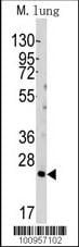
 WB (Western Blot)
(Western blot analysis of lysate from mouse lung tissue lysate, using Mlkl Antibody (C-term). AAA28711 was diluted at 1:1000. A goat anti-rabbit IgG H&L(HRP) at 1:10000 dilution was used as the secondary antibody. Lysate at 20ug.)
WB (Western Blot)
(Western blot analysis of lysate from mouse lung tissue lysate, using Mlkl Antibody (C-term). AAA28711 was diluted at 1:1000. A goat anti-rabbit IgG H&L(HRP) at 1:10000 dilution was used as the secondary antibody. Lysate at 20ug.)
M Mlkl, Polyclonal Antibody (Cat# AAA28711)
Full Name
M Mlkl Antibody (C-term)
Gene Names
Mlkl; 9130019I15Rik
Reactivity
Mouse
Applications
Western Blot
Purity
Peptide Affinity Purified Rabbit Polyclonal Antibody (Pab)
Pricing


 FCM (Flow Cytometry)
(SEPT9 Antibody (A555) flow cytometric analysis of HepG2 cells (right histogram) compared to a negative control cell (left histogram).FITC-conjugated goat-anti-rabbit secondary antibodies were used for the analysis.)
FCM (Flow Cytometry)
(SEPT9 Antibody (A555) flow cytometric analysis of HepG2 cells (right histogram) compared to a negative control cell (left histogram).FITC-conjugated goat-anti-rabbit secondary antibodies were used for the analysis.)
SEPT9, Polyclonal Antibody (Cat# AAA28793)
Full Name
SEPT9 Antibody (C-term)
Gene Names
SEPT9; MSF; MSF1; NAPB; SINT1; PNUTL4; SeptD1; AF17q25
Reactivity
Human
Applications
Western Blot, Immunohistochemistry, Flow Cytometry
Purity
Purified Rabbit Polyclonal Antibody (Pab)
Pricing


 WB (Western Blot)
(Western Blot Analysis: Representative lot data. Lysate from HEK-293 cells was resolved by electrophoresis, transferred to PVDF and probed with anti-PI3 Kinase, p110beta (0.05 ug/mL). Proteins were visualized using donkey anti-rabbit secondary antibody conjugated to HRP and chemiluminescence detection. Arrow indicates PI3 Kinase, p110beta (~110kD).)
WB (Western Blot)
(Western Blot Analysis: Representative lot data. Lysate from HEK-293 cells was resolved by electrophoresis, transferred to PVDF and probed with anti-PI3 Kinase, p110beta (0.05 ug/mL). Proteins were visualized using donkey anti-rabbit secondary antibody conjugated to HRP and chemiluminescence detection. Arrow indicates PI3 Kinase, p110beta (~110kD).)
Phosphoinositide 3 Kinase, p110 beta, Polyclonal Antibody (Cat# AAA26903)
Full Name
Phosphoinositide 3 Kinase, p110 beta (DKFZp779K1237, MGC133043, p110beta, Phosphatidylinositol 3 Kinase Catalytic beta Polypeptide, Phosphatidylinositol-4,5-bisphosphate 3-kinase Catalytic Subunit beta Isoform, Phosphoinositide 3 Kinase Catalytic beta Pol
Gene Names
PIK3CA; MCM; CWS5; MCAP; PI3K; CLOVE; MCMTC; p110-alpha
Reactivity
Human
Applications
Immunocytochemistry, Western Blot, Immunoprecipitation, Immunohistochemistry
Purity
Purified by Immunoaffinity chromatography.
Pricing


 WB (Western Blot)
(TFAM Antibody (C-term) western blot analysis in Hela,Jurkat,K562,MCF-7 cell line lysates (35ug/lane).This demonstrates the TFAM antibody detected the TFAM protein (arrow).)
WB (Western Blot)
(TFAM Antibody (C-term) western blot analysis in Hela,Jurkat,K562,MCF-7 cell line lysates (35ug/lane).This demonstrates the TFAM antibody detected the TFAM protein (arrow).)
TFAM, Polyclonal Antibody (Cat# AAA28696)
Full Name
TFAM Antibody (C-term)
Gene Names
TFAM; TCF6; MTTF1; MTTFA; TCF6L1; TCF6L2; TCF6L3
Reactivity
Human
Applications
Immunohistochemistry, Flow Cytometry, Immunofluorescence, Western Blot
Purity
Peptide Affinity Purified Rabbit Polyclonal Antibody (Pab)
Pricing


 Application Data
(C:FGFR2/isolectinB4 (C) and FGFR1/isolectinB4 (D) staining of apparent mesenchymal cells and the subpopulation of endothelial cells. Virtually all other dispersed apparent mesenchymal cells express FGFR1 and FGFR2 (merged image in E). F: FGFR2 (F) and FGFR1 (G) staining in clustered cells of epithelial origin (inferred by morphology here) demonstrating that epithelial cells express both FGFR1 and FGFR2 (merged image with DAPI staining in H).)
Application Data
(C:FGFR2/isolectinB4 (C) and FGFR1/isolectinB4 (D) staining of apparent mesenchymal cells and the subpopulation of endothelial cells. Virtually all other dispersed apparent mesenchymal cells express FGFR1 and FGFR2 (merged image in E). F: FGFR2 (F) and FGFR1 (G) staining in clustered cells of epithelial origin (inferred by morphology here) demonstrating that epithelial cells express both FGFR1 and FGFR2 (merged image with DAPI staining in H).)
FGFR2, Polyclonal Antibody (Cat# AAA26853)
Full Name
FGFR2, NT (FGFR2, BEK, KGFR, KSAM, Fibroblast growth factor receptor 2, K-sam, Keratinocyte growth factor receptor, CD332) (Biotin)
Gene Names
FGFR2; BEK; JWS; BBDS; CEK3; CFD1; ECT1; KGFR; TK14; TK25; BFR-1; CD332; K-SAM
Reactivity
Human, Monkey, Mouse, Rat
Applications
FC/FACS, EIA, IF, IHC, WB
Purity
Purified by Protein G Affinity Chromatography.
Pricing


 IHC (Immunohistchemistry)
(Immunohistochemistry Analysis: Representative lot data. (Fig. 1 and 2) Paraffin-embedded mouse and human brain tissue was prepared using heat-induced epitope retrieval in citrate buffer, pH 6.0. Immunostaining was performed using a 1:100 dilution. Reactivity was detected using the IHC-Select Detection Kit. Staining pattern appears as cytoplasmic. (Fig. 3 and 4) Paraffin-embedded mouse and mouse olfactory lobe and cerebellum brain tissue was prepared using heat-induced epitope retrieval in citrate buffer, pH 6.0. Immunostaining was performed using a Chicken IgY Antibody 1:100 dilution of Cat. No. AB15894, anti-Tbr2. Reactivity was detected using the IHC-Select Detection Kit. Immunoreactivity seen here is mostly nuclear.)
IHC (Immunohistchemistry)
(Immunohistochemistry Analysis: Representative lot data. (Fig. 1 and 2) Paraffin-embedded mouse and human brain tissue was prepared using heat-induced epitope retrieval in citrate buffer, pH 6.0. Immunostaining was performed using a 1:100 dilution. Reactivity was detected using the IHC-Select Detection Kit. Staining pattern appears as cytoplasmic. (Fig. 3 and 4) Paraffin-embedded mouse and mouse olfactory lobe and cerebellum brain tissue was prepared using heat-induced epitope retrieval in citrate buffer, pH 6.0. Immunostaining was performed using a Chicken IgY Antibody 1:100 dilution of Cat. No. AB15894, anti-Tbr2. Reactivity was detected using the IHC-Select Detection Kit. Immunoreactivity seen here is mostly nuclear.)
EOMES, Polyclonal Antibody (Cat# AAA26893)
Full Name
EOMES (Eomesodermin Homolog, T-box Brain Protein 2, TBR2, T-brain-2, TBR-2)
Gene Names
Eomes; Tbr2; TBR-2; C77258
Reactivity
Mouse, Human, Rat
Applications
Immunohistochemistry, Western Blot
Purity
Purified by affinity chromatography.
Pricing


 IHC (Immunohistchemistry)
(Immunohistochemistry Analysis: Representative lot data. (Fig. 1 and 2) Paraffin-embedded mouse and human brain tissue was prepared using heat-induced epitope retrieval in citrate buffer, pH 6.0. Immunostaining was performed using a 1:100 dilution. Reactivity was detected using the IHC-Select Detection Kit. Staining pattern appears as cytoplasmic. (Fig. 3 and 4) Paraffin-embedded mouse and mouse olfactory lobe and cerebellum brain tissue was prepared using heat-induced epitope retrieval in citrate buffer, pH 6.0. Immunostaining was performed using a Chicken IgY Antibody 1:100 dilution of Cat. No. AB15894, anti-Tbr2. Reactivity was detected using the IHC-Select Detection Kit. Immunoreactivity seen here is mostly nuclear.)
IHC (Immunohistchemistry)
(Immunohistochemistry Analysis: Representative lot data. (Fig. 1 and 2) Paraffin-embedded mouse and human brain tissue was prepared using heat-induced epitope retrieval in citrate buffer, pH 6.0. Immunostaining was performed using a 1:100 dilution. Reactivity was detected using the IHC-Select Detection Kit. Staining pattern appears as cytoplasmic. (Fig. 3 and 4) Paraffin-embedded mouse and mouse olfactory lobe and cerebellum brain tissue was prepared using heat-induced epitope retrieval in citrate buffer, pH 6.0. Immunostaining was performed using a Chicken IgY Antibody 1:100 dilution of Cat. No. AB15894, anti-Tbr2. Reactivity was detected using the IHC-Select Detection Kit. Immunoreactivity seen here is mostly nuclear.)
EOMES, Polyclonal Antibody (Cat# AAA26883)
Full Name
EOMES (Eomesodermin Homolog, T-box Brain Protein 2, TBR2, T-brain-2, TBR-2)
Gene Names
Eomes; Tbr2; TBR-2; C77258
Reactivity
Mouse, Human, Rat
Applications
Immunohistochemistry, Western Blot
Purity
Purified by affinity chromatography.
Pricing


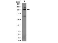
 WB (Western Blot)
(Western blot analysis of Bid (arrow) using rabbit polyclonal Bid Antibody (BH3). 293 cell lysates (2 ug/lane) either nontransfected (Lane 1) or transiently transfected (Lane 2) with the Bid gene.)
WB (Western Blot)
(Western blot analysis of Bid (arrow) using rabbit polyclonal Bid Antibody (BH3). 293 cell lysates (2 ug/lane) either nontransfected (Lane 1) or transiently transfected (Lane 2) with the Bid gene.)
Bid, Polyclonal Antibody (Cat# AAA28769)
Full Name
Bid Antibody (BH3 Domain Specific)
Gene Names
BID; FP497
Reactivity
Human, mouse
Applications
Western Blot, Immunohistochemistry
Purity
Purified Rabbit Polyclonal Antibody (Pab)
Pricing


 WB (Western Blot)
(Western blot of Latexin (arrow) in 293 cell lysates (2 ug/lane) either nontransfected (Lane 1) or transiently transfected with the LXN gene (Lane 2))
WB (Western Blot)
(Western blot of Latexin (arrow) in 293 cell lysates (2 ug/lane) either nontransfected (Lane 1) or transiently transfected with the LXN gene (Lane 2))
Latexin / MUM, Polyclonal Antibody (Cat# AAA12323)
Full Name
Rabbit Polyclonal to Human Latexin / MUM
Gene Names
LXN; ECI; TCI
Reactivity
Human
Applications
Immunohistochemistry, Western Blot, Flow Cytometry
Purity
Immunoaffinity Purified
Pricing


 WB (Western Blot)
(Formalin-fixed and paraffin-embedded human brain tissue reacted with OLR1 Antibody (Center), which was peroxidase-conjugated to the secondary antibody, followed by DAB staining. This data demonstrates the use of this antibody for immunohistochemistry; clinical relevance has not been evaluated.)
WB (Western Blot)
(Formalin-fixed and paraffin-embedded human brain tissue reacted with OLR1 Antibody (Center), which was peroxidase-conjugated to the secondary antibody, followed by DAB staining. This data demonstrates the use of this antibody for immunohistochemistry; clinical relevance has not been evaluated.)
OLR1, Polyclonal Antibody (Cat# AAA28737)
Full Name
OLR1 Antibody (Center)
Gene Names
OLR1; LOX1; LOXIN; SLOX1; CLEC8A; SCARE1
Reactivity
Human
Applications
Western Blot, Immunohistochemistry
Purity
Peptide Affinity Purified Rabbit Polyclonal Antibody (Pab)
Pricing


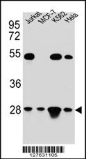
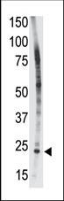
 WB (Western Blot)
(Western blot analysis of LIN28B Antibody (N-term) in HL60 cell line lysates (35 ug/lane). LIN28B (arrow) was detected using the purified Pab.)
WB (Western Blot)
(Western blot analysis of LIN28B Antibody (N-term) in HL60 cell line lysates (35 ug/lane). LIN28B (arrow) was detected using the purified Pab.)
LIN28B, Polyclonal Antibody (Cat# AAA12320)
Full Name
Rabbit Polyclonal to Human LIN28B
Gene Names
LIN28B; CSDD2
Reactivity
Human
Applications
Immunohistochemistry, Immunofluorescence, Western Blot, Flow Cytometry
Purity
Ammonium sulfate precipitation
Pricing


 IF (Immunofluorescence)
(Fluorescent confocal image of MCF-7 cell stained with BHLH3 Antibody (N-term). MCF-7 cells were fixed with 4% PFA (20 min), permeabilized with Triton X-100 (0.1%, 10 min), then incubated with BHLH3 primary antibody (1:25, 1 h at 37 degree). For secondary antibody, Alexa Fluor 488 conjugated donkey anti-rabbit antibody (green) was used (1:400, 50 min at 37 degree).Cytoplasmic actin was counterstained with Alexa Fluor 555 (red) conjugated Phalloidin (7units/ml, 1 h at 37 degree). Nuclei were counterstained with DAPI (blue) (10 ug/ml, 10 min).BHLH3 immunoreactivity is localized to nucleus significantly.)
IF (Immunofluorescence)
(Fluorescent confocal image of MCF-7 cell stained with BHLH3 Antibody (N-term). MCF-7 cells were fixed with 4% PFA (20 min), permeabilized with Triton X-100 (0.1%, 10 min), then incubated with BHLH3 primary antibody (1:25, 1 h at 37 degree). For secondary antibody, Alexa Fluor 488 conjugated donkey anti-rabbit antibody (green) was used (1:400, 50 min at 37 degree).Cytoplasmic actin was counterstained with Alexa Fluor 555 (red) conjugated Phalloidin (7units/ml, 1 h at 37 degree). Nuclei were counterstained with DAPI (blue) (10 ug/ml, 10 min).BHLH3 immunoreactivity is localized to nucleus significantly.)
BHLH3, Polyclonal Antibody (Cat# AAA28734)
Full Name
BHLH3 Antibody (N-term)
Gene Names
BHLHE41; DEC2; hDEC2; BHLHB3; SHARP1
Reactivity
Human, Mouse
Applications
Flow Cytometry, Immunofluorescence, Western Blot
Purity
This antibody is purified through a protein A column, followed by peptide affinity purification.
Pricing


 IHC (Immunohistchemistry)
(Immunohistochemistry Analysis: Representative lot data. (Fig. 1 and 2) Paraffin-embedded mouse and human brain tissue was prepared using heat-induced epitope retrieval in citrate buffer, pH 6.0. Immunostaining was performed using a 1:100 dilution. Reactivity was detected using the IHC-Select Detection Kit. Staining pattern appears as cytoplasmic. (Fig. 3 and 4) Paraffin-embedded mouse and mouse olfactory lobe and cerebellum brain tissue was prepared using heat-induced epitope retrieval in citrate buffer, pH 6.0. Immunostaining was performed using a Chicken IgY Antibody 1:100 dilution of Cat. No. AB15894, anti-Tbr2. Reactivity was detected using the IHC-Select Detection Kit. Immunoreactivity seen here is mostly nuclear.)
IHC (Immunohistchemistry)
(Immunohistochemistry Analysis: Representative lot data. (Fig. 1 and 2) Paraffin-embedded mouse and human brain tissue was prepared using heat-induced epitope retrieval in citrate buffer, pH 6.0. Immunostaining was performed using a 1:100 dilution. Reactivity was detected using the IHC-Select Detection Kit. Staining pattern appears as cytoplasmic. (Fig. 3 and 4) Paraffin-embedded mouse and mouse olfactory lobe and cerebellum brain tissue was prepared using heat-induced epitope retrieval in citrate buffer, pH 6.0. Immunostaining was performed using a Chicken IgY Antibody 1:100 dilution of Cat. No. AB15894, anti-Tbr2. Reactivity was detected using the IHC-Select Detection Kit. Immunoreactivity seen here is mostly nuclear.)
EOMES, Polyclonal Antibody (Cat# AAA26884)
Full Name
EOMES (Eomesodermin Homolog, T-box Brain Protein 2, TBR2, T-brain-2, TBR-2)
Gene Names
Eomes; Tbr2; TBR-2; C77258
Reactivity
Mouse, Human, Rat
Applications
Immunohistochemistry, Western Blot
Purity
Purified by affinity chromatography.
Pricing


 WB (Western Blot)
(Western blot analysis of CARD6 Antibody (Center) in MDA-MB231 cell line lysates (35ug/lane). CARD6 (arrow) was detected using the purified Pab.)
WB (Western Blot)
(Western blot analysis of CARD6 Antibody (Center) in MDA-MB231 cell line lysates (35ug/lane). CARD6 (arrow) was detected using the purified Pab.)
CARD6, Polyclonal Antibody (Cat# AAA28742)
Full Name
CARD6 Antibody (Center)
Gene Names
CARD6; CINCIN1
Reactivity
Human
Applications
Immunohistochemistry, Western Blot
Purity
Peptide Affinity Purified Rabbit Polyclonal Antibody (Pab)
Pricing

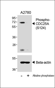


 IHC (Immunohistchemistry)
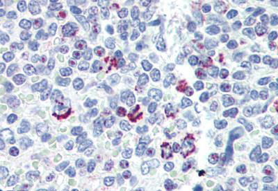
(Immunohistochemical analysis of (AAA28653) on paraffin-embedded Human breastcarcinoma tissue. Tissue was fixed withformaldehyde at room temperature. Heatinduced epitope retrieval was performed byEDTA buffer (pH9. 0). Samples wereincubated with primary antibody(1:100) for 1hour at room temperature. Undiluted CRFAnti-Polyvalent HRP Polymer antibody wasused as the secondary antibody.)
IHC (Immunohistchemistry)
(Immunohistochemical analysis of (AAA28653) on paraffin-embedded Human breastcarcinoma tissue. Tissue was fixed withformaldehyde at room temperature. Heatinduced epitope retrieval was performed byEDTA buffer (pH9. 0). Samples wereincubated with primary antibody(1:100) for 1hour at room temperature. Undiluted CRFAnti-Polyvalent HRP Polymer antibody wasused as the secondary antibody.)
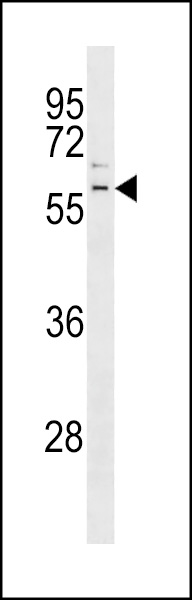
Phospho-CDC25A (S124), Polyclonal Antibody (Cat# AAA28653)
Full Name
Phospho-CDC25A (S124) Antibody
Gene Names
CDC25A; CDC25A2
Reactivity
Human (Predicted Reactivity: Rat)
Applications
WB, EIA, IHC
Purity
Peptide Affinity Purified Rabbit Polyclonal Antibody (Pab)
Pricing


 IHC (Immunohistchemistry)
(Immunohistochemistry Analysis: Representative lot data. (Fig. 1 and 2) Paraffin-embedded mouse and human brain tissue was prepared using heat-induced epitope retrieval in citrate buffer, pH 6.0. Immunostaining was performed using a 1:100 dilution. Reactivity was detected using the IHC-Select Detection Kit. Staining pattern appears as cytoplasmic. (Fig. 3 and 4) Paraffin-embedded mouse and mouse olfactory lobe and cerebellum brain tissue was prepared using heat-induced epitope retrieval in citrate buffer, pH 6.0. Immunostaining was performed using a Chicken IgY Antibody 1:100 dilution of Cat. No. AB15894, anti-Tbr2. Reactivity was detected using the IHC-Select Detection Kit. Immunoreactivity seen here is mostly nuclear.)
IHC (Immunohistchemistry)
(Immunohistochemistry Analysis: Representative lot data. (Fig. 1 and 2) Paraffin-embedded mouse and human brain tissue was prepared using heat-induced epitope retrieval in citrate buffer, pH 6.0. Immunostaining was performed using a 1:100 dilution. Reactivity was detected using the IHC-Select Detection Kit. Staining pattern appears as cytoplasmic. (Fig. 3 and 4) Paraffin-embedded mouse and mouse olfactory lobe and cerebellum brain tissue was prepared using heat-induced epitope retrieval in citrate buffer, pH 6.0. Immunostaining was performed using a Chicken IgY Antibody 1:100 dilution of Cat. No. AB15894, anti-Tbr2. Reactivity was detected using the IHC-Select Detection Kit. Immunoreactivity seen here is mostly nuclear.)
EOMES, Polyclonal Antibody (Cat# AAA26891)
Full Name
EOMES (Eomesodermin Homolog, T-box Brain Protein 2, TBR2, T-brain-2, TBR-2)
Gene Names
Eomes; Tbr2; TBR-2; C77258
Reactivity
Mouse, Human, Rat
Applications
Immunohistochemistry, Western Blot
Purity
Purified by affinity chromatography.
Pricing


 IHC (Immunohistchemistry)
(Immunohistochemistry Analysis: Representative lot data. (Fig. 1 and 2) Paraffin-embedded mouse and human brain tissue was prepared using heat-induced epitope retrieval in citrate buffer, pH 6.0. Immunostaining was performed using a 1:100 dilution. Reactivity was detected using the IHC-Select Detection Kit. Staining pattern appears as cytoplasmic. (Fig. 3 and 4) Paraffin-embedded mouse and mouse olfactory lobe and cerebellum brain tissue was prepared using heat-induced epitope retrieval in citrate buffer, pH 6.0. Immunostaining was performed using a Chicken IgY Antibody 1:100 dilution of Cat. No. AB15894, anti-Tbr2. Reactivity was detected using the IHC-Select Detection Kit. Immunoreactivity seen here is mostly nuclear.)
IHC (Immunohistchemistry)
(Immunohistochemistry Analysis: Representative lot data. (Fig. 1 and 2) Paraffin-embedded mouse and human brain tissue was prepared using heat-induced epitope retrieval in citrate buffer, pH 6.0. Immunostaining was performed using a 1:100 dilution. Reactivity was detected using the IHC-Select Detection Kit. Staining pattern appears as cytoplasmic. (Fig. 3 and 4) Paraffin-embedded mouse and mouse olfactory lobe and cerebellum brain tissue was prepared using heat-induced epitope retrieval in citrate buffer, pH 6.0. Immunostaining was performed using a Chicken IgY Antibody 1:100 dilution of Cat. No. AB15894, anti-Tbr2. Reactivity was detected using the IHC-Select Detection Kit. Immunoreactivity seen here is mostly nuclear.)
EOMES, Polyclonal Antibody (Cat# AAA26892)
Full Name
EOMES (Eomesodermin Homolog, T-box Brain Protein 2, TBR2, T-brain-2, TBR-2)
Gene Names
Eomes; Tbr2; TBR-2; C77258
Reactivity
Mouse, Human, Rat
Applications
Immunohistochemistry, Western Blot
Purity
Purified by affinity chromatography.
Pricing


 Application Data
(C:FGFR2/isolectinB4 (C) and FGFR1/isolectinB4 (D) staining of apparent mesenchymal cells and the subpopulation of endothelial cells. Virtually all other dispersed apparent mesenchymal cells express FGFR1 and FGFR2 (merged image in E). F: FGFR2 (F) and FGFR1 (G) staining in clustered cells of epithelial origin (inferred by morphology here) demonstrating that epithelial cells express both FGFR1 and FGFR2 (merged image with DAPI staining in H).)
Application Data
(C:FGFR2/isolectinB4 (C) and FGFR1/isolectinB4 (D) staining of apparent mesenchymal cells and the subpopulation of endothelial cells. Virtually all other dispersed apparent mesenchymal cells express FGFR1 and FGFR2 (merged image in E). F: FGFR2 (F) and FGFR1 (G) staining in clustered cells of epithelial origin (inferred by morphology here) demonstrating that epithelial cells express both FGFR1 and FGFR2 (merged image with DAPI staining in H).)
FGFR2, Polyclonal Antibody (Cat# AAA26855)
Full Name
FGFR2, NT (FGFR2, BEK, KGFR, KSAM, Fibroblast growth factor receptor 2, K-sam, Keratinocyte growth factor receptor, CD332) (Azide free) (HRP)
Gene Names
FGFR2; BEK; JWS; BBDS; CEK3; CFD1; ECT1; KGFR; TK14; TK25; BFR-1; CD332; K-SAM
Reactivity
Human, Monkey, Mouse, Rat
Applications
IHC, EIA, WB
Purity
Purified by Protein G Affinity Chromatography.
Pricing


 IHC (Immunohistchemistry)
(Formalin-fixed and paraffin-embedded human testis tissue reacted with PARK2 (Parkin) antibody (C-term) , which was peroxidase-conjugated to the secondary antibody, followed by DAB staining. This data demonstrates the use of this antibody for immunohistochemistry; clinical relevance has not been evaluated.)
IHC (Immunohistchemistry)
(Formalin-fixed and paraffin-embedded human testis tissue reacted with PARK2 (Parkin) antibody (C-term) , which was peroxidase-conjugated to the secondary antibody, followed by DAB staining. This data demonstrates the use of this antibody for immunohistochemistry; clinical relevance has not been evaluated.)
Parkin, Polyclonal Antibody (Cat# AAA28668)
Full Name
Parkin Antibody (C-term)
Gene Names
PARK2; PDJ; PRKN; AR-JP; LPRS2
Reactivity
Human, mouse
Applications
WB, EIA, IF, FC/FACS, IHC
Purity
Purified Rabbit Polyclonal Antibody (Pab)
Pricing


 IP (Immunoprecipitation)
(Immunoprecipitation(IP) of CRYAB by using monoclonal anti-CRYAB antibodies (Negative control: IP without adding anti-CRYAB antibody.). For each experiment, 500ul of DDK tagged CRYAB overexpression lysates (at 1:5 dilution with HEK293T lysate), 2 ug of anti-CRYAB antibody and 20ul (0.1 mg) of goat anti-mouse conjugated magnetic beads were mixed and incubated overnight. After extensive wash to remove any non-specific binding, the immuno-precipitated products were analyzed with rabbit anti-DDK polyclonal antibody.)
IP (Immunoprecipitation)
(Immunoprecipitation(IP) of CRYAB by using monoclonal anti-CRYAB antibodies (Negative control: IP without adding anti-CRYAB antibody.). For each experiment, 500ul of DDK tagged CRYAB overexpression lysates (at 1:5 dilution with HEK293T lysate), 2 ug of anti-CRYAB antibody and 20ul (0.1 mg) of goat anti-mouse conjugated magnetic beads were mixed and incubated overnight. After extensive wash to remove any non-specific binding, the immuno-precipitated products were analyzed with rabbit anti-DDK polyclonal antibody.)
CRYAB / Alpha B Crystallin, Monoclonal Antibody (Cat# AAA12373)
Full Name
Mouse Monoclonal [clone 6D11] (IgG1) to Human CRYAB / Alpha B Crystallin
Gene Names
CRYAB; MFM2; CRYA2; CTPP2; HSPB5; CMD1II; CTRCT16; HEL-S-101
Reactivity
Human, Monkey, Rat
Applications
Immunohistochemistry, Immunofluorescence, Western Blot, Immunoprecipitation, Flow Cytometry
Purity
Protein A/G Purified
Pricing


 IHC (Immunohistchemistry)
(Formalin-fixed and paraffin-embedded human cancer tissue reacted with the primary antibody, which was peroxidase-conjugated to the secondary antibody, followed by DAB staining. This data demonstrates the use of this antibody for immunohistochemistry; clinical relevance has not been evaluated. BC = breast carcinoma; HC = hepatocarcinoma.)
IHC (Immunohistchemistry)
(Formalin-fixed and paraffin-embedded human cancer tissue reacted with the primary antibody, which was peroxidase-conjugated to the secondary antibody, followed by DAB staining. This data demonstrates the use of this antibody for immunohistochemistry; clinical relevance has not been evaluated. BC = breast carcinoma; HC = hepatocarcinoma.)
PACSIN2, Polyclonal Antibody (Cat# AAA28758)
Full Name
PACSIN2 Antibody (C-term)
Gene Names
PACSIN2; SDPII
Reactivity
Human, mouse
Applications
Immunohistochemistry, Western Blot, Immunofluorescence
Purity
Purified Rabbit Polyclonal Antibody (Pab)
Pricing


 IHC (Immunohistchemistry)
(Immunohistochemistry Analysis: Representative lot data. (Fig. 1 and 2) Paraffin-embedded mouse and human brain tissue was prepared using heat-induced epitope retrieval in citrate buffer, pH 6.0. Immunostaining was performed using a 1:100 dilution. Reactivity was detected using the IHC-Select Detection Kit. Staining pattern appears as cytoplasmic. (Fig. 3 and 4) Paraffin-embedded mouse and mouse olfactory lobe and cerebellum brain tissue was prepared using heat-induced epitope retrieval in citrate buffer, pH 6.0. Immunostaining was performed using a Chicken IgY Antibody 1:100 dilution of Cat. No. AB15894, anti-Tbr2. Reactivity was detected using the IHC-Select Detection Kit. Immunoreactivity seen here is mostly nuclear.)
IHC (Immunohistchemistry)
(Immunohistochemistry Analysis: Representative lot data. (Fig. 1 and 2) Paraffin-embedded mouse and human brain tissue was prepared using heat-induced epitope retrieval in citrate buffer, pH 6.0. Immunostaining was performed using a 1:100 dilution. Reactivity was detected using the IHC-Select Detection Kit. Staining pattern appears as cytoplasmic. (Fig. 3 and 4) Paraffin-embedded mouse and mouse olfactory lobe and cerebellum brain tissue was prepared using heat-induced epitope retrieval in citrate buffer, pH 6.0. Immunostaining was performed using a Chicken IgY Antibody 1:100 dilution of Cat. No. AB15894, anti-Tbr2. Reactivity was detected using the IHC-Select Detection Kit. Immunoreactivity seen here is mostly nuclear.)
EOMES, Polyclonal Antibody (Cat# AAA26886)
Full Name
EOMES (Eomesodermin Homolog, T-box Brain Protein 2, TBR2, T-brain-2, TBR-2)
Gene Names
Eomes; Tbr2; TBR-2; C77258
Reactivity
Mouse, Human, Rat
Applications
Immunohistochemistry, Western Blot
Purity
Purified by affinity chromatography.
Pricing


 IF (Immunofluorescence)
(Immunofluorescent analysis of 4% paraformaldehyde-fixed, 0.1% Triton X-100 permeabilized MCF-7 (human breast cancer cell line) cells labeling Pdx1 with at 1:25 dilution, followed by DyLight 488-conjugated IgG goat anti-rabbit secondary antibody at 1:200 dilution (green). Immunofluorescence image showing cytoplasm staining on MCF-7 cell line. Cytoplasmic actin is detected with DyLight 554 Phalloidin (PD18466410) at 1:100 dilution (red). The nuclear counter stain is DAPI (blue).)
IF (Immunofluorescence)
(Immunofluorescent analysis of 4% paraformaldehyde-fixed, 0.1% Triton X-100 permeabilized MCF-7 (human breast cancer cell line) cells labeling Pdx1 with at 1:25 dilution, followed by DyLight 488-conjugated IgG goat anti-rabbit secondary antibody at 1:200 dilution (green). Immunofluorescence image showing cytoplasm staining on MCF-7 cell line. Cytoplasmic actin is detected with DyLight 554 Phalloidin (PD18466410) at 1:100 dilution (red). The nuclear counter stain is DAPI (blue).)
OPN-a/b, Polyclonal Antibody (Cat# AAA26865)
Full Name
OPN-a/b, NT (SPP1, BNSP, OPN, Osteopontin, Bone sialoprotein 1, Nephropontin, Secreted phosphoprotein 1, Urinary stone protein, Uropontin) (PE)
Gene Names
SPP1; OPN; BNSP; BSPI; ETA-1
Reactivity
Human
Applications
WB, IHC, IF
Purity
Purified by Protein A and Peptide Affinity Chromatography.
Pricing


 IHC (Immunohistchemistry)
(Immunohistochemistry Analysis: Representative lot data. (Fig. 1 and 2) Paraffin-embedded mouse and human brain tissue was prepared using heat-induced epitope retrieval in citrate buffer, pH 6.0. Immunostaining was performed using a 1:100 dilution of AAA14728. Reactivity was detected using the IHC-Select Detection Kit. Staining pattern appears as cytoplasmic. (Fig. 3 and 4) Paraffin-embedded mouse and mouse olfactory lobe and cerebellum brain tissue was prepared using heat-induced epitope retrieval in citrate buffer, pH 6.0. Immunostaining was performed using a Chicken IgY Antibody 1:100 dilution of Cat. No. AB15894, anti-Tbr2. Reactivity was detected using the IHC-Select Detection Kit . Immunoreactivity seen here is mostly nuclear.)
IHC (Immunohistchemistry)
(Immunohistochemistry Analysis: Representative lot data. (Fig. 1 and 2) Paraffin-embedded mouse and human brain tissue was prepared using heat-induced epitope retrieval in citrate buffer, pH 6.0. Immunostaining was performed using a 1:100 dilution of AAA14728. Reactivity was detected using the IHC-Select Detection Kit. Staining pattern appears as cytoplasmic. (Fig. 3 and 4) Paraffin-embedded mouse and mouse olfactory lobe and cerebellum brain tissue was prepared using heat-induced epitope retrieval in citrate buffer, pH 6.0. Immunostaining was performed using a Chicken IgY Antibody 1:100 dilution of Cat. No. AB15894, anti-Tbr2. Reactivity was detected using the IHC-Select Detection Kit . Immunoreactivity seen here is mostly nuclear.)
EOMES, Polyclonal Antibody (Cat# AAA14728)
Full Name
EOMES, NT (Eomesodermin Homolog, T-box Brain Protein 2, TBR2, T-brain-2, TBR-2)
Gene Names
EOMES; TBR2
Reactivity
Human, Mouse, Rat
Applications
Western Blot, Immunohistochemistry
Purity
Affinity Purified
Purified by affinity chromatography.
Purified by affinity chromatography.
Pricing


 FCM (Flow Cytometry)
(AGXT Antibody (Center) flow cytometric analysis of HepG2 cells (right histogram) compared to a negative control cell (left histogram).FITC-conjugated goat-anti-rabbit secondary antibodies were used for the analysis.)
FCM (Flow Cytometry)
(AGXT Antibody (Center) flow cytometric analysis of HepG2 cells (right histogram) compared to a negative control cell (left histogram).FITC-conjugated goat-anti-rabbit secondary antibodies were used for the analysis.)
AGXT, Polyclonal Antibody (Cat# AAA28732)
Full Name
AGXT Antibody (Center)
Gene Names
AGXT; AGT; PH1; SPT; AGT1; SPAT; TLH6; AGXT1
Reactivity
Human
Applications
Western Blot, Immunohistochemistry, Flow Cytometry
Purity
Peptide Affinity Purified Rabbit Polyclonal Antibody (Pab)
Pricing


 WB (Western Blot)
(Western blot analysis of anti-NEK2 Antibody (Center) in HL60 cell line lysates (35ug/lane). NEK2(arrow) was detected using the purified Pab.)
WB (Western Blot)
(Western blot analysis of anti-NEK2 Antibody (Center) in HL60 cell line lysates (35ug/lane). NEK2(arrow) was detected using the purified Pab.)
NEK2, Polyclonal Antibody (Cat# AAA28694)
Full Name
NEK2 Antibody (Center)
Gene Names
NEK2; NLK1; RP67; NEK2A; HsPK21; PPP1R111
Reactivity
Human
Applications
Western Blot, Immunofluorescence, Immunohistochemistry
Purity
Purified Rabbit Polyclonal Antibody (Pab)
Pricing

 WB (Western Blot)
(WB of AAA24032 with 1:500 antibody dilution in DiluObuffer . Apparent MW is 78kDa.)
WB (Western Blot)
(WB of AAA24032 with 1:500 antibody dilution in DiluObuffer . Apparent MW is 78kDa.)
SPNS2, Polyclonal Antibody (Cat# AAA24032)
Full Name
SPNS2 Antibody
Reactivity
D. melanogaster, Monkey, Mouse, Rat
Applications
EIA, IHC, IP, WB
Pricing


 IF (Immunofluorescence)
(Immunofluorescent analysis of 4% paraformaldehyde-fixed, 0.1% Triton X-100 permeabilized MCF-7 (human breast cancer cell line) cells labeling Pdx1 with at 1:25 dilution, followed by DyLight 488-conjugated IgG goat anti-rabbit secondary antibody at 1:200 dilution (green). Immunofluorescence image showing cytoplasm staining on MCF-7 cell line. Cytoplasmic actin is detected with DyLight 554 Phalloidin (PD18466410) at 1:100 dilution (red). The nuclear counter stain is DAPI (blue).)
IF (Immunofluorescence)
(Immunofluorescent analysis of 4% paraformaldehyde-fixed, 0.1% Triton X-100 permeabilized MCF-7 (human breast cancer cell line) cells labeling Pdx1 with at 1:25 dilution, followed by DyLight 488-conjugated IgG goat anti-rabbit secondary antibody at 1:200 dilution (green). Immunofluorescence image showing cytoplasm staining on MCF-7 cell line. Cytoplasmic actin is detected with DyLight 554 Phalloidin (PD18466410) at 1:100 dilution (red). The nuclear counter stain is DAPI (blue).)
OPN-a/b, Polyclonal Antibody (Cat# AAA26857)
Full Name
OPN-a/b, NT (SPP1, BNSP, OPN, Osteopontin, Bone sialoprotein 1, Nephropontin, Secreted phosphoprotein 1, Urinary stone protein, Uropontin) (APC)
Gene Names
SPP1; OPN; BNSP; BSPI; ETA-1
Reactivity
Human
Applications
WB, IHC, IF
Purity
Purified by Protein A and Peptide Affinity Chromatography.
Pricing


 FCM (Flow Cytometry)
(SYP Antibody (C-term) flow cytometric analysis of Neuro-2a cells (right histogram) compared to a negative control cell (left histogram).FITC-conjugated goat-anti-rabbit secondary antibodies were used for the analysis.)
FCM (Flow Cytometry)
(SYP Antibody (C-term) flow cytometric analysis of Neuro-2a cells (right histogram) compared to a negative control cell (left histogram).FITC-conjugated goat-anti-rabbit secondary antibodies were used for the analysis.)
SYP, Polyclonal Antibody (Cat# AAA28723)
Full Name
SYP Antibody (C-term)
Gene Names
SYP; MRX96; MRXSYP
Reactivity
Human, mouse (Predicted Reactivity: Rat)
Applications
Flow Cytometry, Immunofluorescence, Immunohistochemistry, Western Blot
Purity
Peptide Affinity Purified Rabbit Polyclonal Antibody (Pab)
Pricing


 IF (Immunofluorescence)
(Immunofluorescent analysis of 4% paraformaldehyde-fixed, 0.1% Triton X-100 permeabilized MCF-7 (human breast cancer cell line) cells labeling Pdx1 with at 1:25 dilution, followed by DyLight 488-conjugated IgG goat anti-rabbit secondary antibody at 1:200 dilution (green). Immunofluorescence image showing cytoplasm staining on MCF-7 cell line. Cytoplasmic actin is detected with DyLight 554 Phalloidin (PD18466410) at 1:100 dilution (red). The nuclear counter stain is DAPI (blue).)
IF (Immunofluorescence)
(Immunofluorescent analysis of 4% paraformaldehyde-fixed, 0.1% Triton X-100 permeabilized MCF-7 (human breast cancer cell line) cells labeling Pdx1 with at 1:25 dilution, followed by DyLight 488-conjugated IgG goat anti-rabbit secondary antibody at 1:200 dilution (green). Immunofluorescence image showing cytoplasm staining on MCF-7 cell line. Cytoplasmic actin is detected with DyLight 554 Phalloidin (PD18466410) at 1:100 dilution (red). The nuclear counter stain is DAPI (blue).)
OPN-a/b, Polyclonal Antibody (Cat# AAA26861)
Full Name
OPN-a/b, NT (SPP1, BNSP, OPN, Osteopontin, Bone sialoprotein 1, Nephropontin, Secreted phosphoprotein 1, Urinary stone protein, Uropontin) (MaxLight 490)
Gene Names
SPP1; OPN; BNSP; BSPI; ETA-1
Reactivity
Human
Applications
WB, IHC, IF
Purity
Purified by Protein A and Peptide Affinity Chromatography.
Pricing


 Application Data
(C:FGFR2/isolectinB4 (C) and FGFR1/isolectinB4 (D) staining of apparent mesenchymal cells and the subpopulation of endothelial cells. Virtually all other dispersed apparent mesenchymal cells express FGFR1 and FGFR2 (merged image in E). F: FGFR2 (F) and FGFR1 (G) staining in clustered cells of epithelial origin (inferred by morphology here) demonstrating that epithelial cells express both FGFR1 and FGFR2 (merged image with DAPI staining in H).)
Application Data
(C:FGFR2/isolectinB4 (C) and FGFR1/isolectinB4 (D) staining of apparent mesenchymal cells and the subpopulation of endothelial cells. Virtually all other dispersed apparent mesenchymal cells express FGFR1 and FGFR2 (merged image in E). F: FGFR2 (F) and FGFR1 (G) staining in clustered cells of epithelial origin (inferred by morphology here) demonstrating that epithelial cells express both FGFR1 and FGFR2 (merged image with DAPI staining in H).)
FGFR2, Polyclonal Antibody (Cat# AAA14790)
Full Name
FGFR2, NT (FGFR2, BEK, KGFR, KSAM, Fibroblast growth factor receptor 2, K-sam, Keratinocyte growth factor receptor, CD332)
Gene Names
FGFR2; BEK; JWS; BBDS; CEK3; CFD1; ECT1; KGFR; TK14; TK25; BFR-1; CD332; K-SAM
Reactivity
Human, Monkey, Mouse, Rat
Applications
EL/EIA, WB, IHC, FC/FACS, IF
Purity
Affinity Purified
Purified by Protein A affinity chromatography.
Purified by Protein A affinity chromatography.
Pricing


 WB (Western Blot)
(The anti-SUMO2/3 C-term Pab is used in Western blot to detect SUMO2/3 in HeLa cell lysate.)
WB (Western Blot)
(The anti-SUMO2/3 C-term Pab is used in Western blot to detect SUMO2/3 in HeLa cell lysate.)
SUMO2/3, Polyclonal Antibody (Cat# AAA28663)
Full Name
SUMO2/3 Antibody (C-term)
Gene Names
SUMO3; SMT3A; Smt3B; SMT3H1; SUMO-3
Reactivity
Human, mouse (Predicted Reactivity: Xenopus, Zebrafish, Bovine, Chicken, Hamster, Monkey, Pig, Rat)
Applications
IF, EIA, WB, IHC
Purity
Purified Rabbit Polyclonal Antibody (Pab)
Pricing


 IF (Immunofluorescence)
(Immunofluorescent analysis of 4% paraformaldehyde-fixed, 0.1% Triton X-100 permeabilized MCF-7 (human breast cancer cell line) cells labeling Pdx1 with at 1:25 dilution, followed by DyLight 488-conjugated IgG goat anti-rabbit secondary antibody at 1:200 dilution (green). Immunofluorescence image showing cytoplasm staining on MCF-7 cell line. Cytoplasmic actin is detected with DyLight 554 Phalloidin (PD18466410) at 1:100 dilution (red). The nuclear counter stain is DAPI (blue).)
IF (Immunofluorescence)
(Immunofluorescent analysis of 4% paraformaldehyde-fixed, 0.1% Triton X-100 permeabilized MCF-7 (human breast cancer cell line) cells labeling Pdx1 with at 1:25 dilution, followed by DyLight 488-conjugated IgG goat anti-rabbit secondary antibody at 1:200 dilution (green). Immunofluorescence image showing cytoplasm staining on MCF-7 cell line. Cytoplasmic actin is detected with DyLight 554 Phalloidin (PD18466410) at 1:100 dilution (red). The nuclear counter stain is DAPI (blue).)
OPN-a/b, Polyclonal Antibody (Cat# AAA26863)
Full Name
OPN-a/b, NT (SPP1, BNSP, OPN, Osteopontin, Bone sialoprotein 1, Nephropontin, Secreted phosphoprotein 1, Urinary stone protein, Uropontin) (MaxLight 650)
Gene Names
SPP1; OPN; BNSP; BSPI; ETA-1
Reactivity
Human
Applications
WB, IHC, IF
Purity
Purified by Protein A and Peptide Affinity Chromatography.
Pricing


 IF (Immunofluorescence)
(Immunofluorescent analysis of 4% paraformaldehyde-fixed, 0.1% Triton X-100 permeabilized MCF-7 (human breast cancer cell line) cells labeling Pdx1 with at 1:25 dilution, followed by DyLight 488-conjugated IgG goat anti-rabbit secondary antibody at 1:200 dilution (green). Immunofluorescence image showing cytoplasm staining on MCF-7 cell line. Cytoplasmic actin is detected with DyLight 554 Phalloidin (PD18466410) at 1:100 dilution (red). The nuclear counter stain is DAPI (blue).)
IF (Immunofluorescence)
(Immunofluorescent analysis of 4% paraformaldehyde-fixed, 0.1% Triton X-100 permeabilized MCF-7 (human breast cancer cell line) cells labeling Pdx1 with at 1:25 dilution, followed by DyLight 488-conjugated IgG goat anti-rabbit secondary antibody at 1:200 dilution (green). Immunofluorescence image showing cytoplasm staining on MCF-7 cell line. Cytoplasmic actin is detected with DyLight 554 Phalloidin (PD18466410) at 1:100 dilution (red). The nuclear counter stain is DAPI (blue).)
OPN-a/b, Polyclonal Antibody (Cat# AAA26864)
Full Name
OPN-a/b, NT (SPP1, BNSP, OPN, Osteopontin, Bone sialoprotein 1, Nephropontin, Secreted phosphoprotein 1, Urinary stone protein, Uropontin) (MaxLight 750)
Gene Names
SPP1; OPN; BNSP; BSPI; ETA-1
Reactivity
Human
Applications
WB, IHC, IF
Purity
Purified by Protein A and Peptide Affinity Chromatography.
Pricing


 IF (Immunofluorescence)
(Immunofluorescent analysis of 4% paraformaldehyde-fixed, 0.1% Triton X-100 permeabilized MCF-7 (human breast cancer cell line) cells labeling Pdx1 with at 1:25 dilution, followed by DyLight 488-conjugated IgG goat anti-rabbit secondary antibody at 1:200 dilution (green). Immunofluorescence image showing cytoplasm staining on MCF-7 cell line. Cytoplasmic actin is detected with DyLight 554 Phalloidin (PD18466410) at 1:100 dilution (red). The nuclear counter stain is DAPI (blue).)
IF (Immunofluorescence)
(Immunofluorescent analysis of 4% paraformaldehyde-fixed, 0.1% Triton X-100 permeabilized MCF-7 (human breast cancer cell line) cells labeling Pdx1 with at 1:25 dilution, followed by DyLight 488-conjugated IgG goat anti-rabbit secondary antibody at 1:200 dilution (green). Immunofluorescence image showing cytoplasm staining on MCF-7 cell line. Cytoplasmic actin is detected with DyLight 554 Phalloidin (PD18466410) at 1:100 dilution (red). The nuclear counter stain is DAPI (blue).)
OPN-a/b, Polyclonal Antibody (Cat# AAA26862)
Full Name
OPN-a/b, NT (SPP1, BNSP, OPN, Osteopontin, Bone sialoprotein 1, Nephropontin, Secreted phosphoprotein 1, Urinary stone protein, Uropontin) (MaxLight 550)
Gene Names
SPP1; OPN; BNSP; BSPI; ETA-1
Reactivity
Human
Applications
WB, IHC, IF
Purity
Purified by Protein A and Peptide Affinity Chromatography.
Pricing


 Application Data
(C:FGFR2/isolectinB4 (C) and FGFR1/isolectinB4 (D) staining of apparent mesenchymal cells and the subpopulation of endothelial cells. Virtually all other dispersed apparent mesenchymal cells express FGFR1 and FGFR2 (merged image in E). F: FGFR2 (F) and FGFR1 (G) staining in clustered cells of epithelial origin (inferred by morphology here) demonstrating that epithelial cells express both FGFR1 and FGFR2 (merged image with DAPI staining in H).)
Application Data
(C:FGFR2/isolectinB4 (C) and FGFR1/isolectinB4 (D) staining of apparent mesenchymal cells and the subpopulation of endothelial cells. Virtually all other dispersed apparent mesenchymal cells express FGFR1 and FGFR2 (merged image in E). F: FGFR2 (F) and FGFR1 (G) staining in clustered cells of epithelial origin (inferred by morphology here) demonstrating that epithelial cells express both FGFR1 and FGFR2 (merged image with DAPI staining in H).)
FGFR2, Polyclonal Antibody (Cat# AAA26854)
Full Name
FGFR2, NT (FGFR2, BEK, KGFR, KSAM, Fibroblast growth factor receptor 2, K-sam, Keratinocyte growth factor receptor, CD332) (FITC)
Gene Names
FGFR2; BEK; JWS; BBDS; CEK3; CFD1; ECT1; KGFR; TK14; TK25; BFR-1; CD332; K-SAM
Reactivity
Human, Monkey, Mouse, Rat
Applications
WB, IHC, IF, FC/FACS
Purity
Purified by Protein G Affinity Chromatography.
Pricing


 WB (Western Blot)
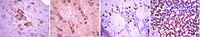
(Formalin-fixed and paraffin-embedded human cancer tissue reacted with the primary antibody, which was peroxidase-conjugated to the secondary antibody, followed by AEC staining. This data demonstrates the use of this antibody for immunohistochemistry; clinical relevance has not been evaluated. BC = breast carcinoma; HC = hepatocarcinoma.)
WB (Western Blot)
(Formalin-fixed and paraffin-embedded human cancer tissue reacted with the primary antibody, which was peroxidase-conjugated to the secondary antibody, followed by AEC staining. This data demonstrates the use of this antibody for immunohistochemistry; clinical relevance has not been evaluated. BC = breast carcinoma; HC = hepatocarcinoma.)
SIRT3, Polyclonal Antibody (Cat# AAA28669)
Full Name
SIRT3 Antibody (C-term)
Gene Names
SIRT3; SIR2L3
Reactivity
Human, mouse
Applications
WB, EIA, IHC
Purity
Purified Rabbit Polyclonal Antibody (Pab)
Pricing