Filters
Clonality
Type
Reactivity
Gene Name
Isotype
Host
Application
Clone
94 results for " Cell Tissue Markers" - showing 50-94


 Application Data
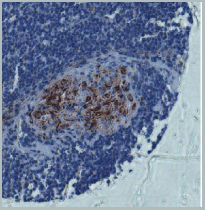
(Published customer image Infiltration of GFP+ BM-cells in infarct and peri-infarct regions. (A-B) Dot plots of viable macrophages/granulocytes (CD11b+CD45high, top right quadrants) and microglia (CD11b+CD45dim, bottom right quadrants) in cortex from BM-chimeric unmanipulated mice and mice exposed to pMCAO. (C) Bar graph showing mean numbers of CD11b+CD45dim microglia and CD11b+CD45high macrophages/granulocytes in BM-chimeric mice 24 hours after pMCAO, subdivided based on expression of GFP (n = 5). Approximately 92% of of the CD45high population were GFP+. (D) Estimation and comparison of mean numbers of CD11b+CD45dim microglia in non-chimeric (n = 10) versus BM-chimeric mice (n = 5) 24 hours after of pMCAO shows significantly fewer CD11b+CD45dim microglial cells in irradiated mice. (E) Overview, showing distribution of infiltrating GFP+ BM-derived cells into infarct (IF) and peri-infarct (P-IF) regions 24 hours after pMCAO. (E-G) By 24 hours, GFP+ single cells (F) and vessel-associated aggregates of GFP+ cells (arrows in G) were observed in infarct and peri-infarct regions. Some of the vessel-associated cells were round, leukocyte-like cells (arrows) while others were elongated cells lining the vasculature (arrow heads in G and in insert). (H) Bar graph showing mean numbers of single GFP+ cells and vessel-associated aggregates of GFP+ cells in ipsi- and contralateral cortex 24 hours after surgery (n = 10). (I-P) Immunohistochemical staining of CD45.1 (I, K), CD45.2 (J, L), IgG2a (M, O) and CD45 (N, P) in ischemic tissue in BM-chimeric (I, J, M, N) and non-chimeric mice (K, L, O, P) 24 hours after pMCAO. N.D, none detected. Scale bars: 200 um (A), 10 um (B, C). 50 um (I-P) *P < 0.05, **P < 0.01, and ***P < 0.001.From: Clausen BH, Lambertsen KL, Babcock AA, Holm TH, Dagnaes-Hansen F, Finsen B. Interleukin-1beta and tumor necrosis factor-alpha are expressed by different subsets of microglia and macrophages after ischemic stroke in mice. J Neuroinflammation. 2008 Oct 23;5:46.)
Application Data
(Published customer image Infiltration of GFP+ BM-cells in infarct and peri-infarct regions. (A-B) Dot plots of viable macrophages/granulocytes (CD11b+CD45high, top right quadrants) and microglia (CD11b+CD45dim, bottom right quadrants) in cortex from BM-chimeric unmanipulated mice and mice exposed to pMCAO. (C) Bar graph showing mean numbers of CD11b+CD45dim microglia and CD11b+CD45high macrophages/granulocytes in BM-chimeric mice 24 hours after pMCAO, subdivided based on expression of GFP (n = 5). Approximately 92% of of the CD45high population were GFP+. (D) Estimation and comparison of mean numbers of CD11b+CD45dim microglia in non-chimeric (n = 10) versus BM-chimeric mice (n = 5) 24 hours after of pMCAO shows significantly fewer CD11b+CD45dim microglial cells in irradiated mice. (E) Overview, showing distribution of infiltrating GFP+ BM-derived cells into infarct (IF) and peri-infarct (P-IF) regions 24 hours after pMCAO. (E-G) By 24 hours, GFP+ single cells (F) and vessel-associated aggregates of GFP+ cells (arrows in G) were observed in infarct and peri-infarct regions. Some of the vessel-associated cells were round, leukocyte-like cells (arrows) while others were elongated cells lining the vasculature (arrow heads in G and in insert). (H) Bar graph showing mean numbers of single GFP+ cells and vessel-associated aggregates of GFP+ cells in ipsi- and contralateral cortex 24 hours after surgery (n = 10). (I-P) Immunohistochemical staining of CD45.1 (I, K), CD45.2 (J, L), IgG2a (M, O) and CD45 (N, P) in ischemic tissue in BM-chimeric (I, J, M, N) and non-chimeric mice (K, L, O, P) 24 hours after pMCAO. N.D, none detected. Scale bars: 200 um (A), 10 um (B, C). 50 um (I-P) *P < 0.05, **P < 0.01, and ***P < 0.001.From: Clausen BH, Lambertsen KL, Babcock AA, Holm TH, Dagnaes-Hansen F, Finsen B. Interleukin-1beta and tumor necrosis factor-alpha are expressed by different subsets of microglia and macrophages after ischemic stroke in mice. J Neuroinflammation. 2008 Oct 23;5:46.)
CD11b, Monoclonal Antibody (Cat# AAA12181)
Full Name
RAT ANTI MOUSE CD11b
Gene Names
Itgam; CR3; CR3A; MAC1; Cd11b; Ly-40; Mac-1; Mac-1a; CD11b/CD18; F730045J24Rik
Reactivity
Human
Applications
Immunohistochemistry, Flow Cytometry, Immunofluorescence, Immunoprecipitation
Pricing


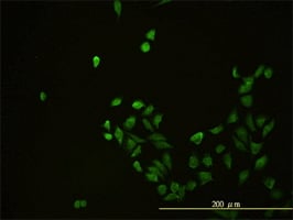
 IHC (Immunohistochemistry)
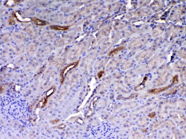
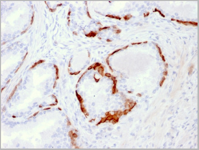
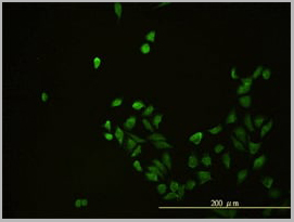
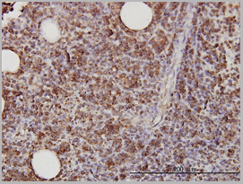
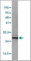
(Figure 7. IHC analysis of GALNS using anti-GALNS antibody (AAA19164).GALNS was detected in paraffin-embedded section of rat small intestine tissue. Heat mediated antigen retrieval was performed in citrate buffer (pH6, epitope retrieval solution) for 20 mins. The tissue section was blocked with 10% goat serum. The tissue section was then incubated with 1ug/ml rabbit anti-GALNS Antibody (AAA19164) overnight at 4 degree C. Biotinylated goat anti-rabbit IgG was used as secondary antibody and incubated for 30 minutes at 37 degree C. The tissue section was developed using Strepavidin-Biotin-Complex (SABC) with DAB as the chromogen.)
IHC (Immunohistochemistry)
(Figure 7. IHC analysis of GALNS using anti-GALNS antibody (AAA19164).GALNS was detected in paraffin-embedded section of rat small intestine tissue. Heat mediated antigen retrieval was performed in citrate buffer (pH6, epitope retrieval solution) for 20 mins. The tissue section was blocked with 10% goat serum. The tissue section was then incubated with 1ug/ml rabbit anti-GALNS Antibody (AAA19164) overnight at 4 degree C. Biotinylated goat anti-rabbit IgG was used as secondary antibody and incubated for 30 minutes at 37 degree C. The tissue section was developed using Strepavidin-Biotin-Complex (SABC) with DAB as the chromogen.)
GALNS, Antibody (Cat# AAA19164)
Full Name
Anti-GALNS Picoband antibody
Gene Names
GALNS; GAS; MPS4A; GalN6S; GALNAC6S
Reactivity
Reacts with: Human, Mouse, Rat
Applications
WB, EIA
Purity
Immunogen affinity purified
Pricing


 FCM (Flow Cytometry)
(Dual staining of pig peripheral blood lymphocytes with Mouse anti Pig CD335 detected with Goat anti Mouse IgG (H/L):FITC (STAR117F), and Mouse anti Pig wCD8a:RPE)
FCM (Flow Cytometry)
(Dual staining of pig peripheral blood lymphocytes with Mouse anti Pig CD335 detected with Goat anti Mouse IgG (H/L):FITC (STAR117F), and Mouse anti Pig wCD8a:RPE)
CD335, Monoclonal Antibody (Cat# AAA12251)
Full Name
MOUSE ANTI PIG CD335: APC
Reactivity
Pig
Applications
Flow Cytometry
Pricing


 Application Data
(Staining of NALM6 cells with MOUSE ANTI HUMAN CD10:ALEXA 488)
Application Data
(Staining of NALM6 cells with MOUSE ANTI HUMAN CD10:ALEXA 488)
CD10, Monoclonal Antibody (Cat# AAA26803)
Full Name
CD10 (CALLA, CD 10, Common Acute Lymphocytic Leukemia Antigen, Enkephalinase, gp100, Membrane Metalloendopeptidase, MME, Neprilysin, Neutral Endopeptidase, Pmel17) (MaxLight 550)
Gene Names
MME; NEP; SFE; CD10; CALLA
Reactivity
Human
Applications
Flow Cytometry, Immunohistochemistry, Immunoprecipitation, Western Blot
Purity
Purified by protein G affinity chromatography from tissue culture supernatant.
Pricing


 IHC (Immunohistchemistry)
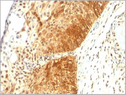
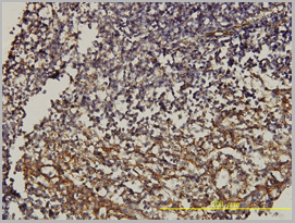
(Figure 6. IHC analysis of HE4 using anti-HE4 antibody (AAA19161).HE4 was detected in paraffin-embedded section of rat small intestine tissue. Heat mediated antigen retrieval was performed in citrate buffer (pH6, epitope retrieval solution) for 20 mins. The tissue section was blocked with 10% goat serum. The tissue section was then incubated with 1ug/ml rabbit anti-HE4 Antibody (AAA19161) overnight at 4 degree C. Biotinylated goat anti-rabbit IgG was used as secondary antibody and incubated for 30 minutes at 37 degree C. The tissue section was developed using Strepavidin-Biotin-Complex (SABC) with DAB as the chromogen.)
IHC (Immunohistchemistry)
(Figure 6. IHC analysis of HE4 using anti-HE4 antibody (AAA19161).HE4 was detected in paraffin-embedded section of rat small intestine tissue. Heat mediated antigen retrieval was performed in citrate buffer (pH6, epitope retrieval solution) for 20 mins. The tissue section was blocked with 10% goat serum. The tissue section was then incubated with 1ug/ml rabbit anti-HE4 Antibody (AAA19161) overnight at 4 degree C. Biotinylated goat anti-rabbit IgG was used as secondary antibody and incubated for 30 minutes at 37 degree C. The tissue section was developed using Strepavidin-Biotin-Complex (SABC) with DAB as the chromogen.)
HE4, Polyclonal Antibody (Cat# AAA19161)
Full Name
Anti-HE4 Picoband Antibody
Gene Names
Wfdc2; re4
Reactivity
Mouse, Rat
No cross reactivity with other proteins.
No cross reactivity with other proteins.
Applications
IHC, WB
Purity
Immunogen affinity purified
Pricing


 Application Data
(Analysis of Protein Array containing more than 19, 000 full-length human proteins using Mesothelin Mouse Monoclonal Antibody (MSLN/2131). Z- and S- Score: The Z-score represents the strength of a signal that a monoclonal antibody (MAb) (in combination with a fluorescently-tagged anti-IgG secondary antibody) produces when binding to a particular protein on the HuProtTM array. Z-scores are described in units of standard deviations (SD's) above the mean value of all signals generated on that array. If targets on HuProtTM are arranged in descending order of the Z-score, the S-score is the difference (also in units of SD's) between the Z-score. S-score therefore represents the relative target specificity of a MAb to its intended target. A MAb is considered to specific to its intended target, if the MAb has an S-score of at least 2.5. For example, if a MAb binds to protein X with a Z-score of 43 and to protein Y with a Z-score of 14, then the S-score for the binding of that MAb to protein X is equal to 29.)
Application Data
(Analysis of Protein Array containing more than 19, 000 full-length human proteins using Mesothelin Mouse Monoclonal Antibody (MSLN/2131). Z- and S- Score: The Z-score represents the strength of a signal that a monoclonal antibody (MAb) (in combination with a fluorescently-tagged anti-IgG secondary antibody) produces when binding to a particular protein on the HuProtTM array. Z-scores are described in units of standard deviations (SD's) above the mean value of all signals generated on that array. If targets on HuProtTM are arranged in descending order of the Z-score, the S-score is the difference (also in units of SD's) between the Z-score. S-score therefore represents the relative target specificity of a MAb to its intended target. A MAb is considered to specific to its intended target, if the MAb has an S-score of at least 2.5. For example, if a MAb binds to protein X with a Z-score of 43 and to protein Y with a Z-score of 14, then the S-score for the binding of that MAb to protein X is equal to 29.)
Mesothelin, Monoclonal Antibody (Cat# AAA23897)
Full Name
Mesothelin (Mesothelial Marker)
Gene Names
MSLN; MPF; SMRP
Reactivity
Human, Mouse, Rat. Others not known.
Applications
Immunohistochemistry
Pricing


 Application Data
(Staining of human peripheral blood lymphocytes with MOUSE ANTI HUMAN CD4:ALEXA 405)
Application Data
(Staining of human peripheral blood lymphocytes with MOUSE ANTI HUMAN CD4:ALEXA 405)
CD4, Monoclonal Antibody (Cat# AAA26847)
Full Name
CD4 (CD4 Antigen, CD4 Molecule, CD4 Receptor, CD4mut, T Cell Surface Antigen T4/Leu3, T Cell Antigen T4, T Cell Surface Glycoprotein CD4) (APC)
Gene Names
CD4; CD4mut
Purity
Purified by protein A affinity chromatography from tissue culture supernatant.
Pricing


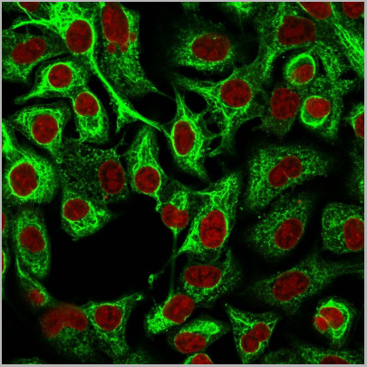
 Application Data
(C:FGFR2/isolectinB4 (C) and FGFR1/isolectinB4 (D) staining of apparent mesenchymal cells and the subpopulation of endothelial cells. Virtually all other dispersed apparent mesenchymal cells express FGFR1 and FGFR2 (merged image in E). F: FGFR2 (F) and FGFR1 (G) staining in clustered cells of epithelial origin (inferred by morphology here) demonstrating that epithelial cells express both FGFR1 and FGFR2 (merged image with DAPI staining in H).)
Application Data
(C:FGFR2/isolectinB4 (C) and FGFR1/isolectinB4 (D) staining of apparent mesenchymal cells and the subpopulation of endothelial cells. Virtually all other dispersed apparent mesenchymal cells express FGFR1 and FGFR2 (merged image in E). F: FGFR2 (F) and FGFR1 (G) staining in clustered cells of epithelial origin (inferred by morphology here) demonstrating that epithelial cells express both FGFR1 and FGFR2 (merged image with DAPI staining in H).)
FGFR2, Polyclonal Antibody (Cat# AAA26854)
Full Name
FGFR2, NT (FGFR2, BEK, KGFR, KSAM, Fibroblast growth factor receptor 2, K-sam, Keratinocyte growth factor receptor, CD332) (FITC)
Gene Names
FGFR2; BEK; JWS; BBDS; CEK3; CFD1; ECT1; KGFR; TK14; TK25; BFR-1; CD332; K-SAM
Reactivity
Human, Monkey, Mouse, Rat
Applications
WB, IHC, IF, FC/FACS
Purity
Purified by Protein G Affinity Chromatography.
Pricing


 Application Data
(C:FGFR2/isolectinB4 (C) and FGFR1/isolectinB4 (D) staining of apparent mesenchymal cells and the subpopulation of endothelial cells. Virtually all other dispersed apparent mesenchymal cells express FGFR1 and FGFR2 (merged image in E). F: FGFR2 (F) and FGFR1 (G) staining in clustered cells of epithelial origin (inferred by morphology here) demonstrating that epithelial cells express both FGFR1 and FGFR2 (merged image with DAPI staining in H).)
Application Data
(C:FGFR2/isolectinB4 (C) and FGFR1/isolectinB4 (D) staining of apparent mesenchymal cells and the subpopulation of endothelial cells. Virtually all other dispersed apparent mesenchymal cells express FGFR1 and FGFR2 (merged image in E). F: FGFR2 (F) and FGFR1 (G) staining in clustered cells of epithelial origin (inferred by morphology here) demonstrating that epithelial cells express both FGFR1 and FGFR2 (merged image with DAPI staining in H).)
FGFR2, Polyclonal Antibody (Cat# AAA26851)
Full Name
FGFR2, NT (FGFR2, BEK, KGFR, KSAM, Fibroblast growth factor receptor 2, K-sam, Keratinocyte growth factor receptor, CD332) (AP)
Gene Names
FGFR2; BEK; JWS; BBDS; CEK3; CFD1; ECT1; KGFR; TK14; TK25; BFR-1; CD332; K-SAM
Reactivity
Human, Monkey, Mouse, Rat
Applications
IF, EIA, IHC, WB
Purity
Purified by Protein G Affinity Chromatography.
Pricing


 Application Data
(Staining of human peripheral blood lymphocytes with MOUSE ANTI HUMAN CD45RA:FITC (MCA88F))
Application Data
(Staining of human peripheral blood lymphocytes with MOUSE ANTI HUMAN CD45RA:FITC (MCA88F))
CD45RA, Monoclonal Antibody (Cat# AAA26848)
Full Name
CD45RA (CD45 Antigen, B220, GP180, Leukocyte Common Antigen, LCA, L-CA, LY5, LY-5, Protein Tyrosine Phosphatase Receptor Type C Polypeptide, PTPRC, T200, T200 Glycoprotein) (APC)
Reactivity
Human, Monkey
Applications
IHC
Purity
Purified by protein G affinity chromatography from tissue culture supernatant.
Pricing


 Application Data
(Staining of human peripheral blood lymphocytes with MOUSE ANTI HUMAN CD45RA:FITC (MCA88F))
Application Data
(Staining of human peripheral blood lymphocytes with MOUSE ANTI HUMAN CD45RA:FITC (MCA88F))
CD45RA, Monoclonal Antibody (Cat# AAA26839)
Full Name
CD45RA (CD45 Antigen, B220, GP180, Leukocyte Common Antigen, LCA, L-CA, LY5, LY-5, Protein Tyrosine Phosphatase Receptor Type C Polypeptide, PTPRC, T200, T200 Glycoprotein) (MaxLight 750)
Reactivity
Human, Monkey
Applications
FC/FACS, IHC
Purity
Purified by protein G affinity chromatography from tissue culture supernatant.
Pricing


 Application Data
(Staining of human peripheral blood lymphocytes with MOUSE ANTI HUMAN CD45RA:FITC (MCA88F))
Application Data
(Staining of human peripheral blood lymphocytes with MOUSE ANTI HUMAN CD45RA:FITC (MCA88F))
CD45RA, Monoclonal Antibody (Cat# AAA26811)
Full Name
CD45RA (CD45 Antigen, B220, GP180, Leukocyte Common Antigen, LCA, L-CA, LY5, LY-5, Protein Tyrosine Phosphatase Receptor Type C Polypeptide, PTPRC, T200, T200 Glycoprotein) (MaxLight 550)
Reactivity
Human, Monkey
Applications
FC/FACS, IHC
Purity
Purified by protein G affinity chromatography from tissue culture supernatant.
Pricing


 Application Data
(Staining of human peripheral blood lymphocytes with MOUSE ANTI HUMAN CD45RA:FITC (MCA88F))
Application Data
(Staining of human peripheral blood lymphocytes with MOUSE ANTI HUMAN CD45RA:FITC (MCA88F))
CD45RA, Monoclonal Antibody (Cat# AAA26825)
Full Name
CD45RA (CD45 Antigen, B220, GP180, Leukocyte Common Antigen, LCA, L-CA, LY5, LY-5, Protein Tyrosine Phosphatase Receptor Type C Polypeptide, PTPRC, T200, T200 Glycoprotein) (MaxLight 650)
Reactivity
Human, Monkey
Applications
FC/FACS, IHC
Purity
Purified by protein G affinity chromatography from tissue culture supernatant.
Pricing


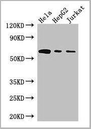
 WB (Western Blot)
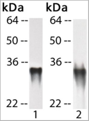
(CD58 monoclonal antibody, Western Blot analysis of CD58 expression in Jurkat.)
WB (Western Blot)
(CD58 monoclonal antibody, Western Blot analysis of CD58 expression in Jurkat.)
CD58, Monoclonal Antibody (Cat# AAA24751)
Full Name
CD58 (Lymphocyte Function-associated Antigen 3, Ag3, Surface Glycoprotein LFA-3, LFA3) (Biotin)
Gene Names
CD58; ag3; LFA3; LFA-3
Reactivity
Human
Applications
EIA, IF, IHC, WB
Purity
Purified by Protein A Affinity Chromatography.
Pricing


 Application Data
(C:FGFR2/isolectinB4 (C) and FGFR1/isolectinB4 (D) staining of apparent mesenchymal cells and the subpopulation of endothelial cells. Virtually all other dispersed apparent mesenchymal cells express FGFR1 and FGFR2 (merged image in E). F: FGFR2 (F) and FGFR1 (G) staining in clustered cells of epithelial origin (inferred by morphology here) demonstrating that epithelial cells express both FGFR1 and FGFR2 (merged image with DAPI staining in H).)
Application Data
(C:FGFR2/isolectinB4 (C) and FGFR1/isolectinB4 (D) staining of apparent mesenchymal cells and the subpopulation of endothelial cells. Virtually all other dispersed apparent mesenchymal cells express FGFR1 and FGFR2 (merged image in E). F: FGFR2 (F) and FGFR1 (G) staining in clustered cells of epithelial origin (inferred by morphology here) demonstrating that epithelial cells express both FGFR1 and FGFR2 (merged image with DAPI staining in H).)
FGFR2, Polyclonal Antibody (Cat# AAA26856)
Full Name
FGFR2, NT (FGFR2, BEK, KGFR, KSAM, Fibroblast growth factor receptor 2, K-sam, Keratinocyte growth factor receptor, CD332) (PE)
Gene Names
FGFR2; BEK; JWS; BBDS; CEK3; CFD1; ECT1; KGFR; TK14; TK25; BFR-1; CD332; K-SAM
Reactivity
Human, Monkey, Mouse, Rat
Applications
WB, IHC, IF, FC/FACS
Purity
Purified by Protein G Affinity Chromatography.
Pricing


 Application Data
(Staining of human peripheral blood monocytes with MOUSE ANTI HUMAN CD274:RPE)
Application Data
(Staining of human peripheral blood monocytes with MOUSE ANTI HUMAN CD274:RPE)
CD274, Monoclonal Antibody (Cat# AAA26834)
Full Name
CD274 (CD274 Antigen, B7 Homolog 1, B7H1, B7-H1, B7-H, MGC142294, MGC142296, OTTHUMP00000021029, Programmed Cell Death 1 Ligand 1, PDCD1 Ligand 1, PDCD1L1, PDCD1LG1, Programmed Death Ligand 1, PDL1, PD-L1, RGD1566211) (MaxLight 750)
Gene Names
CD274; B7-H; B7H1; PDL1; PD-L1; PDCD1L1; PDCD1LG1
Reactivity
Human
Applications
FC/FACS
Purity
Purified by protein G affinity chromatography from tissue culture supernatant.
Pricing


 Application Data
(Staining of human peripheral blood lymphocytes with MOUSE ANTI HUMAN CD4:ALEXA 405)
Application Data
(Staining of human peripheral blood lymphocytes with MOUSE ANTI HUMAN CD4:ALEXA 405)
CD4, Monoclonal Antibody (Cat# AAA26837)
Full Name
CD4 (CD4 Antigen, CD4 Molecule, CD4 Receptor, CD4mut, T Cell Surface Antigen T4/Leu3, T Cell Antigen T4, T Cell Surface Glycoprotein CD4) (MaxLight 750)
Gene Names
CD4; CD4mut
Reactivity
Human
Applications
FC/FACS, IHC
Purity
Purified by protein A affinity chromatography from tissue culture supernatant.
Pricing


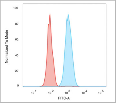
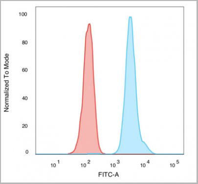
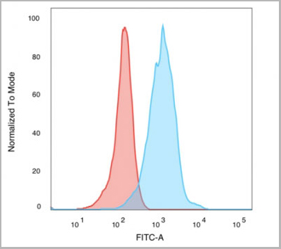
 Application Data
(Staining of NALM6 cells with MOUSE ANTI HUMAN CD10:ALEXA 488)
Application Data
(Staining of NALM6 cells with MOUSE ANTI HUMAN CD10:ALEXA 488)
CD10, Monoclonal Antibody (Cat# AAA26817)
Full Name
CD10 (CALLA, CD 10, Common Acute Lymphocytic Leukemia Antigen, Enkephalinase, gp100, Membrane Metalloendopeptidase, MME, Neprilysin, Neutral Endopeptidase, Pmel17) (MaxLight 650)
Gene Names
MME; NEP; SFE; CD10; CALLA
Reactivity
Human
Applications
FC/FACS, IHC, IP, WB
Purity
Purified by protein G affinity chromatography from tissue culture supernatant.
Pricing
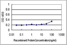
 Standard Curve (Sample)


 Application Data
(Staining of human peripheral blood lymphocytes with MOUSE ANTI HUMAN CD4:ALEXA 405)
Application Data
(Staining of human peripheral blood lymphocytes with MOUSE ANTI HUMAN CD4:ALEXA 405)
CD4, Monoclonal Antibody (Cat# AAA26823)
Full Name
CD4 (CD4 Antigen, CD4 Molecule, CD4 Receptor, CD4mut, T Cell Surface Antigen T4/Leu3, T Cell Antigen T4, T Cell Surface Glycoprotein CD4) (MaxLight 650)
Gene Names
CD4; CD4mut
Reactivity
Human
Applications
FC/FACS, IHC
Purity
Purified by protein A affinity chromatography from tissue culture supernatant.
Pricing


 Application Data
(Staining of human peripheral blood monocytes with MOUSE ANTI HUMAN CD274:RPE)
Application Data
(Staining of human peripheral blood monocytes with MOUSE ANTI HUMAN CD274:RPE)
CD274, Monoclonal Antibody (Cat# AAA26820)
Full Name
CD274 (CD274 Antigen, B7 Homolog 1, B7H1, B7-H1, B7-H, MGC142294, MGC142296, OTTHUMP00000021029, Programmed Cell Death 1 Ligand 1, PDCD1 Ligand 1, PDCD1L1, PDCD1LG1, Programmed Death Ligand 1, PDL1, PD-L1, RGD1566211) (MaxLight 650)
Gene Names
CD274; B7-H; B7H1; PDL1; PD-L1; PDCD1L1; PDCD1LG1
Reactivity
Human
Applications
FC/FACS
Purity
Purified by protein G affinity chromatography from tissue culture supernatant.
Pricing


 Application Data
(Staining of human peripheral blood lymphocytes with MOUSE ANTI HUMAN CD4:ALEXA 405)
Application Data
(Staining of human peripheral blood lymphocytes with MOUSE ANTI HUMAN CD4:ALEXA 405)
CD4, Monoclonal Antibody (Cat# AAA26809)
Full Name
CD4 (CD4 Antigen, CD4 Molecule, CD4 Receptor, CD4mut, T Cell Surface Antigen T4/Leu3, T Cell Antigen T4, T Cell Surface Glycoprotein CD4) (MaxLight 550)
Gene Names
CD4; CD4mut
Reactivity
Human
Applications
FC/FACS, IHC
Purity
Purified by protein A affinity chromatography from tissue culture supernatant.
Pricing


 WB (Western Blot)
(CD58 monoclonal antibody, Western Blot analysis of CD58 expression in Jurkat.)
WB (Western Blot)
(CD58 monoclonal antibody, Western Blot analysis of CD58 expression in Jurkat.)
CD58, Monoclonal Antibody (Cat# AAA25048)
Full Name
CD58 (Lymphocyte Function-associated Antigen 3, Ag3, Surface Glycoprotein LFA-3, LFA3) (FITC)
Gene Names
CD58; ag3; LFA3; LFA-3
Reactivity
Human
Applications
EIA, IF, IHC, WB
Purity
Purified by Protein A Affinity Chromatography.
Pricing


 Application Data
(Staining of NALM6 cells with MOUSE ANTI HUMAN CD10:ALEXA 488)
Application Data
(Staining of NALM6 cells with MOUSE ANTI HUMAN CD10:ALEXA 488)
CD10, Monoclonal Antibody (Cat# AAA26831)
Full Name
CD10 (CALLA, CD 10, Common Acute Lymphocytic Leukemia Antigen, Enkephalinase, gp100, Membrane Metalloendopeptidase, MME, Neprilysin, Neutral Endopeptidase, Pmel17) (MaxLight 750)
Gene Names
MME; NEP; SFE; CD10; CALLA
Reactivity
Human
Applications
FC/FACS, IHC, IP, WB
Purity
Purified by protein G affinity chromatography from tissue culture supernatant.
Pricing


 Application Data
(C:FGFR2/isolectinB4 (C) and FGFR1/isolectinB4 (D) staining of apparent mesenchymal cells and the subpopulation of endothelial cells. Virtually all other dispersed apparent mesenchymal cells express FGFR1 and FGFR2 (merged image in E). F: FGFR2 (F) and FGFR1 (G) staining in clustered cells of epithelial origin (inferred by morphology here) demonstrating that epithelial cells express both FGFR1 and FGFR2 (merged image with DAPI staining in H).)
Application Data
(C:FGFR2/isolectinB4 (C) and FGFR1/isolectinB4 (D) staining of apparent mesenchymal cells and the subpopulation of endothelial cells. Virtually all other dispersed apparent mesenchymal cells express FGFR1 and FGFR2 (merged image in E). F: FGFR2 (F) and FGFR1 (G) staining in clustered cells of epithelial origin (inferred by morphology here) demonstrating that epithelial cells express both FGFR1 and FGFR2 (merged image with DAPI staining in H).)
FGFR2, Polyclonal Antibody (Cat# AAA26852)
Full Name
FGFR2, NT (FGFR2, BEK, KGFR, KSAM, Fibroblast growth factor receptor 2, K-sam, Keratinocyte growth factor receptor, CD332) (APC)
Gene Names
FGFR2; BEK; JWS; BBDS; CEK3; CFD1; ECT1; KGFR; TK14; TK25; BFR-1; CD332; K-SAM
Reactivity
Human, Monkey, Mouse, Rat
Applications
FC/FACS, IF, IHC, WB
Purity
Purified by Protein G Affinity Chromatography.
Pricing
CD58, Monoclonal Antibody (Cat# AAA24161)
Full Name
CD58 (Lymphocyte Function-associated Antigen 3, Ag3, Surface Glycoprotein LFA-3, LFA3) (AP)
Gene Names
CD58; ag3; LFA3; LFA-3
Reactivity
Human
Applications
Immunohistochemistry, Western Blot
Purity
Purified by Protein A Affinity Chromatography.
Pricing
 Application Data
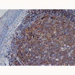
(Figure: Biopsy of tumor lesion from a patient with lymphoma (DLBCL) stained with MAb to APRIL (Aprily-2) . The biopsy was stained by standard immunohistochemistry. The figure shows the selective accumulation of APRIL around tumor cells.Picture courtesy of Dr. Bertrand Huard (University of Geneva).)
Application Data
(Figure: Biopsy of tumor lesion from a patient with lymphoma (DLBCL) stained with MAb to APRIL (Aprily-2) . The biopsy was stained by standard immunohistochemistry. The figure shows the selective accumulation of APRIL around tumor cells.Picture courtesy of Dr. Bertrand Huard (University of Geneva).)
APRIL, Monoclonal Antibody (Cat# AAA14518)
Full Name
APRIL (human) monoclonal antibody (Aprily-2)
Gene Names
TNFSF13; APRIL; CD256; TALL2; ZTNF2; TALL-2; TNLG7B; TRDL-1; UNQ383/PRO715
Reactivity
Human
Applications
Immunohistochemistry
Pricing
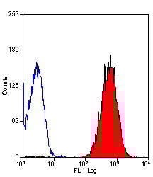
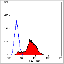
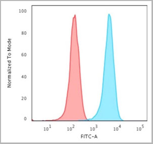
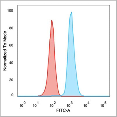
 FCM (Flow Cytometry)
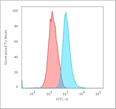
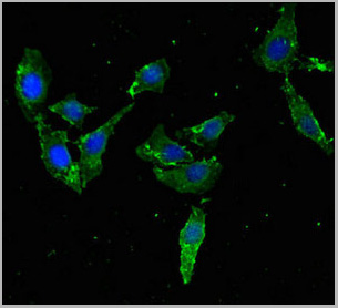
(Flow Cytometric Analysis of trypsinized MeOH-fixed HeLa cells using Cytokeratin 14 Mouse Monoclonal Antibody (LL002) followed by Goat anti-Mouse IgG-CF488 (Blue); Isotype Control (Red).)
FCM (Flow Cytometry)
(Flow Cytometric Analysis of trypsinized MeOH-fixed HeLa cells using Cytokeratin 14 Mouse Monoclonal Antibody (LL002) followed by Goat anti-Mouse IgG-CF488 (Blue); Isotype Control (Red).)
Cytokeratin 14 (KRT14), Monoclonal Antibody (Cat# AAA13813)
Full Name
Cytokeratin 14 (KRT14) (Squamous Cell Marker) Mouse Monoclonal Antibody
Gene Names
KRT14; K14; NFJ; CK14; EBS3; EBS4
Reactivity
Human
Applications
Flow Cytometry, Immunofluorescence, Immunohistochemistry
Pricing
 SDS-PAGE
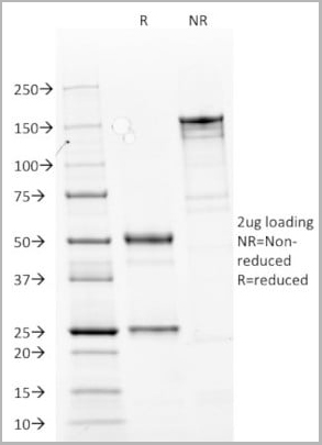
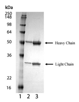
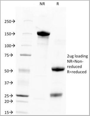
(SDS-PAGE analysis of Prod.(AAA14516): Lane 1: MW marker, Lane 2: 1µg , Lane 3: 2µg BCMA (mouse) monoclonal antibody (Vicky-2), integrity of antibody is shown as both light chain and heavy chain are present.)
SDS-PAGE
(SDS-PAGE analysis of Prod.(AAA14516): Lane 1: MW marker, Lane 2: 1µg , Lane 3: 2µg BCMA (mouse) monoclonal antibody (Vicky-2), integrity of antibody is shown as both light chain and heavy chain are present.)
BCMA, Monoclonal Antibody (Cat# AAA14516)
Full Name
BCMA (mouse) monoclonal antibody (Vicky-2)
Gene Names
Tnfrsf17; BCM; BCMA; Tnfrsf13; Tnfrsf13a
Reactivity
Mouse
Applications
Flow Cytometry, Immunoprecipitation, Western Blot
Purity
Protein G-affinity purified.
Pricing
 WB (Western Blot)
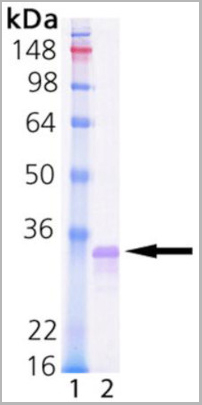
(Western Blot analysis of 10 ng Lin28a protein (Prod. No. AAA14519) probed withLane 1: polyclonal anti-Lin28 antibodyLane 2: anti-6x His tag antibody.)
WB (Western Blot)
(Western Blot analysis of 10 ng Lin28a protein (Prod. No. AAA14519) probed withLane 1: polyclonal anti-Lin28 antibodyLane 2: anti-6x His tag antibody.)
Lin28a, Recombinant Protein (Cat# AAA14519)
Full Name
Lin28a (full-length) (human), (recombinant)
Gene Names
LIN28A; CSDD1; LIN28; LIN-28; ZCCHC1; lin-28A
Applications
Western Blot
Purity
>95% (SDS-PAGE)
Purified by multi-step chromatography
Purified by multi-step chromatography
Pricing
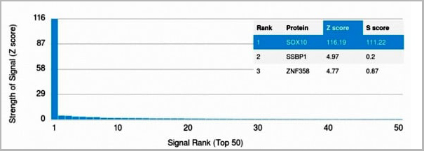 Application Data
(Z- and S- Score: The Z-score represents the strength of a signal that a monoclonal antibody (MAb) (in combination with a fluorescently-tagged anti-IgG secondary antibody) produces when binding to a particular protein on the HuProtTM array. Z-scores are described in units of standard deviations (SD's) above the mean value of all signals generated on that array. If targets on HuProtTM are arranged in descending order of the Z-score, the S-score is the difference (also in units of SD's) between the Z-score. S-score therefore represents the relative target specificity of a MAb to its intended target. A MAb is considered to specific to its intended target, if the MAb has an S-score of at least 2.5. For example, if a MAb binds to protein X with a Z-score of 43 and to protein Y with a Z-score of 14, then the S-score for the binding of that MAb to protein X is equal to 29.)
Application Data
(Z- and S- Score: The Z-score represents the strength of a signal that a monoclonal antibody (MAb) (in combination with a fluorescently-tagged anti-IgG secondary antibody) produces when binding to a particular protein on the HuProtTM array. Z-scores are described in units of standard deviations (SD's) above the mean value of all signals generated on that array. If targets on HuProtTM are arranged in descending order of the Z-score, the S-score is the difference (also in units of SD's) between the Z-score. S-score therefore represents the relative target specificity of a MAb to its intended target. A MAb is considered to specific to its intended target, if the MAb has an S-score of at least 2.5. For example, if a MAb binds to protein X with a Z-score of 43 and to protein Y with a Z-score of 14, then the S-score for the binding of that MAb to protein X is equal to 29.)
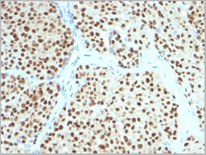
SOX10, Monoclonal Antibody (Cat# AAA13845)
Full Name
SOX10 (Melanoma Marker) Mouse Monoclonal Antibody
Gene Names
SOX10; DOM; WS4; PCWH; WS2E; WS4C
Reactivity
Human, Mouse
Applications
Flow Cytometry, Immunofluorescence, Western Blot, Immunohistochemistry
Pricing
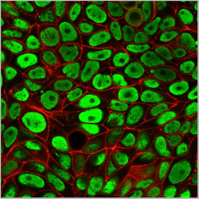
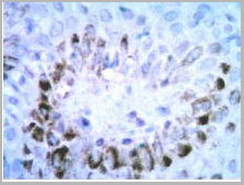 IHC (Immunohistochemistry)
(High magnification: Paraffin-embedded human skin tissue was prepared using heat-induced epitope retrieval in citrate buffer, pH 6.0. Immunostaining was performed using a 1:100 dilution of AAA14653. Cells lining up under the epidermis consist of numerous mast cells (The dermis has a moderate diffuse inflammatory cell infiltrate).)
IHC (Immunohistochemistry)
(High magnification: Paraffin-embedded human skin tissue was prepared using heat-induced epitope retrieval in citrate buffer, pH 6.0. Immunostaining was performed using a 1:100 dilution of AAA14653. Cells lining up under the epidermis consist of numerous mast cells (The dermis has a moderate diffuse inflammatory cell infiltrate).)
Mast Cell Chymase, Monoclonal Antibody (Cat# AAA14653)
Full Name
Mast Cell Chymase
Gene Names
CMA1; CYH; MCT1; chymase; MGC119890; MGC119891
Reactivity
Human
Applications
Immunohistochemistry
Purity
Purified by Protein G affinity chromatography
Pricing
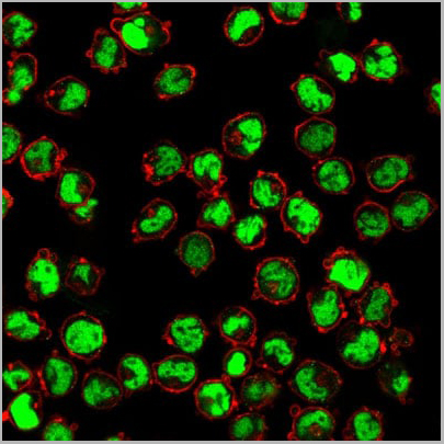
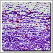 IHC (Immunohistochemistry)
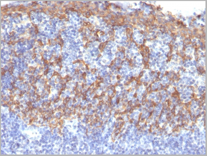
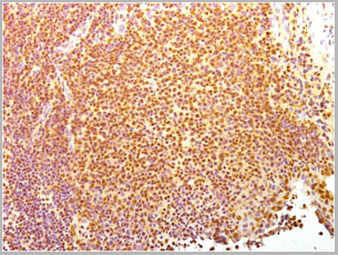
(Immunohistochemistry of formalin-fixed, paraffin-embedded human tonsil using AAA14660 and following by peroxidase-conjugate and AEC chromogen. Note cytoplasmic staining of mast cells.)
IHC (Immunohistochemistry)
(Immunohistochemistry of formalin-fixed, paraffin-embedded human tonsil using AAA14660 and following by peroxidase-conjugate and AEC chromogen. Note cytoplasmic staining of mast cells.)
Mast Cell Chymase, Monoclonal Antibody (Cat# AAA14660)
Full Name
Mast Cell Chymase (BSA & Azide Free)
Gene Names
CMA1; CYH; MCT1; chymase; MGC119890; MGC119891
Reactivity
Human
Applications
Western Blot, Immunohistochemistry
Purity
Purified by Protein G affinity chromatography.
Pricing


 Application Data
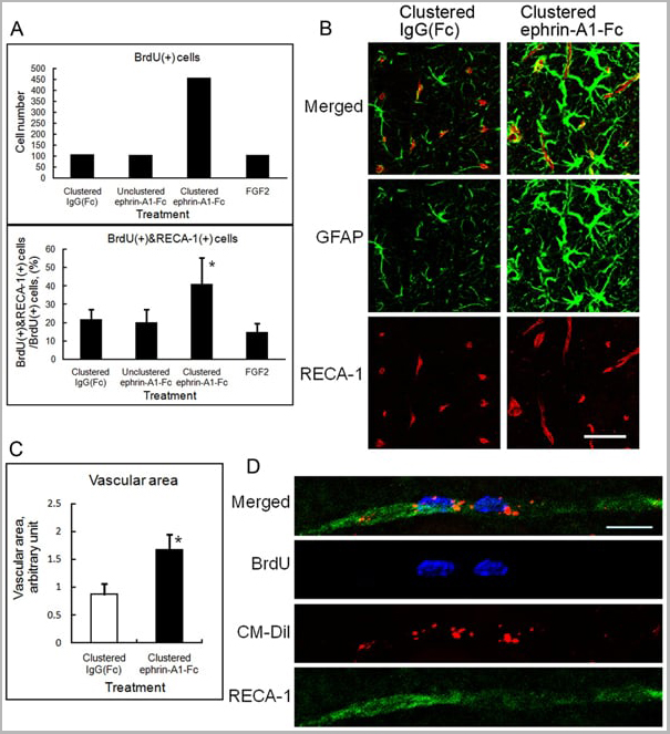
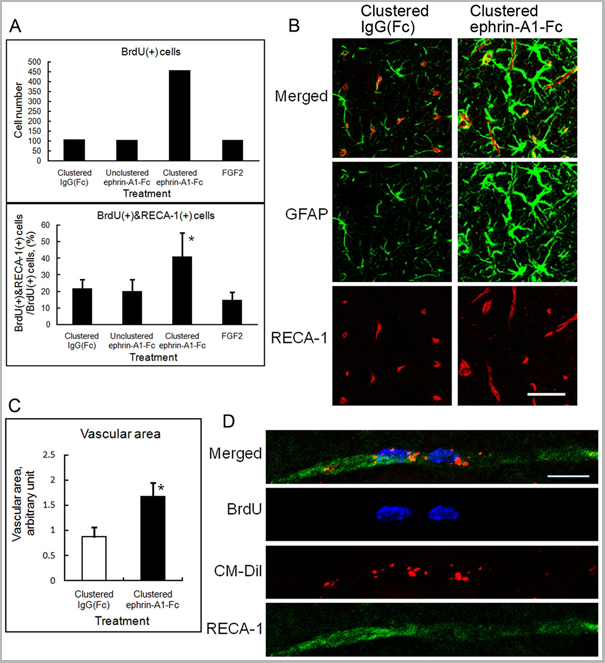
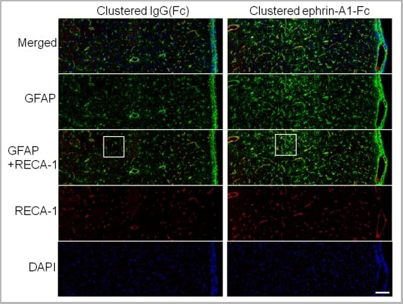
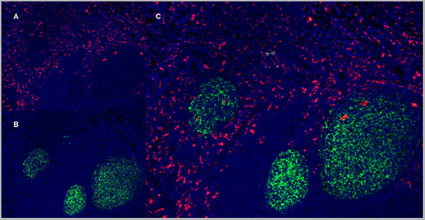
(Published customer image: Effect of clustered ephrin-A1-Fc on vascular formation in the rat striatum. Clustered ephrin-A1-Fc was injected into the lesioned side of the lateral ventricle in the unilaterally lesioned rats. Brains taken 6 weeks after injection were sectioned coronally and stained for GFAP (green) and RECA-1 (red) and with DAPI (nuclei; blue). The rectangular insets are shown in Fig. 8B. Scale bar: 100 um.From: Jing X, Miwa H, Sawada T, Nakanishi I, Kondo T, et al. (2012) Ephrin-A1-Mediated Dopaminergic Neurogenesis and Angiogenesis in a Rat Model of Parkinson's Disease. PLoS ONE 7(2): e32019.)
Application Data
(Published customer image: Effect of clustered ephrin-A1-Fc on vascular formation in the rat striatum. Clustered ephrin-A1-Fc was injected into the lesioned side of the lateral ventricle in the unilaterally lesioned rats. Brains taken 6 weeks after injection were sectioned coronally and stained for GFAP (green) and RECA-1 (red) and with DAPI (nuclei; blue). The rectangular insets are shown in Fig. 8B. Scale bar: 100 um.From: Jing X, Miwa H, Sawada T, Nakanishi I, Kondo T, et al. (2012) Ephrin-A1-Mediated Dopaminergic Neurogenesis and Angiogenesis in a Rat Model of Parkinson's Disease. PLoS ONE 7(2): e32019.)
RECA-1, Monoclonal Antibody (Cat# AAA12018)
Full Name
MOUSE ANTI RAT RECA-1
Applications
Immunohistochemistry, Immunofluorescence
Pricing


 Application Data
(Staining of mouse spleen with Hamster anti Mouse CD81: Alexa Fluor 488)
Application Data
(Staining of mouse spleen with Hamster anti Mouse CD81: Alexa Fluor 488)
CD81, Monoclonal Antibody (Cat# AAA11869)
Full Name
HAMSTER ANTI MOUSE CD81:FITC
Gene Names
Cd81; Tapa1; Tapa-1; Tspan28
Applications
Flow Cytometry
Pricing


 Application Data
(Staining of mouse spleen with Hamster anti Mouse CD81: Alexa Fluor 488)
Application Data
(Staining of mouse spleen with Hamster anti Mouse CD81: Alexa Fluor 488)
CD81, Monoclonal Antibody (Cat# AAA12033)
Full Name
HAMSTER ANTI MOUSE CD81:RPE
Gene Names
Cd81; Tapa1; Tapa-1; Tspan28
Applications
Flow Cytometry
Pricing


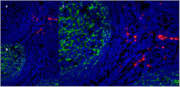
 Application Data
(Published customer image: Effect of clustered ephrin-A1-Fc on vascular formation in the rat striatum. Clustered ephrin-A1-Fc was injected into the lesioned side of the lateral ventricle in the unilaterally lesioned rats. Brains taken 6 weeks after injection were sectioned coronally and stained for GFAP (green) and RECA-1 (red) and with DAPI (nuclei; blue). The rectangular insets are shown in Fig. 8B. Scale bar: 100 um.From: Jing X, Miwa H, Sawada T, Nakanishi I, Kondo T, et al. (2012) Ephrin-A1-Mediated Dopaminergic Neurogenesis and Angiogenesis in a Rat Model of Parkinson's Disease. PLoS ONE 7(2): e32019.)
Application Data
(Published customer image: Effect of clustered ephrin-A1-Fc on vascular formation in the rat striatum. Clustered ephrin-A1-Fc was injected into the lesioned side of the lateral ventricle in the unilaterally lesioned rats. Brains taken 6 weeks after injection were sectioned coronally and stained for GFAP (green) and RECA-1 (red) and with DAPI (nuclei; blue). The rectangular insets are shown in Fig. 8B. Scale bar: 100 um.From: Jing X, Miwa H, Sawada T, Nakanishi I, Kondo T, et al. (2012) Ephrin-A1-Mediated Dopaminergic Neurogenesis and Angiogenesis in a Rat Model of Parkinson's Disease. PLoS ONE 7(2): e32019.)
RECA-1, Monoclonal Antibody (Cat# AAA12019)
Full Name
MOUSE ANTI RAT RECA-1
Applications
Immunohistochemistry, Immunofluorescence
Pricing
 Application Data
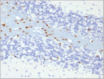
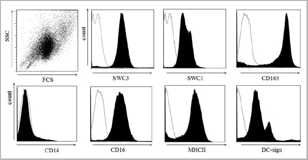
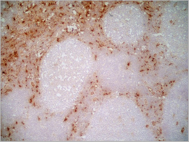
(Immunoperoxidase staining of a human tonsil cryosection with Mouse anti Human CD163 antibody, clone EDHu-1 followed by the Histar detection system . Low power)
Application Data
(Immunoperoxidase staining of a human tonsil cryosection with Mouse anti Human CD163 antibody, clone EDHu-1 followed by the Histar detection system . Low power)
CD163, Monoclonal Antibody (Cat# AAA11943)
Full Name
MOUSE ANTI HUMAN CD163
Gene Names
CD163; M130; MM130
Reactivity
Guinea Pig, Pig, Rhesus Monkey, Sheep
Applications
Immunohistochemistry, Immunohistochemistry, Flow Cytometry, Immunofluorescence, Immunoassay, Western Blot
Pricing


 Application Data
(Staining of mouse spleen with Hamster anti Mouse CD81: Alexa Fluor 488)
Application Data
(Staining of mouse spleen with Hamster anti Mouse CD81: Alexa Fluor 488)
CD81, Monoclonal Antibody (Cat# AAA11942)
Full Name
HAMSTER ANTI MOUSE CD81
Gene Names
Cd81; Tapa1; Tapa-1; Tspan28
Reactivity
Rat
Applications
Immunohistochemistry, Flow Cytometry, Immunoprecipitation, Western Blot
Pricing
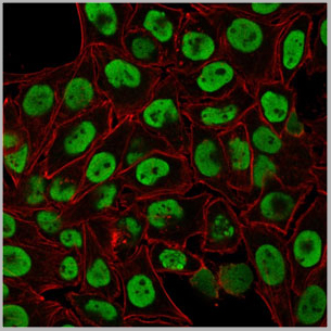
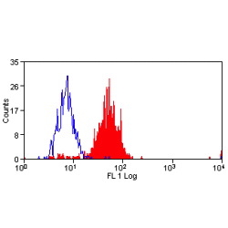
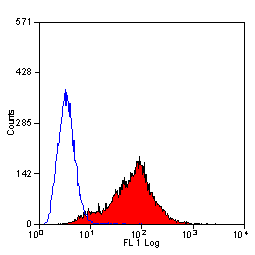
 FCM (Flow Cytometry)
(Flow Cytometric Analysis of PFA-fixed Raji cells with Human Nuclear Antigen Mouse Monoclonal Antibody (235-1); followed by goat anti-mouse IgG-CF488 (Blue); Isotype Control (Red).)
FCM (Flow Cytometry)
(Flow Cytometric Analysis of PFA-fixed Raji cells with Human Nuclear Antigen Mouse Monoclonal Antibody (235-1); followed by goat anti-mouse IgG-CF488 (Blue); Isotype Control (Red).)
Human Nuclear Antigen, Monoclonal Antibody (Cat# AAA13804)
Full Name
Human Nuclear Antigen
Reactivity
Human and Non-human primates. Does not react with mouse, rat, and chicken. Others not known.
Applications
Flow Cytometry, Immunofluorescence, Immunoprecipitation, Immunocytochemistry, Immunohistochemistry
Pricing
 WB (Western Blot)
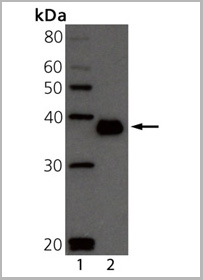
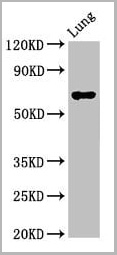
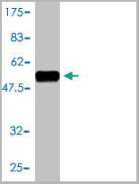
(Western BlotPositive WB detected in: Rat lung tissueAll lanes: TFEB antibody at 3ug/mlSecondaryGoat polyclonal to rabbit IgG at 1/50000 dilutionPredicted band size: 53, 44 KDaObserved band size: 70 KDa)
WB (Western Blot)
(Western BlotPositive WB detected in: Rat lung tissueAll lanes: TFEB antibody at 3ug/mlSecondaryGoat polyclonal to rabbit IgG at 1/50000 dilutionPredicted band size: 53, 44 KDaObserved band size: 70 KDa)
TFEB, Polyclonal Antibody (Cat# AAA26969)
Full Name
TFEB Antibody
Gene Names
TFEB; TCFEB; BHLHE35; ALPHATFEB
Reactivity
Human, Rat
Applications
Western Blot, Immunohistochemistry, Immunofluorescence
Purity
>95%, Protein G purified
Pricing
 WB (Western Blot)
(CD58 monoclonal antibody, Western Blot analysis of CD58 expression in Jurkat.)
WB (Western Blot)
(CD58 monoclonal antibody, Western Blot analysis of CD58 expression in Jurkat.)
CD58, Monoclonal Antibody (Cat# AAA25344)
Full Name
CD58 (Lymphocyte Function-associated Antigen 3, Ag3, Surface Glycoprotein LFA-3, LFA3) (HRP)
Gene Names
CD58; ag3; LFA3; LFA-3
Reactivity
Human
Applications
Immunohistochemistry, Western Blot
Purity
Purified by Protein A Affinity Chromatography.
Pricing
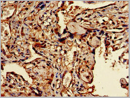
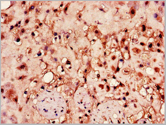 IHC (Immunohistochemistry)
(Immunohistochemistry of paraffin-embedded human placenta tissue using AAA26966 at dilution of 1:100)
IHC (Immunohistochemistry)
(Immunohistochemistry of paraffin-embedded human placenta tissue using AAA26966 at dilution of 1:100)
HPSE, Polyclonal Antibody (Cat# AAA26966)
Full Name
HPSE Antibody
Gene Names
HPSE; HPA; HPA1; HPR1; HSE1; HPSE1
Reactivity
Human
Applications
Western Blot, Immunohistochemistry
Purity
>95%, Protein G purified
Pricing
CD58, Monoclonal Antibody (Cat# AAA24456)
Full Name
CD58 (Lymphocyte Function-associated Antigen 3, Ag3, Surface Glycoprotein LFA-3, LFA3) APC
Gene Names
CD58; ag3; LFA3; LFA-3
Reactivity
Human
Applications
EIA, IF, IHC, WB
Purity
Purified by Protein A Affinity Chromatography.
Pricing