Filters
Clonality
Type
Reactivity
Gene Name
Isotype
Host
Application
Clone
923 results for "Biotin Antibody" - showing 850-900
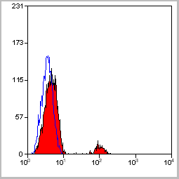
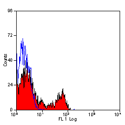 Application Data
(Staining of human peripheral blood lymphocytes with Mouse anti Human CD4: Pacific Blue)
Application Data
(Staining of human peripheral blood lymphocytes with Mouse anti Human CD4: Pacific Blue)
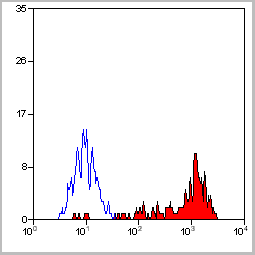
CD4, Monoclonal Antibody (Cat# AAA12071)
Full Name
MOUSE ANTI HUMAN CD4:FITC
Gene Names
CD4; CD4mut
Reactivity
Human
Applications
Flow Cytometry
Pricing


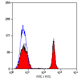
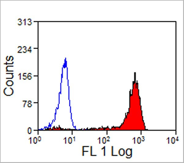
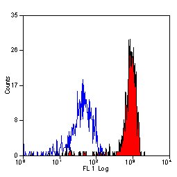
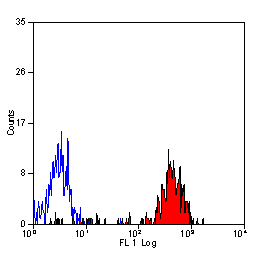
 FCM (Flow Cytometry)
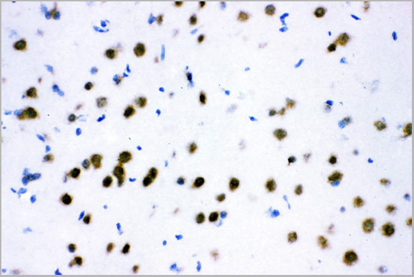
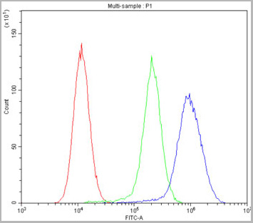
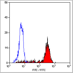
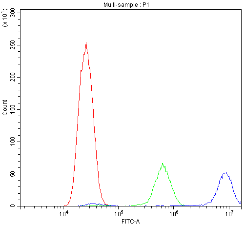
(FACS analysis of ES-2 cells stained with STIP1 monoclonal antibody clone 2E11 (Green) and non-stained ES-2 cells (Black) as negative control.)
FCM (Flow Cytometry)
(FACS analysis of ES-2 cells stained with STIP1 monoclonal antibody clone 2E11 (Green) and non-stained ES-2 cells (Black) as negative control.)
STIP1, Monoclonal Antibody (Cat# AAA26275)
Full Name
STIP1 (Stress-Induced-Phosphoprotein 1, HOP, IEF-SSP-3521, P60, STI1, STI1L) (Biotin)
Gene Names
STIP1; HOP; P60; STI1; STI1L; HEL-S-94n; IEF-SSP-3521
Applications
Flow Cytometry, Immunofluorescence, Immunohistochemistry, Western Blot
Purity
Purified
Pricing

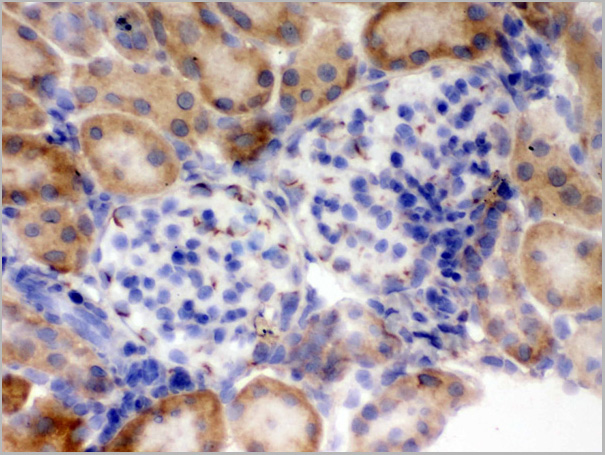
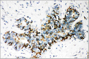
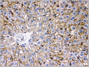
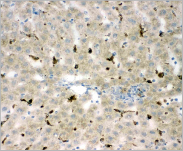
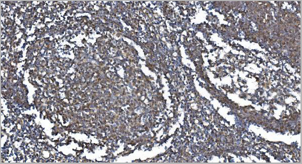
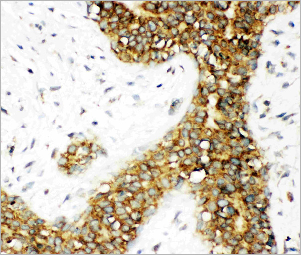
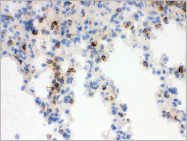
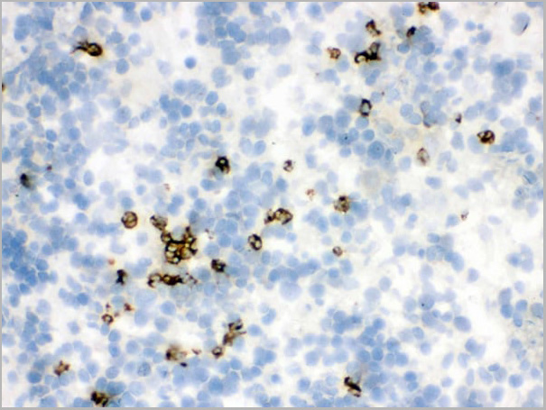 IHC (Immunohistochemistry)
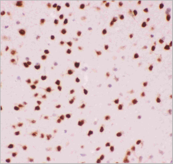
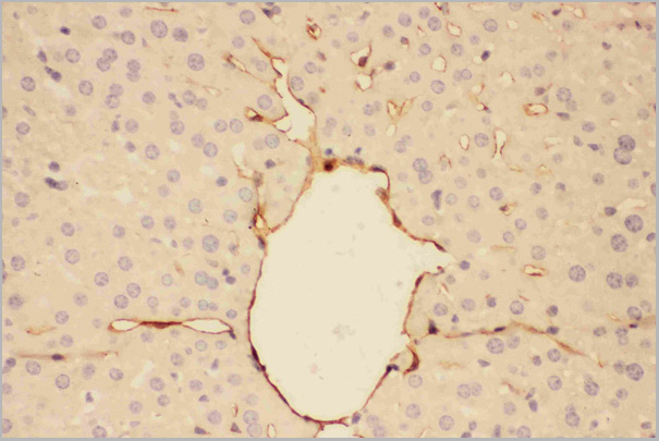
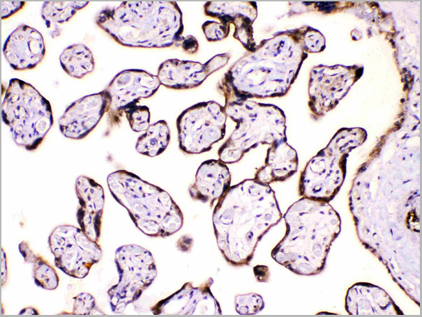
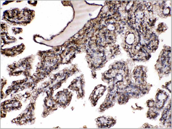
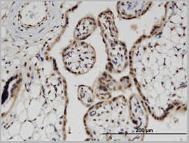
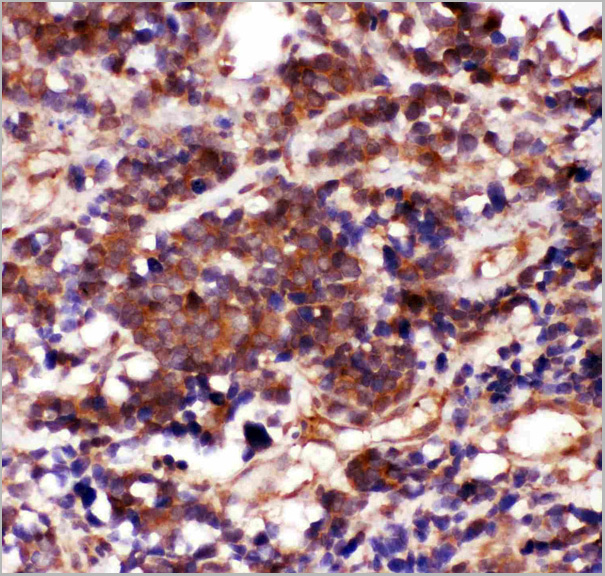
(Figure 7. IHC analysis of Lipocalin 2 using anti-Lipocalin 2 antibody (AAA11663).Lipocalin 2 was detected in frozen section of rat spleen tissue. Heat mediated antigen retrieval was performed in citrate buffer (pH6, epitope retrieval solution) for 20 mins. The tissue section was blocked with 10% goat serum. The tissue section was then incubated with 1ug/ml rabbit anti-Lipocalin 2 Antibody (AAA11663) overnight at 4 degree C. Biotinylated goat anti-rabbit IgG was used as secondary antibody and incubated for 30 minutes at 37 degree C. The tissue section was developed using Strepavidin-Biotin-Complex (SABC) with DAB as the chromogen.)
IHC (Immunohistochemistry)
(Figure 7. IHC analysis of Lipocalin 2 using anti-Lipocalin 2 antibody (AAA11663).Lipocalin 2 was detected in frozen section of rat spleen tissue. Heat mediated antigen retrieval was performed in citrate buffer (pH6, epitope retrieval solution) for 20 mins. The tissue section was blocked with 10% goat serum. The tissue section was then incubated with 1ug/ml rabbit anti-Lipocalin 2 Antibody (AAA11663) overnight at 4 degree C. Biotinylated goat anti-rabbit IgG was used as secondary antibody and incubated for 30 minutes at 37 degree C. The tissue section was developed using Strepavidin-Biotin-Complex (SABC) with DAB as the chromogen.)
Lipocalin 2, Polyclonal Antibody (Cat# AAA11663)
Full Name
Anti-Lipocalin 2 Antibody
Gene Names
Lcn2; Sip24
Reactivity
Human, Mouse, Rat
Applications
Western Blot, Immunohistochemistry, Flow Cytometry
Purity
Immunogen Affinity Purified
Pricing
CD105, Monoclonal Antibody (Cat# AAA14292)
Full Name
CD105 antibody (biotin)
Reactivity
To be determined by end-user.
Applications
Flow Cytometry, Immunohistochemistry
Purity
>95% pure
CD105 antibody (biotin) was purified by Protein A chromatography.
CD105 antibody (biotin) was purified by Protein A chromatography.
Pricing


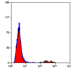
 Application Data
(Staining of mouse peripheral blood lymphocytes with Rat anti Mouse CD102: Biotin)
Application Data
(Staining of mouse peripheral blood lymphocytes with Rat anti Mouse CD102: Biotin)
CD102, Monoclonal Antibody (Cat# AAA11904)
Full Name
RAT ANTI MOUSE CD102:Low Endotoxin
Gene Names
Icam2; CD102; Ly-60; Icam-2
Applications
Flow Cytometry, Functional Assay, Immunoprecipitation
Pricing
 WB (Western Blot)
(HSPA1L monoclonal antibody Western Blot analysis of HSPA1L expression in PC-12.)
WB (Western Blot)
(HSPA1L monoclonal antibody Western Blot analysis of HSPA1L expression in PC-12.)
HSPA1L, Monoclonal Antibody (Cat# AAA24833)
Full Name
HSPA1L (Heat Shock 70kD Protein 1-like, HSP70-Hom, Heat shock 70kD protein 1L, Heat Shock 70kD Protein 1-Hom) (Biotin)
Gene Names
HSPA1L; HSP70T; hum70t; HSP70-1L; HSP70-HOM
Reactivity
Human, Rat
Applications
Immunohistochemistry, Western Blot
Purity
Purified by Protein A Affinity Chromatography.
Pricing
COVID 19 Spike RBD Coronavirus, Recombinant Protein (Cat# AAA13542)
Full Name
Recombinant SARS-Cov-2 Spike RBD Protein (Detection)
Applications
Lateral Flow
Purity
>95% by SDS-PAGE
Pricing
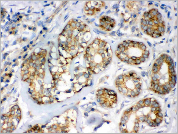
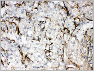
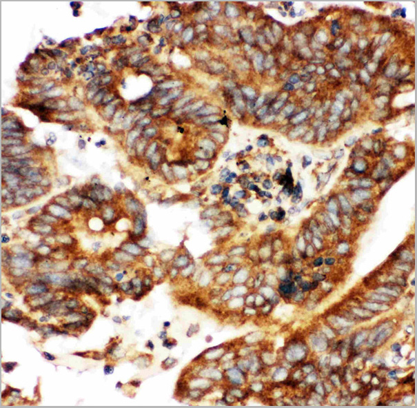
 IHC (Immunohistochemistry)
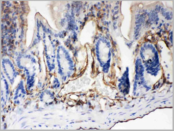
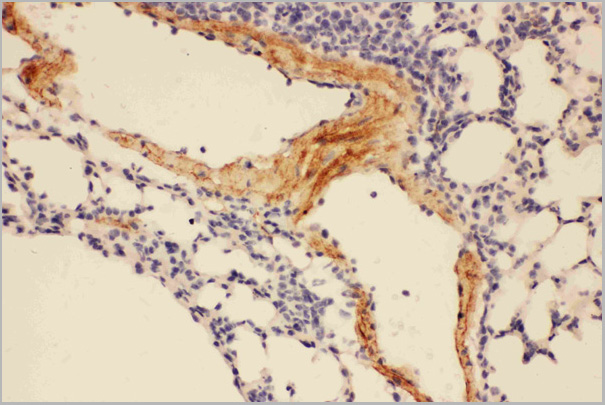
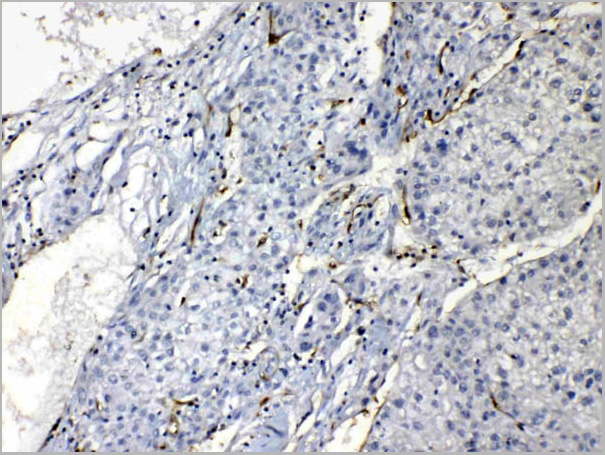
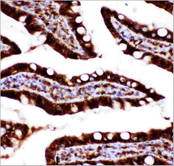
(Figure 7. IHC analysis of Flotillin 2 using anti-Flotillin 2 antibody (AAA11664).Flotillin 2 was detected in frozen section of rat small intestine tissue. Heat mediated antigen retrieval was performed in citrate buffer (pH6, epitope retrieval solution) for 20 mins. The tissue section was blocked with 10% goat serum. The tissue section was then incubated with 1ug/ml rabbit anti-Flotillin 2 Antibody (AAA11664) overnight at 4 degree C. Biotinylated goat anti-rabbit IgG was used as secondary antibody and incubated for 30 minutes at 37 degree C. The tissue section was developed using Strepavidin-Biotin-Complex (SABC) with DAB as the chromogen.)
IHC (Immunohistochemistry)
(Figure 7. IHC analysis of Flotillin 2 using anti-Flotillin 2 antibody (AAA11664).Flotillin 2 was detected in frozen section of rat small intestine tissue. Heat mediated antigen retrieval was performed in citrate buffer (pH6, epitope retrieval solution) for 20 mins. The tissue section was blocked with 10% goat serum. The tissue section was then incubated with 1ug/ml rabbit anti-Flotillin 2 Antibody (AAA11664) overnight at 4 degree C. Biotinylated goat anti-rabbit IgG was used as secondary antibody and incubated for 30 minutes at 37 degree C. The tissue section was developed using Strepavidin-Biotin-Complex (SABC) with DAB as the chromogen.)
Flotillin 2, Polyclonal Antibody (Cat# AAA11664)
Full Name
Anti-Flotillin 2 Antibody
Gene Names
FLOT2; ESA; ECS1; ESA1; ECS-1; M17S1
Reactivity
Human, Mouse, Rat
Applications
Western Blot, Immunohistochemistry
Purity
Immunogen Affinity Purified
Pricing
CXCR3, Polyclonal Antibody (Cat# AAA14406)
Full Name
CXCR3 Antibody BIOTIN-Conjugated
Gene Names
Cxcr3; Gpr9
Reactivity
Mouse, Rat
Applications
Confocal Microscopy, Immunocytochemistry, Immunofluorescence, Immunohistochemistry, Immunohistochemistry, Western Blot
Pricing


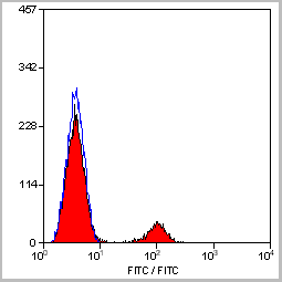
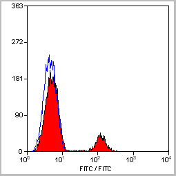
 Application Data
(Staining of mouse peritoneal macrophages with Rat anti Mouse Beta-glucan Receptor: FITC)
Application Data
(Staining of mouse peritoneal macrophages with Rat anti Mouse Beta-glucan Receptor: FITC)
DECTIN-1, Monoclonal Antibody (Cat# AAA12128)
Full Name
RAT ANTI MOUSE DECTIN-1:Biotin
Gene Names
Clec7a; BGR; beta-GR; Clecsf12
Applications
Flow Cytometry
Pricing
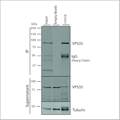
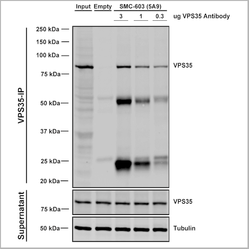
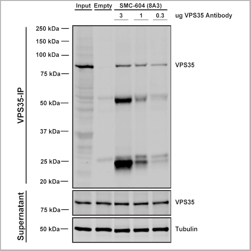
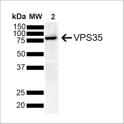
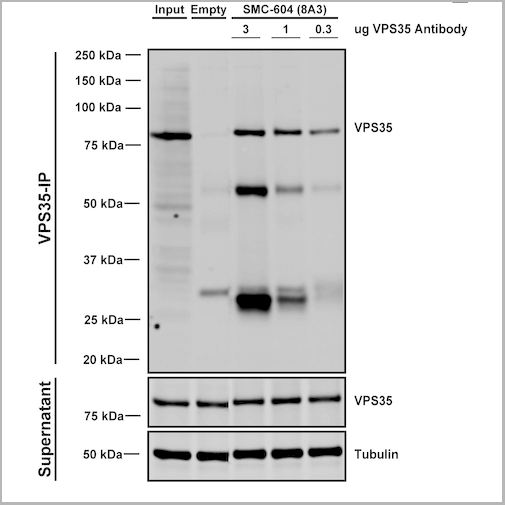
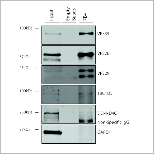
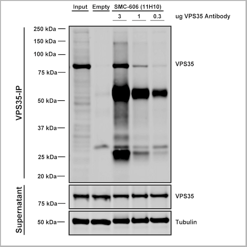 IP (Immunoprecipitation)
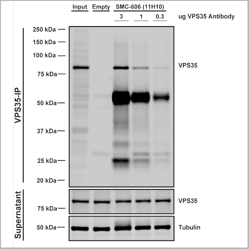
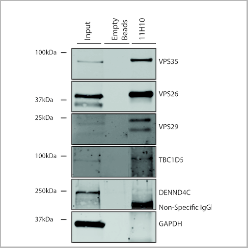
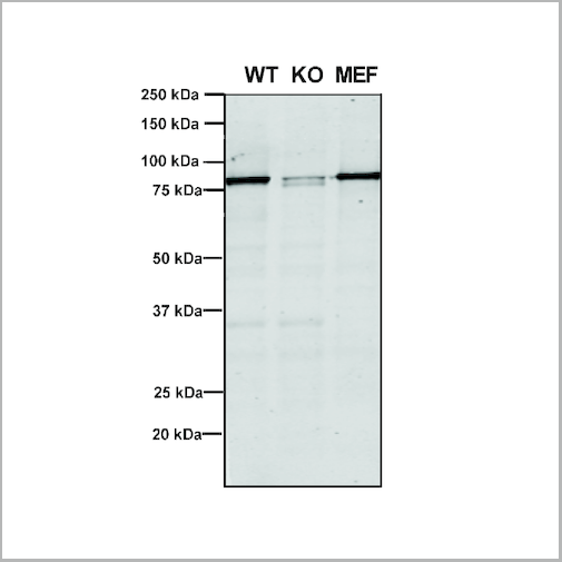
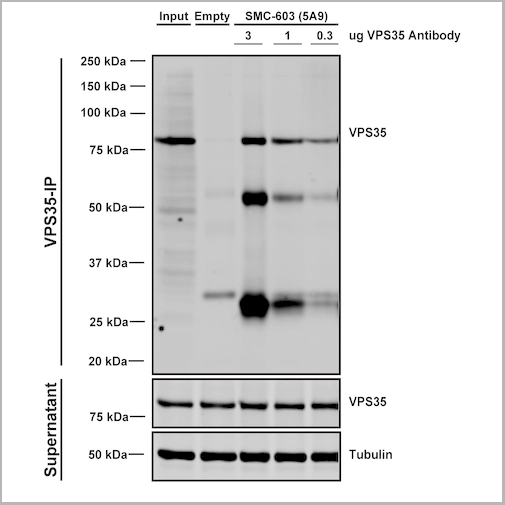
(Immunoprecipitation analysis using Mouse Anti-VPS35 Monoclonal Antibody, Clone 11H10. Tissue: embryonic fibroblast. Species: Mouse. Primary Antibody: Mouse Anti-VPS35 Monoclonal Antibody. Three amounts of (3, 1 and 0.3 ug) were non-covalently coupled to 10uL of A/G sepharose beads for 1 hour at 4 degree C and next incubated with 250ug of MEF lysate for 2 hours at 4 degree C.)
IP (Immunoprecipitation)
(Immunoprecipitation analysis using Mouse Anti-VPS35 Monoclonal Antibody, Clone 11H10. Tissue: embryonic fibroblast. Species: Mouse. Primary Antibody: Mouse Anti-VPS35 Monoclonal Antibody. Three amounts of (3, 1 and 0.3 ug) were non-covalently coupled to 10uL of A/G sepharose beads for 1 hour at 4 degree C and next incubated with 250ug of MEF lysate for 2 hours at 4 degree C.)
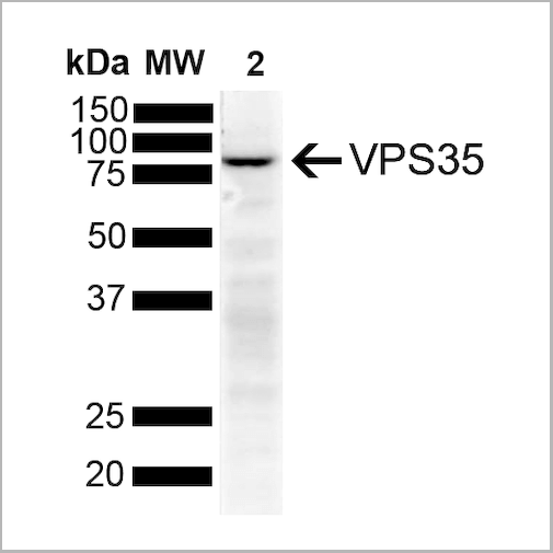
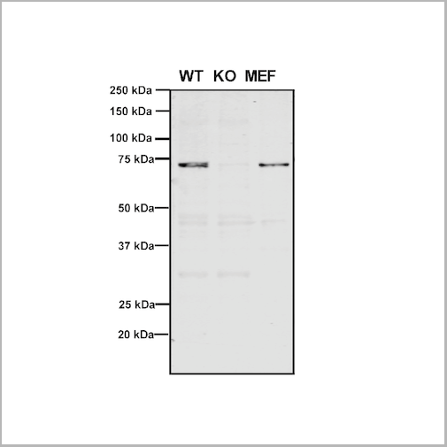
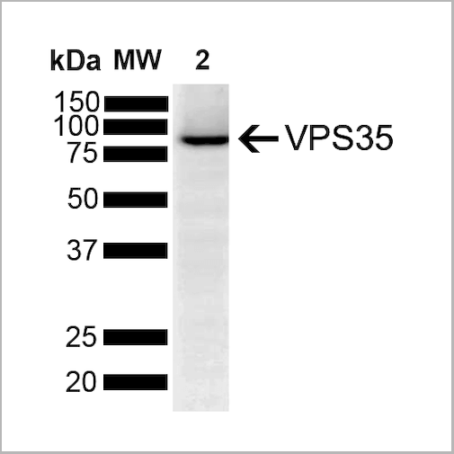
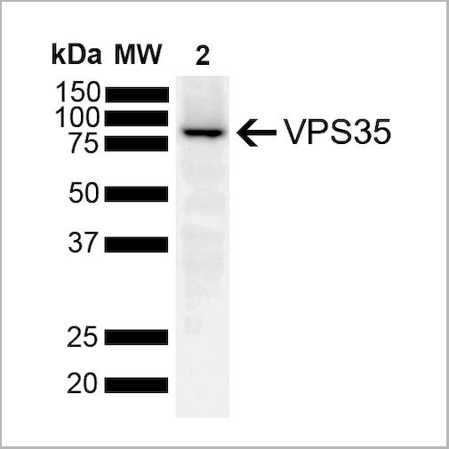
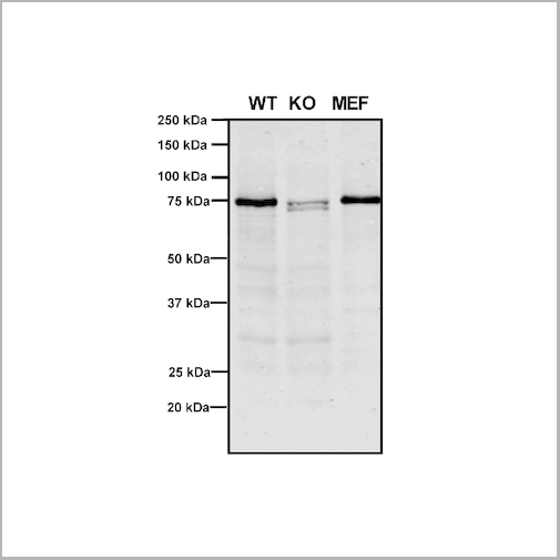
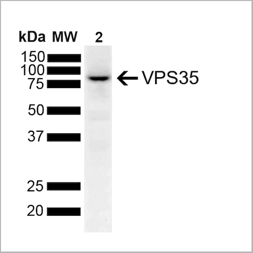
VPS35, Monoclonal Antibody (Cat# AAA27704)
Full Name
VPS35 Antibody, Clone 11H10: Biotin
Gene Names
VPS35; MEM3; PARK17
Reactivity
Human, Mouse, Rat
Applications
Western Blot, Immunoprecipitation
Purity
Protein G Purified
Pricing
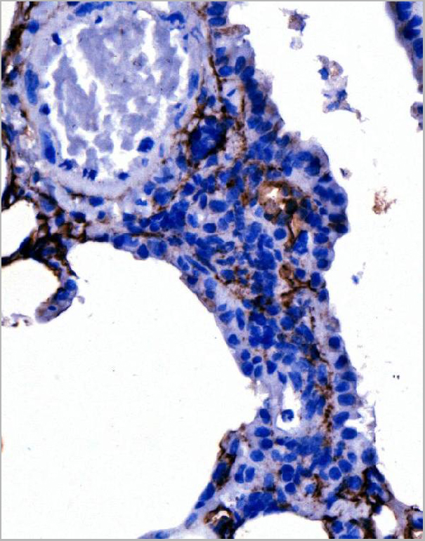
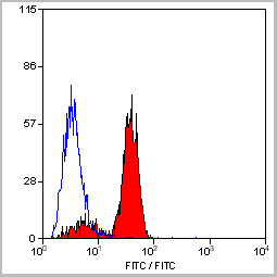
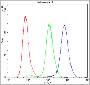
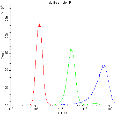
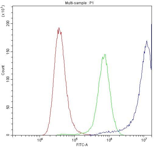
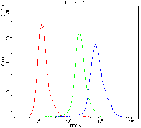
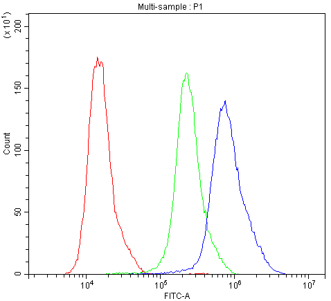
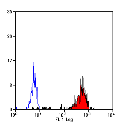
 FCM (Flow Cytometry)
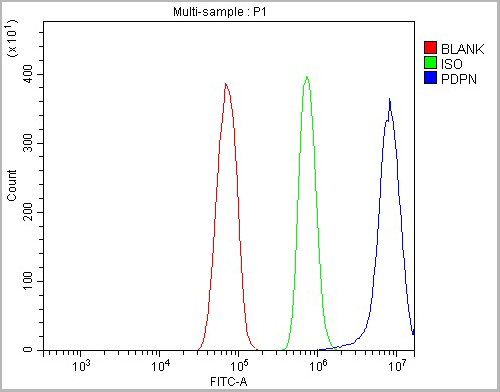
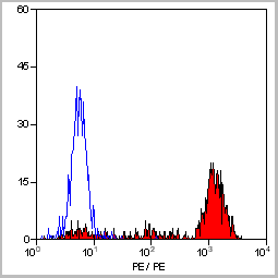
(Figure 5. Flow Cytometry analysis of RH35 cells using anti-Podoplanin/gp36/Pdpn antibody (AAA19233).Overlay histogram showing RH35 cells stained with AAA19233 (Blue line). The cells were blocked with 10% normal goat serum. And then incubated with rabbit anti-Podoplanin/gp36/Pdpn Antibody (AAA19233, 1μg/1x106 cells) for 30 min at 20 degree C. DyLight®488 conjugated goat anti-rabbit IgG (5-10μg/1x106 cells) was used as secondary antibody for 30 minutes at 20 degree C. Isotype control antibody (Green line) was rabbit IgG (1μg/1x106) used under the same conditions. Unlabelled sample (Red line) was also used as a control.)
FCM (Flow Cytometry)
(Figure 5. Flow Cytometry analysis of RH35 cells using anti-Podoplanin/gp36/Pdpn antibody (AAA19233).Overlay histogram showing RH35 cells stained with AAA19233 (Blue line). The cells were blocked with 10% normal goat serum. And then incubated with rabbit anti-Podoplanin/gp36/Pdpn Antibody (AAA19233, 1μg/1x106 cells) for 30 min at 20 degree C. DyLight®488 conjugated goat anti-rabbit IgG (5-10μg/1x106 cells) was used as secondary antibody for 30 minutes at 20 degree C. Isotype control antibody (Green line) was rabbit IgG (1μg/1x106) used under the same conditions. Unlabelled sample (Red line) was also used as a control.)
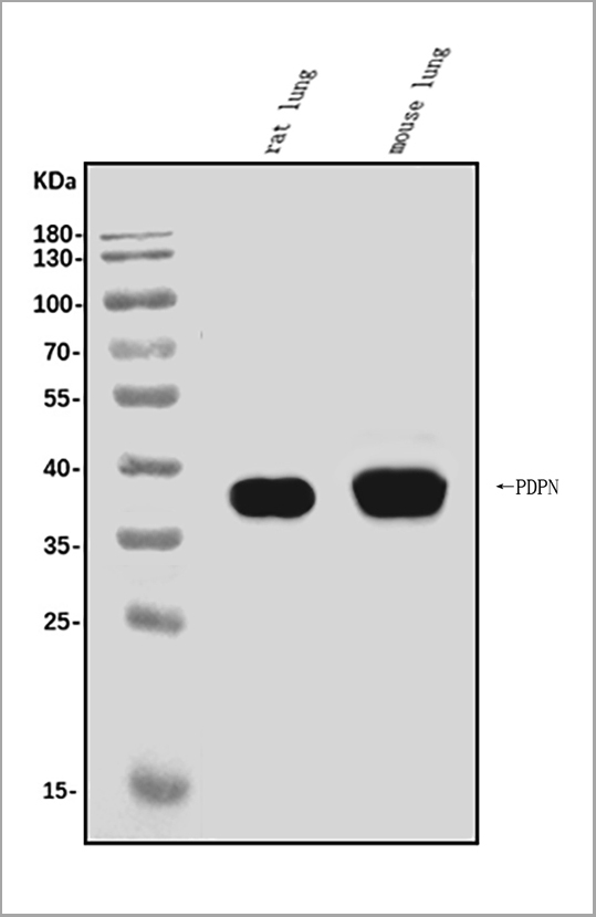
Podoplanin/gp36/Pdpn, Polyclonal Antibody (Cat# AAA19233)
Full Name
Anti-Podoplanin/gp36/Pdpn Antibody
Gene Names
Pdpn; T1a; Gp38; OTS-8; T1alpha; RANDAM-2; T1-alpha
Reactivity
Mouse, Rat
Applications
Western Blot, Immunohistochemistry, Immunocytochemistry, Immunofluorescence, Flow Cytometry, Direct ELISA
Purity
Immunogen affinity purified.
Pricing
 Application Data
(Staining of New Zealand Black mouse peripheral blood granulocytes with Rat anti Mouse Ly-6B.2 conjugated to FITC Data)
Application Data
(Staining of New Zealand Black mouse peripheral blood granulocytes with Rat anti Mouse Ly-6B.2 conjugated to FITC Data)
Ly-6B.2 ALLOANTIGEN, Monoclonal Antibody (Cat# AAA12188)
Full Name
RAT ANTI MOUSE Ly-6B.2 ALLOANTIGEN:Biotin
Applications
Flow Cytometry
Pricing
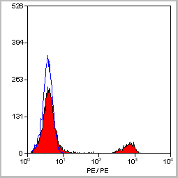
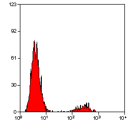
 Application Data
(Staining of human peripheral blood lymphocytes with Mouse anti Human CD19: RPE- Alexa Fluor 647)
Application Data
(Staining of human peripheral blood lymphocytes with Mouse anti Human CD19: RPE- Alexa Fluor 647)
CD19, Monoclonal Antibody (Cat# AAA11870)
Full Name
MOUSE ANTI HUMAN CD19:FITC
Gene Names
CD19; B4; CVID3
Applications
Flow Cytometry
Pricing


 Application Data
(Staining of mouse peritoneal macrophages with Rat anti Mouse Beta-glucan Receptor: FITC)
Application Data
(Staining of mouse peritoneal macrophages with Rat anti Mouse Beta-glucan Receptor: FITC)
DECTIN-1, Monoclonal Antibody (Cat# AAA12127)
Full Name
RAT ANTI MOUSE DECTIN-1:Biotin
Gene Names
Clec7a; BGR; beta-GR; Clecsf12
Applications
Flow Cytometry
Pricing


 Application Data
(Staining of human peripheral blood monocytes with Mouse anti Human CD14:RPE)
Application Data
(Staining of human peripheral blood monocytes with Mouse anti Human CD14:RPE)
CD14, Monoclonal Antibody (Cat# AAA12179)
Full Name
MOUSE ANTI HUMAN CD14
Applications
Immunohistochemistry, Flow Cytometry, Immunoprecipitation
Pricing
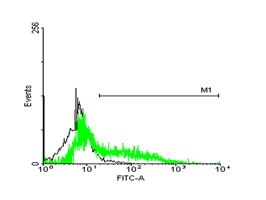
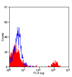
 FCM (Flow Cytometry)
(Figure 8. Flow Cytometry analysis of HepG2 cells using anti-ATF2 antibody (AAA11634).Overlay histogram showing HepG2 cells stained with AAA11634 (Blue line).The cells were blocked with 10% normal goat serum. And then incubated with rabbit anti-ATF2 Antibody (AAA11634,1ug/1x10^6 cells) for 30 min at 20 degree C. DyLight ®488 conjugated goat anti-rabbit IgG (5-10ug/1x10^6 cells) was used as secondary antibody for 30 minutes at 20 degree C. Isotype control antibody (Green line) was rabbit IgG (1ug/1x106) used under the same conditions. Unlabelled sample (Red line) was also used as a control.)
FCM (Flow Cytometry)
(Figure 8. Flow Cytometry analysis of HepG2 cells using anti-ATF2 antibody (AAA11634).Overlay histogram showing HepG2 cells stained with AAA11634 (Blue line).The cells were blocked with 10% normal goat serum. And then incubated with rabbit anti-ATF2 Antibody (AAA11634,1ug/1x10^6 cells) for 30 min at 20 degree C. DyLight ®488 conjugated goat anti-rabbit IgG (5-10ug/1x10^6 cells) was used as secondary antibody for 30 minutes at 20 degree C. Isotype control antibody (Green line) was rabbit IgG (1ug/1x106) used under the same conditions. Unlabelled sample (Red line) was also used as a control.)
Cyclic AMP-dependent transcription factor ATF-2, Polyclonal Antibody (Cat# AAA11634)
Full Name
Anti-ATF2 Antibody
Gene Names
ATF2; HB16; CREB2; TREB7; CREB-2; CRE-BP1
Reactivity
Human, Mouse, Rat. No cross reactivity with other proteins.
Applications
Western Blot, Immunohistochemistry, Immunohistochemistry
Purity
Immunogen affinity purified.
Pricing
 FCM (Flow Cytometry)
(Figure 6. Flow Cytometry analysis of A549 cells using anti-MDR3 antibody (AAA11652).Overlay histogram showing A549 cells stained with AAA11652 (Blue line).The cells were blocked with 10% normal goat serum. And then incubated with rabbit anti-MDR3 Antibody (AAA11652,1ug/1x106 cells) for 30 min at 20°C. DyLight®488 conjugated goat anti-rabbit IgG (5-10ug/1x106 cells) was used as secondary antibody for 30 minutes at 20°C. Isotype control antibody (Green line) was rabbit IgG (1ug/1x106) used under the same conditions. Unlabelled sample (Red line) was also used as a control.)
FCM (Flow Cytometry)
(Figure 6. Flow Cytometry analysis of A549 cells using anti-MDR3 antibody (AAA11652).Overlay histogram showing A549 cells stained with AAA11652 (Blue line).The cells were blocked with 10% normal goat serum. And then incubated with rabbit anti-MDR3 Antibody (AAA11652,1ug/1x106 cells) for 30 min at 20°C. DyLight®488 conjugated goat anti-rabbit IgG (5-10ug/1x106 cells) was used as secondary antibody for 30 minutes at 20°C. Isotype control antibody (Green line) was rabbit IgG (1ug/1x106) used under the same conditions. Unlabelled sample (Red line) was also used as a control.)
ABCB4, Polyclonal Antibody (Cat# AAA11652)
Full Name
Anti-ABCB4 Antibody
Gene Names
ABCB4; GBD1; ICP3; MDR2; MDR3; PGY3; ABC21; MDR2/3; PFIC-3
Reactivity
Human, Mouse, Rat
Applications
Western Blot, Immunohistochemistry, Immunofluorescence, Flow Cytometry
Purity
Immunogen Affinity Purified
Pricing
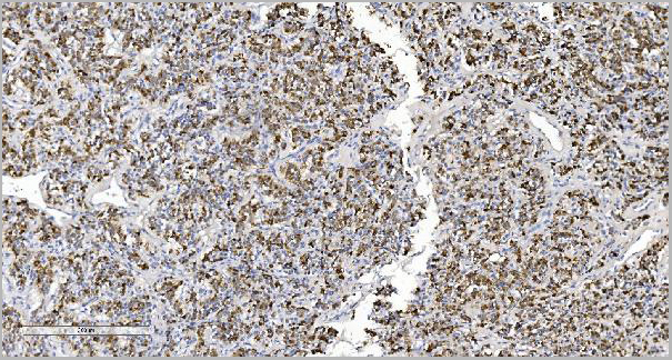
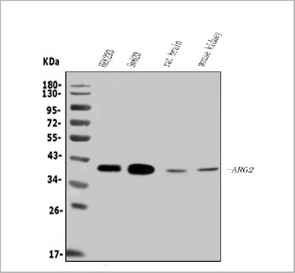
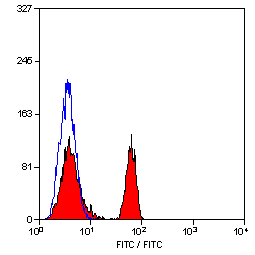
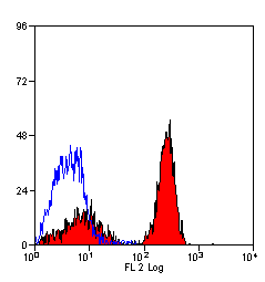
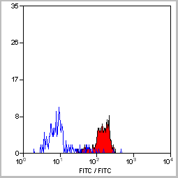
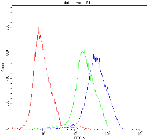
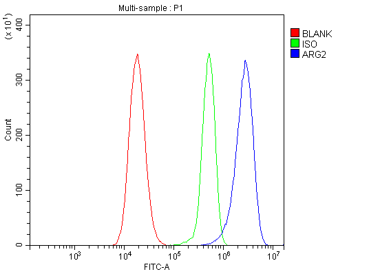 FCM (Flow Cytometry)
(Figure 7. Flow Cytometry analysis of 293T cells using anti-ARG2 antibody (AAA19256).Overlay histogram showing 293T cells stained with AAA19256 (Blue line). The cells were blocked with 10% normal goat serum. And then incubated with rabbit anti-ARG2 Antibody (AAA19256, 1μg/1x106 cells) for 30 min at 20 degree C. DyLight®488 conjugated goat anti-rabbit IgG (5-10μg/1x106 cells) was used as secondary antibody for 30 minutes at 20 degree C. Isotype control antibody (Green line) was rabbit IgG (1μg/1x106) used under the same conditions. Unlabelled sample (Red line) was also used as a control.)
FCM (Flow Cytometry)
(Figure 7. Flow Cytometry analysis of 293T cells using anti-ARG2 antibody (AAA19256).Overlay histogram showing 293T cells stained with AAA19256 (Blue line). The cells were blocked with 10% normal goat serum. And then incubated with rabbit anti-ARG2 Antibody (AAA19256, 1μg/1x106 cells) for 30 min at 20 degree C. DyLight®488 conjugated goat anti-rabbit IgG (5-10μg/1x106 cells) was used as secondary antibody for 30 minutes at 20 degree C. Isotype control antibody (Green line) was rabbit IgG (1μg/1x106) used under the same conditions. Unlabelled sample (Red line) was also used as a control.)
ARG2, Polyclonal Antibody (Cat# AAA19256)
Full Name
Anti-ARG2 Antibody
Reactivity
Human, Mouse, Rat
Applications
Western Blot, Immunohistochemistry, Immunocytochemistry, Immunofluorescence, Flow Cytometry, Direct ELISA
Purity
Immunogen affinity purified.
Pricing
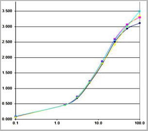
 Application Data
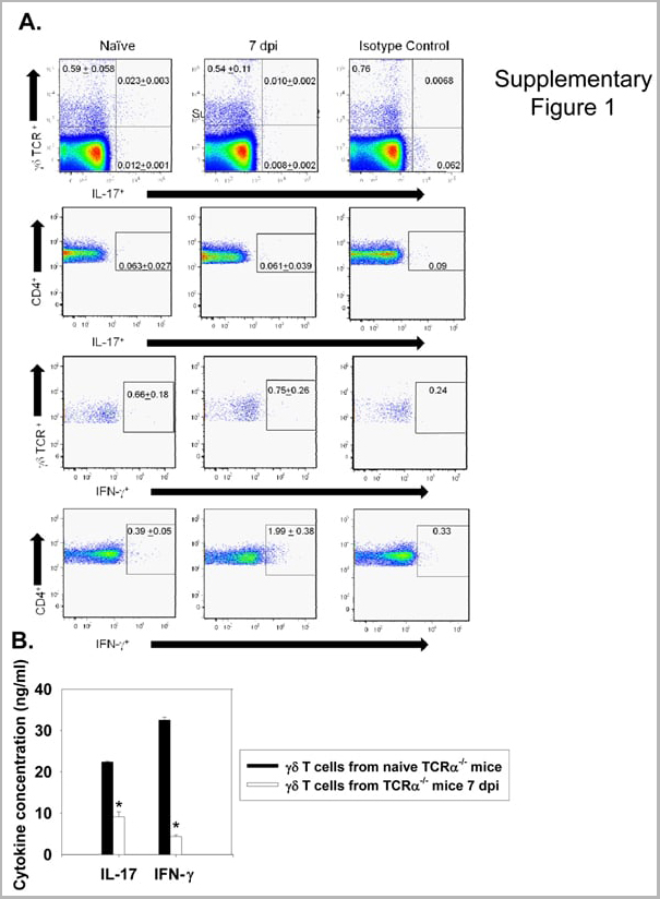
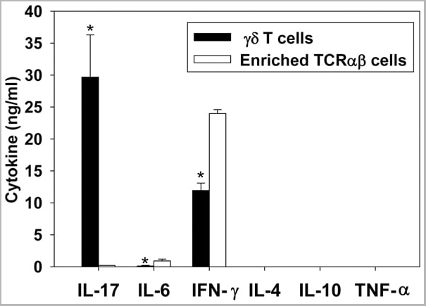
(Published customer image: gamma delta T cells are the primary source of IL-17 during B. abortus infection. C57BL/6 mice were infected i.p. with 5x10⁴ CFUs of B. abortus 2308, and two weeks later gamma delta T cells (>95% purity) and an enriched TCRalphabeta (~55% CD4+, 25% CD8+) cell fraction were isolated from the spleens of infected mice. Cells were stimulated with 500 ng/ml ionomycin and 50 ng/ml PMA for three days, and cell-free supernatants from triplicate wells were assayed for cytokine production via ELISA. The mean +/- SD is shown; * P)
Application Data
(Published customer image: gamma delta T cells are the primary source of IL-17 during B. abortus infection. C57BL/6 mice were infected i.p. with 5x10⁴ CFUs of B. abortus 2308, and two weeks later gamma delta T cells (>95% purity) and an enriched TCRalphabeta (~55% CD4+, 25% CD8+) cell fraction were isolated from the spleens of infected mice. Cells were stimulated with 500 ng/ml ionomycin and 50 ng/ml PMA for three days, and cell-free supernatants from triplicate wells were assayed for cytokine production via ELISA. The mean +/- SD is shown; * P)
IFN GAMMA, Monoclonal Antibody (Cat# AAA12094)
Full Name
MOUSE ANTI BOVINE INTERFERON GAMMA:Biotin
Applications
Flow Cytometry
Pricing


 Application Data
(Staining of human peripheral blood monocytes with Mouse anti Human CD14:RPE)
Application Data
(Staining of human peripheral blood monocytes with Mouse anti Human CD14:RPE)
CD14, Monoclonal Antibody (Cat# AAA11998)
Full Name
MOUSE ANTI HUMAN CD14
Applications
Immunohistochemistry, Flow Cytometry, Immunofluorescence, Immunoprecipitation
Pricing
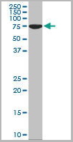
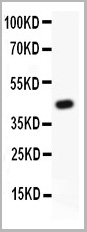
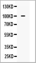
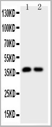
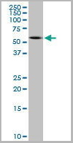
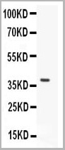
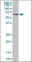
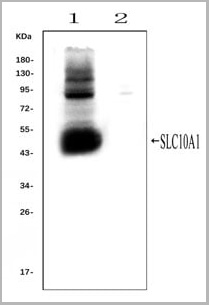 WB (Western Blot)
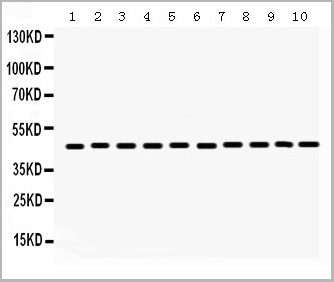
(Western blot analysis of SLC10A1 using anti- SLC10A1 antibody (AAA11653).Electrophoresis was performed on a 5-20% SDS-PAGE gel at 70V (Stacking gel) / 90V (Resolving gel) for 2-3 hours. The sample well of each lane was loaded with 50ug of sample under reducing conditions.Lane 1: rat liver tissue lysates (positive control), Lane 2: rat kidney tissue lysates, (negative control)After Electrophoresis, proteins were transferred to a Nitrocellulose membrane at 150mA for 50-90 minutes. Blocked the membrane with 5% Non-fat Milk/ TBS for 1.5 hour at RT. The membrane was incubated with rabbit anti-SLC10A1 antigen affinity purified polyclonal antibody (AAA11653) at 0.25ug/mL overnight at 4°C, then washed with TBS-0.1%Tween 3 times with 5 minutes each and probed with a goat anti-rabbit IgG-HRP secondary antibody at a dilution of 1:10000 for 1.5 hour at RT. The signal is developed using an Enhanced Chemiluminescent detection (ECL) kit with Tanon 5200 system. A specific band was detected for SLC10A1 at approximately 50KD. The expected band size for SLC10A1 is at 38KD.)
WB (Western Blot)
(Western blot analysis of SLC10A1 using anti- SLC10A1 antibody (AAA11653).Electrophoresis was performed on a 5-20% SDS-PAGE gel at 70V (Stacking gel) / 90V (Resolving gel) for 2-3 hours. The sample well of each lane was loaded with 50ug of sample under reducing conditions.Lane 1: rat liver tissue lysates (positive control), Lane 2: rat kidney tissue lysates, (negative control)After Electrophoresis, proteins were transferred to a Nitrocellulose membrane at 150mA for 50-90 minutes. Blocked the membrane with 5% Non-fat Milk/ TBS for 1.5 hour at RT. The membrane was incubated with rabbit anti-SLC10A1 antigen affinity purified polyclonal antibody (AAA11653) at 0.25ug/mL overnight at 4°C, then washed with TBS-0.1%Tween 3 times with 5 minutes each and probed with a goat anti-rabbit IgG-HRP secondary antibody at a dilution of 1:10000 for 1.5 hour at RT. The signal is developed using an Enhanced Chemiluminescent detection (ECL) kit with Tanon 5200 system. A specific band was detected for SLC10A1 at approximately 50KD. The expected band size for SLC10A1 is at 38KD.)
SLC10A1, Polyclonal Antibody (Cat# AAA11653)
Full Name
Anti-SLC10A1 Antibody
Gene Names
Slc10a1; Ntcp
Reactivity
Mouse, Rat
Applications
Flow Cytometry, Immunofluorescence, Immunohistochemistry, Immunohistochemistry, Immunocytochemistry, Western Blot
Purity
Immunogen affinity purified.
Pricing
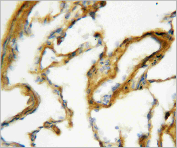
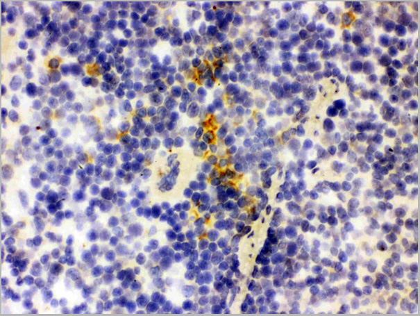
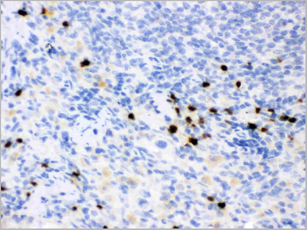
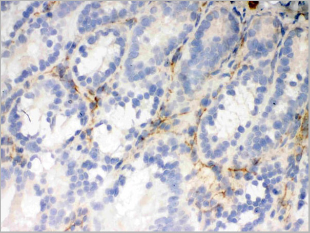
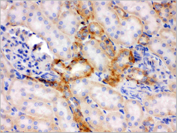
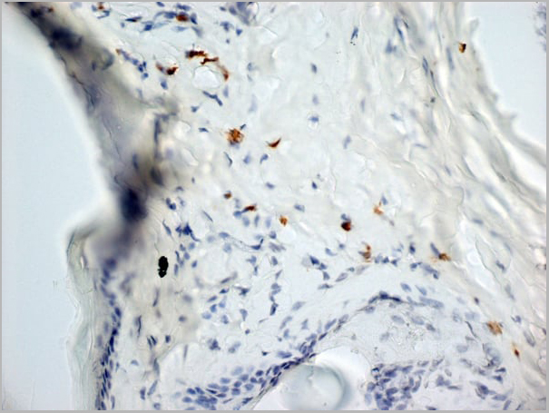
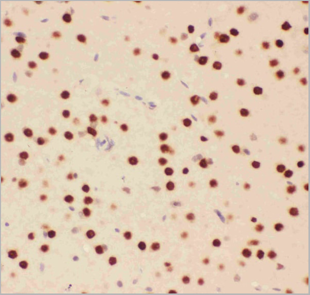
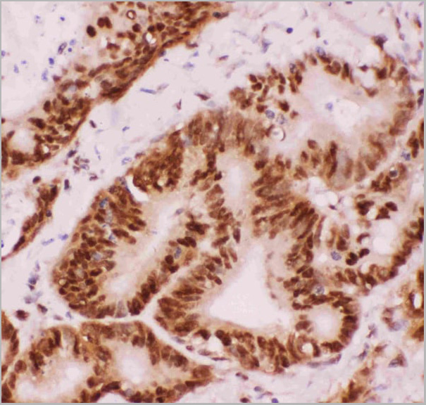
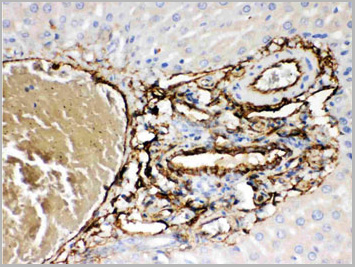
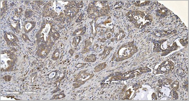
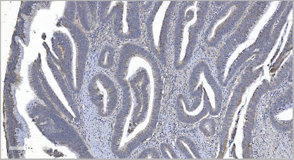
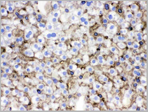
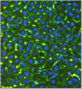
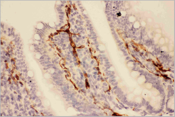
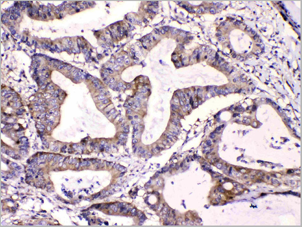
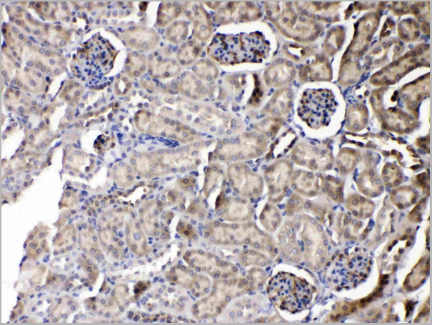
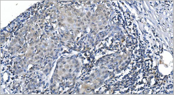
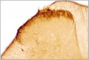
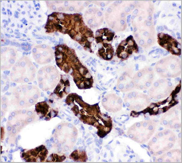
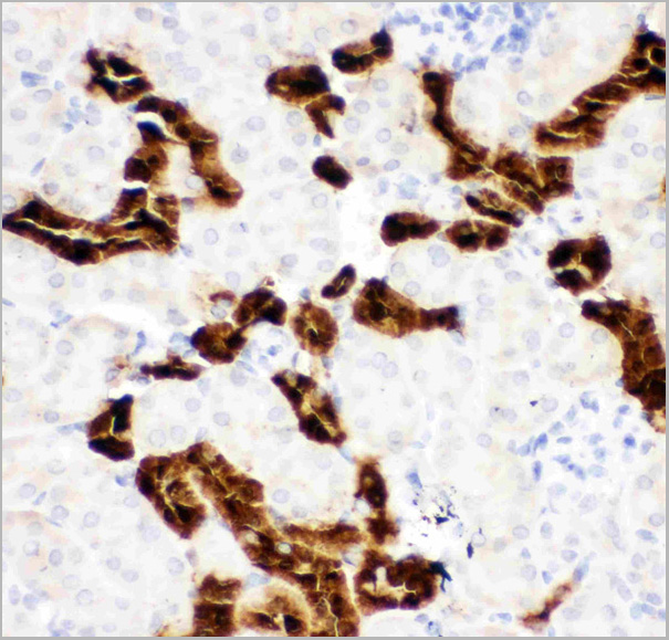
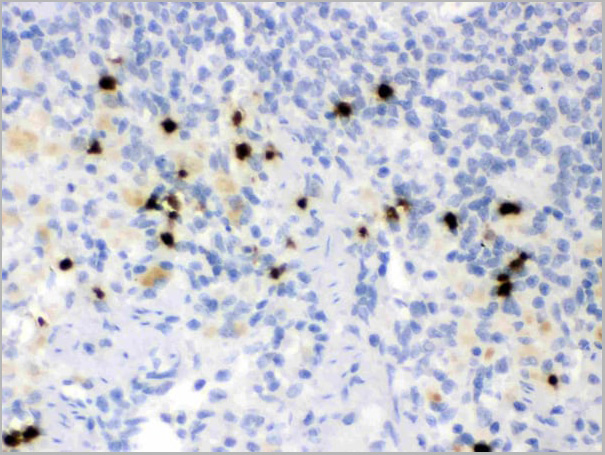
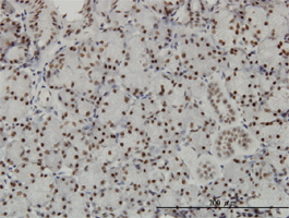
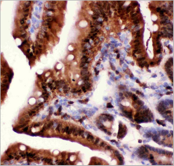
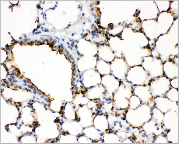 IHC (Immunohistochemistry)
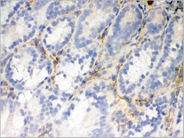
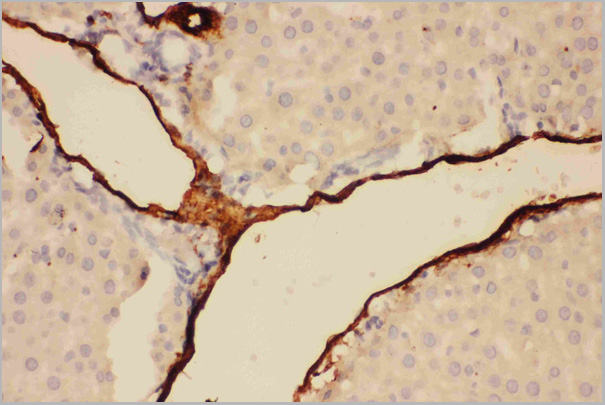
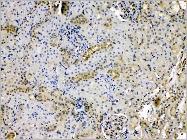
(Figure 3. IHC analysis of SFTPA1 using anti- SFTPA1 antibody (AAA11521).SFTPA1 was detected in paraffin-embedded section of mouse lung tissues. Heat mediated antigen retrieval was performed in citrate buffer (pH6, epitope retrieval solution) for 20 mins. The tissue section was blocked with 10% goat serum. The tissue section was then incubated with 1ug/ml rabbit anti- SFTPA1 Antibody (AAA11521) overnight at 4 degree C. Biotinylated goat anti-rabbit IgG was used as secondary antibody and incubated for 30 minutes at 37 degree C. The tissue section was developed using Strepavidin-Biotin-Complex (SABC) with DAB as the chromogen.)
IHC (Immunohistochemistry)
(Figure 3. IHC analysis of SFTPA1 using anti- SFTPA1 antibody (AAA11521).SFTPA1 was detected in paraffin-embedded section of mouse lung tissues. Heat mediated antigen retrieval was performed in citrate buffer (pH6, epitope retrieval solution) for 20 mins. The tissue section was blocked with 10% goat serum. The tissue section was then incubated with 1ug/ml rabbit anti- SFTPA1 Antibody (AAA11521) overnight at 4 degree C. Biotinylated goat anti-rabbit IgG was used as secondary antibody and incubated for 30 minutes at 37 degree C. The tissue section was developed using Strepavidin-Biotin-Complex (SABC) with DAB as the chromogen.)
Surfactant Protein A, Polyclonal Antibody (Cat# AAA11521)
Full Name
Polyclonal Anti-SFTPA1 Antibody
Gene Names
Sftpa1; SP-A; Sftp1; Sftp-1
Reactivity
Mouse, Rat
Applications
Western Blot, Immunohistochemistry
Purity
Immunogen affinity purified.
Pricing
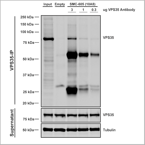
 IP (Immunoprecipitation)
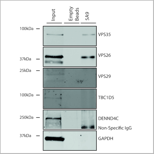
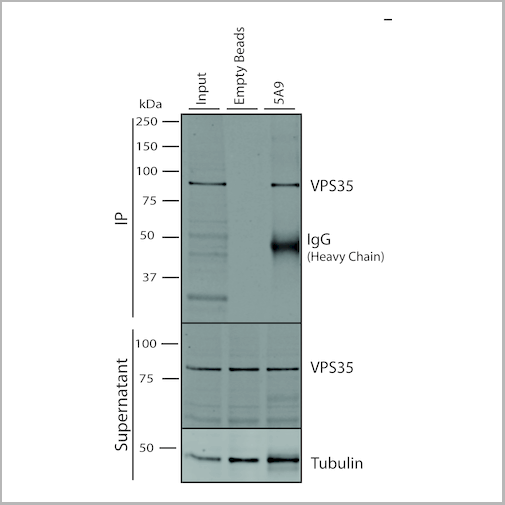
(Immunoprecipitation analysis using Mouse Anti-VPS35 Monoclonal Antibody, Clone 5A9. Tissue: embryonic fibroblast. Species: Mouse. Primary Antibody: Mouse Anti-VPS35 Monoclonal Antibody. Three amounts of (3, 1 and 0.3 ug) were non-covalently coupled to 10uL of A/G sepharose beads for 1 hour at 4 degree C and next incubated with 250ug of MEF lysate for 2 hours at 4 degree C.)
IP (Immunoprecipitation)
(Immunoprecipitation analysis using Mouse Anti-VPS35 Monoclonal Antibody, Clone 5A9. Tissue: embryonic fibroblast. Species: Mouse. Primary Antibody: Mouse Anti-VPS35 Monoclonal Antibody. Three amounts of (3, 1 and 0.3 ug) were non-covalently coupled to 10uL of A/G sepharose beads for 1 hour at 4 degree C and next incubated with 250ug of MEF lysate for 2 hours at 4 degree C.)
VPS35, Monoclonal Antibody (Cat# AAA27677)
Full Name
VPS35 Antibody, Clone 5A9: Biotin
Gene Names
VPS35; MEM3; PARK17
Reactivity
Human, Mouse, Rat
Applications
Western Blot, Immunocytochemistry, Immunofluorescence, Immunoprecipitation
Purity
Protein G Purified
Pricing
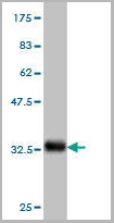
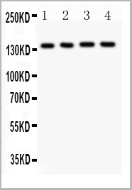
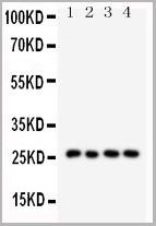
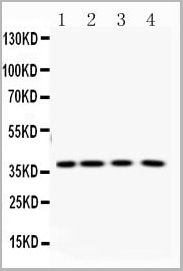
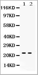
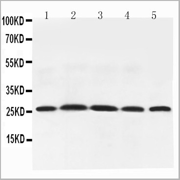 WB (Western Blot)
(Figure 6. Western blot analysis of Adiponectin using anti-Adiponectin antibody (AAA11602).Electrophoresis was performed on a 5-20% SDS-PAGE gel at 70V (Stacking gel) / 90V (Resolving gel) for 2-3 hours. The sample well of each lane was loaded with 50ug of sample under reducing conditions.Lane 1: Rat Testis Tissue Lysate,Lane 2: Rat Spleen Tissue Lysate,Lane 3: Rat Skeletal Muscle Tissue Lysate,Lane 4: Rat Cardiac Muscle Tissue Lysate,Lane 5: Rat Liver Tissue Lysate,After Electrophoresis, proteins were transferred to a Nitrocellulose membrane at 150mA for 50-90 minutes. Blocked the membrane with 5% Non-fat Milk/ TBS for 1.5 hour at RT. The membrane was incubated with rabbit anti-Adiponectin antigen affinity purified polyclonal antibody at 0.5ug/mL overnight at 4 degree C, then washed with TBS-0.1%Tween 3 times with 5 minutes each and probed with a goat anti-rabbit IgG-HRP secondary antibody at a dilution of 1:10000 for 1.5 hour at RT. The signal is developed using an Enhanced Chemiluminescent detection (ECL) kit with Tanon 5200 system. A specific band was detected for Adiponectin at approximately 26KD. The expected band size for Adiponectin is at 26KD.)
WB (Western Blot)
(Figure 6. Western blot analysis of Adiponectin using anti-Adiponectin antibody (AAA11602).Electrophoresis was performed on a 5-20% SDS-PAGE gel at 70V (Stacking gel) / 90V (Resolving gel) for 2-3 hours. The sample well of each lane was loaded with 50ug of sample under reducing conditions.Lane 1: Rat Testis Tissue Lysate,Lane 2: Rat Spleen Tissue Lysate,Lane 3: Rat Skeletal Muscle Tissue Lysate,Lane 4: Rat Cardiac Muscle Tissue Lysate,Lane 5: Rat Liver Tissue Lysate,After Electrophoresis, proteins were transferred to a Nitrocellulose membrane at 150mA for 50-90 minutes. Blocked the membrane with 5% Non-fat Milk/ TBS for 1.5 hour at RT. The membrane was incubated with rabbit anti-Adiponectin antigen affinity purified polyclonal antibody at 0.5ug/mL overnight at 4 degree C, then washed with TBS-0.1%Tween 3 times with 5 minutes each and probed with a goat anti-rabbit IgG-HRP secondary antibody at a dilution of 1:10000 for 1.5 hour at RT. The signal is developed using an Enhanced Chemiluminescent detection (ECL) kit with Tanon 5200 system. A specific band was detected for Adiponectin at approximately 26KD. The expected band size for Adiponectin is at 26KD.)
Adiponectin, Polyclonal Antibody (Cat# AAA11602)
Full Name
Anti-Adiponectin antibody
Gene Names
Adipoq; Acdc; Acrp30
Reactivity
Mouse, Rat
Applications
Western Blot, Immunohistochemistry
Purity
Immunogen affinity purified.
Pricing
GFAP, Antibody (Cat# AAA13651)
Full Name
Goat anti-GFAP Antibody
Gene Names
GFAP; ALXDRD
Reactivity
Tested: Human, Rat
Expected from sequence similarity: Human, Mouse, Rat, Dog
Expected from sequence similarity: Human, Mouse, Rat, Dog
Applications
Peptide ELISA, Western Blot
Purity
Purified from goat serum by ammonium sulphate precipitation followed by antigen affinity chromatography using the immunizing peptide.
Pricing
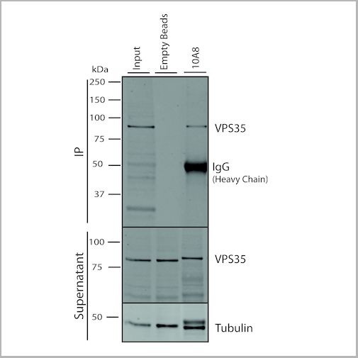
 IP (Immunoprecipitation)
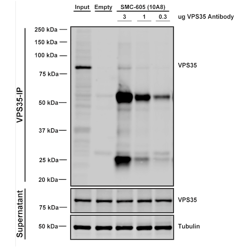
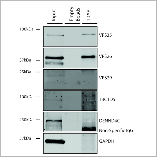
(Immunoprecipitation analysis using Mouse Anti-VPS35 Monoclonal Antibody, Clone 10A8. Tissue: embryonic fibroblast. Species: Mouse. Primary Antibody: Mouse Anti-VPS35 Monoclonal Antibody. Three amounts of (3, 1 and 0.3 ug) were non-covalently coupled to 10uL of A/G sepharose beads for 1 hour at 4 degree C and next incubated with 250ug of MEF lysate for 2 hours at 4 degree C.)
IP (Immunoprecipitation)
(Immunoprecipitation analysis using Mouse Anti-VPS35 Monoclonal Antibody, Clone 10A8. Tissue: embryonic fibroblast. Species: Mouse. Primary Antibody: Mouse Anti-VPS35 Monoclonal Antibody. Three amounts of (3, 1 and 0.3 ug) were non-covalently coupled to 10uL of A/G sepharose beads for 1 hour at 4 degree C and next incubated with 250ug of MEF lysate for 2 hours at 4 degree C.)
VPS35, Monoclonal Antibody (Cat# AAA27695)
Full Name
VPS35 Antibody, Clone 10A8: Biotin
Gene Names
VPS35; MEM3; PARK17
Reactivity
Human, Mouse, Rat
Applications
Western Blot, Immunocytochemistry, Immunofluorescence, Immunoprecipitation
Purity
Protein G Purified
Pricing
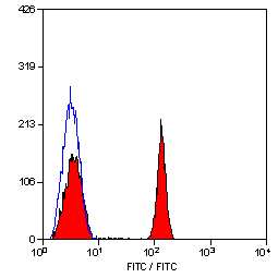
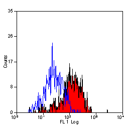
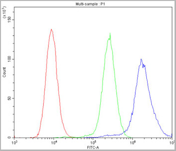
 FCM (Flow Cytometry)
(Figure 6. Flow Cytometry analysis of U20S cells using anti-Calpastatin antibody (AAA11679).Overlay histogram showing U20S cells stained with AAA11679 (Blue line).The cells were blocked with 10% normal goat serum. And then incubated with rabbit anti-Calpastatin Antibody (AAA11679,1ug/1x10^6 cells) for 30 min at 20 degree C. DyLight ®488 conjugated goat anti-rabbit IgG (5-10ug/1x10^6 cells) was used as secondary antibody for 30 minutes at 20 degree C. Isotype control antibody (Green line) was rabbit IgG (1ug/1x106) used under the same conditions. Unlabelled sample (Red line) was also used as a control.)
FCM (Flow Cytometry)
(Figure 6. Flow Cytometry analysis of U20S cells using anti-Calpastatin antibody (AAA11679).Overlay histogram showing U20S cells stained with AAA11679 (Blue line).The cells were blocked with 10% normal goat serum. And then incubated with rabbit anti-Calpastatin Antibody (AAA11679,1ug/1x10^6 cells) for 30 min at 20 degree C. DyLight ®488 conjugated goat anti-rabbit IgG (5-10ug/1x10^6 cells) was used as secondary antibody for 30 minutes at 20 degree C. Isotype control antibody (Green line) was rabbit IgG (1ug/1x106) used under the same conditions. Unlabelled sample (Red line) was also used as a control.)
Calpastatin, Polyclonal Antibody (Cat# AAA11679)
Full Name
Anti-Calpastatin Antibody
Gene Names
CAST; BS-17; PLACK
Reactivity
Human
Applications
Western Blot, Immunohistochemistry
Purity
Immunogen affinity purified.
Pricing

 FCM (Flow Cytometry)
(Figure 7. Flow Cytometry analysis of U251 cells using anti-PNP antibody (AAA11680).Overlay histogram showing U251 cells stained with AAA11680 (Blue line).The cells were blocked with 10% normal goat serum. And then incubated with rabbit anti-PNP Antibody (AAA11680,1ug/1x10^6 cells) for 30 min at 20 degree C. DyLight ®488 conjugated goat anti-rabbit IgG (5-10ug/1x10^6 cells) was used as secondary antibody for 30 minutes at 20 degree C. Isotype control antibody (Green line) was rabbit IgG (1ug/1x106) used under the same conditions. Unlabelled sample (Red line) was also used as a control.)
FCM (Flow Cytometry)
(Figure 7. Flow Cytometry analysis of U251 cells using anti-PNP antibody (AAA11680).Overlay histogram showing U251 cells stained with AAA11680 (Blue line).The cells were blocked with 10% normal goat serum. And then incubated with rabbit anti-PNP Antibody (AAA11680,1ug/1x10^6 cells) for 30 min at 20 degree C. DyLight ®488 conjugated goat anti-rabbit IgG (5-10ug/1x10^6 cells) was used as secondary antibody for 30 minutes at 20 degree C. Isotype control antibody (Green line) was rabbit IgG (1ug/1x106) used under the same conditions. Unlabelled sample (Red line) was also used as a control.)
PNP, Polyclonal Antibody (Cat# AAA11680)
Full Name
Anti-PNP Antibody
Gene Names
PNP; NP; PUNP; PRO1837
Reactivity
Human, Mouse, Rat
Applications
Western Blot, Immunohistochemistry
Purity
Immunogen affinity purified.
Pricing
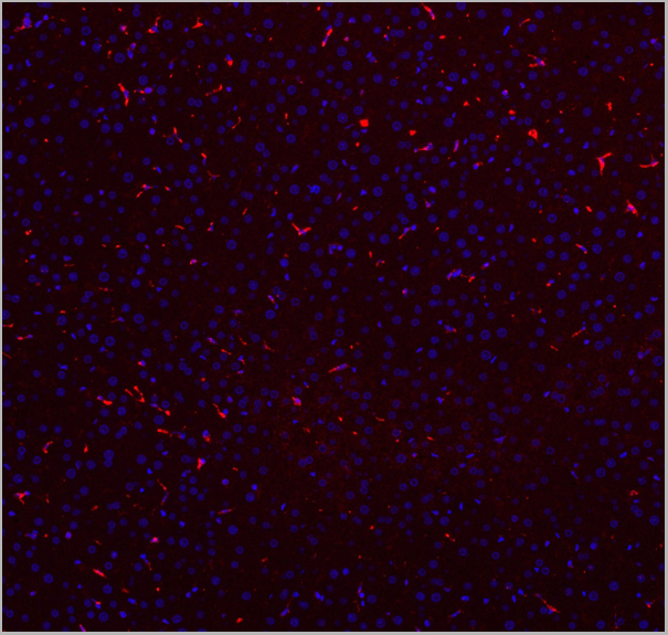
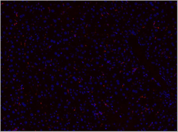

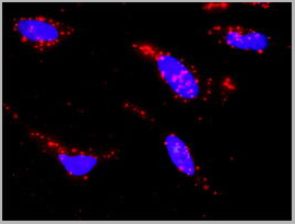
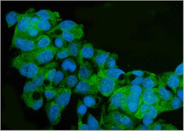
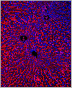
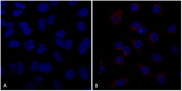
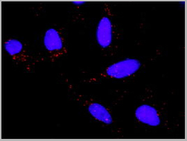
 IF (Immunofluorescence)
(Figure 7. IF analysis of CD68 using anti- CD68 antibody CD68 was detected in paraffin-embedded section of mouse liver tissues. Heat mediated antigen retrieval was performed in citrate buffer (pH6, epitope retrieval solution) for 20 mins. The tissue section was blocked with 10% goat serum. The tissue section was then incubated with 1ug/mL rabbit anti- CD68 Antibody overnight at 4 degree C. Cy3 Conjugated Goat Anti-Rabbit IgG was used as secondary antibody at 1:100 dilution and incubated for 30 minutes at 37 degree C. The section was counterstained with DAPI. Visualize using a fluorescence microscope and filter sets appropriate for the label used.)
IF (Immunofluorescence)
(Figure 7. IF analysis of CD68 using anti- CD68 antibody CD68 was detected in paraffin-embedded section of mouse liver tissues. Heat mediated antigen retrieval was performed in citrate buffer (pH6, epitope retrieval solution) for 20 mins. The tissue section was blocked with 10% goat serum. The tissue section was then incubated with 1ug/mL rabbit anti- CD68 Antibody overnight at 4 degree C. Cy3 Conjugated Goat Anti-Rabbit IgG was used as secondary antibody at 1:100 dilution and incubated for 30 minutes at 37 degree C. The section was counterstained with DAPI. Visualize using a fluorescence microscope and filter sets appropriate for the label used.)
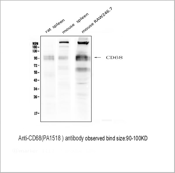
CD68, Polyclonal Antibody (Cat# AAA11512)
Full Name
Anti-CD68 antibody
Gene Names
Cd68; Lamp4; gp110; Scard1
Reactivity
Mouse, Rat
Applications
Western Blot, Immunohistochemistry, Immunohistochemistry
Purity
Immunogen affinity purified.
Pricing


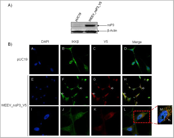 Application Data
(Published customer image: Mouse anti V5 tag antibody, clone SV5-Pk1 used for the detection of V5 tagged WEEV_nsP3 protein by western blotting and immunofluorescenceImage caption: WEEV nsP3 interaction with host IKKbeta. A) U87MGs were transfected in a 6-well plate with 5 ug of pUC19 and WEEV_nsP3_HA for 24 hours. Cell lysates were resolved using SDS-PAGE and subsequently immunoblotted with V5 antibody and beta-actin served as a loading control. B) U87MGs were transfected with WEEV_nsP3_V5; cells were fixed after 24 hours and stained with antibodies against the endogenous IKKbeta and the V5 tag. Cells were incubated with appropriate secondary Alexa Fluor antibodies and the nuclei stained with DAPI. Co-localization of IKKbeta with WEEV_nsP3_V5 (yellow) was observed as shown by the arrows. B) Panels E -H serve as an example of transfected cells in a given field of view that show co-localization of IKKbeta and WEEV_nsP3_V5 24 hours post transfection. Panels I-L represent magnified images of other cells showing co-localization of IKKbeta and WEEV_nsP3_V5. Panel M is a magnified image of panel L. The co-localization was confirmed by Z-stack analysis. Co-localization was calculated to be approximately in 61% of cells (163 cells were counted of which 44% demonstrated expression of nsP3. Of those cells that expressed nsP3, 61% showed co-localization of both proteins). Images were taken using Nikon Eclipse TE2000-U at 60x magnification and are representative of 2 independent experiments.From: Amaya M, Voss K, Sampey G, Senina S, de la Fuente C, et al. (2014) The Role of IKKbeta in Venezuelan Equine Encephalitis Virus Infection. PLoS ONE 9(2): e86745.)
Application Data
(Published customer image: Mouse anti V5 tag antibody, clone SV5-Pk1 used for the detection of V5 tagged WEEV_nsP3 protein by western blotting and immunofluorescenceImage caption: WEEV nsP3 interaction with host IKKbeta. A) U87MGs were transfected in a 6-well plate with 5 ug of pUC19 and WEEV_nsP3_HA for 24 hours. Cell lysates were resolved using SDS-PAGE and subsequently immunoblotted with V5 antibody and beta-actin served as a loading control. B) U87MGs were transfected with WEEV_nsP3_V5; cells were fixed after 24 hours and stained with antibodies against the endogenous IKKbeta and the V5 tag. Cells were incubated with appropriate secondary Alexa Fluor antibodies and the nuclei stained with DAPI. Co-localization of IKKbeta with WEEV_nsP3_V5 (yellow) was observed as shown by the arrows. B) Panels E -H serve as an example of transfected cells in a given field of view that show co-localization of IKKbeta and WEEV_nsP3_V5 24 hours post transfection. Panels I-L represent magnified images of other cells showing co-localization of IKKbeta and WEEV_nsP3_V5. Panel M is a magnified image of panel L. The co-localization was confirmed by Z-stack analysis. Co-localization was calculated to be approximately in 61% of cells (163 cells were counted of which 44% demonstrated expression of nsP3. Of those cells that expressed nsP3, 61% showed co-localization of both proteins). Images were taken using Nikon Eclipse TE2000-U at 60x magnification and are representative of 2 independent experiments.From: Amaya M, Voss K, Sampey G, Senina S, de la Fuente C, et al. (2014) The Role of IKKbeta in Venezuelan Equine Encephalitis Virus Infection. PLoS ONE 9(2): e86745.)
V5-TAG, Monoclonal Antibody (Cat# AAA11850)
Full Name
MOUSE ANTI V5-TAG:Biotin
Applications
Immunohistochemistry, Western Blot
Pricing
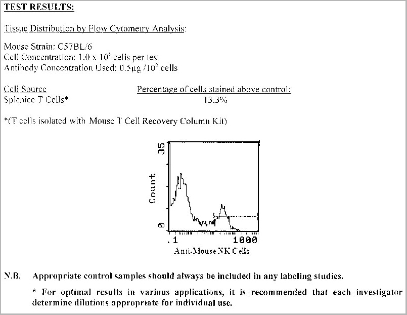
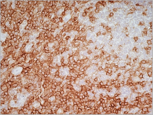
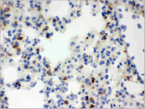
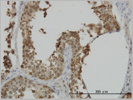
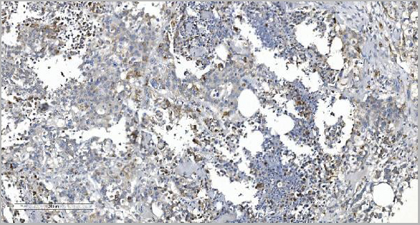
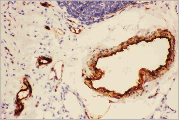
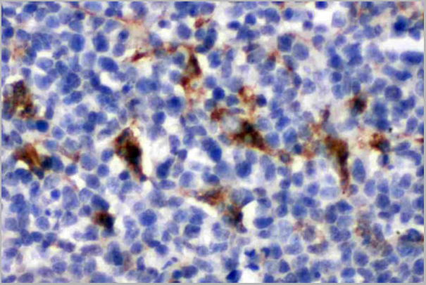
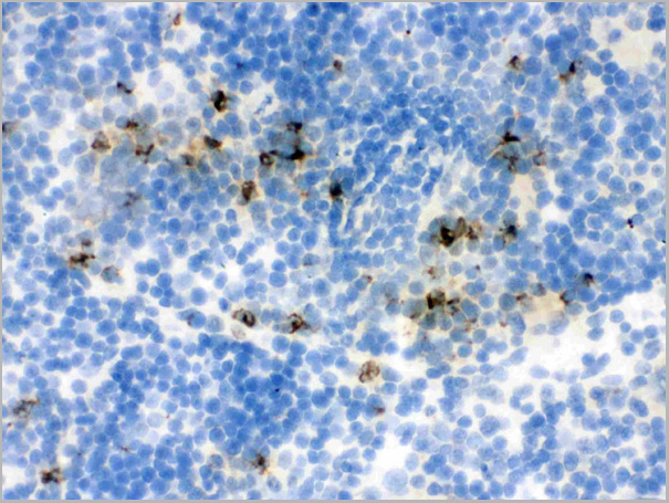
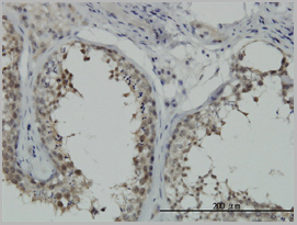
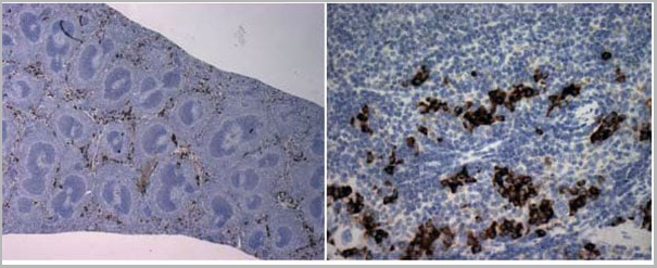 IHC (Immunohistochemistry)
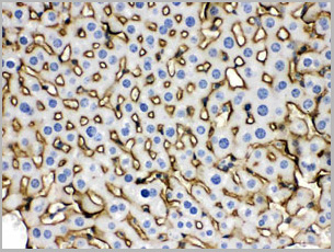
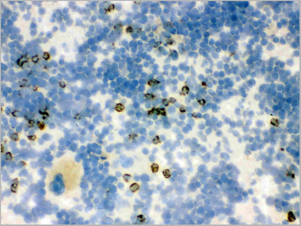
(Purified Anti-Mouse NK Cells (NK1.1) staining of paraformaldehyde-fixed paraffin-embedded mouse spleen sections at 2.5x (Left) and 40x (Right) magnification. (Anti-mouse NK Cells (NK1.1) was used at a dilution of 1:100 and stained with DAB for 1 minute))
IHC (Immunohistochemistry)
(Purified Anti-Mouse NK Cells (NK1.1) staining of paraformaldehyde-fixed paraffin-embedded mouse spleen sections at 2.5x (Left) and 40x (Right) magnification. (Anti-mouse NK Cells (NK1.1) was used at a dilution of 1:100 and stained with DAB for 1 minute))
NK Cells (NK1.1), Monoclonal Antibody (Cat# AAA14274)
Full Name
Anti-Mouse NK Cells (NK1.1), PE (Clone PK136) (mouse IgG2a)
Applications
Immunohistochemistry
Pricing
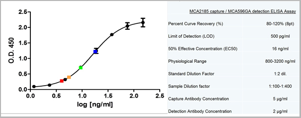
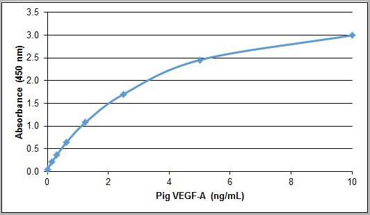 ELISA
(Pig VEGF-A ELISA Recombinant Pig VEGF-A detected using Goat anti Pig VEGF-A as the capture reagent and Goat anti Pig VEGF-A:Biotin as the detection reagent followed by Streptavidin:HRP)
ELISA
(Pig VEGF-A ELISA Recombinant Pig VEGF-A detected using Goat anti Pig VEGF-A as the capture reagent and Goat anti Pig VEGF-A:Biotin as the detection reagent followed by Streptavidin:HRP)
VEGF-A, Polyclonal Antibody (Cat# AAA12254)
Full Name
Goat anti Pig VEGF-A: Biotin
Gene Names
VEGFA; VEGF
Applications
ELISA
Pricing


 Application Data
(Staining of mouse peripheral blood platelets with Hamster anti Mouse CD61:FITC)
Application Data
(Staining of mouse peripheral blood platelets with Hamster anti Mouse CD61:FITC)
CD61, Monoclonal Antibody (Cat# AAA12041)
Full Name
HAMSTER ANTI MOUSE CD61:RPE
Gene Names
Itgb3; CD61; GP3A; INGRB3
Applications
Flow Cytometry
Pricing
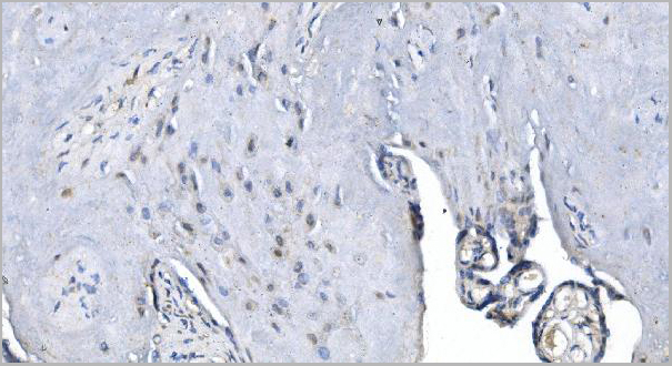
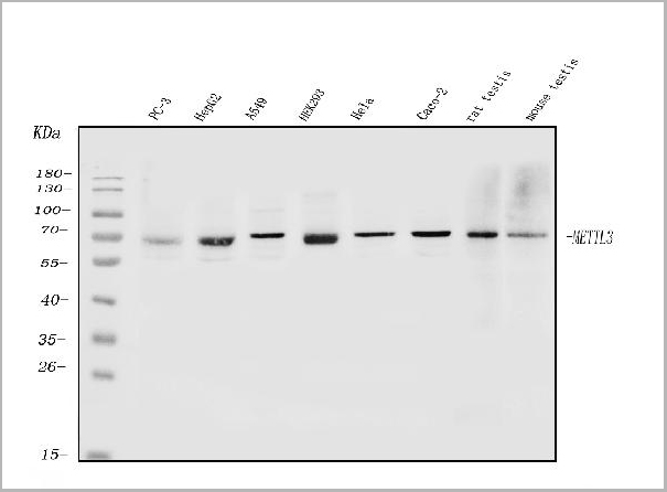
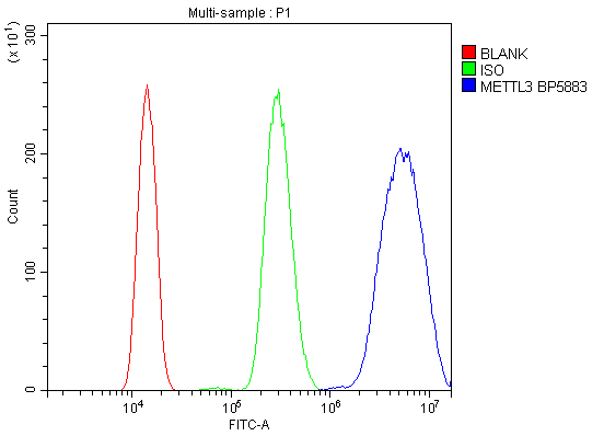 FCM (Flow Cytometry)
(Figure 6. Flow Cytometry analysis of HL-60 cells using anti-METTL3 antibody (AAA19247).Overlay histogram showing HL-60 cells stained with AAA19247 (Blue line). The cells were blocked with 10% normal goat serum. And then incubated with rabbit anti-METTL3 Antibody (AAA19247, 1μg/1x106 cells) for 30 min at 20 degree C. DyLight®488 conjugated goat anti-rabbit IgG (5-10μg/1x106 cells) was used as secondary antibody for 30 minutes at 20 degree C. Isotype control antibody (Green line) was rabbit IgG (1μg/1x106) used under the same conditions. Unlabelled sample (Red line) was also used as a control.)
FCM (Flow Cytometry)
(Figure 6. Flow Cytometry analysis of HL-60 cells using anti-METTL3 antibody (AAA19247).Overlay histogram showing HL-60 cells stained with AAA19247 (Blue line). The cells were blocked with 10% normal goat serum. And then incubated with rabbit anti-METTL3 Antibody (AAA19247, 1μg/1x106 cells) for 30 min at 20 degree C. DyLight®488 conjugated goat anti-rabbit IgG (5-10μg/1x106 cells) was used as secondary antibody for 30 minutes at 20 degree C. Isotype control antibody (Green line) was rabbit IgG (1μg/1x106) used under the same conditions. Unlabelled sample (Red line) was also used as a control.)
METTL3, Polyclonal Antibody (Cat# AAA19247)
Full Name
Anti-METTL3 Antibody
Gene Names
METTL3; M6A; IME4; Spo8; MT-A70
Reactivity
Human, Mouse, Rat
Applications
Western Blot, Immunohistochemistry, Immunocytochemistry, Immunofluorescence, Flow Cytometry, Direct ELISA
Purity
Immunogen affinity purified.
Pricing
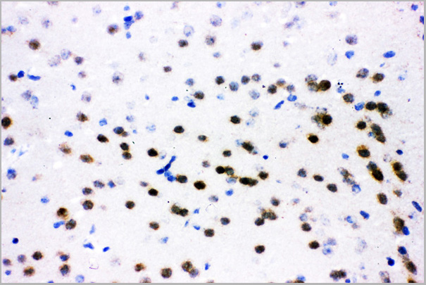
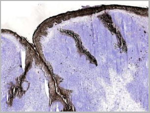
 Application Data
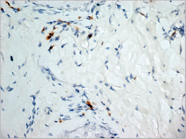
(Image: Cholecystokinin 8 terminals and fibers in rat dorsal horn (spinal cord), vibratome sections, ABC detection.)
Application Data
(Image: Cholecystokinin 8 terminals and fibers in rat dorsal horn (spinal cord), vibratome sections, ABC detection.)
CCK-8, Antibody (Cat# AAA14443)
Full Name
CCK-8 (Cholecystokinin Octapeptide) Antibody
Reactivity
Mouse, Rat
Applications
Immunofluorescence
Pricing
 IP (Immunoprecipitation)
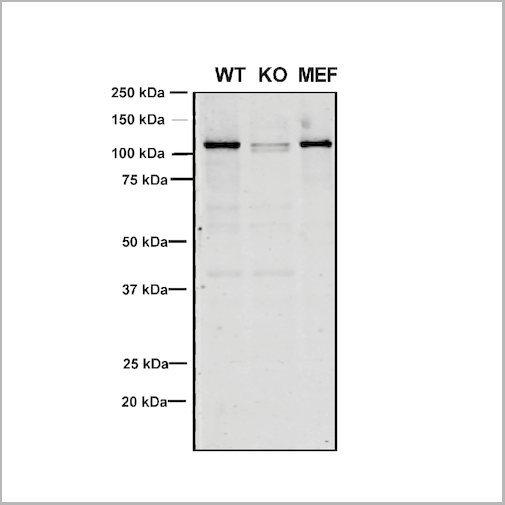
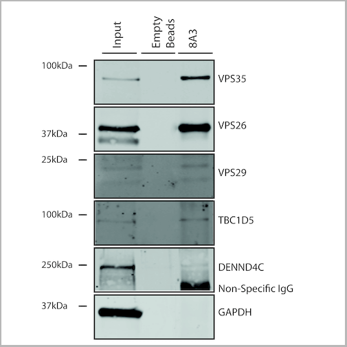
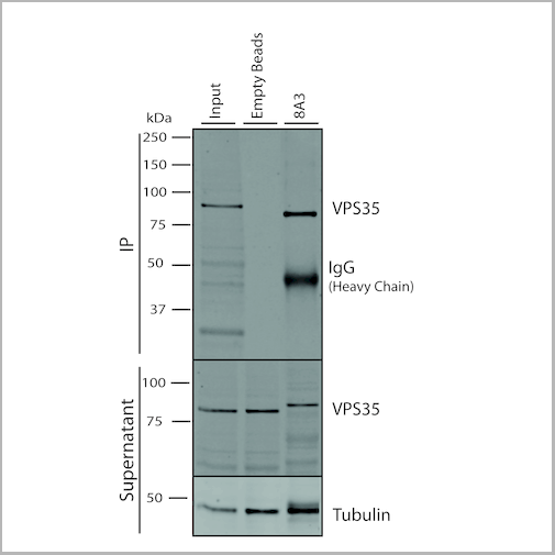
(Immunoprecipitation analysis using Mouse Anti-VPS35 Monoclonal Antibody, Clone 8A3. Tissue: embryonic fibroblast. Species: Mouse. Primary Antibody: Mouse Anti-VPS35 Monoclonal Antibody. Three amounts of (3, 1 and 0.3 ug) were non-covalently coupled to 10uL of A/G sepharose beads for 1 hour at 4 degree C and next incubated with 250ug of MEF lysate for 2 hours at 4 degree C.)
IP (Immunoprecipitation)
(Immunoprecipitation analysis using Mouse Anti-VPS35 Monoclonal Antibody, Clone 8A3. Tissue: embryonic fibroblast. Species: Mouse. Primary Antibody: Mouse Anti-VPS35 Monoclonal Antibody. Three amounts of (3, 1 and 0.3 ug) were non-covalently coupled to 10uL of A/G sepharose beads for 1 hour at 4 degree C and next incubated with 250ug of MEF lysate for 2 hours at 4 degree C.)
VPS35, Monoclonal Antibody (Cat# AAA27686)
Full Name
VPS35 Antibody, Clone 8A3: Biotin
Gene Names
VPS35; MEM3; PARK17
Reactivity
Human, Mouse, Rat
Applications
Western Blot, Immunocytochemistry, Immunofluorescence, Immunoprecipitation
Purity
Protein G Purified
Pricing
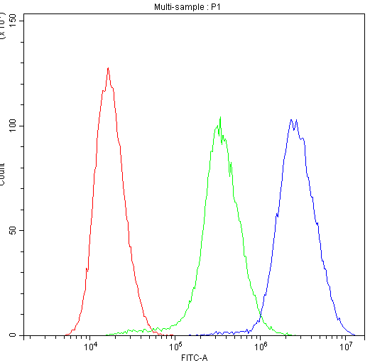
 FCM (Flow Cytometry)
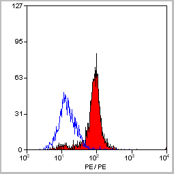
(Figure 7. Flow Cytometry analysis of Hela cells using anti-Calbindin antibody (AAA11603).Overlay histogram showing Hela cells stained with AAA11603 (Blue line).The cells were blocked with 10% normal goat serum. And then incubated with rabbit anti-Calbindin Antibody (AAA11603,1ug/1x10^6 cells) for 30 min at 20 degree C. DyLight®488 conjugated goat anti-rabbit IgG (5-10ug/1x10^6 cells) was used as secondary antibody for 30 minutes at 20 degree C. Isotype control antibody (Green line) was rabbit IgG (1ug/1x106) used under the same conditions. Unlabelled sample (Red line) was also used as a control.)
FCM (Flow Cytometry)
(Figure 7. Flow Cytometry analysis of Hela cells using anti-Calbindin antibody (AAA11603).Overlay histogram showing Hela cells stained with AAA11603 (Blue line).The cells were blocked with 10% normal goat serum. And then incubated with rabbit anti-Calbindin Antibody (AAA11603,1ug/1x10^6 cells) for 30 min at 20 degree C. DyLight®488 conjugated goat anti-rabbit IgG (5-10ug/1x10^6 cells) was used as secondary antibody for 30 minutes at 20 degree C. Isotype control antibody (Green line) was rabbit IgG (1ug/1x106) used under the same conditions. Unlabelled sample (Red line) was also used as a control.)
Calbindin, Polyclonal Antibody (Cat# AAA11603)
Full Name
Anti-Calbindin Antibody
Gene Names
CALB1; CALB
Reactivity
Human, Mouse, Rat
Applications
Western Blot, Immunohistochemistry
Purity
Immunogen affinity purified.
Pricing
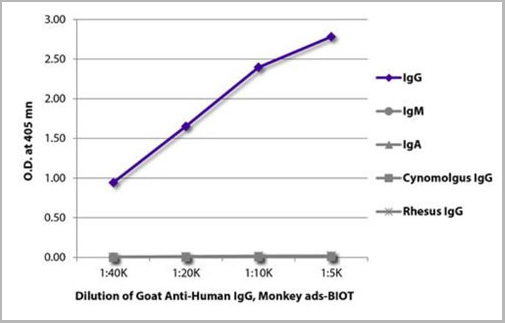 ELISA
(ELISA plate was coated with purified human IgG, IgM, and IgA, cynomolgus IgG, and rhesus IgG. Immunoglobulins were detected with serially diluted Goat Anti-Human IgG, Monkey ads-BIOT (AAA14911) followed by Streptavidin-HRP (please inquire).)
ELISA
(ELISA plate was coated with purified human IgG, IgM, and IgA, cynomolgus IgG, and rhesus IgG. Immunoglobulins were detected with serially diluted Goat Anti-Human IgG, Monkey ads-BIOT (AAA14911) followed by Streptavidin-HRP (please inquire).)
Goat Anti-Human IgG, Monkey ads, Secondary Antibody (Cat# AAA14911)
Full Name
Goat Anti-Human IgG, Monkey ads-BIOT
Reactivity
Human
Applications
ELISA, FLISA
Purity
Affinity chromatography on Human IgG covalently linked to agarose.
Pricing
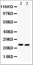
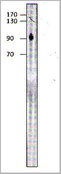 WB (Western Blot)
(WB of with THP on 10% 50S gel. 1:250 antibody dilution in OiluObuffer for 1 hour at room temperature. Apparent MW is 95 kDa .)
WB (Western Blot)
(WB of with THP on 10% 50S gel. 1:250 antibody dilution in OiluObuffer for 1 hour at room temperature. Apparent MW is 95 kDa .)
Progesterone Receptor, Polyclonal Antibody (Cat# AAA14415)
Full Name
Progesterone Receptor Antibody BIOTIN
Gene Names
PGR; PR; NR3C3
Reactivity
Cat, Human, Monkey, Pig
Applications
Immunoprecipitation, Western Blot
Pricing
 IA (Immuno Assay)
(used as capture antibody. Different concentrations were tested (0.5-2 ug/ml).)
IA (Immuno Assay)
(used as capture antibody. Different concentrations were tested (0.5-2 ug/ml).)
Calprotectin, Monoclonal Antibody (Cat# AAA14609)
Full Name
Calprotectin, Human, mAb 27E10, Biotin
Reactivity
Mouse: No; Rhesus Monkey: Yes (subpopulations of macrophages)
Applications
Immunohistochemistry, Immunofluorescence, Flow Cytometry, Immunoassay, Immunoprecipitation, Western Blot
Purity
Protein G
Pricing
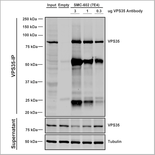

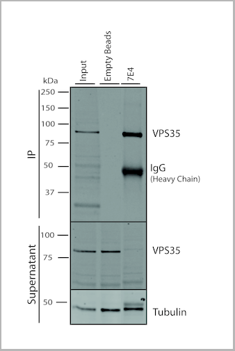
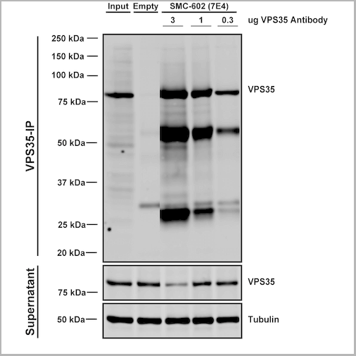 IP (Immunoprecipitation)
(Immunoprecipitation analysis using Mouse Anti-VPS35 Monoclonal Antibody, Clone 7E4. Tissue: embryonic fibroblast. Species: Mouse. Primary Antibody: Mouse Anti-VPS35 Monoclonal Antibody. Three amounts (3, 1 and 0.3 ug) were non-covalently coupled to 10uL of A/G sepharose beads for 1 hour at 4 degree C and next incubated with 250ug of MEF lysate for 2 hours at 4 degree C.)
IP (Immunoprecipitation)
(Immunoprecipitation analysis using Mouse Anti-VPS35 Monoclonal Antibody, Clone 7E4. Tissue: embryonic fibroblast. Species: Mouse. Primary Antibody: Mouse Anti-VPS35 Monoclonal Antibody. Three amounts (3, 1 and 0.3 ug) were non-covalently coupled to 10uL of A/G sepharose beads for 1 hour at 4 degree C and next incubated with 250ug of MEF lysate for 2 hours at 4 degree C.)
VPS35, Monoclonal Antibody (Cat# AAA27668)
Full Name
VPS35 Antibody, Clone 7E4: Biotin
Gene Names
VPS35; MEM3; PARK17
Reactivity
Human, Mouse, Rat
Applications
Western Blot, Immunocytochemistry, Immunofluorescence, Immunoprecipitation
Purity
Protein G Purified
Pricing

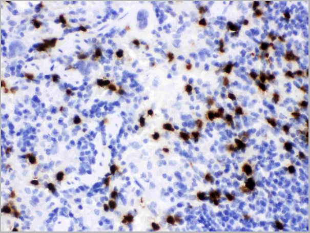
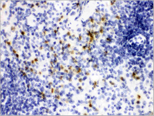
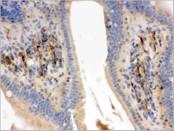
 IHC (Immunohistochemistry)
(Figure 7. IHC analysis of Lipocalin 2 using anti-Lipocalin 2 antibody (AAA11670).Lipocalin 2 was detected in frozen section of rat spleen tissue. Heat mediated antigen retrieval was performed in citrate buffer (pH6, epitope retrieval solution) for 20 mins. The tissue section was blocked with 10% goat serum. The tissue section was then incubated with 1ug/ml rabbit anti-Lipocalin 2 Antibody (AAA11670) overnight at 4 degree C. Biotinylated goat anti-rabbit IgG was used as secondary antibody and incubated for 30 minutes at 37 degree C. The tissue section was developed using Strepavidin-Biotin-Complex (SABC) with DAB as the chromogen.)
IHC (Immunohistochemistry)
(Figure 7. IHC analysis of Lipocalin 2 using anti-Lipocalin 2 antibody (AAA11670).Lipocalin 2 was detected in frozen section of rat spleen tissue. Heat mediated antigen retrieval was performed in citrate buffer (pH6, epitope retrieval solution) for 20 mins. The tissue section was blocked with 10% goat serum. The tissue section was then incubated with 1ug/ml rabbit anti-Lipocalin 2 Antibody (AAA11670) overnight at 4 degree C. Biotinylated goat anti-rabbit IgG was used as secondary antibody and incubated for 30 minutes at 37 degree C. The tissue section was developed using Strepavidin-Biotin-Complex (SABC) with DAB as the chromogen.)
Lipocalin 2, Polyclonal Antibody (Cat# AAA11670)
Full Name
Anti-Lipocalin 2 Antibody
Gene Names
Lcn2; 24p3; Sip24; AW212229
Reactivity
Mouse, Rat
Applications
Western Blot, Immunohistochemistry
Purity
Immunogen Affinity Purified
Pricing
Carbamyl-lysine, Antibody (Cat# AAA13603)
Full Name
Biotinylated Goat Anti Carbamyl-lysine
Applications
Immunoblot
Pricing


 Application Data
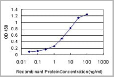
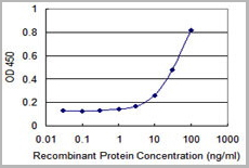
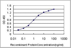
(Detection limit for recombinant GST tagged TCEA3 is approximately 0.03ng/ml as a capture antibody.)
Application Data
(Detection limit for recombinant GST tagged TCEA3 is approximately 0.03ng/ml as a capture antibody.)
TCEA3, Monoclonal Antibody (Cat# AAA26209)
Full Name
TCEA3 (Transcription Elongation Factor A (SII), 3, TFIIS, TFIIS.H) (Biotin)
Applications
Immunofluorescence, Immunohistochemistry, Western Blot
Purity
Purified
Pricing
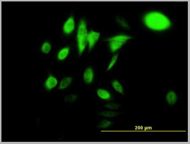
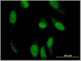 IF (Immunofluorescence)
(Immunofluorescence of on HeLa cell using AAA24920 (10ug/ml).)
IF (Immunofluorescence)
(Immunofluorescence of on HeLa cell using AAA24920 (10ug/ml).)
PRPH, Monoclonal Antibody (Cat# AAA24920)
Full Name
PRPH (Peripherin, Neurofilament 4, NEF4, PRPH1) (Biotin)
Gene Names
PRPH; NEF4; PRPH1
Reactivity
Human
Applications
Immunofluorescence, Immunohistochemistry, Western Blot
Purity
Purified by Protein A Affinity Chromatography.
Pricing
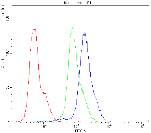
 FCM (Flow Cytometry)
(Figure 7. Flow Cytometry analysis of K562 cells using anti-CASP3 antibody (AAA11633).Overlay histogram showing K562 cells stained with AAA11633 (Blue line).The cells were blocked with 10% normal goat serum. And then incubated with rabbit anti-CASP3 Antibody (AAA11633,1ug/1x10^6 cells) for 30 min at 20 degree C. DyLight ®488 conjugated goat anti-rabbit IgG (5-10ug/1x10^6 cells) was used as secondary antibody for 30 minutes at 20 degree C. Isotype control antibody (Green line) was rabbit IgG (1ug/1x106) used under the same conditions. Unlabelled sample (Red line) was also used as a control.)
FCM (Flow Cytometry)
(Figure 7. Flow Cytometry analysis of K562 cells using anti-CASP3 antibody (AAA11633).Overlay histogram showing K562 cells stained with AAA11633 (Blue line).The cells were blocked with 10% normal goat serum. And then incubated with rabbit anti-CASP3 Antibody (AAA11633,1ug/1x10^6 cells) for 30 min at 20 degree C. DyLight ®488 conjugated goat anti-rabbit IgG (5-10ug/1x10^6 cells) was used as secondary antibody for 30 minutes at 20 degree C. Isotype control antibody (Green line) was rabbit IgG (1ug/1x106) used under the same conditions. Unlabelled sample (Red line) was also used as a control.)
Caspase-3, Polyclonal Antibody (Cat# AAA11633)
Full Name
Anti-Caspase-3 Antibody
Gene Names
CASP3; CPP32; SCA-1; CPP32B
Reactivity
Human, Mouse, Rat. No cross reactivity with other proteins.
Applications
Western Blot, Immunohistochemistry
Purity
Immunogen affinity purified.
Pricing
 Application Data
(Staining of human peripheral blood monocytes probed with Mouse anti Human CD14:FITC)
Application Data
(Staining of human peripheral blood monocytes probed with Mouse anti Human CD14:FITC)
CD14, Monoclonal Antibody (Cat# AAA12224)
Full Name
MOUSE ANTI HUMAN CD14:Biotin
Applications
Flow Cytometry
Pricing


 Application Data
(Detection limit for recombinant GST tagged ROCK2 is 0.3 ng/ml as a capture antibody.)
Application Data
(Detection limit for recombinant GST tagged ROCK2 is 0.3 ng/ml as a capture antibody.)
ROCK2, Monoclonal Antibody (Cat# AAA26224)
Full Name
ROCK2 (Rho-Associated, coiled-coil Containing Protein Kinase 2, KIAA0619) (Biotin)
Gene Names
ROCK2; ROCK-II
Applications
Immunofluorescence, Immunohistochemistry, Western Blot
Purity
Purified
Pricing
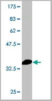
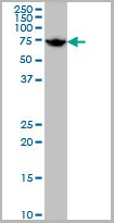
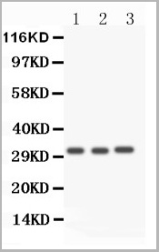
 WB (Western Blot)
(HSPA1B monoclonal antibody Western Blot analysis of HSPA1B expression in Raw 264.7.)
WB (Western Blot)
(HSPA1B monoclonal antibody Western Blot analysis of HSPA1B expression in Raw 264.7.)
HSPA1A, Monoclonal Antibody (Cat# AAA24832)
Full Name
HSPA1A (Heat Shock 70kD Protein 1A/1B, Heat Shock 70kD Protein 1/2, HSP70.1/HSP70.2, HSP70-1/HSP70-2, HSPA1, HSPA1B) (Biotin)
Gene Names
HSPA1A; HSP72; HSPA1; HSP70I; HSP70-1; HSP70.1; HSP70-1A; HEL-S-103
Reactivity
Human, Mouse, Rat
Applications
Immunofluorescence, Immunohistochemistry, Western Blot
Purity
Purified by Protein A Affinity Chromatography.
Pricing