Filters
Clonality
Type
Reactivity
Gene Name
Isotype
Host
Application
Clone
884 results for "Control Antibodies" - showing 800-850
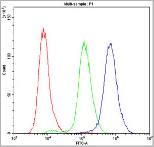
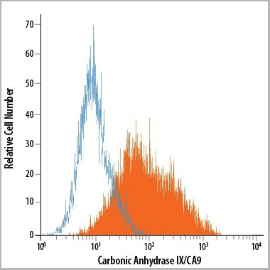 FCM (Flow Cytometry)
(Detection of Carbonic Anhydrase IX/CA9 in U‑87 MG Human Cell Line by Flow Cytometry using AAA14680.)
FCM (Flow Cytometry)
(Detection of Carbonic Anhydrase IX/CA9 in U‑87 MG Human Cell Line by Flow Cytometry using AAA14680.)
Carbonic Anhydrase IX, Monoclonal Antibody (Cat# AAA14680)
Full Name
Carbonic Anhydrase IX (CA9) (PE)
Gene Names
CA9; MN; CAIX
Applications
Flow Cytometry
Purity
Affinity Purified
Purified by Protein A affinity chromatography from hybridoma culture supernant.
Purified by Protein A affinity chromatography from hybridoma culture supernant.
Pricing
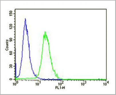
 FCM (Flow Cytometry)
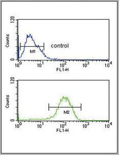
(PCSK9 Antibody (N-term) (Cat. #AAA28774) flow cytometry analysis of Jurkat cells (bottom histogram) compared to a negative control cell(top histogram).FITC-conjugated goat-anti-rabbit secondary antibodies were used for the analysis.)
FCM (Flow Cytometry)
(PCSK9 Antibody (N-term) (Cat. #AAA28774) flow cytometry analysis of Jurkat cells (bottom histogram) compared to a negative control cell(top histogram).FITC-conjugated goat-anti-rabbit secondary antibodies were used for the analysis.)
PCSK9, Polyclonal Antibody (Cat# AAA28774)
Full Name
PCSK9 Antibody (N-term)
Gene Names
PCSK9; FH3; PC9; NARC1; LDLCQ1; NARC-1; HCHOLA3
Reactivity
Human
Applications
Western Blot, Immunohistochemistry, Flow Cytometry
Pricing


 Application Data
(Staining of human peripheral blood lymphocytes with Mouse anti Human CD40:RPE)
Application Data
(Staining of human peripheral blood lymphocytes with Mouse anti Human CD40:RPE)
CD40, Monoclonal Antibody (Cat# AAA12089)
Full Name
MOUSE ANTI HUMAN CD40:RPE
Gene Names
CD40; p50; Bp50; CDW40; TNFRSF5
Applications
Flow Cytometry
Pricing
 IHC (Immunohistochemistry)
(High magnification: Paraffin-embedded human skin tissue was prepared using heat-induced epitope retrieval in citrate buffer, pH 6.0. Immunostaining was performed using a 1:100 dilution of AAA14653. Cells lining up under the epidermis consist of numerous mast cells (The dermis has a moderate diffuse inflammatory cell infiltrate).)
IHC (Immunohistochemistry)
(High magnification: Paraffin-embedded human skin tissue was prepared using heat-induced epitope retrieval in citrate buffer, pH 6.0. Immunostaining was performed using a 1:100 dilution of AAA14653. Cells lining up under the epidermis consist of numerous mast cells (The dermis has a moderate diffuse inflammatory cell infiltrate).)
Mast Cell Chymase, Monoclonal Antibody (Cat# AAA14653)
Full Name
Mast Cell Chymase
Gene Names
CMA1; CYH; MCT1; chymase; MGC119890; MGC119891
Reactivity
Human
Applications
Immunohistochemistry
Purity
Purified by Protein G affinity chromatography
Pricing


 Application Data
(Staining of human peripheral blood lymphocytes with Mouse anti Human CD40:RPE)
Application Data
(Staining of human peripheral blood lymphocytes with Mouse anti Human CD40:RPE)
CD40, Monoclonal Antibody (Cat# AAA12031)
Full Name
MOUSE ANTI HUMAN CD40:RPE
Gene Names
CD40; p50; Bp50; CDW40; TNFRSF5
Applications
Flow Cytometry
Pricing
 Application Data
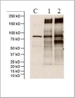
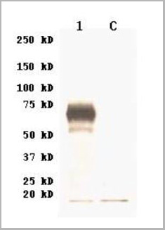
(C: 293 Cell extract control1: 293 cell expressing Spike protein(SARS-CoV BJ02)2: 293 cell expressing Spike protein (Civet SARS-CoV SZ3/2003))
Application Data
(C: 293 Cell extract control1: 293 cell expressing Spike protein(SARS-CoV BJ02)2: 293 cell expressing Spike protein (Civet SARS-CoV SZ3/2003))
Spike (SARS-CoV), Polyclonal Antibody (Cat# AAA13726)
Full Name
Anti-Spike (SARS-CoV)
Applications
Western Blot
Purity
Immunoaffinity chromatography.
Pricing
 WB (Western Blot)
(C: 293 cell extract control1: 293 cell expressing H3 (A/Wyoming/3/03)(H3N2)2: 293 cell expressing H3 (A/Brisbane/10/07)(H3N2))
WB (Western Blot)
(C: 293 cell extract control1: 293 cell expressing H3 (A/Wyoming/3/03)(H3N2)2: 293 cell expressing H3 (A/Brisbane/10/07)(H3N2))
HAs (A/Wyoming/3/03(H3N2), A/Wisconsin/67/X-161/2005(H3), A/Brisbane/10/2007(H3N2)), Polyclonal Antibody (Cat# AAA13730)
Full Name
Anti-HAs (A/Wyoming/3/03(H3N2), A/Wisconsin/67/X-161/2005(H3), A/Brisbane/10/2007(H3N2)), rabbit IgG
Applications
Western Blot
Purity
Immunoaffinity chromatography
Pricing


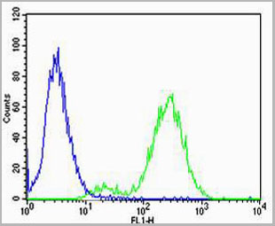
 FCM (Flow Cytometry)
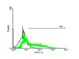
(FACS analysis of ES-2 cells stained with STIP1 monoclonal antibody clone 2E11 (Green) and non-stained ES-2 cells (Black) as negative control.)
FCM (Flow Cytometry)
(FACS analysis of ES-2 cells stained with STIP1 monoclonal antibody clone 2E11 (Green) and non-stained ES-2 cells (Black) as negative control.)
STIP1, Monoclonal Antibody (Cat# AAA26275)
Full Name
STIP1 (Stress-Induced-Phosphoprotein 1, HOP, IEF-SSP-3521, P60, STI1, STI1L) (Biotin)
Gene Names
STIP1; HOP; P60; STI1; STI1L; HEL-S-94n; IEF-SSP-3521
Applications
Flow Cytometry, Immunofluorescence, Immunohistochemistry, Western Blot
Purity
Purified
Pricing
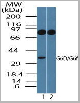
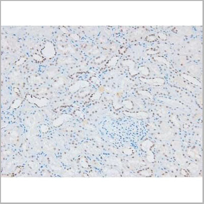
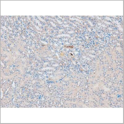
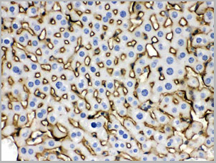
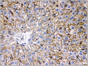
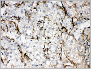
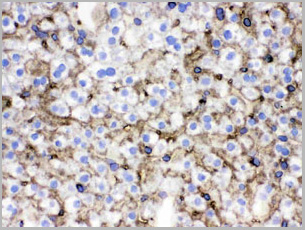
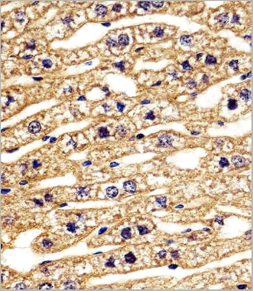
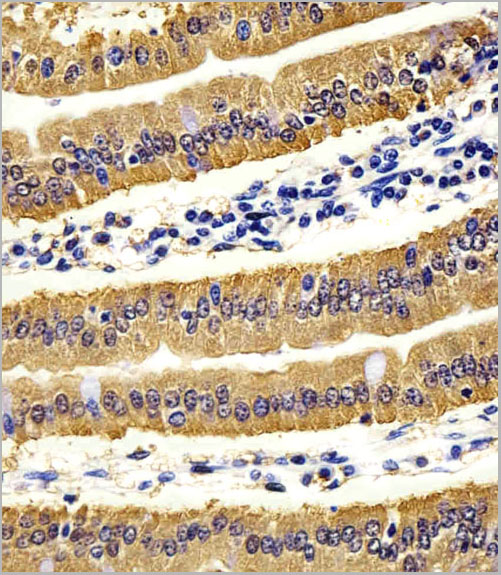
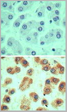 IHC (Immunohistochemistry)
(Immunohistochemical analysis of G6D in formalin-fixed, paraffin-embedded human liver tissue using an isotype control (top) and (bottom) AAA24064 at 5ug/ml.)
IHC (Immunohistochemistry)
(Immunohistochemical analysis of G6D in formalin-fixed, paraffin-embedded human liver tissue using an isotype control (top) and (bottom) AAA24064 at 5ug/ml.)
LY6G6D, Polyclonal Antibody (Cat# AAA24064)
Full Name
LY6G6D (G6D, Lymphocyte Antigen 6 Complex Locus Protein G6f, C6orf21, G6F, LY6G6D, NG32)
Gene Names
LY6G6D; G6D; NG25; LY6-D; MEGT1; C6orf23
Reactivity
Human
Applications
Western Blot, Immunohistochemistry
Purity
Purified by Protein A affinity chromatography.
Pricing
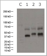 WB (Western Blot)
(C: 293 cell extract control1: 293 cell expressing H5 (1997) subtype2: 293 cell expressing H5 (2004) subtype3: 293 cell expressing H5 (2005) subtype)
WB (Western Blot)
(C: 293 cell extract control1: 293 cell expressing H5 (1997) subtype2: 293 cell expressing H5 (2004) subtype3: 293 cell expressing H5 (2005) subtype)
HA (H5N1, Human)(A/Vietnam/1203/2004), Antibody (Cat# AAA13715)
Full Name
Anti-HA (H5N1, Human)(A/Vietnam/1203/2004)
Applications
Western Blot
Purity
Immunoaffinity chromatography
Pricing
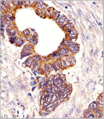
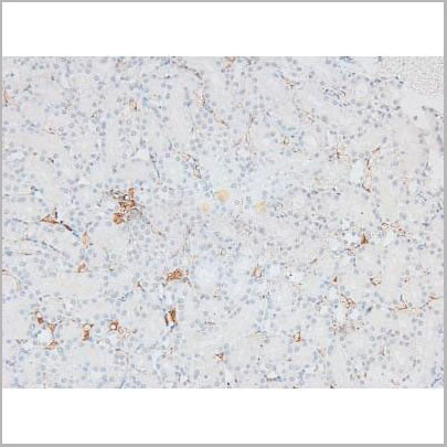
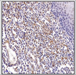
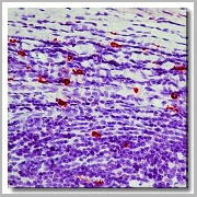 IHC (Immunohistochemistry)
(Immunohistochemistry of formalin-fixed, paraffin-embedded human tonsil using AAA14660 and following by peroxidase-conjugate and AEC chromogen. Note cytoplasmic staining of mast cells.)
IHC (Immunohistochemistry)
(Immunohistochemistry of formalin-fixed, paraffin-embedded human tonsil using AAA14660 and following by peroxidase-conjugate and AEC chromogen. Note cytoplasmic staining of mast cells.)
Mast Cell Chymase, Monoclonal Antibody (Cat# AAA14660)
Full Name
Mast Cell Chymase (BSA & Azide Free)
Gene Names
CMA1; CYH; MCT1; chymase; MGC119890; MGC119891
Reactivity
Human
Applications
Western Blot, Immunohistochemistry
Purity
Purified by Protein G affinity chromatography.
Pricing
 WB (Western Blot)
(Western blot analysis of ZO-1 using various lysatesLanes 1 - 2: Merged signal (red and green). Green - AAA31075 observed at 195 kDa. Red - loading control, , observed at 55 kDa. Blots were developed with Goat Anti- Rabbit IgG(H+L) FITC–conjugated and Goat Anti-Mouse IgG(H+L) Alexa Fluor 594–conjugated secondary antibodies)
WB (Western Blot)
(Western blot analysis of ZO-1 using various lysatesLanes 1 - 2: Merged signal (red and green). Green - AAA31075 observed at 195 kDa. Red - loading control, , observed at 55 kDa. Blots were developed with Goat Anti- Rabbit IgG(H+L) FITC–conjugated and Goat Anti-Mouse IgG(H+L) Alexa Fluor 594–conjugated secondary antibodies)
ZO 1, Polyclonal Antibody (Cat# AAA31075)
Full Name
ZO 1 Antibody
Gene Names
TJP1; ZO-1
Reactivity
Human, Mouse, Rat, Pig, Monkey
Applications
Western Blot, Immunohistochemistry, Immunofluorescence, Immunocytochemistry
Purity
The antiserum was purified by peptide affinity chromatography using SulfoLink Coupling Resin
Pricing
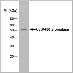
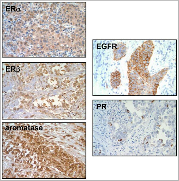
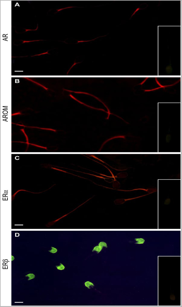
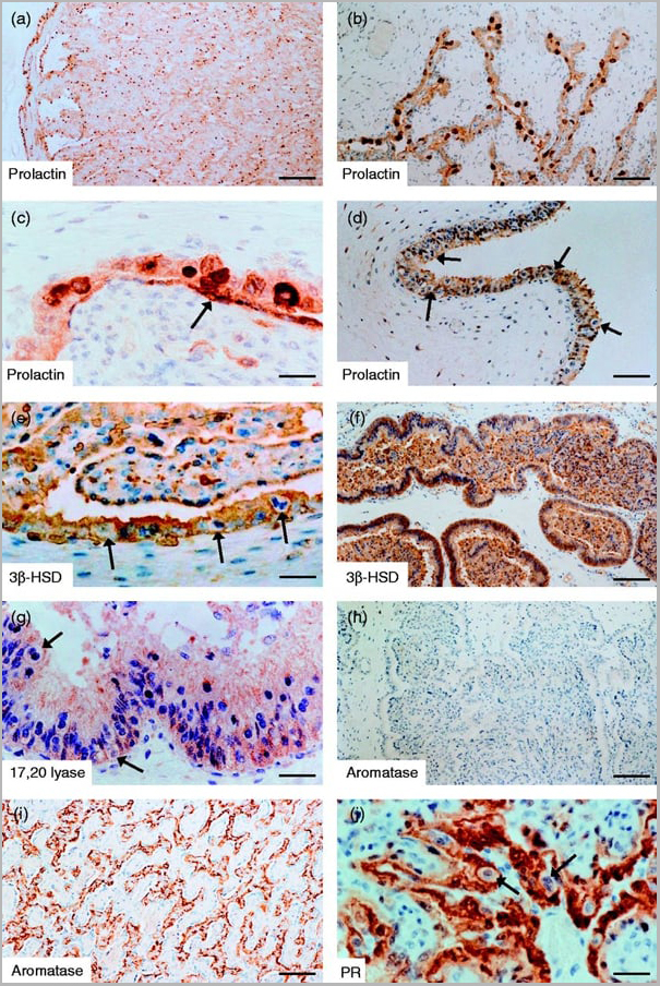
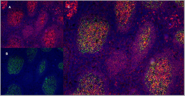
 Application Data
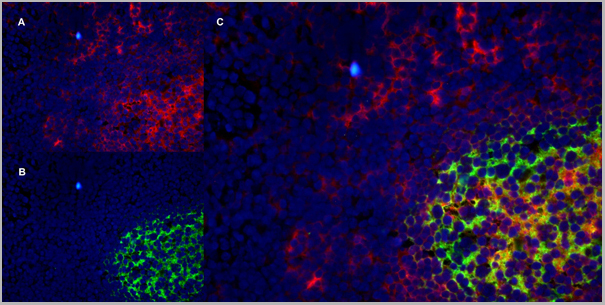
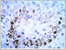
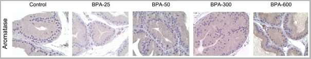
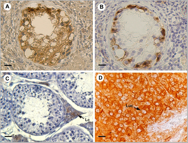
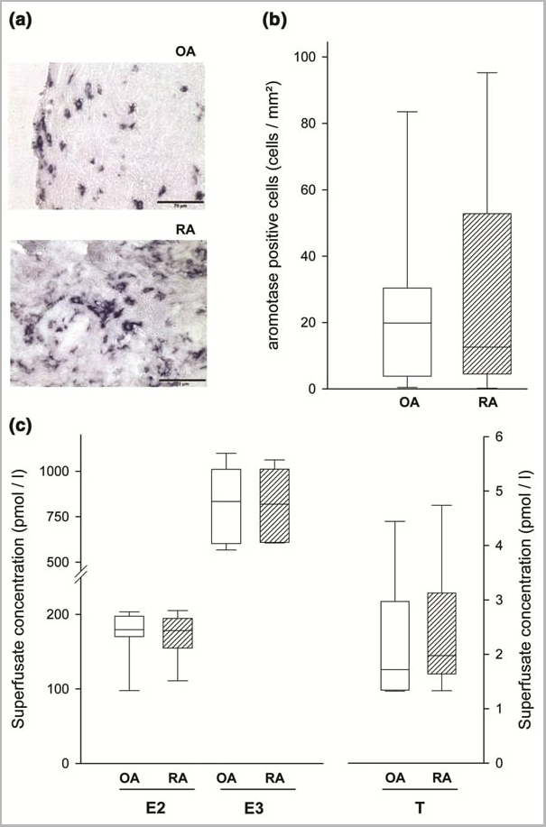
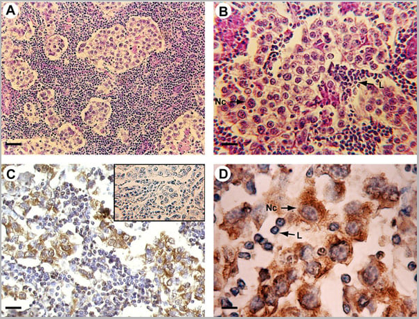
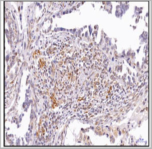
(Published customer image: Mouse anti Human cytochrome p450 aromatase antibody, clone H4 used for the detaction of aromatase in human tissues by Immunohistochemistry on paraffin sectionsImage caption:Morphology and P450 arom immunoreactivity of tumoral region in human testis with seminoma. A-B: Haematoxylin-eosin staining. C-D: Strong P450 arom immunoreactivity in cytoplasm of neoplastic cells (Nc) and unstained lymphocytes (L). Insert: absorption control. Scale bars: A, 20 um; B-C, 12.5 um; D, 5 um.From: Rago V, Romeo F, Aquila S, Montanaro D, And S, Carpino A. Cytochrome P450 aromatase expression in human seminoma. Reprod Biol Endocrinol. 2005 Dec 22;3:72.)
Application Data
(Published customer image: Mouse anti Human cytochrome p450 aromatase antibody, clone H4 used for the detaction of aromatase in human tissues by Immunohistochemistry on paraffin sectionsImage caption:Morphology and P450 arom immunoreactivity of tumoral region in human testis with seminoma. A-B: Haematoxylin-eosin staining. C-D: Strong P450 arom immunoreactivity in cytoplasm of neoplastic cells (Nc) and unstained lymphocytes (L). Insert: absorption control. Scale bars: A, 20 um; B-C, 12.5 um; D, 5 um.From: Rago V, Romeo F, Aquila S, Montanaro D, And S, Carpino A. Cytochrome P450 aromatase expression in human seminoma. Reprod Biol Endocrinol. 2005 Dec 22;3:72.)
CYTOCHROME P450 AROMATASE, Monoclonal Antibody (Cat# AAA12113)
Full Name
MOUSE ANTI HUMAN CYTOCHROME P450 AROMATASE
Gene Names
CYP19A1; ARO; ARO1; CPV1; CYAR; CYP19; CYPXIX; P-450AROM
Applications
Immunofluorescence, Immunohistochemistry, Western Blot
Pricing


 Application Data
(Published customer image: Mouse anti V5 tag antibody, clone SV5-Pk1 used for the detection of V5 tagged WEEV_nsP3 protein by western blotting and immunofluorescenceImage caption: WEEV nsP3 interaction with host IKKbeta. A) U87MGs were transfected in a 6-well plate with 5 ug of pUC19 and WEEV_nsP3_HA for 24 hours. Cell lysates were resolved using SDS-PAGE and subsequently immunoblotted with V5 antibody and beta-actin served as a loading control. B) U87MGs were transfected with WEEV_nsP3_V5; cells were fixed after 24 hours and stained with antibodies against the endogenous IKKbeta and the V5 tag. Cells were incubated with appropriate secondary Alexa Fluor antibodies and the nuclei stained with DAPI. Co-localization of IKKbeta with WEEV_nsP3_V5 (yellow) was observed as shown by the arrows. B) Panels E -H serve as an example of transfected cells in a given field of view that show co-localization of IKKbeta and WEEV_nsP3_V5 24 hours post transfection. Panels I-L represent magnified images of other cells showing co-localization of IKKbeta and WEEV_nsP3_V5. Panel M is a magnified image of panel L. The co-localization was confirmed by Z-stack analysis. Co-localization was calculated to be approximately in 61% of cells (163 cells were counted of which 44% demonstrated expression of nsP3. Of those cells that expressed nsP3, 61% showed co-localization of both proteins). Images were taken using Nikon Eclipse TE2000-U at 60x magnification and are representative of 2 independent experiments.From: Amaya M, Voss K, Sampey G, Senina S, de la Fuente C, et al. (2014) The Role of IKKbeta in Venezuelan Equine Encephalitis Virus Infection. PLoS ONE 9(2): e86745.)
Application Data
(Published customer image: Mouse anti V5 tag antibody, clone SV5-Pk1 used for the detection of V5 tagged WEEV_nsP3 protein by western blotting and immunofluorescenceImage caption: WEEV nsP3 interaction with host IKKbeta. A) U87MGs were transfected in a 6-well plate with 5 ug of pUC19 and WEEV_nsP3_HA for 24 hours. Cell lysates were resolved using SDS-PAGE and subsequently immunoblotted with V5 antibody and beta-actin served as a loading control. B) U87MGs were transfected with WEEV_nsP3_V5; cells were fixed after 24 hours and stained with antibodies against the endogenous IKKbeta and the V5 tag. Cells were incubated with appropriate secondary Alexa Fluor antibodies and the nuclei stained with DAPI. Co-localization of IKKbeta with WEEV_nsP3_V5 (yellow) was observed as shown by the arrows. B) Panels E -H serve as an example of transfected cells in a given field of view that show co-localization of IKKbeta and WEEV_nsP3_V5 24 hours post transfection. Panels I-L represent magnified images of other cells showing co-localization of IKKbeta and WEEV_nsP3_V5. Panel M is a magnified image of panel L. The co-localization was confirmed by Z-stack analysis. Co-localization was calculated to be approximately in 61% of cells (163 cells were counted of which 44% demonstrated expression of nsP3. Of those cells that expressed nsP3, 61% showed co-localization of both proteins). Images were taken using Nikon Eclipse TE2000-U at 60x magnification and are representative of 2 independent experiments.From: Amaya M, Voss K, Sampey G, Senina S, de la Fuente C, et al. (2014) The Role of IKKbeta in Venezuelan Equine Encephalitis Virus Infection. PLoS ONE 9(2): e86745.)
V5-TAG, Monoclonal Antibody (Cat# AAA12081)
Full Name
MOUSE ANTI V5-TAG:HRP
Applications
Western Blot
Pricing
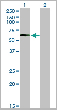
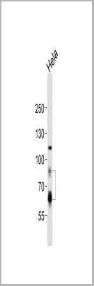
 WB (Western Blot)
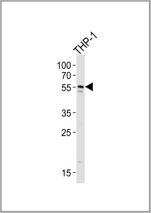
(STK33 monoclonal antibody, Western Blot analysis of STK33 expression in HeLa.)
WB (Western Blot)
(STK33 monoclonal antibody, Western Blot analysis of STK33 expression in HeLa.)
STK33, Monoclonal Antibody (Cat# AAA25856)
Full Name
STK33 (Serine/Threonine Kinase 33) (PE)
Reactivity
Human
Applications
Immunofluorescence, Immunoprecipitation, Western Blot
Purity
Purified by Protein A Affinity Chromatography.
Pricing
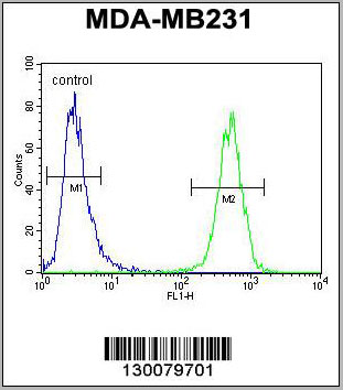
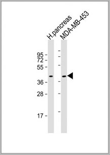
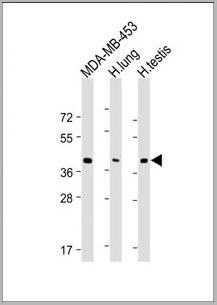
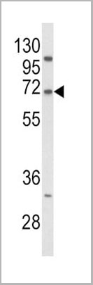
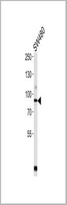
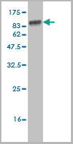
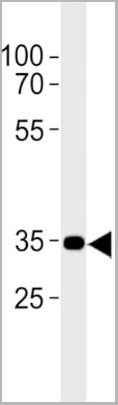
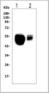
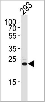
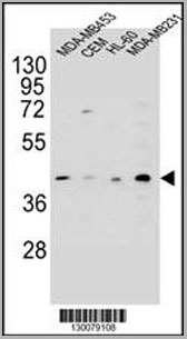 WB (Western Blot)
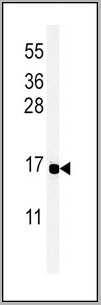
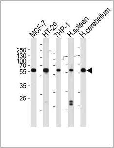
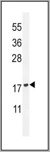
(HHLA2 Antibody (N-term) western blot analysis in MDA-MB453,CEM,HL-60,MDA-MB231 cell line lysates (35ug/lane).This demonstrates the HHLA2 antibody detected the HHLA2 protein (arrow).)
WB (Western Blot)
(HHLA2 Antibody (N-term) western blot analysis in MDA-MB453,CEM,HL-60,MDA-MB231 cell line lysates (35ug/lane).This demonstrates the HHLA2 antibody detected the HHLA2 protein (arrow).)
HHLA2, Polyclonal Antibody (Cat# AAA28722)
Full Name
HHLA2 Antibody (N-term)
Gene Names
HHLA2; B7H7; B7-H7
Reactivity
Human
Applications
Western Blot, Immunohistochemistry, Flow Cytometry
Purity
Peptide Affinity Purified Rabbit Polyclonal Antibody (Pab)
Pricing
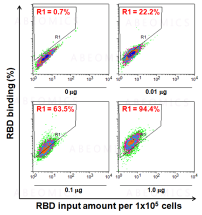
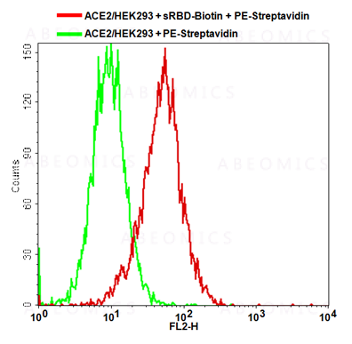
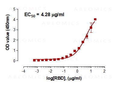
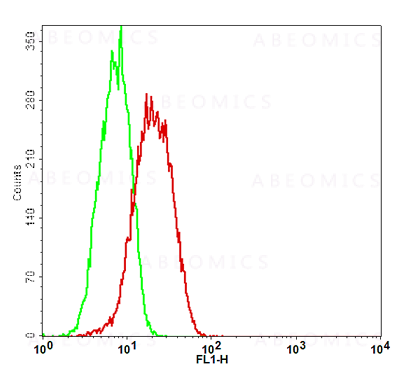 Application Data
(Fig-5: Cell Surface staining of human ACE2 in the ACE2/HEK293 stable cell line. ATTO 488-conjugated anti-hACE2 antibody (clone AC18F; was used at 1ug/ 1x10^6 cells (Red). ATTO 488-conjugated mouse IgG1 was used as isotype control at 1ug/ 1x10^6 cells (Green).)
Application Data
(Fig-5: Cell Surface staining of human ACE2 in the ACE2/HEK293 stable cell line. ATTO 488-conjugated anti-hACE2 antibody (clone AC18F; was used at 1ug/ 1x10^6 cells (Red). ATTO 488-conjugated mouse IgG1 was used as isotype control at 1ug/ 1x10^6 cells (Green).)
ACE2, Cell Line (Cat# AAA14888)
Full Name
ACE2/HEK293 Stable Cell Line
Gene Names
ACE2; ACEH
Applications
Functional Assay
Pricing


 Application Data
(Staining of mouse spleen with Hamster anti Mouse CD81: Alexa Fluor 488)
Application Data
(Staining of mouse spleen with Hamster anti Mouse CD81: Alexa Fluor 488)
CD81, Monoclonal Antibody (Cat# AAA11869)
Full Name
HAMSTER ANTI MOUSE CD81:FITC
Gene Names
Cd81; Tapa1; Tapa-1; Tspan28
Applications
Flow Cytometry
Pricing


 Application Data
(Staining of mouse spleen with Hamster anti Mouse CD81: Alexa Fluor 488)
Application Data
(Staining of mouse spleen with Hamster anti Mouse CD81: Alexa Fluor 488)
CD81, Monoclonal Antibody (Cat# AAA12033)
Full Name
HAMSTER ANTI MOUSE CD81:RPE
Gene Names
Cd81; Tapa1; Tapa-1; Tspan28
Applications
Flow Cytometry
Pricing
 FCM (Flow Cytometry)
(PE conjugated polystyrene beads were stained with (filled histogram) or rabbit IgG, FITC isotype control (open histogram).)
FCM (Flow Cytometry)
(PE conjugated polystyrene beads were stained with (filled histogram) or rabbit IgG, FITC isotype control (open histogram).)
R-PE, Antibody (Cat# AAA14282)
Full Name
Purified Rabbit Anti-R-PE, Polyclonal Antibody
Applications
Flow Cytometry, Immunohistochemistry
Pricing
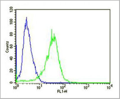
 FCM (Flow Cytometry)
(CB2 Antibody (C-term) flow cytometric analysis of Jurkat cells (right histogram) compared to a negative control cell (left histogram).FITC-conjugated goat-anti-rabbit secondary antibodies were used for the analysis.)
FCM (Flow Cytometry)
(CB2 Antibody (C-term) flow cytometric analysis of Jurkat cells (right histogram) compared to a negative control cell (left histogram).FITC-conjugated goat-anti-rabbit secondary antibodies were used for the analysis.)
CB2, Polyclonal Antibody (Cat# AAA28672)
Full Name
CB2 Antibody (C-term)
Gene Names
CNR2; CB2; CX5; CB-2
Reactivity
Human
Applications
Immunohistochemistry, Flow Cytometry, Western Blot
Purity
This antibody is purified through a protein A column, followed by peptide affinity purification.
Pricing
 WB (Western Blot)
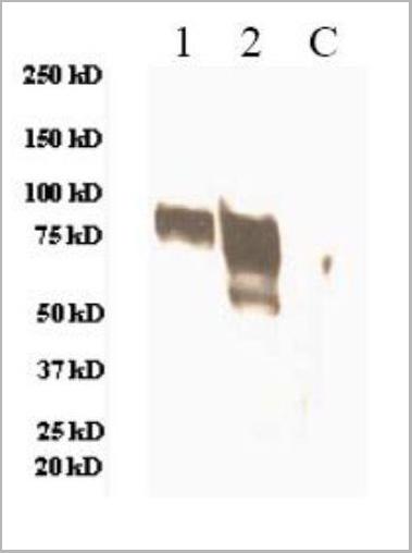
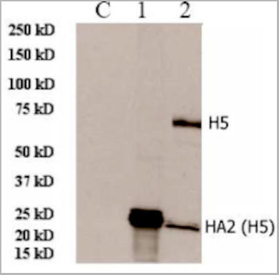
(lane C, 293 cell extract controlLane 1, 293 cell expressing HA2 (H5N1)Lane 2, 293 cell expressing HA (H5N1))
WB (Western Blot)
(lane C, 293 cell extract controlLane 1, 293 cell expressing HA2 (H5N1)Lane 2, 293 cell expressing HA (H5N1))
HA2 (A/Vietnam/1203/2004)(H5N1), Polyclonal Antibody (Cat# AAA13718)
Full Name
Anti-HA2 (A/Vietnam/1203/2004)(H5N1), rabbit IgG
Applications
Western Blot
Purity
Immunoaffinity chromatography
Pricing
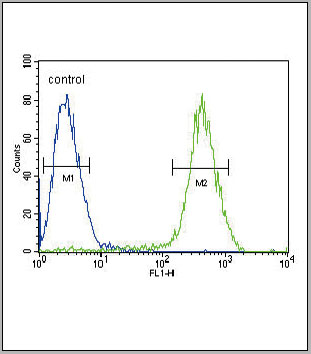
 Application Data
H5 (A/Hong Kong/483/97)(H5N1), Polyclonal Antibody (Cat# AAA13731)
Full Name
Anti-H5 (A/Hong Kong/483/97)(H5N1), rabbit IgG
Reactivity
Has not been tested.
Applications
Western Blot
Purity
Immunoaffinity chromatography
Pricing
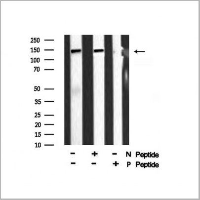
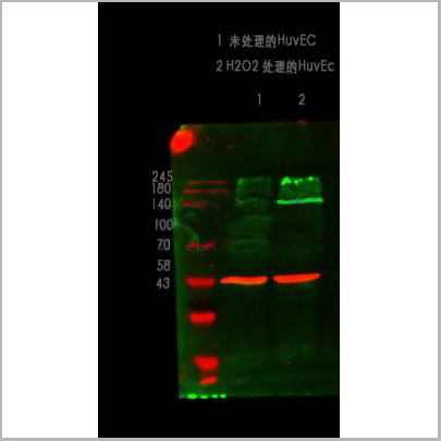
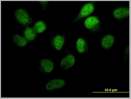
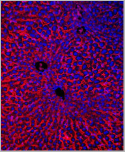
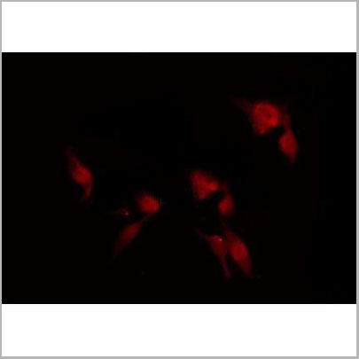 Application Data
(At 25 degree C. The primary antibody was diluted at 1/200 and incubated with the sample for 1 hour at 37 degree C. An Alexa Fluor 594 conjugated goat anti-rabbit IgG (H+L) Ab, diluted at 1/600, was used as the secondary antibody.)
Application Data
(At 25 degree C. The primary antibody was diluted at 1/200 and incubated with the sample for 1 hour at 37 degree C. An Alexa Fluor 594 conjugated goat anti-rabbit IgG (H+L) Ab, diluted at 1/600, was used as the secondary antibody.)
eNOS, Polyclonal Antibody (Cat# AAA31394)
Full Name
Phospho-eNOS (Ser1179) Antibody
Gene Names
NOS3; eNOS; ECNOS
Reactivity
Human, Mouse, Rat
Predicted Reactivity: Pig (100%), Bovine (100%), Rabbit (100%), Dog (100%)
Predicted Reactivity: Pig (100%), Bovine (100%), Rabbit (100%), Dog (100%)
Applications
Western Blot, Immunohistochemistry, Immunofluorescence, Immunocytochemistry, Peptide ELISA
Purity
The antibody is from purified rabbit serum by affinity purification via sequential chromatography on phospho-peptide and non-phospho-peptide affinity columns.
Pricing
Double-stranded RNA (dsRNA), Monoclonal ELISA Kit (Cat# AAA14576)
Full Name
Double-stranded RNA (dsRNA) ELISA kit (J2 based)
Reactivity
General
Applications
ELISA
Pricing
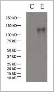 WB (Western Blot)
(Western Blot Data: C: 293 cell extract controlE: 293 cell expressing gp120 (SIV/mac239))
WB (Western Blot)
(Western Blot Data: C: 293 cell extract controlE: 293 cell expressing gp120 (SIV/mac239))
gp120/160 (SIV/mac239), Polyclonal Antibody (Cat# AAA13713)
Full Name
Anti-gp120/160 (SIV-1/mac239) Rabbit Polyclonal Antibody
Applications
Western Blot
Purity
Immunoaffinity chromatography
Pricing
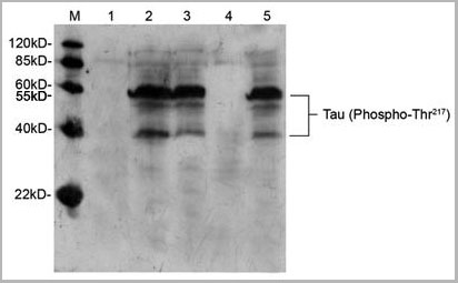
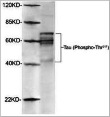 WB (Western Blot)
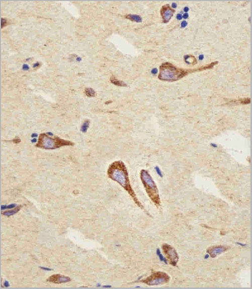
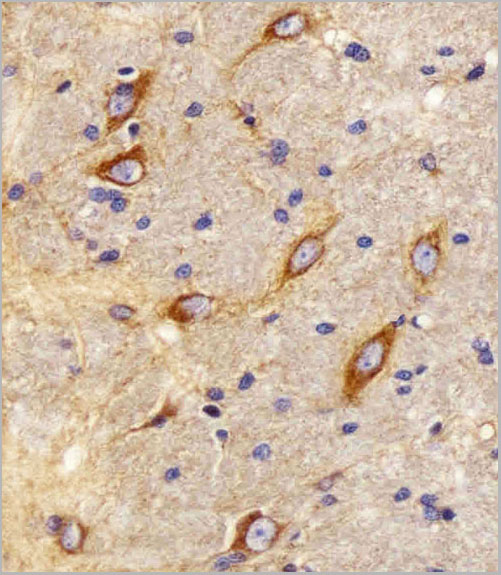
(Western Blot of Rat brain using AAA14727 (1ug/mL). The signal was developed with IRDyeTM800 conjugated giat anti-rabbit IgG)
WB (Western Blot)
(Western Blot of Rat brain using AAA14727 (1ug/mL). The signal was developed with IRDyeTM800 conjugated giat anti-rabbit IgG)
Tau, Polyclonal Antibody (Cat# AAA14727)
Full Name
Tau, phosphorylated (Thr217) (Tau Protein, Microtubule-associated Protein, MAP)
Gene Names
tau; BcDNA:RE16764; CG12881; CG31057; CG5606; DmelCG31057; dtau
Reactivity
Human
Applications
Western Blot
Purity
Purified by Affinity chromatography.
Pricing
 FCM (Flow Cytometry)
(PYY Antibody (C-term) (AAA28733) flow cytometric analysis of MCF-7 cells (bottom histogram) compared to a negative control cell (top histogram).FITC-conjugated goat-anti-rabbit secondary antibodies were used for the analysis.)
FCM (Flow Cytometry)
(PYY Antibody (C-term) (AAA28733) flow cytometric analysis of MCF-7 cells (bottom histogram) compared to a negative control cell (top histogram).FITC-conjugated goat-anti-rabbit secondary antibodies were used for the analysis.)
PYY, Polyclonal Antibody (Cat# AAA28733)
Full Name
PYY Antibody (C-term)
Gene Names
PYY; PYY1; PYY-I
Reactivity
Human
Applications
Western Blot, Immunohistochemistry, Immunofluorescence, Flow Cytometry
Purity
Peptide Affinity Purified Rabbit Polyclonal Antibody (Pab)
Pricing
 WB (Western Blot)
(Western blot analysis of PD-L1. Anti-PD-L1 antibody (Clone: ABM4E54) was tested at 2 ug/ml on h Spleen lysate.)
WB (Western Blot)
(Western blot analysis of PD-L1. Anti-PD-L1 antibody (Clone: ABM4E54) was tested at 2 ug/ml on h Spleen lysate.)
PD-L1, Monoclonal Antibody (Cat# AAA14873)
Full Name
Monoclonal Antibody to PD-L1 (Clone: ABM4E54)
Gene Names
CD274; B7-H; B7H1; PDL1; PD-L1; PDCD1L1; PDCD1LG1
Reactivity
Human
Applications
Immunohistochemistry, Flow Cytometry, Western Blot
Purity
Purified
Protein G Chromatography
Protein G Chromatography
Pricing
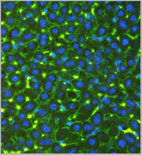
 IS (Immunostaining)
(Immunostaining of cultured E20 rat cortical neurons and glia stained with anti-nestin antibody (AAA14229, red, 1:500) and anti-MAP2 antibody (, green, 1:500). The blue is Hoechst staining for nuclear DNA. The nestin antibody labels developing astrocytes and neuronal stem cells in a clearly filamentous fashion, while the MAP2 antibody stains dendrites and perikarya of mature neurons.)
IS (Immunostaining)
(Immunostaining of cultured E20 rat cortical neurons and glia stained with anti-nestin antibody (AAA14229, red, 1:500) and anti-MAP2 antibody (, green, 1:500). The blue is Hoechst staining for nuclear DNA. The nestin antibody labels developing astrocytes and neuronal stem cells in a clearly filamentous fashion, while the MAP2 antibody stains dendrites and perikarya of mature neurons.)
Nestin, Monoclonal Antibody (Cat# AAA14229)
Full Name
Anti-Nestin
Gene Names
NES; Nbla00170
Applications
Western Blot, Immunofluorescence
Purity
Protein G purified culture supernatant
Pricing
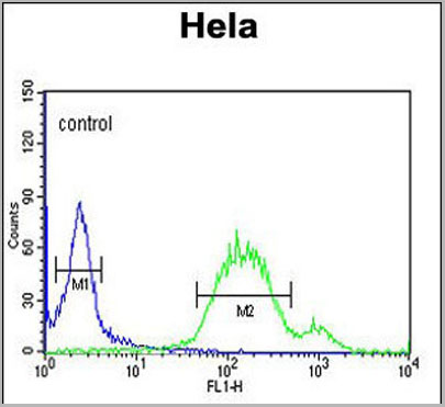
 FCM (Flow Cytometry)
(Overlay histogram showing Hela cells stained with AAA28750 (green line). The cells were fixed with 2% paraformaldehyde (10 min) and then permeabilized with 90% methanol for 10 min. The cells were then icubated in 2% bovine serum albumin to block non-specific protein-protein interactions followed by the antibody (AAA28750, 1:25 dilution) for 60 min at 37ºC. The secondary antibody used was Goat-Anti-Rabbit IgG, DyLight® 488 Conjugated Highly Cross-Adsorbed (1583138) at 1/200 dilution for 40 min at 37°C. Isotype control antibody (blue line) was rabbit IgG1 (1ug/1x106 cells) used under the same conditions. Acquisition of >10, 000 events wasperformed.)
FCM (Flow Cytometry)
(Overlay histogram showing Hela cells stained with AAA28750 (green line). The cells were fixed with 2% paraformaldehyde (10 min) and then permeabilized with 90% methanol for 10 min. The cells were then icubated in 2% bovine serum albumin to block non-specific protein-protein interactions followed by the antibody (AAA28750, 1:25 dilution) for 60 min at 37ºC. The secondary antibody used was Goat-Anti-Rabbit IgG, DyLight® 488 Conjugated Highly Cross-Adsorbed (1583138) at 1/200 dilution for 40 min at 37°C. Isotype control antibody (blue line) was rabbit IgG1 (1ug/1x106 cells) used under the same conditions. Acquisition of >10, 000 events wasperformed.)
WNT5A, Polyclonal Antibody (Cat# AAA28750)
Full Name
WNT5A Antibody (Center)
Gene Names
WNT5A; hWNT5A
Reactivity
Human, Mouse, Rat
Predicted: Rabbit
Predicted: Rabbit
Applications
Immunohistochemistry, Immunohistochemistry, Flow Cytometry, Western Blot
Purity
This antibody is purified through a protein A column, followed by peptide affinity purification.
Pricing
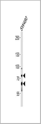
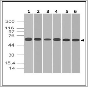
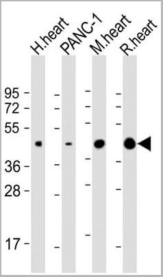
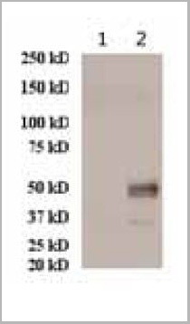
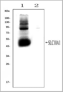 WB (Western Blot)
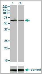
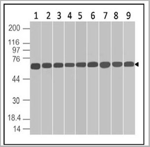
(Western blot analysis of SLC10A1 using anti- SLC10A1 antibody (AAA11653).Electrophoresis was performed on a 5-20% SDS-PAGE gel at 70V (Stacking gel) / 90V (Resolving gel) for 2-3 hours. The sample well of each lane was loaded with 50ug of sample under reducing conditions.Lane 1: rat liver tissue lysates (positive control), Lane 2: rat kidney tissue lysates, (negative control)After Electrophoresis, proteins were transferred to a Nitrocellulose membrane at 150mA for 50-90 minutes. Blocked the membrane with 5% Non-fat Milk/ TBS for 1.5 hour at RT. The membrane was incubated with rabbit anti-SLC10A1 antigen affinity purified polyclonal antibody (AAA11653) at 0.25ug/mL overnight at 4°C, then washed with TBS-0.1%Tween 3 times with 5 minutes each and probed with a goat anti-rabbit IgG-HRP secondary antibody at a dilution of 1:10000 for 1.5 hour at RT. The signal is developed using an Enhanced Chemiluminescent detection (ECL) kit with Tanon 5200 system. A specific band was detected for SLC10A1 at approximately 50KD. The expected band size for SLC10A1 is at 38KD.)
WB (Western Blot)
(Western blot analysis of SLC10A1 using anti- SLC10A1 antibody (AAA11653).Electrophoresis was performed on a 5-20% SDS-PAGE gel at 70V (Stacking gel) / 90V (Resolving gel) for 2-3 hours. The sample well of each lane was loaded with 50ug of sample under reducing conditions.Lane 1: rat liver tissue lysates (positive control), Lane 2: rat kidney tissue lysates, (negative control)After Electrophoresis, proteins were transferred to a Nitrocellulose membrane at 150mA for 50-90 minutes. Blocked the membrane with 5% Non-fat Milk/ TBS for 1.5 hour at RT. The membrane was incubated with rabbit anti-SLC10A1 antigen affinity purified polyclonal antibody (AAA11653) at 0.25ug/mL overnight at 4°C, then washed with TBS-0.1%Tween 3 times with 5 minutes each and probed with a goat anti-rabbit IgG-HRP secondary antibody at a dilution of 1:10000 for 1.5 hour at RT. The signal is developed using an Enhanced Chemiluminescent detection (ECL) kit with Tanon 5200 system. A specific band was detected for SLC10A1 at approximately 50KD. The expected band size for SLC10A1 is at 38KD.)
SLC10A1, Polyclonal Antibody (Cat# AAA11653)
Full Name
Anti-SLC10A1 Antibody
Gene Names
Slc10a1; Ntcp
Reactivity
Mouse, Rat
Applications
Flow Cytometry, Immunofluorescence, Immunohistochemistry, Immunohistochemistry, Immunocytochemistry, Western Blot
Purity
Immunogen affinity purified.
Pricing


 Application Data
(Published customer image: Mouse anti V5 tag antibody, clone SV5-Pk1 used for the detection of V5 tagged WEEV_nsP3 protein by western blotting and immunofluorescenceImage caption: WEEV nsP3 interaction with host IKKbeta. A) U87MGs were transfected in a 6-well plate with 5 ug of pUC19 and WEEV_nsP3_HA for 24 hours. Cell lysates were resolved using SDS-PAGE and subsequently immunoblotted with V5 antibody and beta-actin served as a loading control. B) U87MGs were transfected with WEEV_nsP3_V5; cells were fixed after 24 hours and stained with antibodies against the endogenous IKKbeta and the V5 tag. Cells were incubated with appropriate secondary Alexa Fluor antibodies and the nuclei stained with DAPI. Co-localization of IKKbeta with WEEV_nsP3_V5 (yellow) was observed as shown by the arrows. B) Panels E -H serve as an example of transfected cells in a given field of view that show co-localization of IKKbeta and WEEV_nsP3_V5 24 hours post transfection. Panels I-L represent magnified images of other cells showing co-localization of IKKbeta and WEEV_nsP3_V5. Panel M is a magnified image of panel L. The co-localization was confirmed by Z-stack analysis. Co-localization was calculated to be approximately in 61% of cells (163 cells were counted of which 44% demonstrated expression of nsP3. Of those cells that expressed nsP3, 61% showed co-localization of both proteins). Images were taken using Nikon Eclipse TE2000-U at 60x magnification and are representative of 2 independent experiments.From: Amaya M, Voss K, Sampey G, Senina S, de la Fuente C, et al. (2014) The Role of IKKbeta in Venezuelan Equine Encephalitis Virus Infection. PLoS ONE 9(2): e86745.)
Application Data
(Published customer image: Mouse anti V5 tag antibody, clone SV5-Pk1 used for the detection of V5 tagged WEEV_nsP3 protein by western blotting and immunofluorescenceImage caption: WEEV nsP3 interaction with host IKKbeta. A) U87MGs were transfected in a 6-well plate with 5 ug of pUC19 and WEEV_nsP3_HA for 24 hours. Cell lysates were resolved using SDS-PAGE and subsequently immunoblotted with V5 antibody and beta-actin served as a loading control. B) U87MGs were transfected with WEEV_nsP3_V5; cells were fixed after 24 hours and stained with antibodies against the endogenous IKKbeta and the V5 tag. Cells were incubated with appropriate secondary Alexa Fluor antibodies and the nuclei stained with DAPI. Co-localization of IKKbeta with WEEV_nsP3_V5 (yellow) was observed as shown by the arrows. B) Panels E -H serve as an example of transfected cells in a given field of view that show co-localization of IKKbeta and WEEV_nsP3_V5 24 hours post transfection. Panels I-L represent magnified images of other cells showing co-localization of IKKbeta and WEEV_nsP3_V5. Panel M is a magnified image of panel L. The co-localization was confirmed by Z-stack analysis. Co-localization was calculated to be approximately in 61% of cells (163 cells were counted of which 44% demonstrated expression of nsP3. Of those cells that expressed nsP3, 61% showed co-localization of both proteins). Images were taken using Nikon Eclipse TE2000-U at 60x magnification and are representative of 2 independent experiments.From: Amaya M, Voss K, Sampey G, Senina S, de la Fuente C, et al. (2014) The Role of IKKbeta in Venezuelan Equine Encephalitis Virus Infection. PLoS ONE 9(2): e86745.)
V5-TAG, Monoclonal Antibody (Cat# AAA11864)
Full Name
MOUSE ANTI V5-TAG:FITC
Applications
Immunofluorescence
Pricing


 Application Data
(Published customer image: Mouse anti V5 tag antibody, clone SV5-Pk1 used for the detection of V5 tagged WEEV_nsP3 protein by western blotting and immunofluorescenceImage caption: WEEV nsP3 interaction with host IKKbeta. A) U87MGs were transfected in a 6-well plate with 5 ug of pUC19 and WEEV_nsP3_HA for 24 hours. Cell lysates were resolved using SDS-PAGE and subsequently immunoblotted with V5 antibody and beta-actin served as a loading control. B) U87MGs were transfected with WEEV_nsP3_V5; cells were fixed after 24 hours and stained with antibodies against the endogenous IKKbeta and the V5 tag. Cells were incubated with appropriate secondary Alexa Fluor antibodies and the nuclei stained with DAPI. Co-localization of IKKbeta with WEEV_nsP3_V5 (yellow) was observed as shown by the arrows. B) Panels E -H serve as an example of transfected cells in a given field of view that show co-localization of IKKbeta and WEEV_nsP3_V5 24 hours post transfection. Panels I-L represent magnified images of other cells showing co-localization of IKKbeta and WEEV_nsP3_V5. Panel M is a magnified image of panel L. The co-localization was confirmed by Z-stack analysis. Co-localization was calculated to be approximately in 61% of cells (163 cells were counted of which 44% demonstrated expression of nsP3. Of those cells that expressed nsP3, 61% showed co-localization of both proteins). Images were taken using Nikon Eclipse TE2000-U at 60x magnification and are representative of 2 independent experiments.From: Amaya M, Voss K, Sampey G, Senina S, de la Fuente C, et al. (2014) The Role of IKKbeta in Venezuelan Equine Encephalitis Virus Infection. PLoS ONE 9(2): e86745.)
Application Data
(Published customer image: Mouse anti V5 tag antibody, clone SV5-Pk1 used for the detection of V5 tagged WEEV_nsP3 protein by western blotting and immunofluorescenceImage caption: WEEV nsP3 interaction with host IKKbeta. A) U87MGs were transfected in a 6-well plate with 5 ug of pUC19 and WEEV_nsP3_HA for 24 hours. Cell lysates were resolved using SDS-PAGE and subsequently immunoblotted with V5 antibody and beta-actin served as a loading control. B) U87MGs were transfected with WEEV_nsP3_V5; cells were fixed after 24 hours and stained with antibodies against the endogenous IKKbeta and the V5 tag. Cells were incubated with appropriate secondary Alexa Fluor antibodies and the nuclei stained with DAPI. Co-localization of IKKbeta with WEEV_nsP3_V5 (yellow) was observed as shown by the arrows. B) Panels E -H serve as an example of transfected cells in a given field of view that show co-localization of IKKbeta and WEEV_nsP3_V5 24 hours post transfection. Panels I-L represent magnified images of other cells showing co-localization of IKKbeta and WEEV_nsP3_V5. Panel M is a magnified image of panel L. The co-localization was confirmed by Z-stack analysis. Co-localization was calculated to be approximately in 61% of cells (163 cells were counted of which 44% demonstrated expression of nsP3. Of those cells that expressed nsP3, 61% showed co-localization of both proteins). Images were taken using Nikon Eclipse TE2000-U at 60x magnification and are representative of 2 independent experiments.From: Amaya M, Voss K, Sampey G, Senina S, de la Fuente C, et al. (2014) The Role of IKKbeta in Venezuelan Equine Encephalitis Virus Infection. PLoS ONE 9(2): e86745.)
V5-TAG, Monoclonal Antibody (Cat# AAA11930)
Full Name
MOUSE ANTI V5-TAG
Applications
Immunohistochemistry, Flow Cytometry, Immunofluorescence, Immunoprecipitation, Western Blot, Radioimmunoassay
Pricing
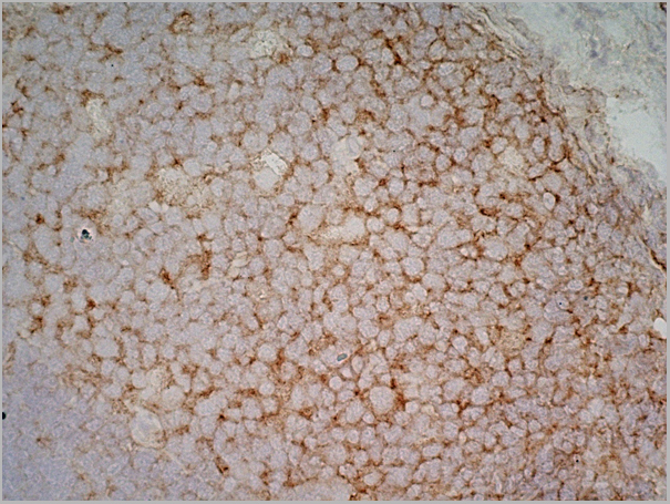
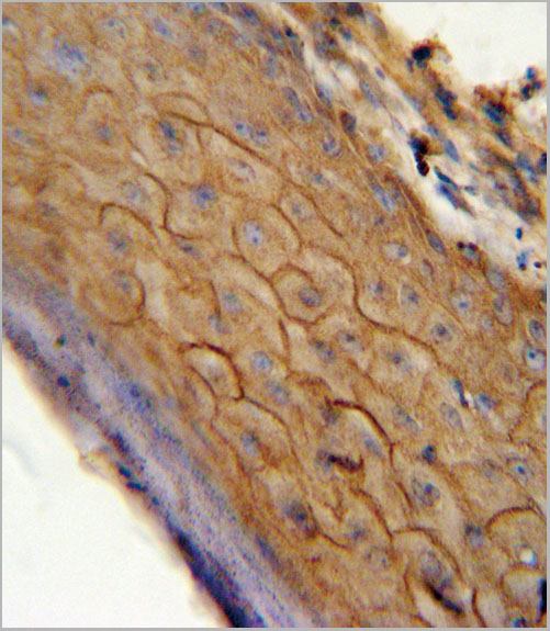
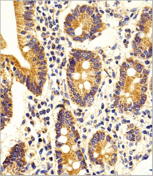
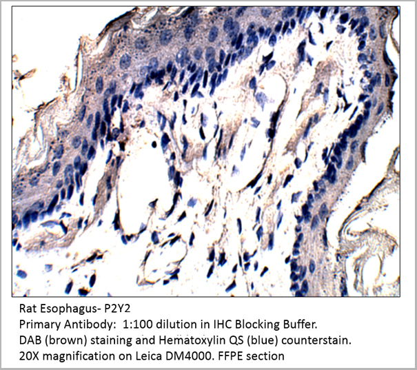 IHC (Immunohistochemistry)
P2Y2, Polyclonal Antibody (Cat# AAA14402)
Full Name
P2Y2 Antibody
Gene Names
P2RY2; P2U; HP2U; P2U1; P2UR; P2Y2; P2RU1; P2Y2R
Reactivity
Cat, Human, Mouse, Rat
Applications
Confocal Microscopy, Immunocytochemistry, Immunofluorescence, Immunohistochemistry, Immunoprecipitation, Western Blot
Pricing


 Application Data
(Published customer image: Mouse anti V5 tag antibody, clone SV5-Pk1 used for the detection of V5 tagged WEEV_nsP3 protein by western blotting and immunofluorescenceImage caption: WEEV nsP3 interaction with host IKKbeta. A) U87MGs were transfected in a 6-well plate with 5 ug of pUC19 and WEEV_nsP3_HA for 24 hours. Cell lysates were resolved using SDS-PAGE and subsequently immunoblotted with V5 antibody and beta-actin served as a loading control. B) U87MGs were transfected with WEEV_nsP3_V5; cells were fixed after 24 hours and stained with antibodies against the endogenous IKKbeta and the V5 tag. Cells were incubated with appropriate secondary Alexa Fluor antibodies and the nuclei stained with DAPI. Co-localization of IKKbeta with WEEV_nsP3_V5 (yellow) was observed as shown by the arrows. B) Panels E -H serve as an example of transfected cells in a given field of view that show co-localization of IKKbeta and WEEV_nsP3_V5 24 hours post transfection. Panels I-L represent magnified images of other cells showing co-localization of IKKbeta and WEEV_nsP3_V5. Panel M is a magnified image of panel L. The co-localization was confirmed by Z-stack analysis. Co-localization was calculated to be approximately in 61% of cells (163 cells were counted of which 44% demonstrated expression of nsP3. Of those cells that expressed nsP3, 61% showed co-localization of both proteins). Images were taken using Nikon Eclipse TE2000-U at 60x magnification and are representative of 2 independent experiments.From: Amaya M, Voss K, Sampey G, Senina S, de la Fuente C, et al. (2014) The Role of IKKbeta in Venezuelan Equine Encephalitis Virus Infection. PLoS ONE 9(2): e86745.)
Application Data
(Published customer image: Mouse anti V5 tag antibody, clone SV5-Pk1 used for the detection of V5 tagged WEEV_nsP3 protein by western blotting and immunofluorescenceImage caption: WEEV nsP3 interaction with host IKKbeta. A) U87MGs were transfected in a 6-well plate with 5 ug of pUC19 and WEEV_nsP3_HA for 24 hours. Cell lysates were resolved using SDS-PAGE and subsequently immunoblotted with V5 antibody and beta-actin served as a loading control. B) U87MGs were transfected with WEEV_nsP3_V5; cells were fixed after 24 hours and stained with antibodies against the endogenous IKKbeta and the V5 tag. Cells were incubated with appropriate secondary Alexa Fluor antibodies and the nuclei stained with DAPI. Co-localization of IKKbeta with WEEV_nsP3_V5 (yellow) was observed as shown by the arrows. B) Panels E -H serve as an example of transfected cells in a given field of view that show co-localization of IKKbeta and WEEV_nsP3_V5 24 hours post transfection. Panels I-L represent magnified images of other cells showing co-localization of IKKbeta and WEEV_nsP3_V5. Panel M is a magnified image of panel L. The co-localization was confirmed by Z-stack analysis. Co-localization was calculated to be approximately in 61% of cells (163 cells were counted of which 44% demonstrated expression of nsP3. Of those cells that expressed nsP3, 61% showed co-localization of both proteins). Images were taken using Nikon Eclipse TE2000-U at 60x magnification and are representative of 2 independent experiments.From: Amaya M, Voss K, Sampey G, Senina S, de la Fuente C, et al. (2014) The Role of IKKbeta in Venezuelan Equine Encephalitis Virus Infection. PLoS ONE 9(2): e86745.)
V5-TAG, Monoclonal Antibody (Cat# AAA12211)
Full Name
MOUSE ANTI V5-TAG
Applications
Immunohistochemistry, Flow Cytometry, Immunofluorescence, Immunoprecipitation, Western Blot, Radioimmunoassay
Pricing
COVID 19 Spike RBD IgG Humanized Coronavirus, Monoclonal Antibody (Cat# AAA13540)
Full Name
Anti-SARS-CoV-2 Spike RBD IgG, Humanized antibody (Positive Control)
Reactivity
Human
Applications
Lateral Flow
Purity
Purity: >95% by HPLC & SDS-PAGE
Purification: Protein A purified
Purification: Protein A purified
Pricing
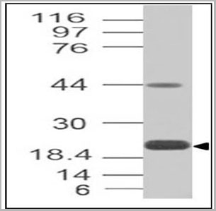
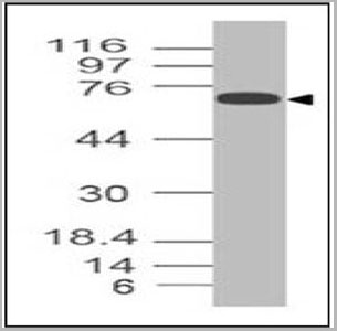
 WB (Western Blot)
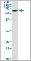
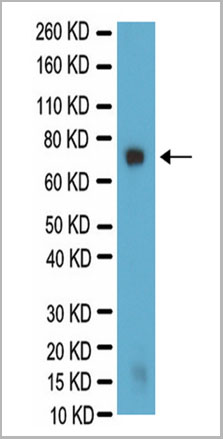
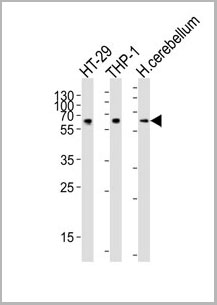
(Western Blot analysis for galactocerebroside in PC12 lysate. PC12 lysate was resolved by electrophoresis, transferred to PVDF membrane and probed with AAA14685 at 1:500 dilution. Proteins were visualized using a goat anti-mouse secondary antibody conjugated to HRP and a chemiluminescence detection system. Arrow indicates protein Galactocerebroside (~75kD).)
WB (Western Blot)
(Western Blot analysis for galactocerebroside in PC12 lysate. PC12 lysate was resolved by electrophoresis, transferred to PVDF membrane and probed with AAA14685 at 1:500 dilution. Proteins were visualized using a goat anti-mouse secondary antibody conjugated to HRP and a chemiluminescence detection system. Arrow indicates protein Galactocerebroside (~75kD).)
Galactocerebroside, Monoclonal Antibody (Cat# AAA14685)
Full Name
Mouse anti-Bovine Galactocerebroside
Applications
Immunohistochemistry, Immunocytochemistry
Purity
Purified by Protein A affinity chromatography.
Pricing
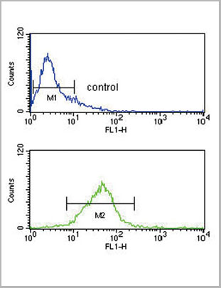
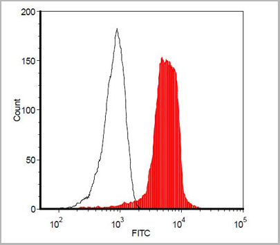
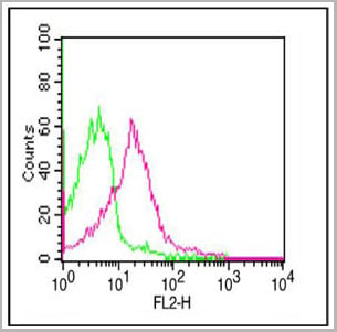
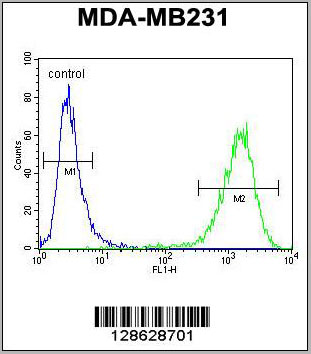
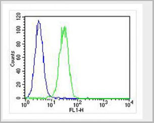
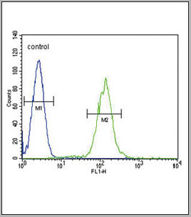
 FCM (Flow Cytometry)
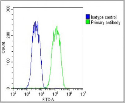
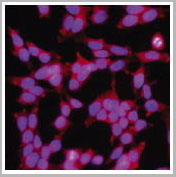
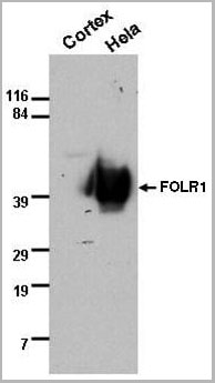
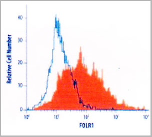
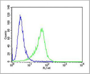
(Flow Cytometry detection of FOLR1 in MCF-7 human cell line using AAA14691. MCF-7 human breast cancer cell line was stained with AAA14691 (filled histogram) or isotype control antibody (open histogram), followed byPhycoerythrin-conjugated Anti-MouseIgG Secondary Antibody.)
FCM (Flow Cytometry)
(Flow Cytometry detection of FOLR1 in MCF-7 human cell line using AAA14691. MCF-7 human breast cancer cell line was stained with AAA14691 (filled histogram) or isotype control antibody (open histogram), followed byPhycoerythrin-conjugated Anti-MouseIgG Secondary Antibody.)
FOLR1, Monoclonal Antibody (Cat# AAA14691)
Full Name
FOLR1 (Folate receptor 1 (adult) Adult folate-binding protein, FBP, Folate receptor, adult, Folate receptor 1, Folate receptor alpha, FOLR, FR-alpha, KB cells FBP, MOv18, Ovarian tumor-associated antigen MOv18)
Gene Names
FOLR1; FBP; FOLR
Reactivity
Human
Applications
Flow Cytometry, Western Blot, Immunocytochemistry
Purity
Purified by Protein G affinity chromatography from hybridoma culture supernatant.
Pricing
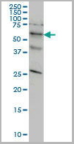
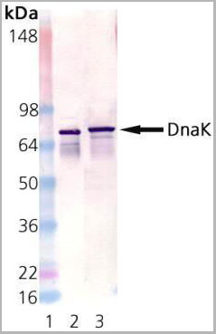 WB (Western Blot)
(Western Blot Analysis using AAA14655: Lane 1: MW Marker; Lane 2: DnaK active recombinant protein, Lane 3: E. coli cell lysate.)
WB (Western Blot)
(Western Blot Analysis using AAA14655: Lane 1: MW Marker; Lane 2: DnaK active recombinant protein, Lane 3: E. coli cell lysate.)
DnaK, Monoclonal Antibody (Cat# AAA14655)
Full Name
DnaK
Reactivity
E Coli
Applications
Western Blot
Purity
Affinity Purified
Purified by Protein G affinity chromatography.
Purified by Protein G affinity chromatography.
Pricing
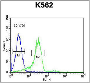 FCM (Flow Cytometry)
(ENAM Antibody (C-term) (AAA28699) flow cytometric analysis of K562 cells (right histogram) compared to a negative control cell (left histogram).FITC-conjugated goat-anti-rabbit secondary antibodies were used for the analysis.)
FCM (Flow Cytometry)
(ENAM Antibody (C-term) (AAA28699) flow cytometric analysis of K562 cells (right histogram) compared to a negative control cell (left histogram).FITC-conjugated goat-anti-rabbit secondary antibodies were used for the analysis.)
ENAM, Polyclonal Antibody (Cat# AAA28699)
Full Name
ENAM Antibody (C-term)
Gene Names
ENAM; ADAI; AI1C; AIH2
Reactivity
Human
Applications
Western Blot, Immunohistochemistry, Flow Cytometry
Purity
Peptide Affinity Purified Rabbit Polyclonal Antibody (Pab)
Pricing
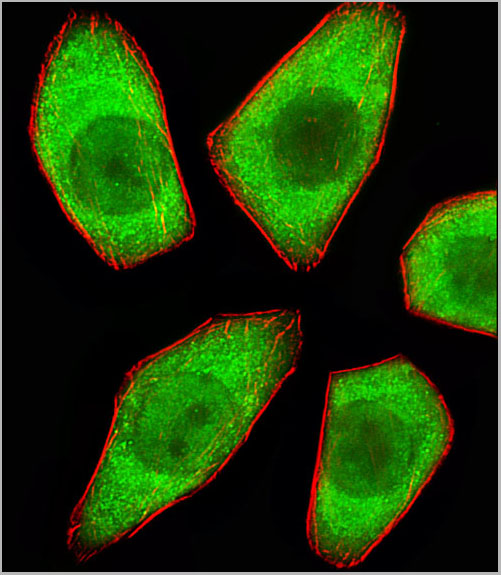
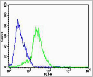
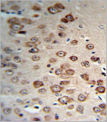
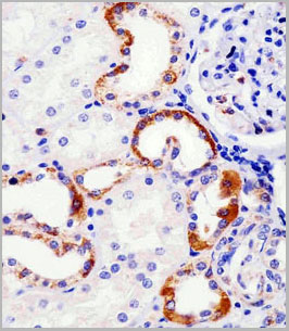
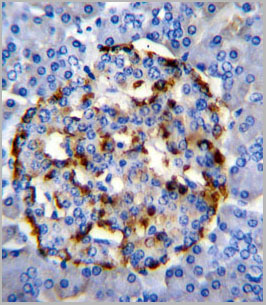
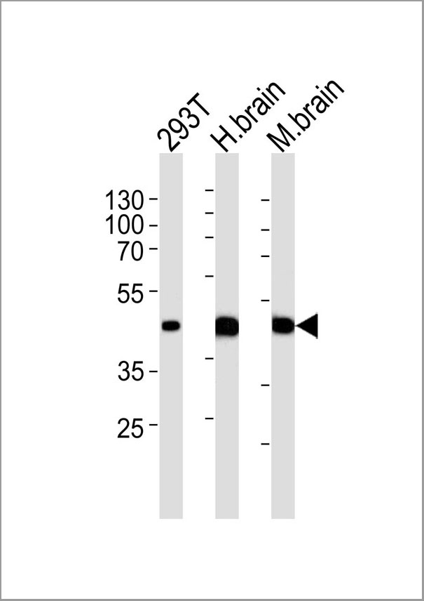 IHC (Immunohistochemistry)
(Western blot analysis of lysates from 293T cell line human brain and mouse brain tissue lysate(from left to right), using STRADA Antibody (C-term). AAA28673 was diluted at 1:1000 at each lane. A goat anti-rabbit IgG H&L(HRP) at 1:5000 dilution was used as the secondary antibody. Lysates at 35ug per lane.)
IHC (Immunohistochemistry)
(Western blot analysis of lysates from 293T cell line human brain and mouse brain tissue lysate(from left to right), using STRADA Antibody (C-term). AAA28673 was diluted at 1:1000 at each lane. A goat anti-rabbit IgG H&L(HRP) at 1:5000 dilution was used as the secondary antibody. Lysates at 35ug per lane.)
STRADA, Polyclonal Antibody (Cat# AAA28673)
Full Name
STRADA Antibody (C-term)
Gene Names
STRADA; LYK5; PMSE; Stlk; STRAD; NY-BR-96
Reactivity
Human, mouse (Predicted Reactivity: Monkey)
Applications
Western Blot, Flow Cytometry, Immunofluorescence, Immunohistochemistry
Purity
Purified Rabbit Polyclonal Antibody (Pab)
Pricing
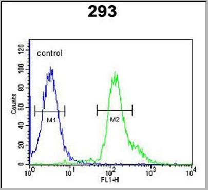 FCM (Flow Cytometry)
(RSPO2 Antibody (C-term) (AAA28708) flow cytometric analysis of 293 cells (right histogram) compared to a negative control cell (left histogram).FITC-conjugated goat-anti-rabbit secondary antibodies were used for the analysis.)
FCM (Flow Cytometry)
(RSPO2 Antibody (C-term) (AAA28708) flow cytometric analysis of 293 cells (right histogram) compared to a negative control cell (left histogram).FITC-conjugated goat-anti-rabbit secondary antibodies were used for the analysis.)
RSPO2, Polyclonal Antibody (Cat# AAA28708)
Full Name
RSPO2 Antibody (C-term)
Gene Names
RSPO2; CRISTIN2
Reactivity
Human (Predicted Reactivity: Mouse)
Applications
Westen Blot, Immunohistochemistry, Flow Cytometry
Purity
This antibody is purified through a protein A column, followed by peptide affinity purification.
Pricing


 Application Data
(Published customer image: Mouse anti V5 tag antibody, clone SV5-Pk1 used for the detection of V5 tagged WEEV_nsP3 protein by western blotting and immunofluorescenceImage caption: WEEV nsP3 interaction with host IKKbeta. A) U87MGs were transfected in a 6-well plate with 5 ug of pUC19 and WEEV_nsP3_HA for 24 hours. Cell lysates were resolved using SDS-PAGE and subsequently immunoblotted with V5 antibody and beta-actin served as a loading control. B) U87MGs were transfected with WEEV_nsP3_V5; cells were fixed after 24 hours and stained with antibodies against the endogenous IKKbeta and the V5 tag. Cells were incubated with appropriate secondary Alexa Fluor antibodies and the nuclei stained with DAPI. Co-localization of IKKbeta with WEEV_nsP3_V5 (yellow) was observed as shown by the arrows. B) Panels E -H serve as an example of transfected cells in a given field of view that show co-localization of IKKbeta and WEEV_nsP3_V5 24 hours post transfection. Panels I-L represent magnified images of other cells showing co-localization of IKKbeta and WEEV_nsP3_V5. Panel M is a magnified image of panel L. The co-localization was confirmed by Z-stack analysis. Co-localization was calculated to be approximately in 61% of cells (163 cells were counted of which 44% demonstrated expression of nsP3. Of those cells that expressed nsP3, 61% showed co-localization of both proteins). Images were taken using Nikon Eclipse TE2000-U at 60x magnification and are representative of 2 independent experiments.From: Amaya M, Voss K, Sampey G, Senina S, de la Fuente C, et al. (2014) The Role of IKKbeta in Venezuelan Equine Encephalitis Virus Infection. PLoS ONE 9(2): e86745.)
Application Data
(Published customer image: Mouse anti V5 tag antibody, clone SV5-Pk1 used for the detection of V5 tagged WEEV_nsP3 protein by western blotting and immunofluorescenceImage caption: WEEV nsP3 interaction with host IKKbeta. A) U87MGs were transfected in a 6-well plate with 5 ug of pUC19 and WEEV_nsP3_HA for 24 hours. Cell lysates were resolved using SDS-PAGE and subsequently immunoblotted with V5 antibody and beta-actin served as a loading control. B) U87MGs were transfected with WEEV_nsP3_V5; cells were fixed after 24 hours and stained with antibodies against the endogenous IKKbeta and the V5 tag. Cells were incubated with appropriate secondary Alexa Fluor antibodies and the nuclei stained with DAPI. Co-localization of IKKbeta with WEEV_nsP3_V5 (yellow) was observed as shown by the arrows. B) Panels E -H serve as an example of transfected cells in a given field of view that show co-localization of IKKbeta and WEEV_nsP3_V5 24 hours post transfection. Panels I-L represent magnified images of other cells showing co-localization of IKKbeta and WEEV_nsP3_V5. Panel M is a magnified image of panel L. The co-localization was confirmed by Z-stack analysis. Co-localization was calculated to be approximately in 61% of cells (163 cells were counted of which 44% demonstrated expression of nsP3. Of those cells that expressed nsP3, 61% showed co-localization of both proteins). Images were taken using Nikon Eclipse TE2000-U at 60x magnification and are representative of 2 independent experiments.From: Amaya M, Voss K, Sampey G, Senina S, de la Fuente C, et al. (2014) The Role of IKKbeta in Venezuelan Equine Encephalitis Virus Infection. PLoS ONE 9(2): e86745.)
V5-TAG, Monoclonal Antibody (Cat# AAA11850)
Full Name
MOUSE ANTI V5-TAG:Biotin
Applications
Immunohistochemistry, Western Blot
Pricing
 WB (Western Blot)
(Western Blot Data: 1: 293 cell extract control 2: 293 cell expressing E2 (HCV))
WB (Western Blot)
(Western Blot Data: 1: 293 cell extract control 2: 293 cell expressing E2 (HCV))
E2 (HCV), Polyclonal Antibody (Cat# AAA13709)
Full Name
Anti-E2 (HCV), rabbit IgG
Applications
Western Blot
Purity
Immunoaffinity chromatography
Pricing
CA 125, Native Protein (Cat# AAA14342)
Full Name
CA 125 Ag protein (preservative free)
Reactivity
CA 19-9:<1.0%, CA 72-4<1.0%, CA15-3:<0.1%
Applications
Control
Purity
> 95% pure
Pricing
 Application Data
(Overlay histogram showing U-2OS cells stained with AP10510c (green line). The cells were fixed with 2% paraformaldehyde (10 min) and then permeabilized with 90% methanol for 10 min. The cells were then icubated n 2% bovine serum albumin to block non-specificprotein-protein interactions followed by the antibody (AP10510c, 1:25 dilution) for 60 min at 37ºC. The secondary antibody used wasGoat-Anti-Rabbit IgG, DyLight® 488 Conjugated Highly Cross-Adsorbed(NA168821) at 1/400 dilution for 40 min at 37ºC. Isotype control antibody (blue line) was rabbit IgG (1?g/1x10^6 cells) used under the same conditions. Acquisition of >10, 000 events wasperformed)
Application Data
(Overlay histogram showing U-2OS cells stained with AP10510c (green line). The cells were fixed with 2% paraformaldehyde (10 min) and then permeabilized with 90% methanol for 10 min. The cells were then icubated n 2% bovine serum albumin to block non-specificprotein-protein interactions followed by the antibody (AP10510c, 1:25 dilution) for 60 min at 37ºC. The secondary antibody used wasGoat-Anti-Rabbit IgG, DyLight® 488 Conjugated Highly Cross-Adsorbed(NA168821) at 1/400 dilution for 40 min at 37ºC. Isotype control antibody (blue line) was rabbit IgG (1?g/1x10^6 cells) used under the same conditions. Acquisition of >10, 000 events wasperformed)
CF150, Polyclonal Antibody (Cat# AAA28789)
Full Name
CF150 Antibody (Center)
Gene Names
MB21D1; cGAS; h-cGAS; C6orf150
Reactivity
Human
Applications
Flow Cytometry, Western Blot
Purity
Peptide Affinity Purified Rabbit Polyclonal Antibody (Pab)
Pricing

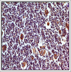
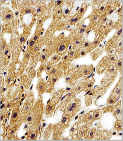
 IHC (Immunohistchemistry)
(Figure 9 Immunohistochemistry Validation of IRF7 in Human Liver Tissue Immunohistochemical analysis of paraffin-embedded human liver tissue using anti-IRF7 antibody (8991) at 2 μg/ml. Tissue was fixed with formaldehyde and blocked with 10% serum for 1 h at RT; antigen retrieval was by heat mediation with a citrate buffer (pH6). Samples were incubated with primary antibody overnight at 4˚C. A goat anti-rabbit IgG H&L (HRP) at 1/250 was used as secondary. Counter stained with Hematoxylin.)
IHC (Immunohistchemistry)
(Figure 9 Immunohistochemistry Validation of IRF7 in Human Liver Tissue Immunohistochemical analysis of paraffin-embedded human liver tissue using anti-IRF7 antibody (8991) at 2 μg/ml. Tissue was fixed with formaldehyde and blocked with 10% serum for 1 h at RT; antigen retrieval was by heat mediation with a citrate buffer (pH6). Samples were incubated with primary antibody overnight at 4˚C. A goat anti-rabbit IgG H&L (HRP) at 1/250 was used as secondary. Counter stained with Hematoxylin.)
IRF7, Polyclonal Antibody (Cat# AAA11032)
Full Name
IRF7 Antibody
Gene Names
IRF7; IRF7A; IRF7B; IRF7C; IRF7H; IRF-7H
Reactivity
Human, Mouse, Rat
Applications
Western Blot, Immunohistochemistry
Purity
IRF7 Antibody is Protein A purified.
Pricing
 FCM (Flow Cytometry)
(PLA2G2D Antibody (C-term) flow cytometric analysis of HL-60 cells (right histogram) compared to a negative control cell (left histogram).FITC-conjugated goat-anti-rabbit secondary antibodies were used for the analysis.)
FCM (Flow Cytometry)
(PLA2G2D Antibody (C-term) flow cytometric analysis of HL-60 cells (right histogram) compared to a negative control cell (left histogram).FITC-conjugated goat-anti-rabbit secondary antibodies were used for the analysis.)
PLA2G2D, Polyclonal Antibody (Cat# AAA28718)
Full Name
PLA2G2D Antibody (C-term)
Gene Names
PLA2G2D; SPLASH; sPLA2S; PLA2IID; sPLA2-IID
Reactivity
Human
Applications
Western Blot, Immunohistochemistry, Flow Cytometry
Purity
Peptide Affinity Purified Rabbit Polyclonal Antibody (Pab)
Pricing
Oxyntomodulin, Polyclonal Antibody (Cat# AAA14714)
Full Name
Oxyntomodulin (OXM)
Applications
ELISA
Purity
Affinity Purified
Purified by immunoaffinity chromatography.
Purified by immunoaffinity chromatography.
Pricing