Filters
Clonality
Type
Reactivity
Gene Name
Isotype
Host
Application
Clone
1038 results for " Polyclonal antibodies" - showing 800-850


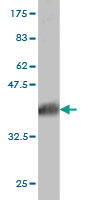
 WB (Western Blot)
(HHIP monoclonal antibody. Western Blot analysis of HHIP expression in MCF-7.)
WB (Western Blot)
(HHIP monoclonal antibody. Western Blot analysis of HHIP expression in MCF-7.)
HHIP, Monoclonal Antibody (Cat# AAA24827)
Full Name
HHIP (Hedgehog-interacting Protein, HHIP, HIP, UNQ5825/PRO19644, FLJ20992, FLJ90230) (Biotin)
Gene Names
HHIP; HIP
Reactivity
Human
Applications
EIA, IHC, IP, WB
Purity
Purified by Protein A Affinity Chromatography.
Pricing


 WB (Western Blot)
(CSK monoclonal antibody. Western Blot analysis of CSK expression in Hela NE.)
WB (Western Blot)
(CSK monoclonal antibody. Western Blot analysis of CSK expression in Hela NE.)
CSK, Monoclonal Antibody (Cat# AAA24769)
Full Name
CSK (Tyrosine-protein Kinase CSK, C-SRC Kinase, Protein-tyrosine Kinase CYL) (Biotin)
Reactivity
Human
Applications
EIA, IHC, IP, WB
Purity
Purified by Protein A Affinity Chromatography.
Pricing


 WB (Western Blot)
(SMURF1 monoclonal antibody, Western Blot analysis of SMURF1 expression in HeLa.)
WB (Western Blot)
(SMURF1 monoclonal antibody, Western Blot analysis of SMURF1 expression in HeLa.)
SMURF1, Monoclonal Antibody (Cat# AAA25255)
Full Name
SMURF1 (E3 Ubiquitin-protein Ligase SMURF1, hSMURF1, SMAD Ubiquitination Regulatory Factor 1, KIAA1625, SMAD-specific E3 Ubiquitin-protein Ligase 1) (FITC)
Reactivity
Human, Mouse
Applications
EIA, IF, IHC, IP, WB
Purity
Purified by Protein A Affinity Chromatography.
Pricing


 WB (Western Blot)
(Western blot analysis of CRKL over-expressed 293 cell line, cotransfected with CRKL Validated Chimera RNAi (Lane 2) or non-transfected control (Lane 1). Blot probed with CRKL monoclonal antibody. GAPDH (36.1kD) used as specificity and loading control.)
WB (Western Blot)
(Western blot analysis of CRKL over-expressed 293 cell line, cotransfected with CRKL Validated Chimera RNAi (Lane 2) or non-transfected control (Lane 1). Blot probed with CRKL monoclonal antibody. GAPDH (36.1kD) used as specificity and loading control.)
CRKL, Monoclonal Antibody (Cat# AAA25064)
Full Name
CRKL (Crk-like Protein) (FITC)
Reactivity
Human
Applications
EIA, IF, WB
Purity
Purified by Protein A Affinity Chromatography.
Pricing


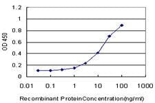
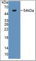
 Application Data
(Detection limit for recombinant GST tagged PLK1 is ~1ng/ml as a capture antibody.)
Application Data
(Detection limit for recombinant GST tagged PLK1 is ~1ng/ml as a capture antibody.)
PLK1, Monoclonal Antibody (Cat# AAA25799)
Full Name
PLK1 (STPK13, Polo-like Kinase 1, Serine/Threonine-protein Kinase 13, Serine/Threonine-protein Kinase PLK1, PLK, PLK-1) (PE)
Gene Names
PLK1; PLK; STPK13
Reactivity
Human
Applications
EIA, IF, IP, WB
Purity
Purified by Protein A Affinity Chromatography.
Pricing


 WB (Western Blot)
(Western blot analysis of CRKL over-expressed 293 cell line, cotransfected with CRKL Validated Chimera RNAi (Lane 2) or non-transfected control (Lane 1). Blot probed with CRKL monoclonal antibody. GAPDH (36.1kD) used as specificity and loading control.)
WB (Western Blot)
(Western blot analysis of CRKL over-expressed 293 cell line, cotransfected with CRKL Validated Chimera RNAi (Lane 2) or non-transfected control (Lane 1). Blot probed with CRKL monoclonal antibody. GAPDH (36.1kD) used as specificity and loading control.)
CRKL, Monoclonal Antibody (Cat# AAA24176)
Full Name
CRKL (Crk-like Protein) (AP)
Reactivity
Human
Applications
EIA, WB
Purity
Purified by Protein A Affinity Chromatography.
Pricing
Goat anti Human IgG Fc, Secondary Antibody (Cat# AAA14316)
Full Name
Goat anti Human IgG Fc
Reactivity
Minimal cross-reaction to Bovine, Horse, and Mouse Serum Proteins.
Purity
Human IgG Fc antibody was purified by affinity chromatography.
Pricing
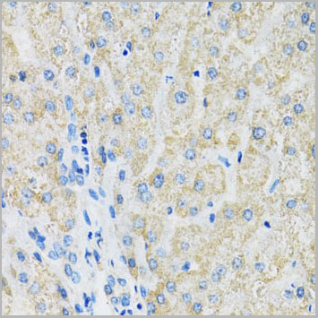
 IHC (Immunohistochemistry)
(DAB staining on IHC-P; Samples: Rat Small intestine Tissue; Primary Ab: 20ug/ml Rabbit Anti-Rat AT Antibody Second Ab: 2ug/mL HRP-Linked Caprine Anti-Rabbit IgG Polyclonal Antibody (Immunohistchemistry))
IHC (Immunohistochemistry)
(DAB staining on IHC-P; Samples: Rat Small intestine Tissue; Primary Ab: 20ug/ml Rabbit Anti-Rat AT Antibody Second Ab: 2ug/mL HRP-Linked Caprine Anti-Rabbit IgG Polyclonal Antibody (Immunohistchemistry))
Antithrombin, Polyclonal Antibody (Cat# AAA21014)
Full Name
Antithrombin (AT) Polyclonal Antibody
Reactivity
Rat
Applications
Westen Blot, Immunohistochemistry, Immunocytochemistry, Immunoprecipitation
Purity
Antigen-specific affinity chromatography followed by Protein A affinity chromatography
Pricing
Adenovirus, Hexon, Polyclonal Antibody (Cat# AAA14723)
Full Name
Adenovirus, Hexon
Applications
Immunohistochemistry, Immunofluorescence, Western Blot
Purity
Purified
Pricing
Wnt3 alpha, Polyclonal Antibody (Cat# AAA14338)
Full Name
Wnt3 alpha antibody
Gene Names
WNT3A; WNT3; WNT-3A
Reactivity
Human Wnt3
Applications
Western Blot
Pricing
PPM1B, Monoclonal Antibody (Cat# AAA24326)
Full Name
PPM1B (Protein Phosphatase 1B, PP2C-beta, Protein Phosphatase 2C Isoform beta, PP2CB, MGC21657) (AP)
Gene Names
PPM1B; PP2CB; PP2CBETA; PP2C-beta; PPC2BETAX; PP2C-beta-X
Reactivity
Human
Applications
Immunoprecipitation, Western Blot
Purity
Purified by Protein A Affinity Chromatography.
Pricing
Homocysteine, Conjugated, Polyclonal Antibody (Cat# AAA14713)
Full Name
Homocysteine, Conjugated
Applications
Immunocytochemistry, Gel Super Shift Assay
Purity
Purified
Antiserum previously preabsobed on protein carriers, and purified
Antiserum previously preabsobed on protein carriers, and purified
Pricing
Survivin, Monoclonal Antibody (Cat# AAA24380)
Full Name
Survivin (Apoptosis Inhibitor 4, API4, Apoptosis Inhibitor Survivin, Baculoviral IAP Repeat Containing Protein 5, BIRC5, EPR-1, IAP4) (AP)
Gene Names
BIRC5; API4; EPR-1
Reactivity
Human
Applications
Immunoprecipitation, Western Blot
Purity
Purified by Protein A Affinity Chromatography.
Pricing
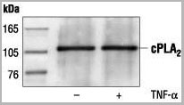
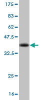
 WB (Western Blot)
(Western blot analysis of extracts from HeLa, C6 and NIH/3T3 cells, using AAA14717.)
WB (Western Blot)
(Western blot analysis of extracts from HeLa, C6 and NIH/3T3 cells, using AAA14717.)
c-Phospholipase A2, Polyclonal Antibody (Cat# AAA14717)
Full Name
c-Phospholipase A2 (Cytosolic PLA2, cPLA2)
Reactivity
Human
Applications
Western Blot, Immunoprecipitation
Purity
Purified by Protein A and peptide affinity chromatography
Pricing
Papillomavirus, Polyclonal Antibody (Cat# AAA13466)
Full Name
Papillomavirus
Reactivity
Crossreactive with HPV types 16 & 11 (other types not tested)
Applications
Immunohistochemistry
Pricing
RAB7B, Monoclonal Antibody (Cat# AAA24333)
Full Name
RAB7B (Ras-related Protein Rab-7b, RAB7) (AP)
Gene Names
RAB7B; RAB7
Reactivity
Human
Applications
Immunoprecipitation, Western Blot
Purity
Purified by Protein A Affinity Chromatography.
Pricing
ACSL5, Monoclonal Antibody (Cat# AAA24121)
Full Name
ACSL5 (Long-chain-fatty-acid-CoA Ligase 5, Long-chain acyl-CoA Synthetase 5, LACS 5, ACS5, FACL5, UNQ633/PRO1250) (AP)
Gene Names
ACSL5; ACS2; ACS5; FACL5
Reactivity
Human
Applications
Immunohistochemistry, Immunoprecipitation, Western Blot
Purity
Purified by Protein A Affinity Chromatography.
Pricing
Gelatin, Skin, Polyclonal Antibody (Cat# AAA14724)
Full Name
Rabbit anti-Porcine Gelatin, Skin
Reactivity
Porcine Gelatine , Skin
Applications
ELISA
Purity
Serum
Pricing
Streptococcus Group A, Polyclonal Antibody (Cat# AAA14322)
Full Name
Streptococcus Group A antibody
Reactivity
Streptococci Group A 100%
No other cross reactants detected.
No other cross reactants detected.
Purity
>95% by SDS-PAGE
Pricing
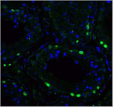
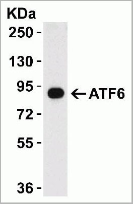
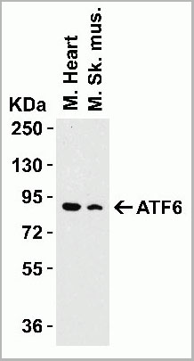
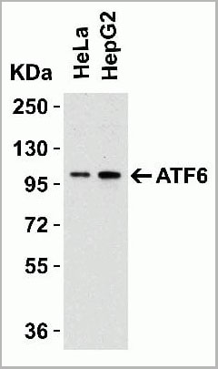
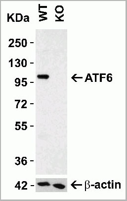
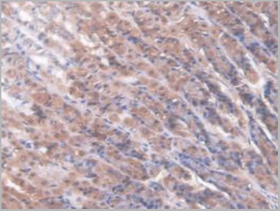
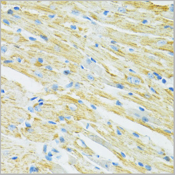
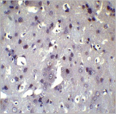 IHC (Immunohistochemistry)
(Figure 8 Immunohistochemistry Validation of ATF6 in Rat Brain Tissue Immunohistochemical analysis of paraffin-embedded Rat brain tissue using anti-ATF6 antibody (AAA10937) at 5 ug/ml. Tissue was fixed with formaldehyde and blocked with 10% serum for 1 h at RT; antigen retrieval was by heat mediation with a citrate buffer (pH6). Samples were incubated with primary antibody overnight at 4°C. A goat anti-rabbit IgG H&L (HRP) at 1/250 was used as secondary. Counter stained with Hematoxylin.)
IHC (Immunohistochemistry)
(Figure 8 Immunohistochemistry Validation of ATF6 in Rat Brain Tissue Immunohistochemical analysis of paraffin-embedded Rat brain tissue using anti-ATF6 antibody (AAA10937) at 5 ug/ml. Tissue was fixed with formaldehyde and blocked with 10% serum for 1 h at RT; antigen retrieval was by heat mediation with a citrate buffer (pH6). Samples were incubated with primary antibody overnight at 4°C. A goat anti-rabbit IgG H&L (HRP) at 1/250 was used as secondary. Counter stained with Hematoxylin.)
ATF6, Polyclonal Antibody (Cat# AAA10937)
Full Name
ATF6 Antibody
Gene Names
ATF6; ATF6A
Reactivity
Human, Mouse, Rat
Applications
Western Blot, Immunohistochemistry, Immunofluorescence
Purity
ATF6 Antibody is affinity chromatography purified via peptide column.
Pricing
C9 Tag, Polyclonal Antibody (Cat# AAA13688)
Full Name
Anti-C9 Tag Polyclonal Antibody, rabbit IgG
Applications
Western Blot
Purity
Protein A immunoaffinity chromatography
Pricing
SMAD3, Monoclonal Antibody (Cat# AAA24362)
Full Name
SMAD3 (Mothers Against Decapentaplegic Homolog 3, SMAD 3, Mothers Against DPP Homolog 3, MAD Homolog 3, Mad3, hMAD-3, SMAD Family Member 3, JV15-2, hSMAD3, MADH3, DKFZp586N0721, DKFZp686J10186, HSPC193, HsT17436, MGC60396) (AP)
Gene Names
SMAD3; LDS3; LDS1C; MADH3; JV15-2; HSPC193; HsT17436
Reactivity
Human, Rat
Applications
Immunohistochemistry, Western Blot
Purity
Purified by Protein A Affinity Chromatography.
Pricing
Clostridium botulinum Type A Neurotoxin Complex, Antibody (Cat# AAA13408)
Full Name
Rabbit anti C. botulinum Neurotoxin Type A
Applications
Dot Blot, Western Blot
Purity
Protein G Sepharose Chromatography
Pricing
ANXA5, Monoclonal Antibody (Cat# AAA24433)
Full Name
ANXA5 (Annexin A5, Annexin-5, Annexin V, Lipocortin V, Endonexin II, Calphobindin I, CBP-I, Placental Anticoagulant Protein I, PAP-I, Placental Anticoagulant Protein 4, PP4, Thromboplastin Inhibitor, Vascular Anticoagulant-alpha, VAC-alpha, Anchorin CII,
Gene Names
ANXA5; PP4; ANX5; ENX2; RPRGL3; HEL-S-7
Reactivity
Human
Applications
Immunofluorescence, Immunohistochemistry, Immunoprecipitation, Western Blot
Purity
Purified by Protein A Affinity Chromatography.
Pricing
SDMA, Polyclonal Antibody (Cat# AAA14719)
Full Name
SDMA (Symmetric Dimethylarginine)
Applications
Western Blot, Immunohistochemistry, Immunocytochemistry, Radioimmunoassay
Purity
Serum
Pricing
CAMKK2, Monoclonal Antibody (Cat# AAA24450)
Full Name
CAMKK2 (Calcium/Calmodulin-dependent Protein Kinase Kinase 2, CaM-kinase Kinase 2, CaM-KK 2, CaMKK 2, Calcium/Calmodulin-dependent Protein Kinase Kinase beta, CaM-kinase Kinase beta, CaM-KK beta, CaMKK beta, CAMKKB, KIAA0787) APC
Gene Names
CAMKK2; CAMKK; CAMKKB
Reactivity
Human
Applications
Immunoprecipitation, Western Blot
Purity
Purified by Protein A Affinity Chromatography.
Pricing
SMAD4, Monoclonal Antibody (Cat# AAA24363)
Full Name
SMAD4 (Mothers Against Decapentaplegic Homolog 4, SMAD 4, Mothers Against DPP Homolog 4, Deletion Target In Pancreatic Carcinoma 4, MAD Homolog 4, SMAD Family Member 4, hSMAD4, DPC4, MADH4) (AP)
Gene Names
SMAD4; JIP; DPC4; MADH4; MYHRS
Reactivity
Human
Applications
Western Blot
Purity
Purified by Protein A Affinity Chromatography.
Pricing
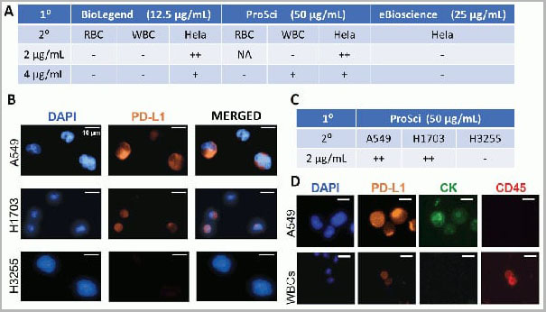
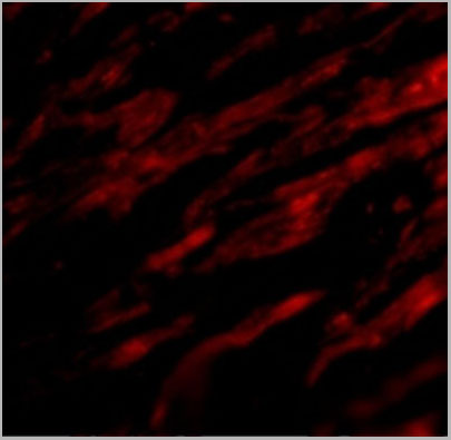
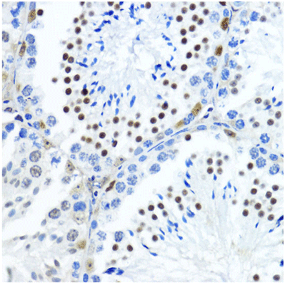
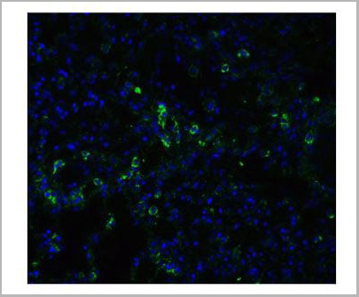
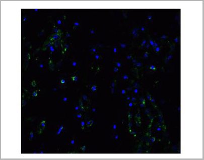
 IF (Immunofluorescence)
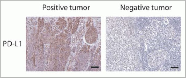
(Figure 12 Immunohistochemistry Validation of PD-L1in Human Tumors (Gadiot et al., 2011)Immunohistochemical analysis of patient tumors labeling PD-L1 with anti-PD-L1 antibodies (AAA10941). Several anti-PD-L1 antibodies were tested for staining, “Only 1 antibody gave no background staining and was competitively blocked by the addition of PD-L1Fc protein (AAA10941)”.)
IF (Immunofluorescence)
(Figure 12 Immunohistochemistry Validation of PD-L1in Human Tumors (Gadiot et al., 2011)Immunohistochemical analysis of patient tumors labeling PD-L1 with anti-PD-L1 antibodies (AAA10941). Several anti-PD-L1 antibodies were tested for staining, “Only 1 antibody gave no background staining and was competitively blocked by the addition of PD-L1Fc protein (AAA10941)”.)
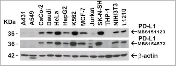
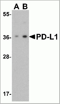
PDL-1, Polyclonal Antibody (Cat# AAA10941)
Full Name
PDL-1 Antibody
Gene Names
CD274; B7-H; B7H1; PDL1; PD-L1; PDCD1L1; PDCD1LG1
Applications
Western Blot, Immunohistochemistry, Immunofluorescence, Flow Cytometry
Purity
PD-L1 Antibody is affinity chromatography purified via peptide column.
Pricing
Myostatin Propeptide, Polyclonal Antibody (Cat# AAA14715)
Full Name
Myostatin Propeptide
Reactivity
Human
Applications
Western Blot
Purity
Affinity Purified
Purified by immunoaffinity chromatography.
Purified by immunoaffinity chromatography.
Pricing
CENPM, Monoclonal Antibody (Cat# AAA24446)
Full Name
CENPM (C22orf18, ICEN39, PANE1, Centromere Protein M, Interphase Centromere Complex Protein 39, Proliferation-associated Nuclear Element Protein 1, MGC861, PANE1, BK250D10.2) APC
Gene Names
CENPM; PANE1; CENP-M; C22orf18
Reactivity
Human
Applications
Immunofluorescence, Immunoprecipitation, Western Blot
Purity
Purified by Protein A Affinity Chromatography.
Pricing
SHC, Monoclonal Antibody (Cat# AAA24358)
Full Name
SHC (SHC-transforming Protein 1, SH2 Domain Protein C1, SHC1, SHC-transforming Protein 3, SHC-transforming Protein A, SHCA, Src Homology 2 Domain-containing-transforming Protein C1, FLJ26504) (AP)
Gene Names
SHC1; SHC; SHCA
Reactivity
Human
Applications
Immunohistochemistry, Western Blot
Purity
Purified by Protein A Affinity Chromatography.
Pricing
ABL2, Monoclonal Antibody (Cat# AAA24411)
Full Name
ABL2 (Tyrosine-protein Kinase ABL2, Abelson Murine Leukemia Viral Oncogene Homolog 2, Abelson-related Gene Protein, Tyrosine Kinase ARG, ABLL, ARG) APC
Gene Names
ABL2; ARG; ABLL
Reactivity
Human
Applications
Western Blot
Purity
Purified by Protein A Affinity Chromatography.
Pricing
CBS, Monoclonal Antibody (Cat# AAA24452)
Full Name
CBS (Cystathionine beta-synthase, Serine Sulfhydrase, Beta-thionase) APC
Gene Names
CBS; HIP4
Reactivity
Human
Applications
Immunohistochemistry, Immunoprecipitation, Western Blot
Purity
Purified by Protein A Affinity Chromatography.
Pricing
ACY1, Monoclonal Antibody (Cat# AAA24124)
Full Name
ACY1 (Aminoacylase 1, Aminoacylase-1, ACY-1, N-acyl-L-amino-acid Amidohydrolase) (AP)
Gene Names
ACY1; ACY-1; ACY1D; HEL-S-5
Reactivity
Human
Applications
Immunohistochemistry, Immunoprecipitation, Western Blot
Purity
Purified by Protein A Affinity Chromatography.
Pricing
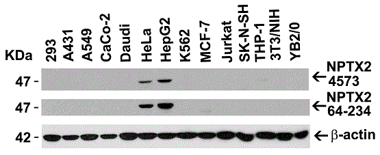
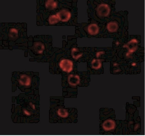
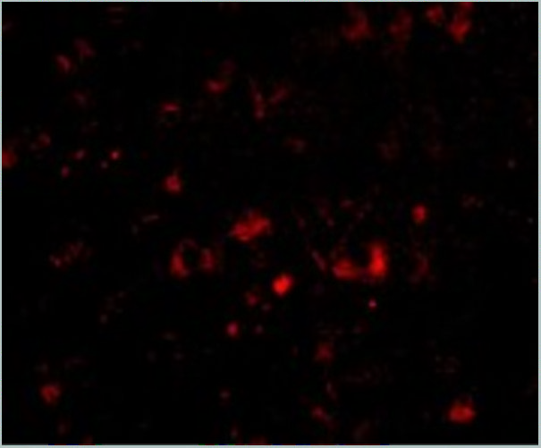
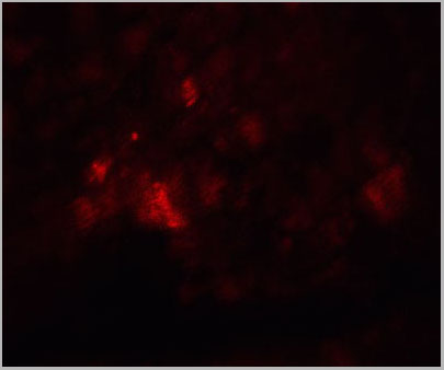
 IF (Immunofluorescence)
(Immunofluorescent analysis of 4% paraformaldehyde-fixed mouse brain issue labeling NPTX2 with AAA10933 at 20ug/mL, followed by goat anti-rabbit IgG secondaryantibody at 1/500 dilution (red) and DAPI staining (blue).)
IF (Immunofluorescence)
(Immunofluorescent analysis of 4% paraformaldehyde-fixed mouse brain issue labeling NPTX2 with AAA10933 at 20ug/mL, followed by goat anti-rabbit IgG secondaryantibody at 1/500 dilution (red) and DAPI staining (blue).)
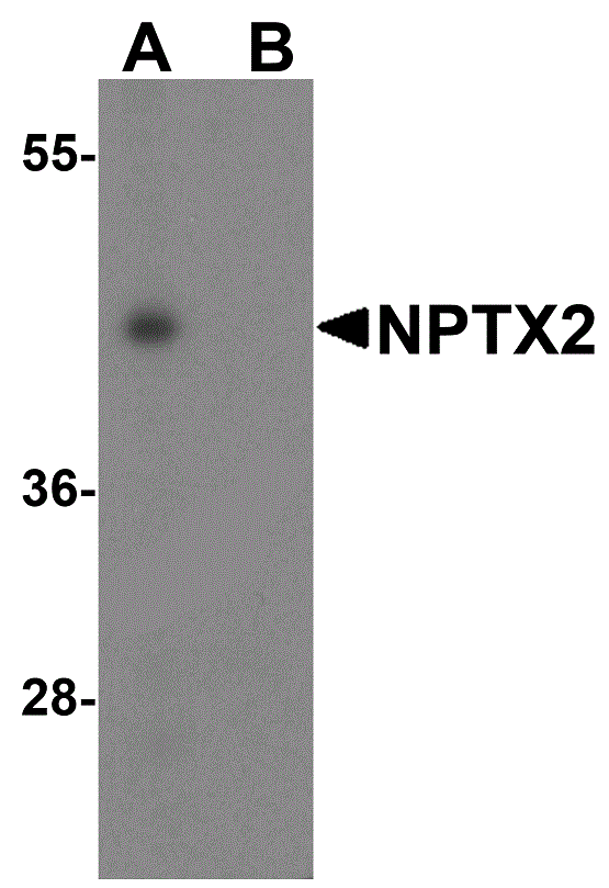
NPTX2, Polyclonal Antibody (Cat# AAA10933)
Full Name
NPTX2 Antibody
Gene Names
NPTX2; NP2; NARP; NP-II
Reactivity
Human, Mouse, Rat
Applications
Western Blot, Immunohistochemistry, Immunofluorescence
Purity
NPTX2 Antibody is affinity chromatography purified via peptide column.
Pricing
CA1, Monoclonal Antibody (Cat# AAA24447)
Full Name
CA1 (Carbonic Anhydrase 1, Carbonic Anhydrase I, CA-I, Carbonate Dehydratase I, Carbonic Anhydrase B, CAB) APC
Gene Names
CA1; CAB; CA-I; Car1; HEL-S-11
Reactivity
Human
Applications
Immunoprecipitation, Western Blot
Purity
Purified
Pricing
SDCBP, Monoclonal Antibody (Cat# AAA24355)
Full Name
SDCBP (Syndecan-binding Protein 1, Melanoma Differentiation-associated Protein 9, MDA9, MDA-9, Pro-TGF-alpha Cytoplasmic Domain-interacting Protein 18, TACIP18, Scaffold Protein Pbp1, SYCL, Syntenin-1, ST1) (AP)
Gene Names
SDCBP; ST1; MDA9; SYCL; MDA-9; TACIP18
Reactivity
Human
Applications
Immunohistochemistry, Immunoprecipitation, Western Blot
Purity
Purified by Protein A Affinity Chromatography.
Pricing
CD142, Monoclonal Antibody (Cat# AAA24455)
Full Name
CD142 (CD142 Antigen, Coagulation Factor III, F3, Tissue Factor, TF, TFA, Thromboplastin) APC
Gene Names
F3; TF; TFA; CD142
Reactivity
Human
Applications
Immunoprecipitation, Western Blot
Purity
Purified by Protein A Affinity Chromatography.
Pricing
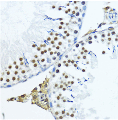
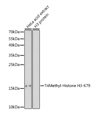
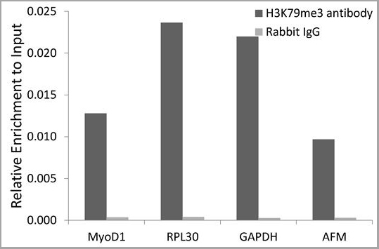
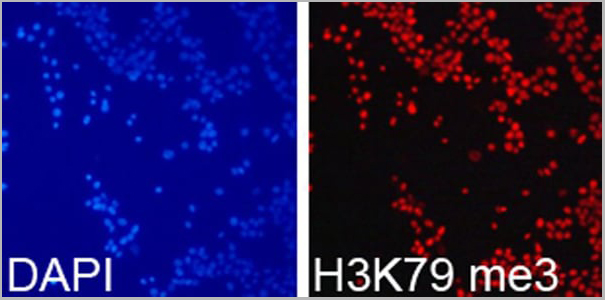
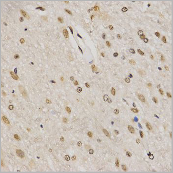

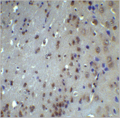
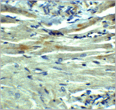
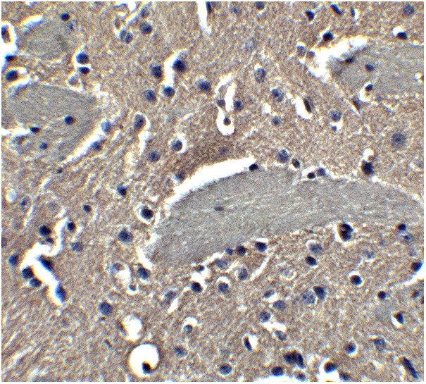
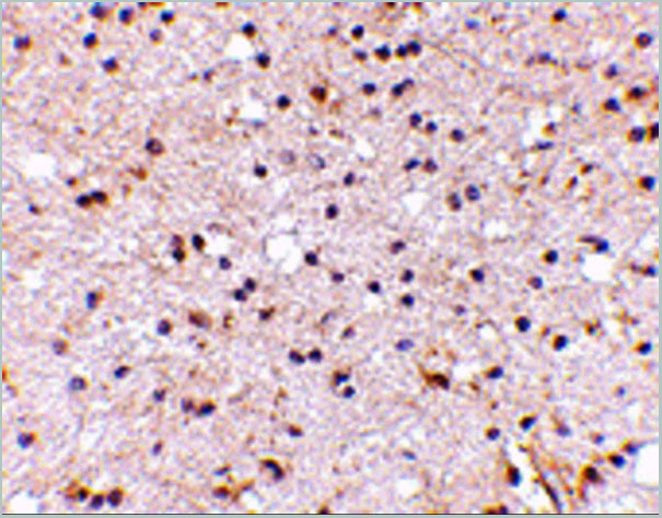
 IHC (Immunohistochemistry)
(Immunohistochemistry of paraffin-embedded mouse testis using TriMethyl-Histone H3-K79 antibody (AAA10656) at dilution of 1:200 (40x lens).)
IHC (Immunohistochemistry)
(Immunohistochemistry of paraffin-embedded mouse testis using TriMethyl-Histone H3-K79 antibody (AAA10656) at dilution of 1:200 (40x lens).)
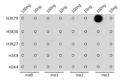
H3K79me3, Antibody (Cat# AAA10656)
Full Name
Histone H3K79me3 Polyclonal Antibody
Gene Names
HIST3H3; H3t; H3.4; H3/g; H3FT
Applications
Western Blot, Immunohistochemistry, Immunofluorescence, Immunoprecipitation, Chromatin Immunoprecipitation, Chromatin Immunoprecipitation
Purity
Affinity Purification
Pricing
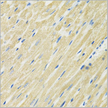
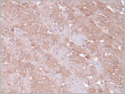
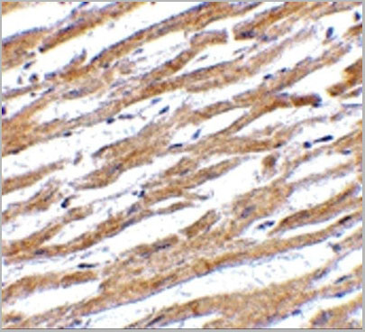
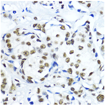
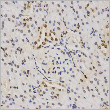
 IHC (Immunohistochemistry)
(Immunohistochemistry of paraffin-embedded mouse heart using SLC19A1 antibody at dilution of 1:100 (40x lens).)
IHC (Immunohistochemistry)
(Immunohistochemistry of paraffin-embedded mouse heart using SLC19A1 antibody at dilution of 1:100 (40x lens).)
SLC19A1, Polyclonal Antibody (Cat# AAA28277)
Full Name
SLC19A1 Polyclonal Antibody
Gene Names
SLC19A1; CHMD; FOLT; IFC1; REFC; RFC1
Applications
Western Blot
Purity
Affinity Purification
Pricing
Fetoprotein, Alpha, Monoclonal Antibody (Cat# AAA24421)
Full Name
Fetoprotein, Alpha (AFP, Alpha1-fetoprotein, Alpha-fetoglobulin, Alpha-fetoprotein precursor, FETA, HPAFP) APC
Gene Names
AFP; AFPD; FETA; HPAFP
Reactivity
Human
Applications
Immunofluorescence, Immunoprecipitation, Western Blot
Purity
Purified by Protein A Affinity Chromatography.
Pricing
RICTOR, Monoclonal Antibody (Cat# AAA24338)
Full Name
RICTOR (Rapamycin-insensitive Companion of mTOR, AVO3 Homolog, hAVO3, KIAA1999, mAVO3, PIA) (AP)
Gene Names
RICTOR; PIA; AVO3; hAVO3
Reactivity
Human
Applications
Immunohistochemistry, Immunoprecipitation, Western Blot
Purity
Purified by Protein A Affinity Chromatography.
Pricing
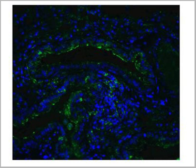
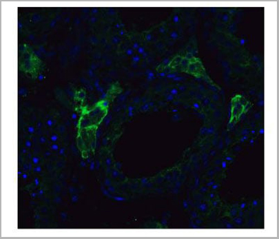
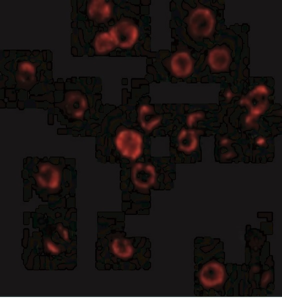
 IF (Immunofluorescence)
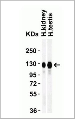
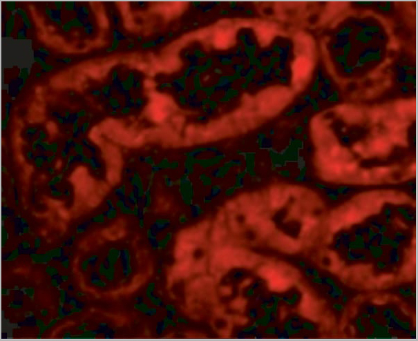
(Figure 11 Immunofluorescence Validation of ACE2 In Caco2 CellsImmunofluorescent analysis of 4% paraformaldehyde-fixed Caco2 cells labeling ACE2 with AAA10945 at 20 ug/mL, followed by goat anti-rabbit IgG secondary antibody at 1/500 dilution (green) and DAPI staining (blue). Imageshowing membrane staining on Caco2 cells.)
IF (Immunofluorescence)
(Figure 11 Immunofluorescence Validation of ACE2 In Caco2 CellsImmunofluorescent analysis of 4% paraformaldehyde-fixed Caco2 cells labeling ACE2 with AAA10945 at 20 ug/mL, followed by goat anti-rabbit IgG secondary antibody at 1/500 dilution (green) and DAPI staining (blue). Imageshowing membrane staining on Caco2 cells.)
ACE2, Polyclonal Antibody (Cat# AAA10945)
Full Name
ACE2 Antibody
Gene Names
ACE2; ACEH
Reactivity
Human, Mouse, Rat
Applications
Western Blot, Immunohistochemistry, Immunofluorescence
Purity
ACE2 Antibody is affinity chromatography purified via peptide column.
Pricing
CA1, Monoclonal Antibody (Cat# AAA24152)
Full Name
CA1 (Carbonic Anhydrase 1, Carbonic Anhydrase I, CA-I, Carbonate Dehydratase I, Carbonic Anhydrase B, CAB) (AP)
Gene Names
CA1; CAB; CA-I; Car1; HEL-S-11
Reactivity
Human
Applications
Immunoprecipitation, Western Blot
Purity
Purified
Pricing
PML, Monoclonal Antibody (Cat# AAA24323)
Full Name
PML (Probable Transcription Factor PML, RING Finger Protein 71, Tripartite Motif-containing Protein 19, PML, MYL, RNF71, TRIM19) (AP)
Gene Names
PML; MYL; RNF71; PP8675; TRIM19
Reactivity
Human, Rat
Applications
Western Blot
Purity
Purified by Protein A Affinity Chromatography.
Pricing
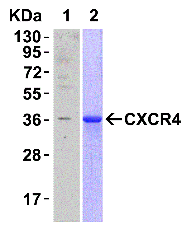

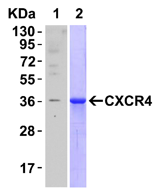
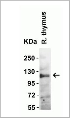
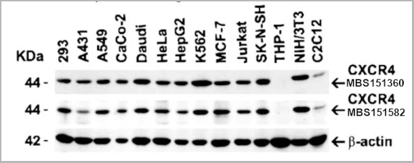
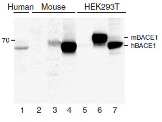
 WB (Western Blot)
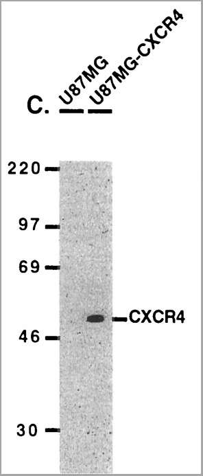
(Figure 9 WB Validation of CXCR4 in metastatic melanoma (Scala et al., 2006)CXCR4 protein was detected in the human metastatic melanoma cell lines and human melanoma cell line (colo38), but not in the human primary melanocytes (MPR1) with anti-CXCR4 antibodies.)
WB (Western Blot)
(Figure 9 WB Validation of CXCR4 in metastatic melanoma (Scala et al., 2006)CXCR4 protein was detected in the human metastatic melanoma cell lines and human melanoma cell line (colo38), but not in the human primary melanocytes (MPR1) with anti-CXCR4 antibodies.)
CXCR4, Polyclonal Antibody (Cat# AAA10962)
Full Name
CXCR4 Antibody
Gene Names
CXCR4; FB22; HM89; LAP3; LCR1; NPYR; WHIM; CD184; LAP-3; LESTR; NPY3R; NPYRL; HSY3RR; NPYY3R; D2S201E
Reactivity
Human, Mouse
Applications
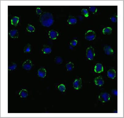
Western Blot, Immunofluorescence
Purity
CXCR4 Antibody is Protein A purified.
Pricing
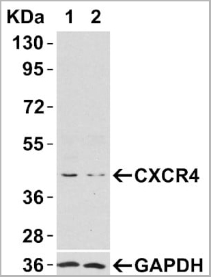
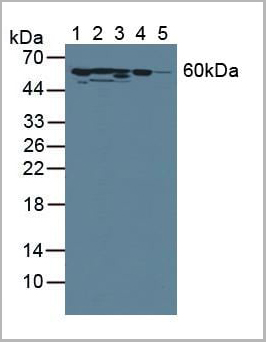
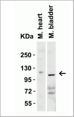
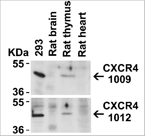
 WB (Western Blot)
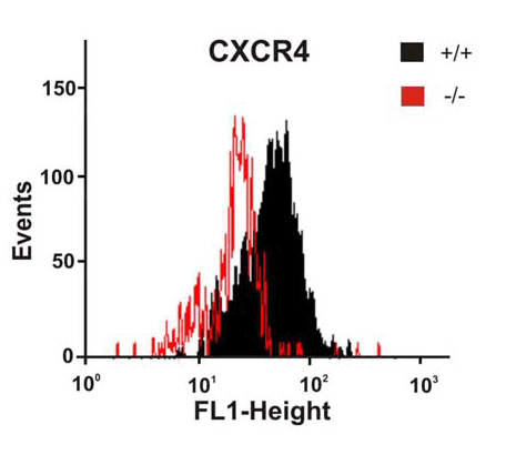
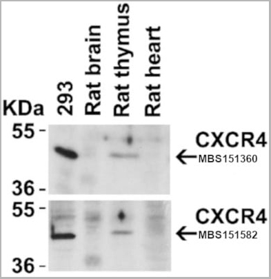
(Figure 4 Animal Species ReactivityLoading: Lysates/proteins at 20 μg per lane. Antibodies: 1009 (2 μg/mL) or 1012 (2 μg/mL). 1 h incubation at RT in 5% NFDM/TBST. Secondary: Goat anti-rabbit IgG HRP conjugate at 1:10000 dilution.)
WB (Western Blot)
(Figure 4 Animal Species ReactivityLoading: Lysates/proteins at 20 μg per lane. Antibodies: 1009 (2 μg/mL) or 1012 (2 μg/mL). 1 h incubation at RT in 5% NFDM/TBST. Secondary: Goat anti-rabbit IgG HRP conjugate at 1:10000 dilution.)
CXCR4, Polyclonal Antibody (Cat# AAA10951)
Full Name
CXCR4 Antibody
Gene Names
CXCR4; FB22; HM89; LAP3; LCR1; NPYR; WHIM; CD184; LAP-3; LESTR; NPY3R; NPYRL; HSY3RR; NPYY3R; D2S201E
Reactivity
Human, Mouse, Rat
Applications
Western Blot, Immunocytochemistry, Immunoprecipitation, Immunofluorescence, Immunohistochemistry, Flow Cytometry
Purity
CXCR4 Antibody is affinity chromatography purified via peptide column.
Pricing
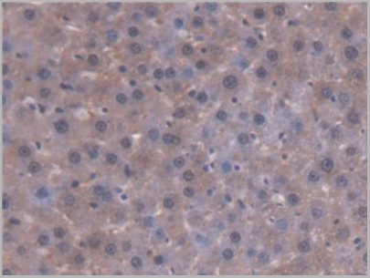
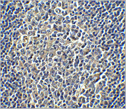
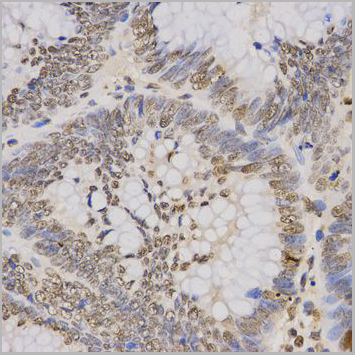
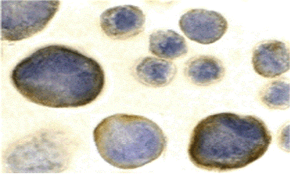
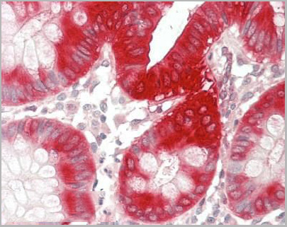 IHC (Immunohistochemistry-Paraffin)
(Human Colon: Formalin-Fixed, Paraffin-Embedded (FFPE))
IHC (Immunohistochemistry-Paraffin)
(Human Colon: Formalin-Fixed, Paraffin-Embedded (FFPE))
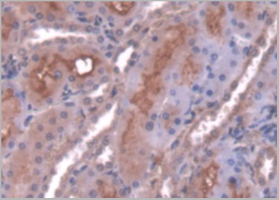
PTGS2 / COX2 / COX-2, Polyclonal Antibody (Cat# AAA12379)
Full Name
Anti-PTGS2 / COX2 / COX-2 Antibody (C-Terminus) IHC-plus
Gene Names
PTGS2; COX2; COX-2; PHS-2; PGG/HS; PGHS-2; hCox-2; GRIPGHS
Reactivity
Human, Mouse, Rat, Bovine, Pig, Sheep
Applications
Immunohistochemistry, Immunocytochemistry, Immunofluorescence, Western Blot
Purity
Immunoaffinity purified
Pricing

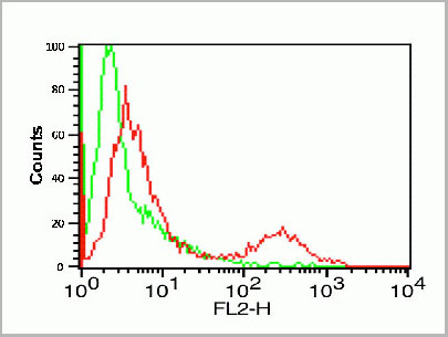
 IF (Immunofluorescence)
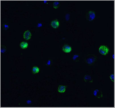
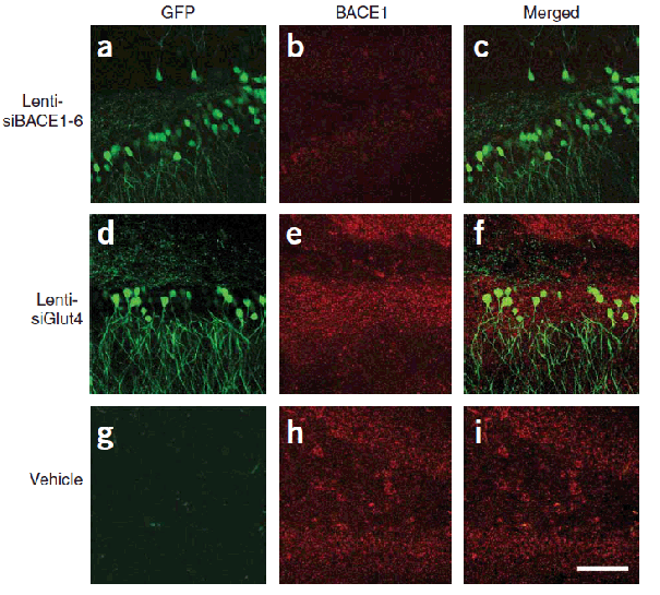
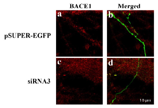
(Figure 10 KD Validation of BACE in DRG (Hyun, 2007)Decreased BACE1 expression in DRG following siRNA3 transfection. DRG neurons were transfected with 1 ug siRNA3 plasmid and incubated for 48 hours in 37°˚C. DRG neurons were stained for BACE1 us¬ing the Anti-BACE antibody (a,b) Neurons transfected with the control plas¬mid pSUPER-EGFP (green) did not display any changes in BACE1 expression (red). (c,d) DRG neurons transfected with siR¬NA3 displayed reduced BACE1 expression in the axon.)
IF (Immunofluorescence)
(Figure 10 KD Validation of BACE in DRG (Hyun, 2007)Decreased BACE1 expression in DRG following siRNA3 transfection. DRG neurons were transfected with 1 ug siRNA3 plasmid and incubated for 48 hours in 37°˚C. DRG neurons were stained for BACE1 us¬ing the Anti-BACE antibody (a,b) Neurons transfected with the control plas¬mid pSUPER-EGFP (green) did not display any changes in BACE1 expression (red). (c,d) DRG neurons transfected with siR¬NA3 displayed reduced BACE1 expression in the axon.)
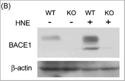
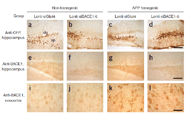
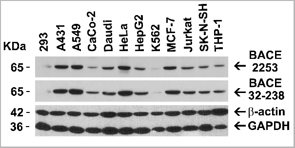
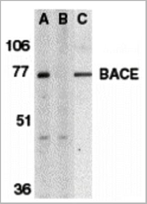
BACE, Polyclonal Antibody (Cat# AAA10918)
Full Name
BACE Antibody
Gene Names
BACE1; ASP2; BACE; HSPC104
Reactivity
Human, Mouse
Applications
Immunocytochemistry, Immunofluorescence, Immunohistochemistry, Western Blot
Purity
BACE Antibody is affinity chromatography purified via peptide column.
Pricing
Dopamine Receptor, D2R, Polyclonal Antibody (Cat# AAA14709)
Full Name
Dopamine Receptor, D2R
Gene Names
DRD2; D2R; D2DR
Reactivity
Rat
Applications
ELISA
Purity
Affinity Purified
Purified by immunoaffinity chromatography.
Purified by immunoaffinity chromatography.
Pricing