Filters
Clonality
Type
Reactivity
Gene Name
Isotype
Host
Application
Clone
8384 results for "Mouse" - showing 7450-7500
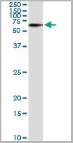
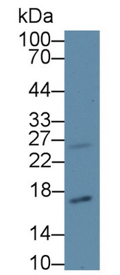
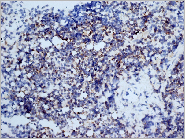
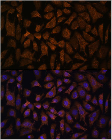
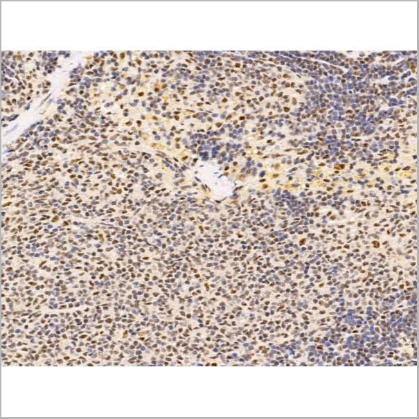
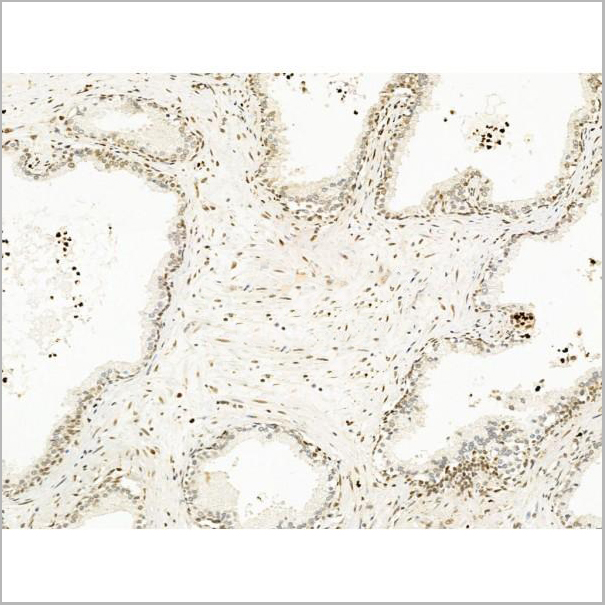
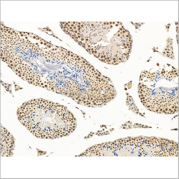
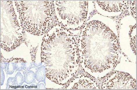
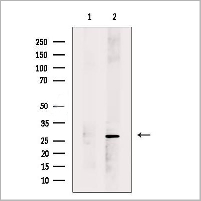
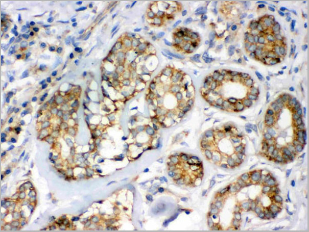
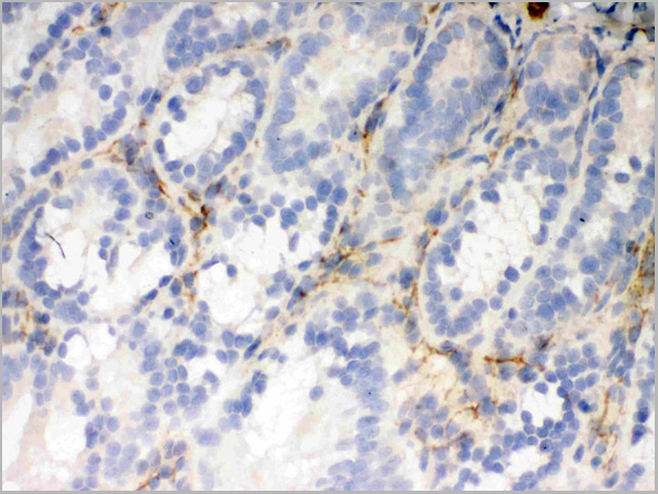
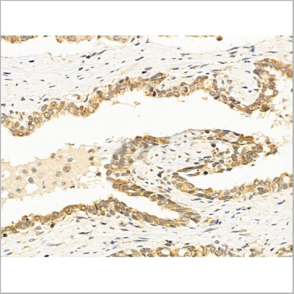
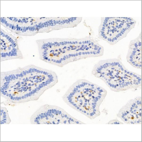
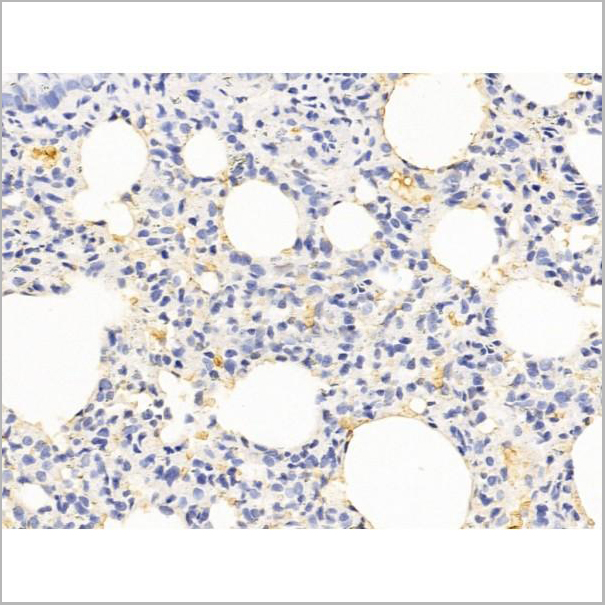
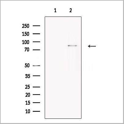
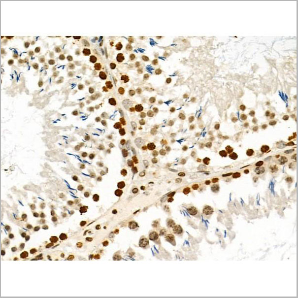
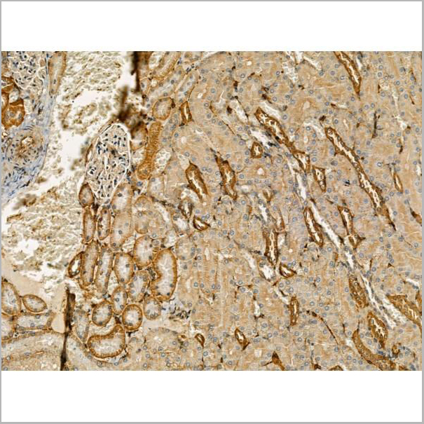
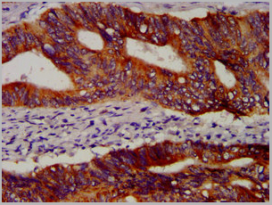
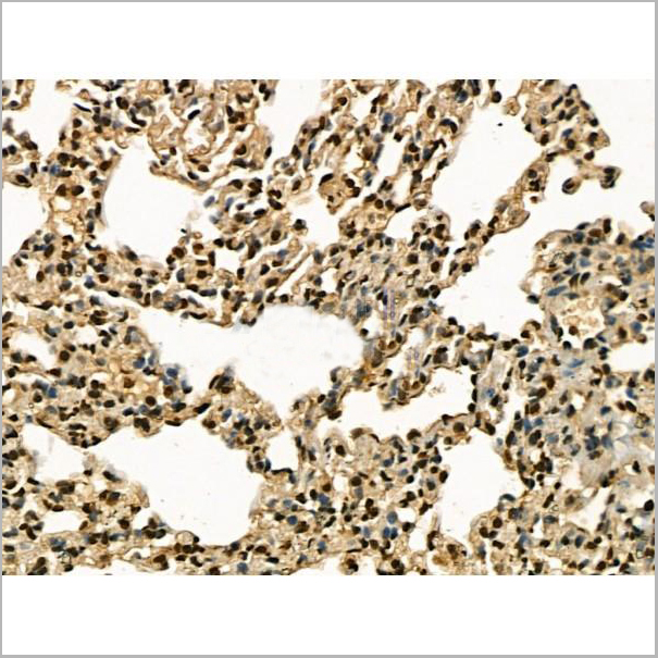
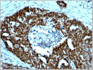
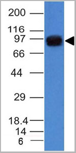
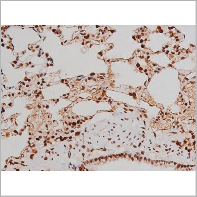
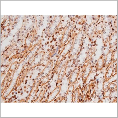
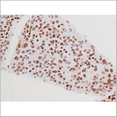
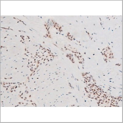
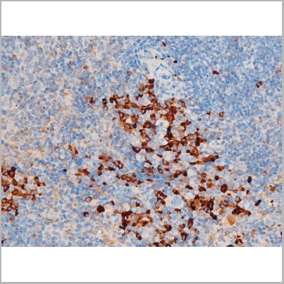
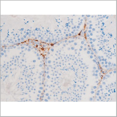
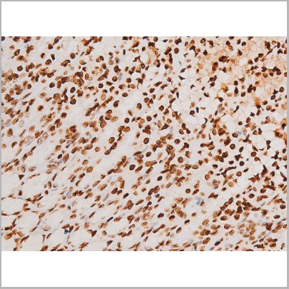
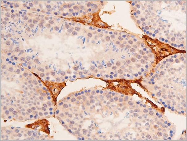
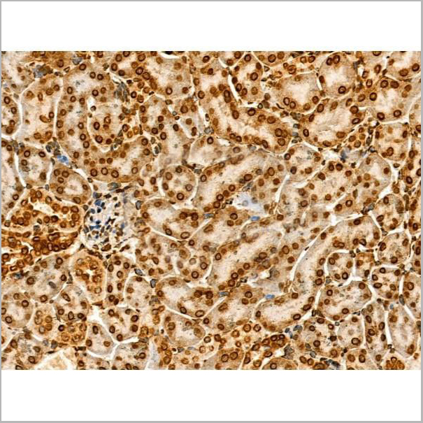
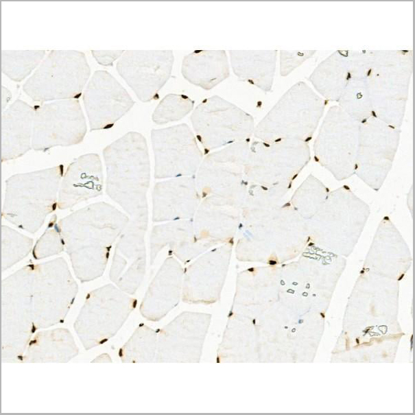
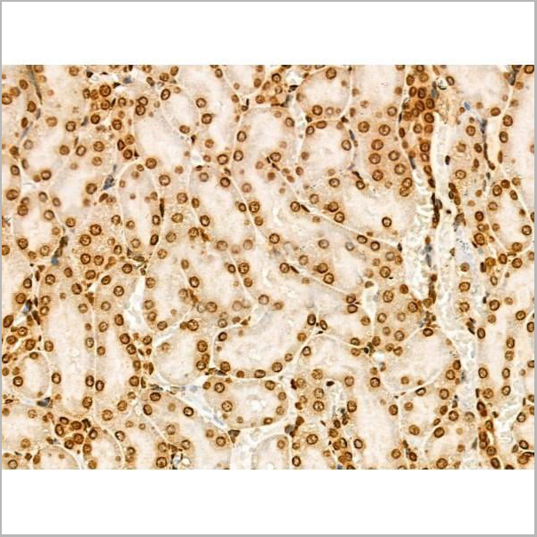
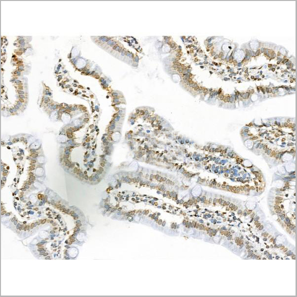
 IHC (Immunohistochemistry)
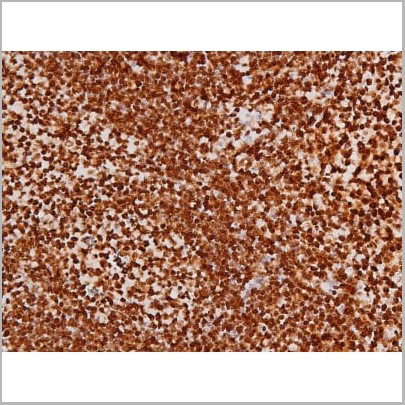
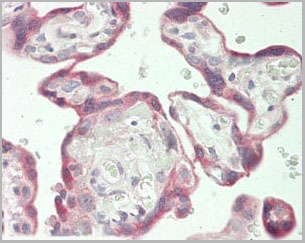
(Immunohistochemical analysis of paraffin-embedded Human Lung Carcinoma using MICU1 Monoclonal Antibody.)
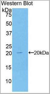
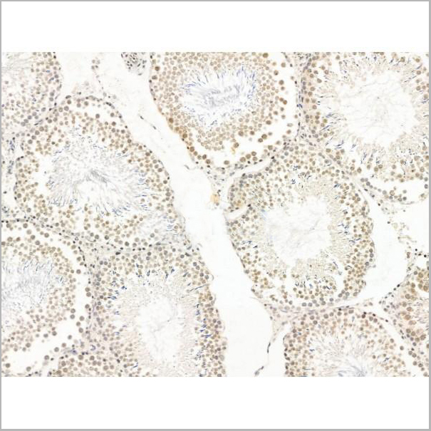
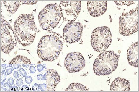
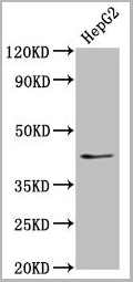
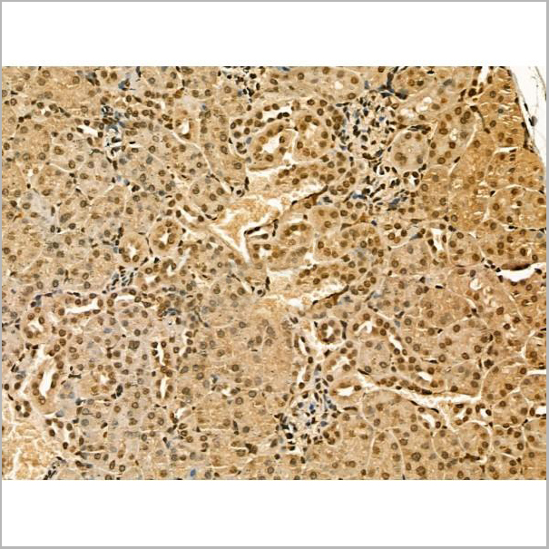
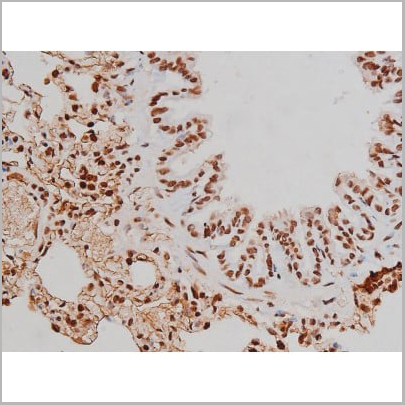
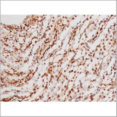
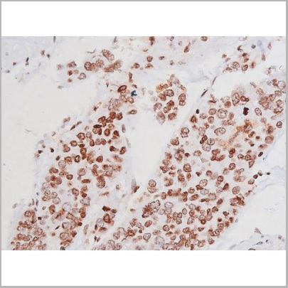
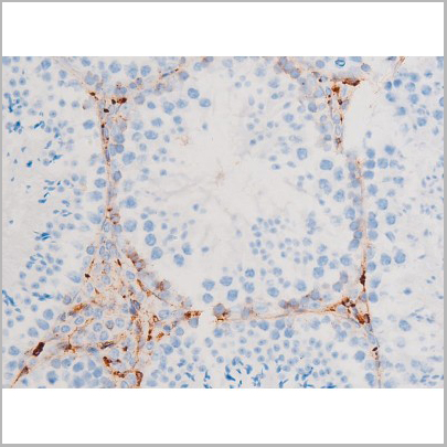
IHC (Immunohistochemistry)
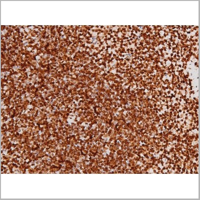
(Immunohistochemical analysis of paraffin-embedded Human Lung Carcinoma using MICU1 Monoclonal Antibody.)
MICU1, Monoclonal Antibody (Cat# AAA29640)
Full Name
MICU1 Monoclonal Antibody
Gene Names
MICU1; CALC; EFHA3; MPXPS; CBARA1
Reactivity
Human, Mouse, Rat
Applications
Western Blot, Immunohistochemistry
Purity
The antibody was affinity-purified from antiserum by affinity-chromatography
Pricing
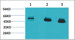
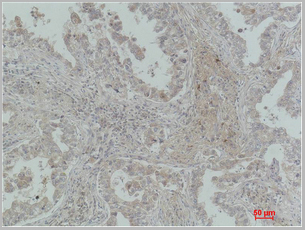
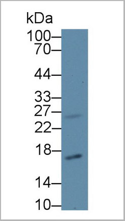
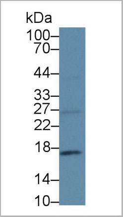
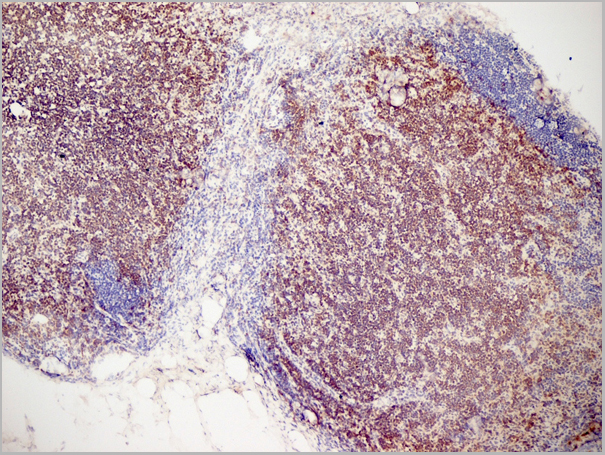
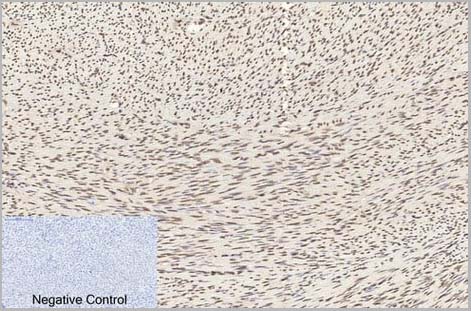
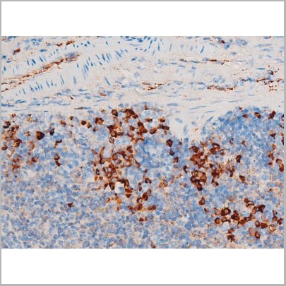
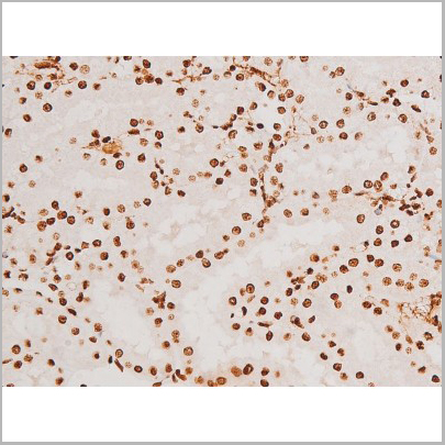
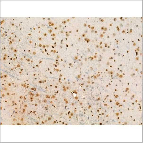
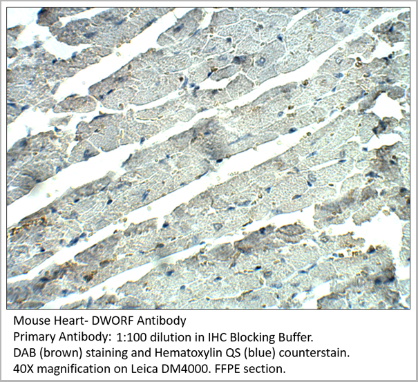 IHC (Immunohistochemistry)
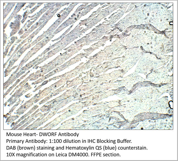
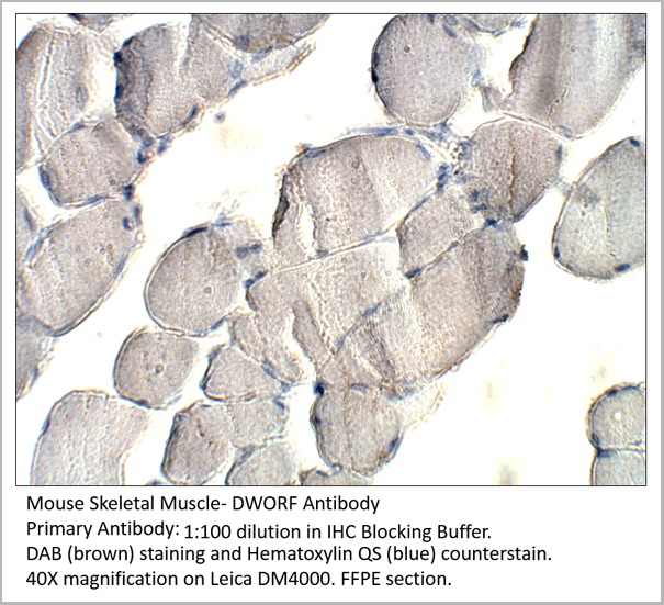
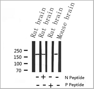
DWORF, Polyclonal Antibody (Cat# AAA14407)
Full Name
DWORF Antibody
Applications
Immunohistochemistry, Immunoprecipitation, Western Blot
Purity
Affinity Purified Immunoglobulins
Pricing
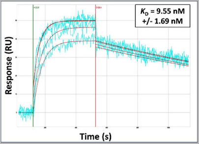 Application Data
(Surface Plasmon Resonance Kinetic Characterization of Polyclonal Antibody Affinity. Purified polyclonal antibodies were immobilized on a Protein A/G coated Carterra LSA sensor chip (PAGH200M) at concentrations of 5, and 50 ug/mL in duplicate. Antibodies on the surface were exposed to interaction with peptides sequentially via microfluidic controlled flow at 333nM peptide concentration for kinetic characterization of the binders for affinity and specificity, followed by curve fitting using the Kinetics software. Kd determinations for both concentrations were averaged and results and standard deviation are shown.)
Application Data
(Surface Plasmon Resonance Kinetic Characterization of Polyclonal Antibody Affinity. Purified polyclonal antibodies were immobilized on a Protein A/G coated Carterra LSA sensor chip (PAGH200M) at concentrations of 5, and 50 ug/mL in duplicate. Antibodies on the surface were exposed to interaction with peptides sequentially via microfluidic controlled flow at 333nM peptide concentration for kinetic characterization of the binders for affinity and specificity, followed by curve fitting using the Kinetics software. Kd determinations for both concentrations were averaged and results and standard deviation are shown.)
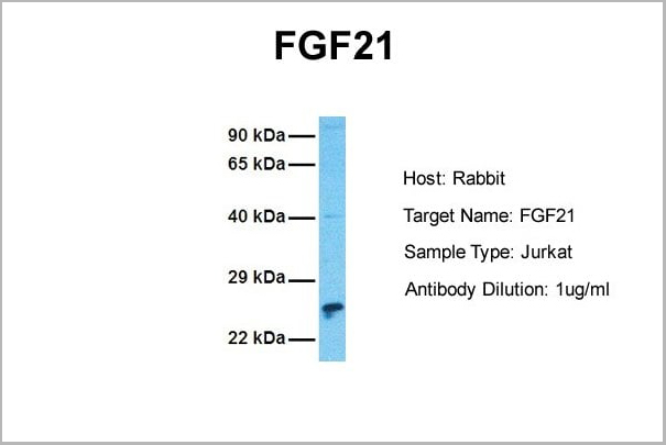
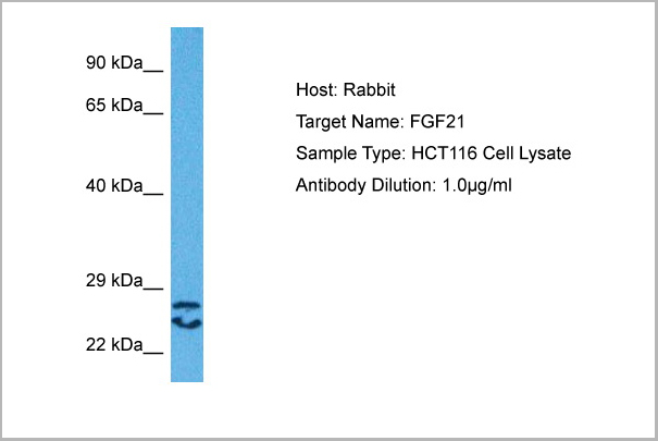
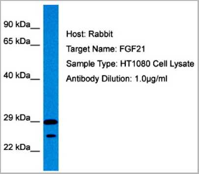
FGF21, Polyclonal Antibody (Cat# AAA23563)
Full Name
FGF21 antibody - N-terminal region
Reactivity
Tested Species Reactivity: Human
Predicted Species Reactivity: Human, Mouse, Rat, Cow, Dog, Guinea Pig, Horse, Rabbit
Predicted Species Reactivity: Human, Mouse, Rat, Cow, Dog, Guinea Pig, Horse, Rabbit
Applications
Immunohistochemistry, Western Blot
Purity
Affinity Purified
Pricing
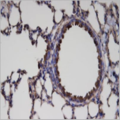
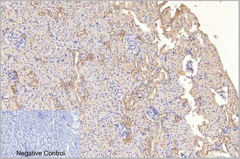
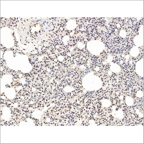
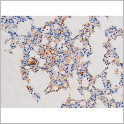
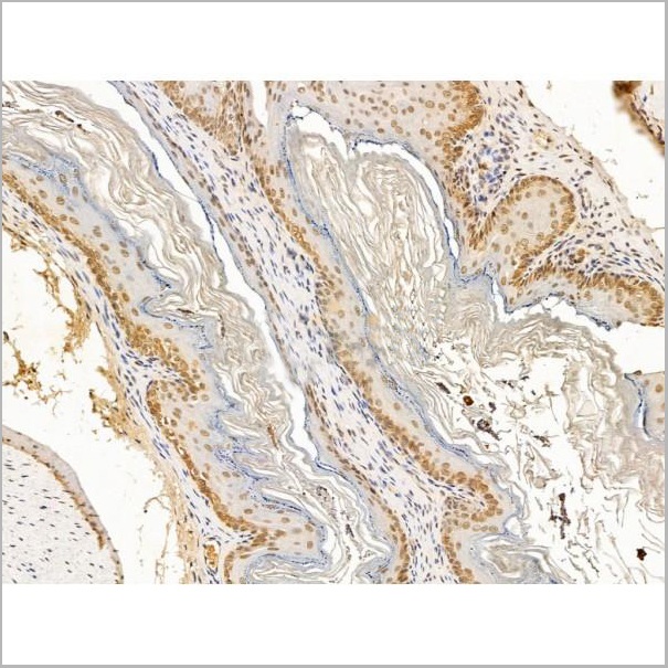
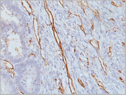
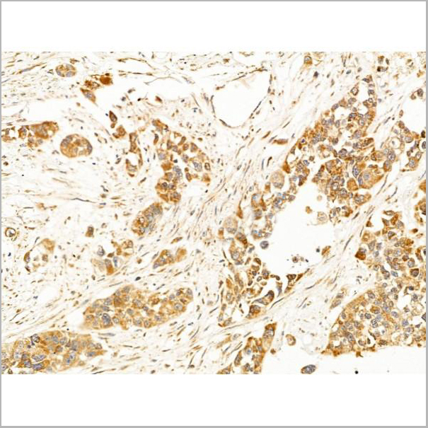
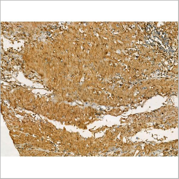
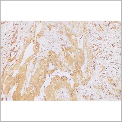
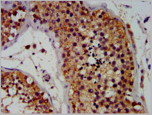
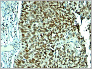
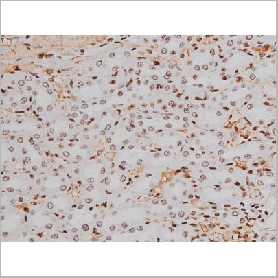
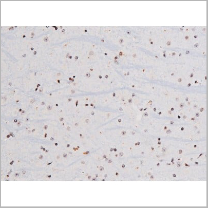
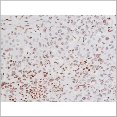
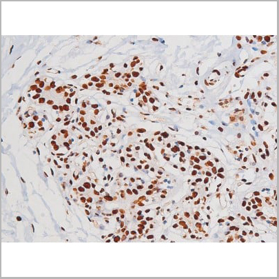
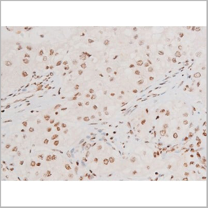
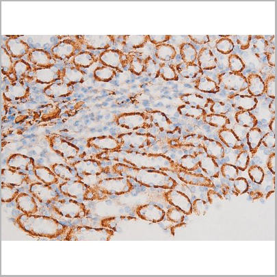
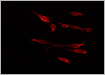
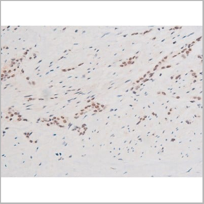
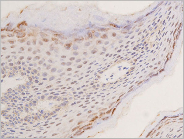
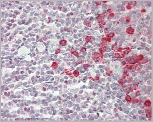
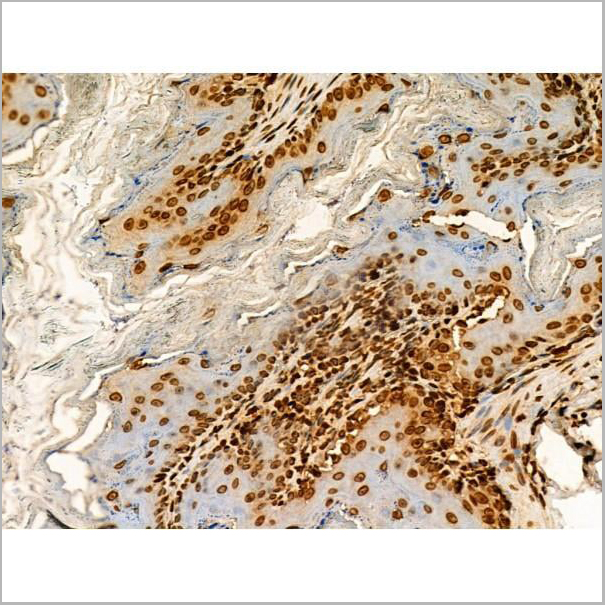
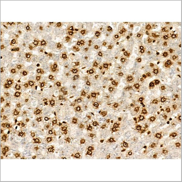
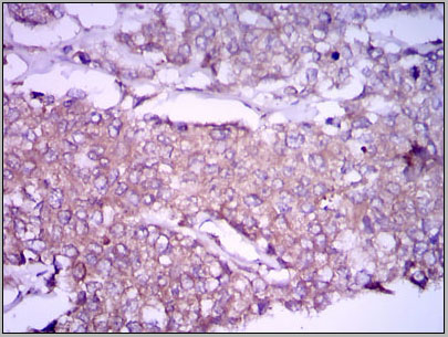 IHC (Immunohistchemistry)
(Immunohistochemical analysis of paraffin-embedded breast cancer tissues using GPC3 mouse mAb with DAB staining.)
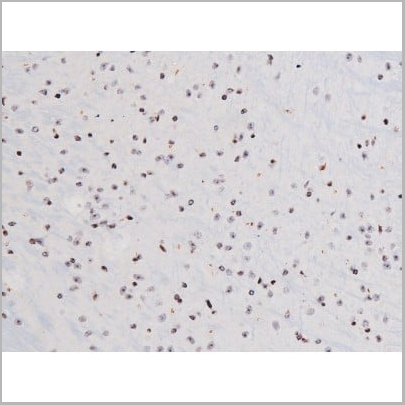
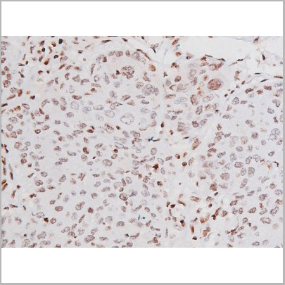
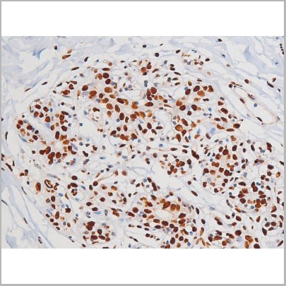
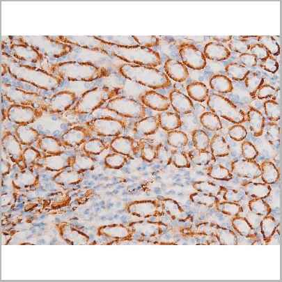
IHC (Immunohistchemistry)
(Immunohistochemical analysis of paraffin-embedded breast cancer tissues using GPC3 mouse mAb with DAB staining.)
GPC3, Monoclonal Antibody (Cat# AAA14118)
Full Name
Anti-GPC3 Mouse mAb
Gene Names
GPC3; SGB; DGSX; MXR7; SDYS; SGBS; OCI-5; SGBS1; GTR2-2
Reactivity
Human, Mouse
Applications
Immunohistochemistry, Immunocytochemistry, Immunofluorescence, Flow Cytometry
Purity
Affinity Purified
Pricing

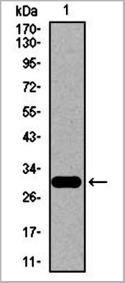
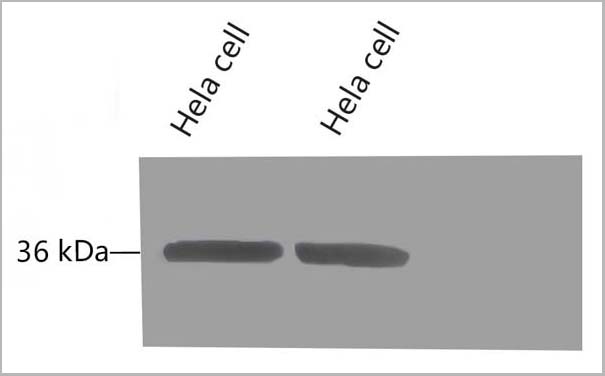
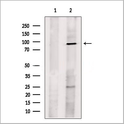
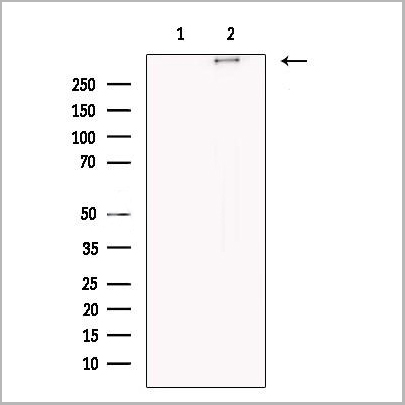
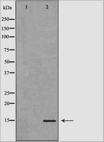
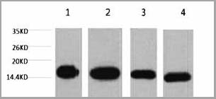
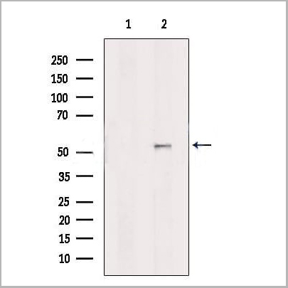
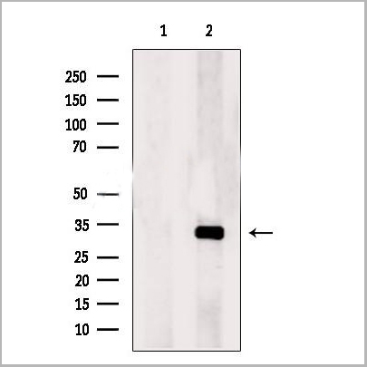
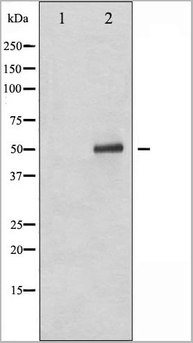
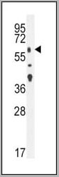
 WB (Western Blot)
(STK33 monoclonal antibody, Western Blot analysis of STK33 expression in HeLa.)
WB (Western Blot)
(STK33 monoclonal antibody, Western Blot analysis of STK33 expression in HeLa.)
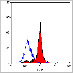
STK33, Monoclonal Antibody (Cat# AAA25856)
Full Name
STK33 (Serine/Threonine Kinase 33) (PE)
Reactivity
Human
Applications
Immunofluorescence, Immunoprecipitation, Western Blot
Purity
Purified by Protein A Affinity Chromatography.
Pricing
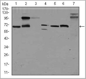
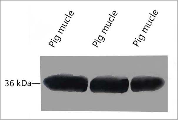
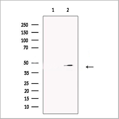
 WB (Western Blot)
(Western Blot:Sample: Recombinant PI3, Human.)
WB (Western Blot)
(Western Blot:Sample: Recombinant PI3, Human.)
Peptidase Inhibitor 3, Monoclonal Antibody (Cat# AAA20831)
Full Name
Monoclonal Antibody to Peptidase Inhibitor 3, Skin Derived (PI3)
Gene Names
PI3; ESI; WAP3; SKALP; WFDC14; cementoin
Reactivity
Human, Pig
Applications
WB, IHC, ICC, IP
Purity
Purification: Protein A + Protein G affinity chromatography
Pricing
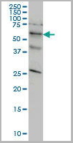
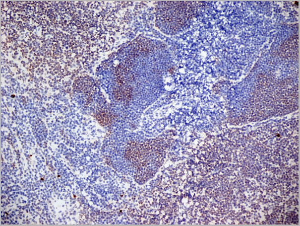
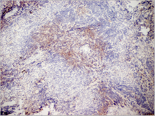
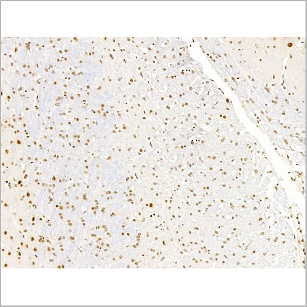
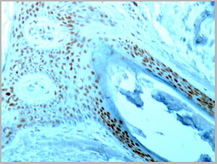
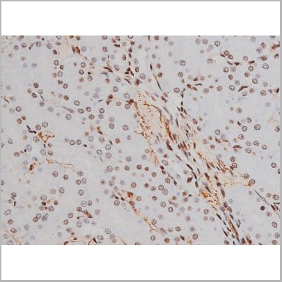
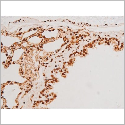
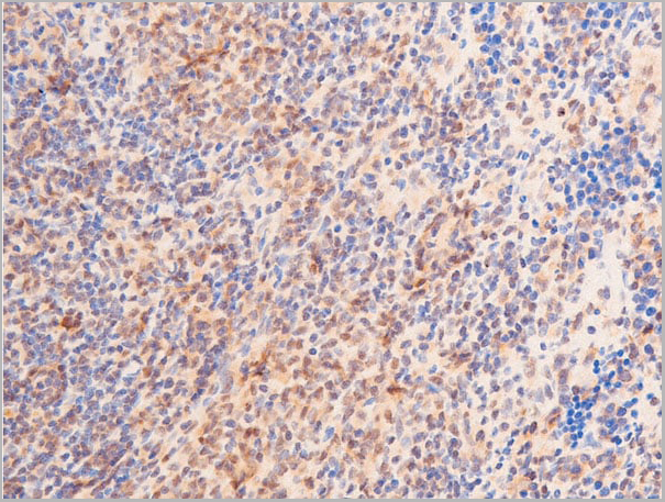
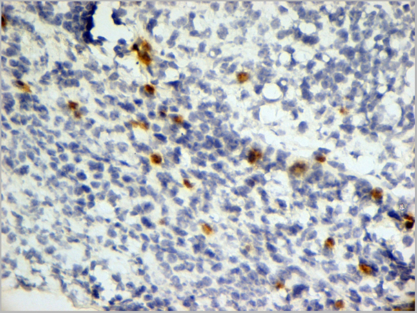
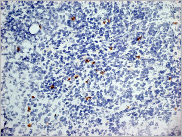
 IHC (Immunohistochemistry)
(Immunohistochemistry: Lung tissue (FFPE) stained with Rabbit anti-ER81 (Cat# AAA14073) at 1:200 for 10 min @ RT. Staining of formalin-fixed tissue requires boiling tissue sections in 10 mM Citrate Buffer, pH 6.0 for 10 min followed by cooling at RT for 20 min.)
IHC (Immunohistochemistry)
(Immunohistochemistry: Lung tissue (FFPE) stained with Rabbit anti-ER81 (Cat# AAA14073) at 1:200 for 10 min @ RT. Staining of formalin-fixed tissue requires boiling tissue sections in 10 mM Citrate Buffer, pH 6.0 for 10 min followed by cooling at RT for 20 min.)
ER81, Polyclonal Antibody (Cat# AAA14073)
Full Name
Rabbit anti ER81 Polyclonal Antibody
Gene Names
ETV1; ER81
Reactivity
Mouse, Human, Rat
Applications
Flow Cytometry, Immunohistochemistry, Western Blot, Immunoprecipitation
Purity
Rabbit IgG is purified by Epitope Affinity Purification
Pricing

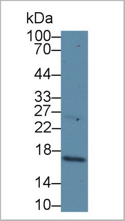
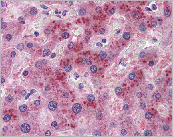
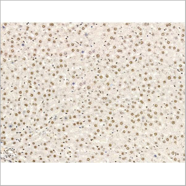
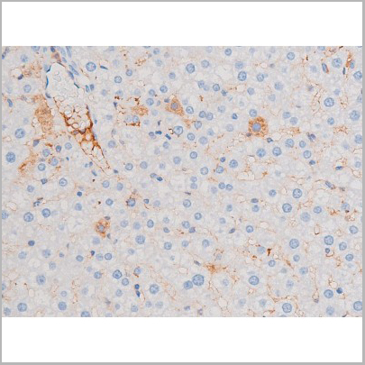
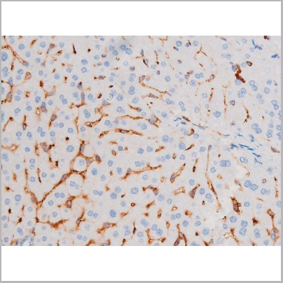
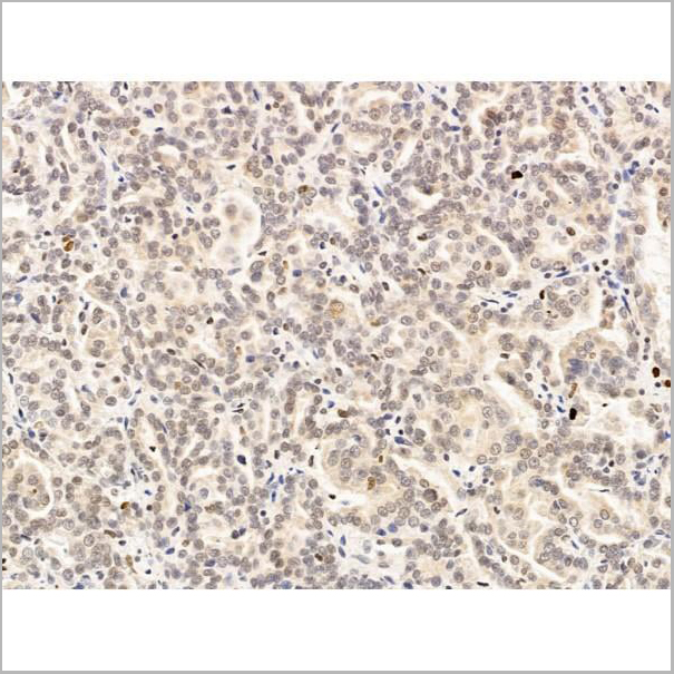
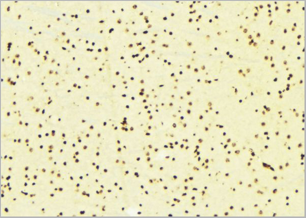
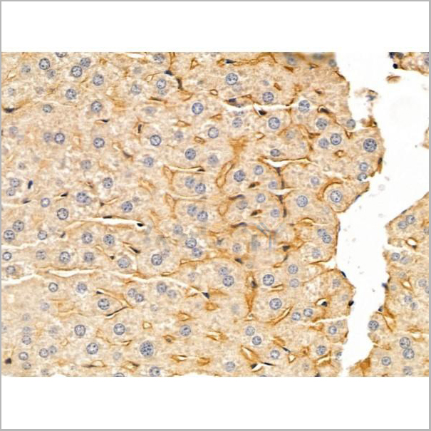
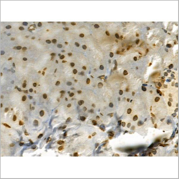
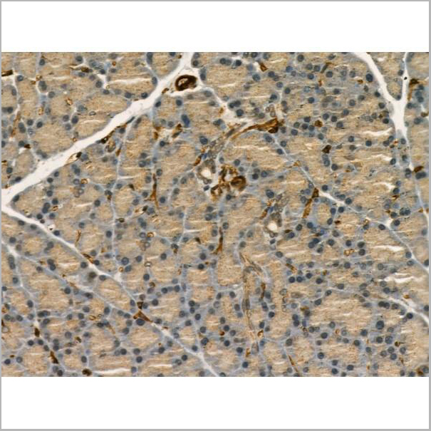
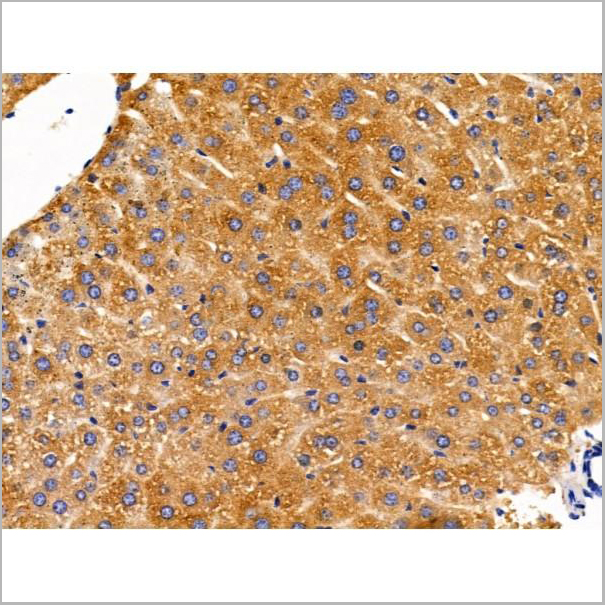
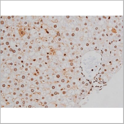
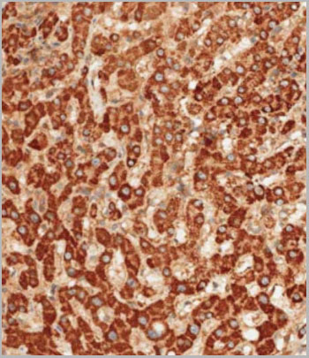
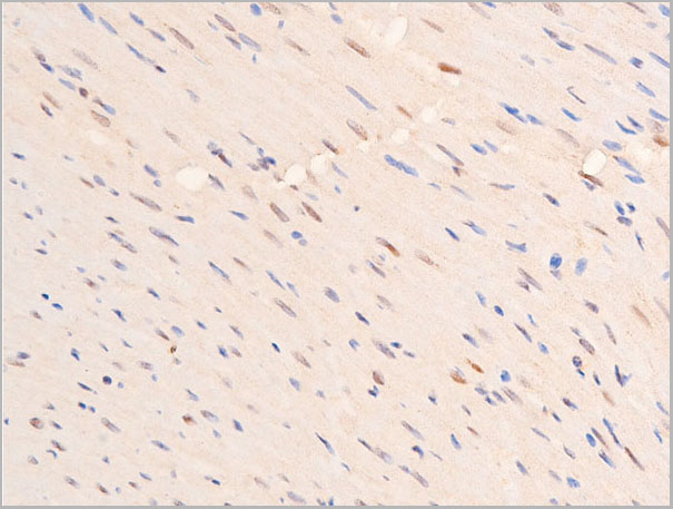
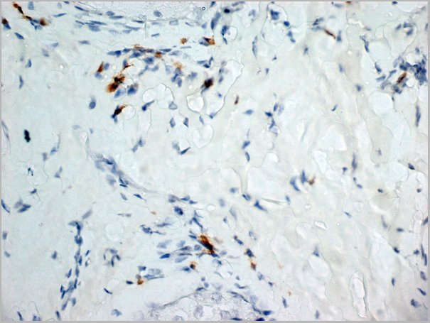
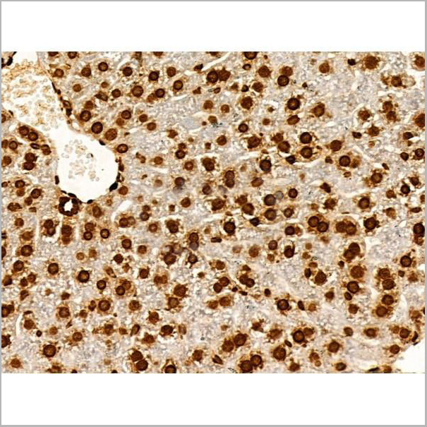
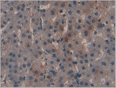 IHC (Immunohistochemistry)
(DAB staining on fromalin fixed paraffin- embedded liver tissue))
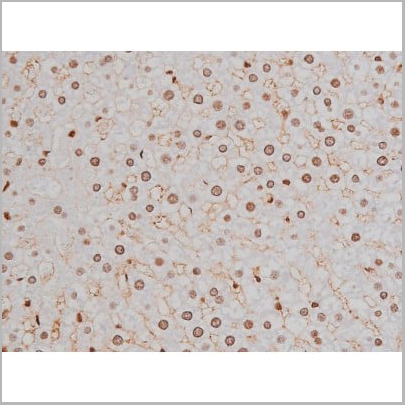
IHC (Immunohistochemistry)
(DAB staining on fromalin fixed paraffin- embedded liver tissue))
Cyclin Dependent Kinase Inhibitor 2A (CDKN2A), Polyclonal Antibody (Cat# AAA20299)
Full Name
Polyclonal Antibody to Cyclin Dependent Kinase Inhibitor 2A (CDKN2A)
Gene Names
CDKN2A; ARF; MLM; P14; P16; P19; CMM2; INK4; MTS1; TP16; CDK4I; CDKN2; INK4A; MTS-1; P14ARF; P19ARF; P16INK4; P16INK4A; P16-INK4A
Reactivity
Human
Applications
Westen Blot, Immunohistochemistry, Immunocytochemistry, Immunoprecipitation
Purity
Antigen-specific affinity chromatography followed by Protein A affinity chromatography
Pricing
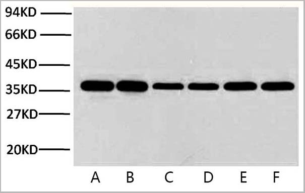
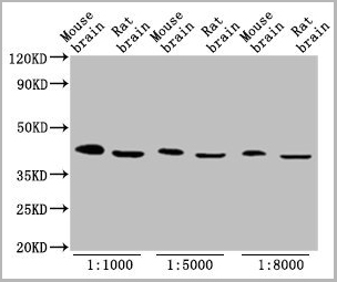
 WB (Western Blot)
(Fig.8 Western blot analysis of Hela, GAPDH Monoclonal Antibody (2B5) was diluted at 1:10000 (25°C, 3h).)
WB (Western Blot)
(Fig.8 Western blot analysis of Hela, GAPDH Monoclonal Antibody (2B5) was diluted at 1:10000 (25°C, 3h).)
GAPDH, Monoclonal Antibody (Cat# AAA31539)
Full Name
Anti-GAPDH Mouse Monoclonal Antibody (2B5)
Gene Names
GAPDH; G3PD; GAPD
Reactivity
Human, Mouse, Rat, Monkey, Dog, Chicken, Rabbit, Pig, Sheep, Yeast, Hamster
Applications
Western Blot, Immunohistochemistry, Immunofluorescence
Purity
The antibody was affinity-purified from mouse ascites by affinity-chromatography using specific immunogen
Pricing
 Application Data
(Staining of mouse spleen cells with Rat anti Mouse CD3 Epsilon (T3): APC)
Application Data
(Staining of mouse spleen cells with Rat anti Mouse CD3 Epsilon (T3): APC)
CD3, Monoclonal Antibody (Cat# AAA11984)
Full Name
RAT ANTI MOUSE CD3
Gene Names
Cd3e; CD3; T3e; AI504783; CD3epsilon
Applications
Immunohistochemistry, Flow Cytometry
Pricing
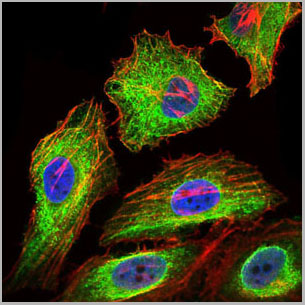
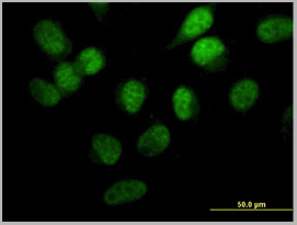
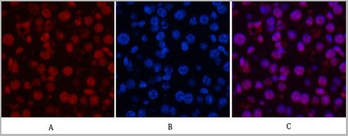
 IF (Immunofluorescence)
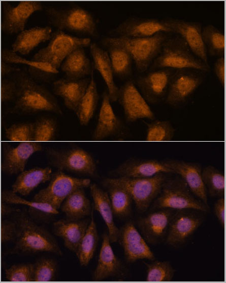
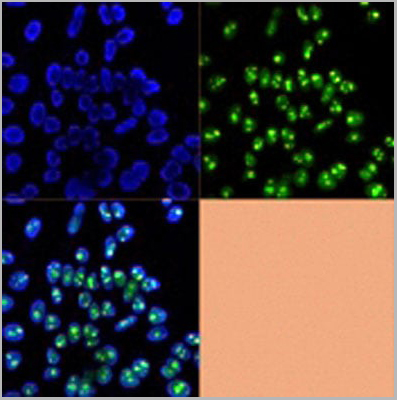
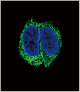
(Immunofluorescence analysis of U-2 OS cells using PP2A-B56δ/PR61δ/PP2A-B56δ/PR61δ/PPP2R5D antibody at dilution of 1:100. Blue: DAPI for nuclear staining.)
IF (Immunofluorescence)
(Immunofluorescence analysis of U-2 OS cells using PP2A-B56δ/PR61δ/PP2A-B56δ/PR61δ/PPP2R5D antibody at dilution of 1:100. Blue: DAPI for nuclear staining.)
PP2A-B56delta/PR61delta/PPP2R5D, Polyclonal Antibody (Cat# AAA28319)
Full Name
PP2A-B56delta/PR61delta/PPP2R5D Rabbit pAb
Gene Names
PPP2R5D; B56D
Reactivity
Human, Mouse, Rat
Applications
Western Blot, Immunohistochemistry, Immunofluorescence
Purity
Affinity purification
Pricing
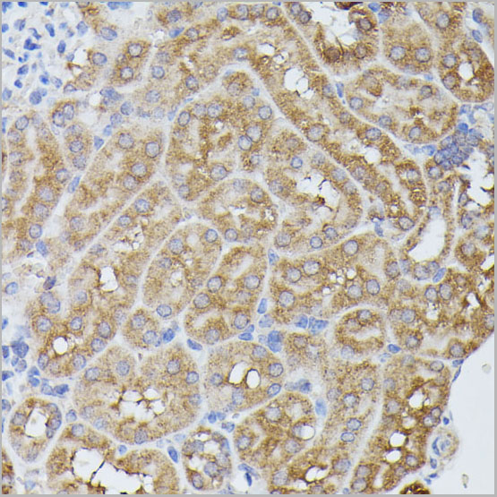
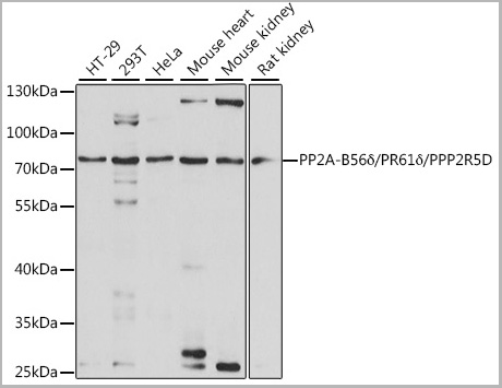
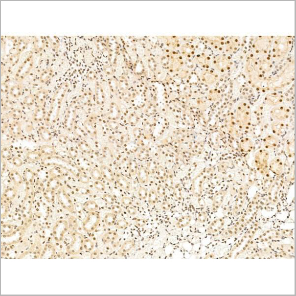
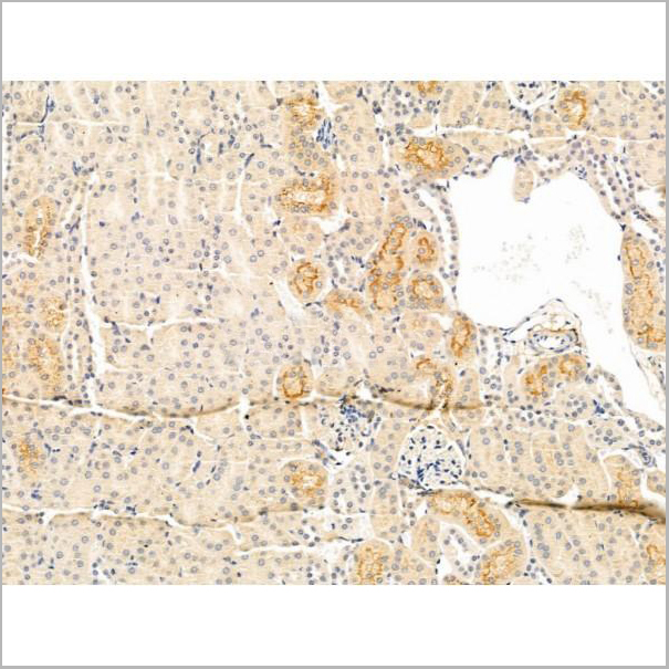
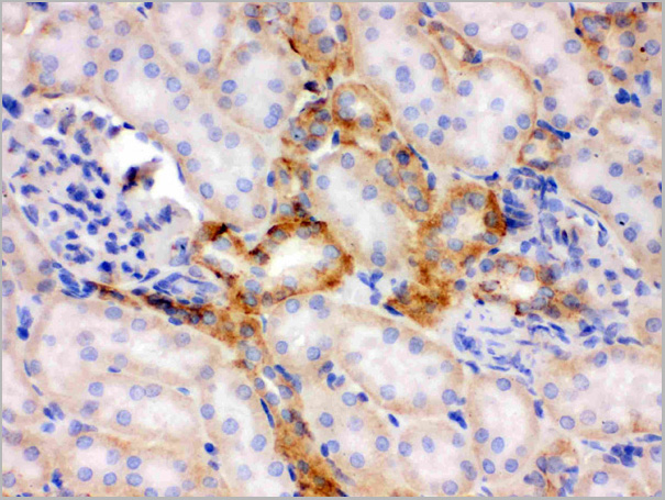
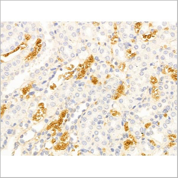
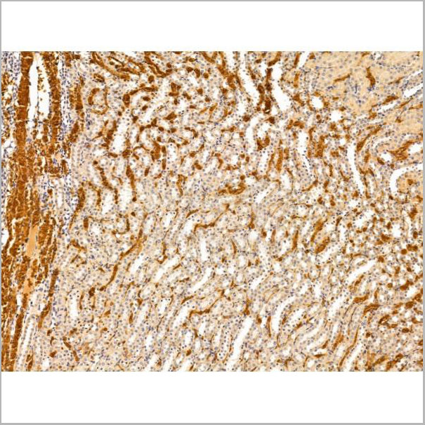
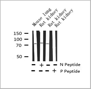
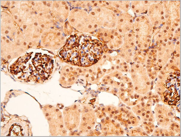
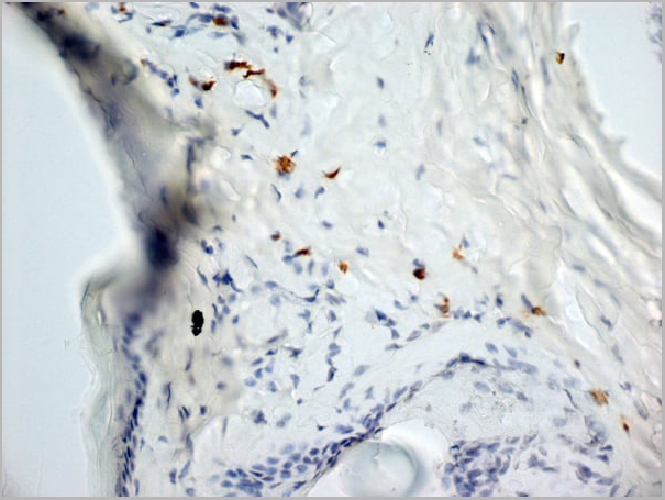
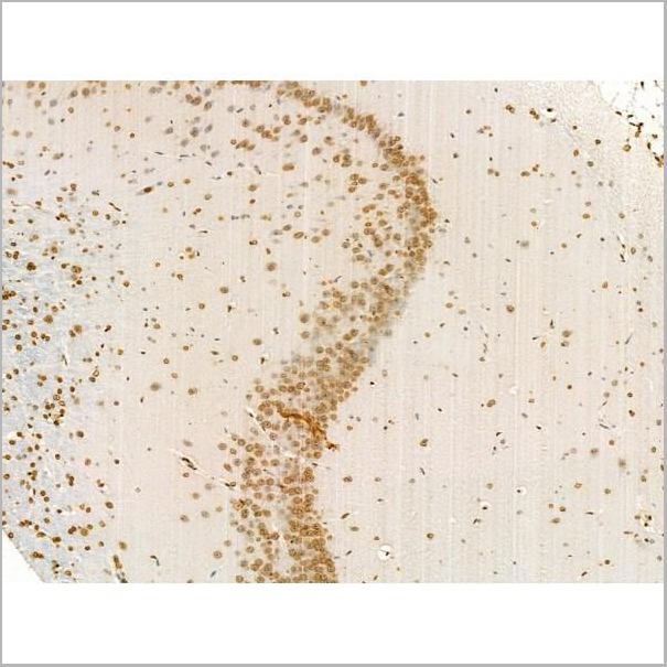
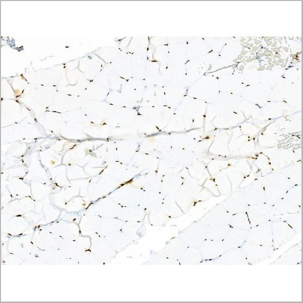
 IHC (Immunohistochemistry)
(AAA31106 at 1/100 staining Rat kidney tissue by IHC-P. The sample was formaldehyde fixed and a heat mediated antigen retrieval step in citrate buffer was performed. The sample was then blocked and incubated with the primary antibody at 4°C overnight. An HRP conjugated anti-Rabbit antibody was used as the secondary antibody.)
IHC (Immunohistochemistry)
(AAA31106 at 1/100 staining Rat kidney tissue by IHC-P. The sample was formaldehyde fixed and a heat mediated antigen retrieval step in citrate buffer was performed. The sample was then blocked and incubated with the primary antibody at 4°C overnight. An HRP conjugated anti-Rabbit antibody was used as the secondary antibody.)
GPR141, Polyclonal Antibody (Cat# AAA31106)
Full Name
GPR141 Antibody
Gene Names
GPR141; PGR13
Reactivity
Human, Mouse, Rat
Applications
Western Blot, Immunohistochemistry
Purity
The antiserum was purified by peptide affinity chromatography using SulfoLinkTM Coupling Resin (Thermo Fisher Scientific).
Pricing
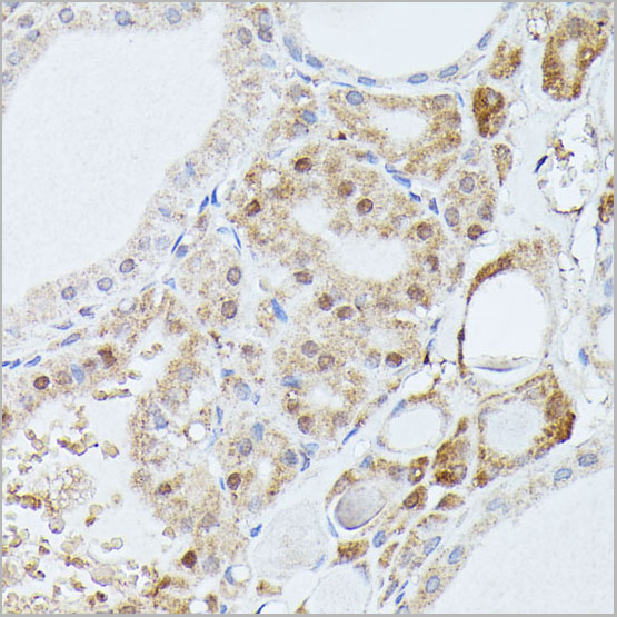
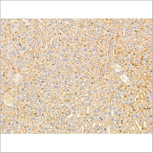
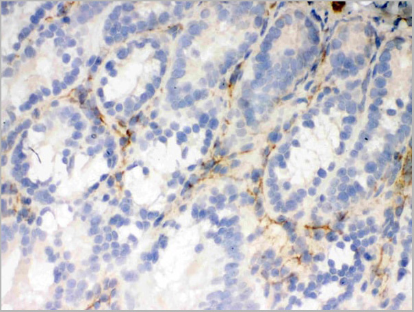
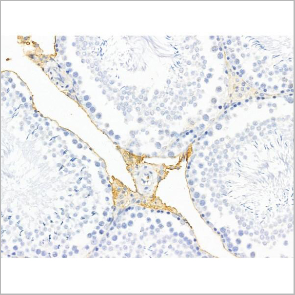
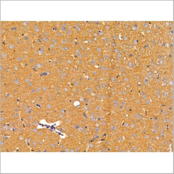
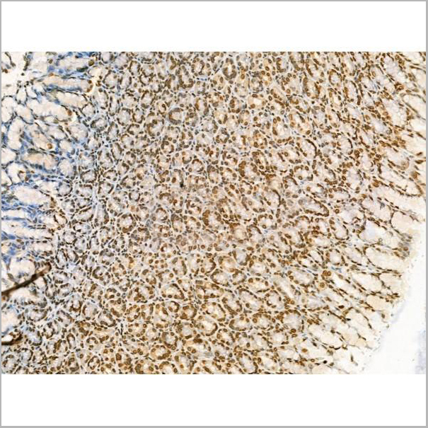
 IHC (Immunohistochemistry-Paraffin)
(AAA31258 at 1/100 staining Rat liver tissue by IHC-P. The sample was formaldehyde fixed and a heat mediated antigen retrieval step in citrate buffer was performed. The sample was then blocked and incubated with the primary antibody at 4°C overnight. An HRP conjugated anti-Rabbit antibody was used as the secondary antibody.)
IHC (Immunohistochemistry-Paraffin)
(AAA31258 at 1/100 staining Rat liver tissue by IHC-P. The sample was formaldehyde fixed and a heat mediated antigen retrieval step in citrate buffer was performed. The sample was then blocked and incubated with the primary antibody at 4°C overnight. An HRP conjugated anti-Rabbit antibody was used as the secondary antibody.)
L3MBTL1, Polyclonal Antibody (Cat# AAA31258)
Full Name
L3MBTL1 Antibody
Gene Names
L3MBTL1; L3MBTL; ZC2HC3; H-L(3)MBT; dJ138B7.3
Reactivity
Human, Mouse, Rat
Predicted Reactivity: Horse(100%), Rabbit(100%), Dog(100%), Chicken(100%)
Predicted Reactivity: Horse(100%), Rabbit(100%), Dog(100%), Chicken(100%)
Applications
ELISA
Purity
The antiserum was purified by peptide affinity chromatography using SulfoLink Coupling Resin (Thermo Fisher Scientific).
Pricing
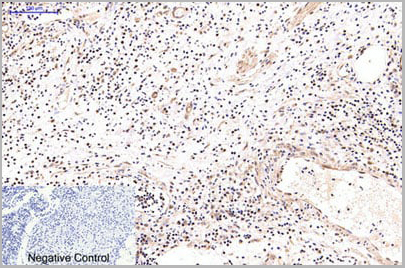
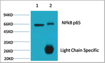
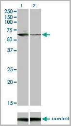
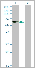
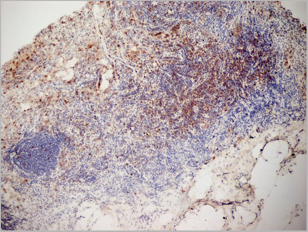
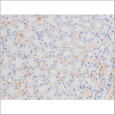
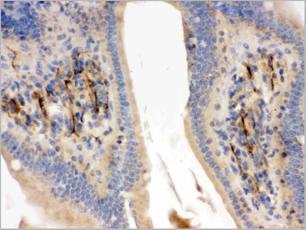
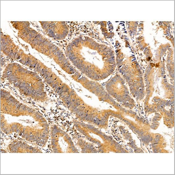
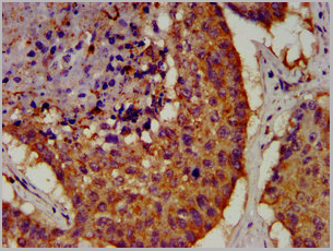
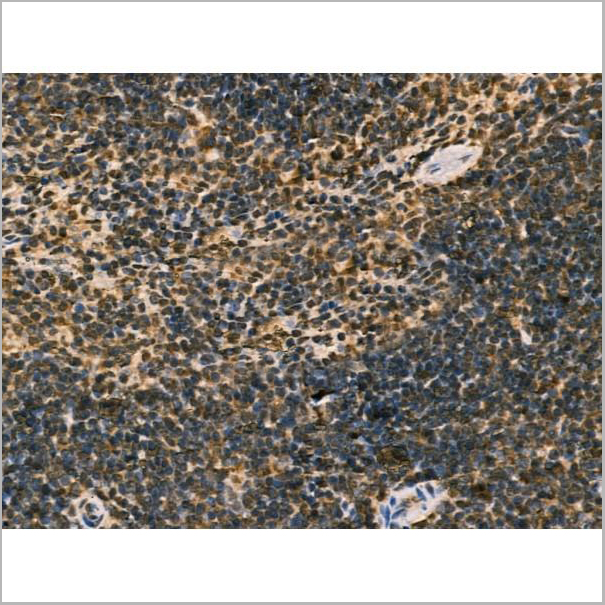
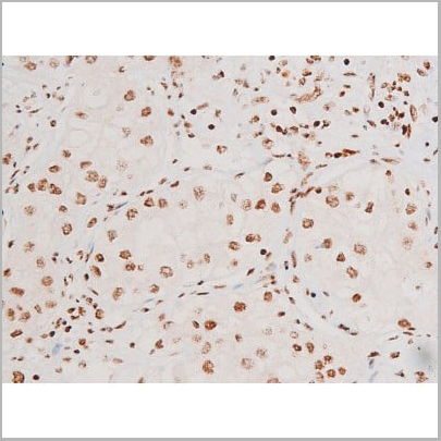
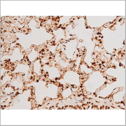
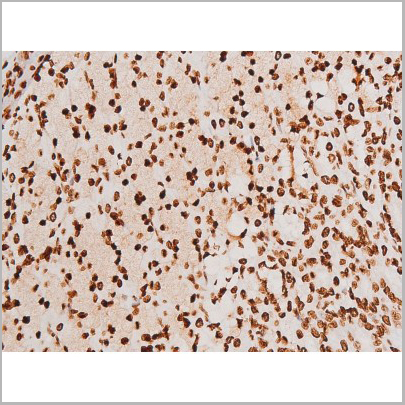
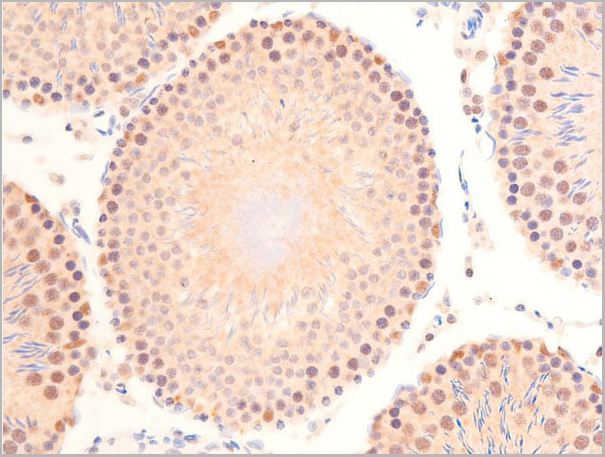
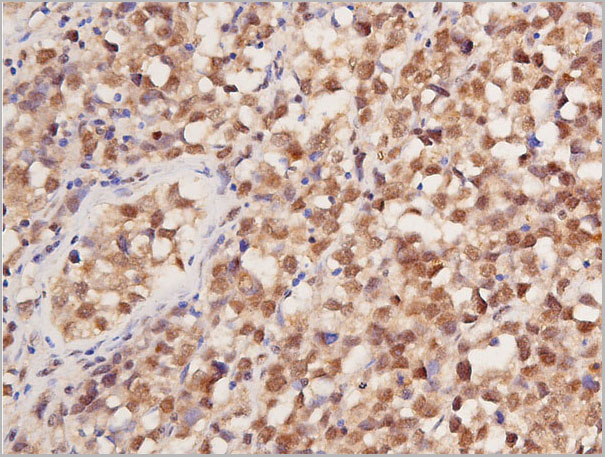
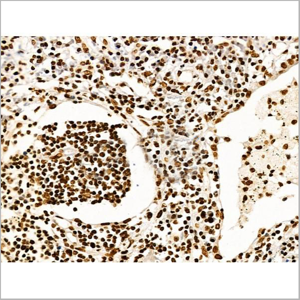
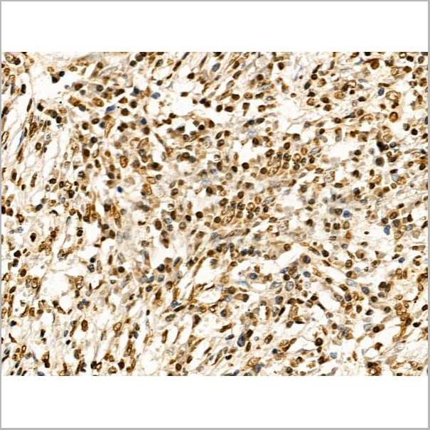
 IHC (Immunohistchemistry)
(Immunohistochemical analysis of paraffin-embedded Rat-lung tissue. 1,NFkB p65 Monoclonal Antibody(5G6) was diluted at 1:200(4C,overnight). 2, Sodium citrate pH 6.0 was used for antibody retrieval(>98C,20min). 3,Secondary antibody was diluted at 1:200(room tempeRature, 30min). Negative control was used by secondary antibody only.)
IHC (Immunohistchemistry)
(Immunohistochemical analysis of paraffin-embedded Rat-lung tissue. 1,NFkB p65 Monoclonal Antibody(5G6) was diluted at 1:200(4C,overnight). 2, Sodium citrate pH 6.0 was used for antibody retrieval(>98C,20min). 3,Secondary antibody was diluted at 1:200(room tempeRature, 30min). Negative control was used by secondary antibody only.)
NFkappaB p65, Monoclonal Antibody (Cat# AAA29642)
Full Name
NFkappaB p65 Mouse Monoclonal Antibody
Gene Names
RELA; p65; NFKB3
Reactivity
Human, Rat, Mouse
Applications
Western Blot, Immunohistochemistry, Immunofluorescence
Purity
Affinity purification using immunogen.
Pricing
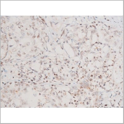
 IHC (Immunohistchemistry)
(AAA31038 at 1/200 staining Rat kidney tissue sections by IHC-P. The tissue was formaldehyde fixed and a heat mediated antigen retrieval step in citrate buffer was performed. The tissue was then blocked and incubated with the antibody for 1.5 hours at 22 degree C. An HRP conjugated goat anti-rabbit antibody was used as the secondary.)
IHC (Immunohistchemistry)
(AAA31038 at 1/200 staining Rat kidney tissue sections by IHC-P. The tissue was formaldehyde fixed and a heat mediated antigen retrieval step in citrate buffer was performed. The tissue was then blocked and incubated with the antibody for 1.5 hours at 22 degree C. An HRP conjugated goat anti-rabbit antibody was used as the secondary.)
IRS-1, Polyclonal Antibody (Cat# AAA31038)
Full Name
Phospho-IRS-1 (Ser639) Antibody
Gene Names
IRS1; HIRS-1
Reactivity
Human, Mouse, Rat, Monkey
Applications
Western Blot, Immunohistochemistry, Immunofluorescence, Immunocytochemistry
Purity
From purified rabbit serum by affinity purification via sequential chromatography on phospho-and non-phospho-peptide affinity columns.
Pricing
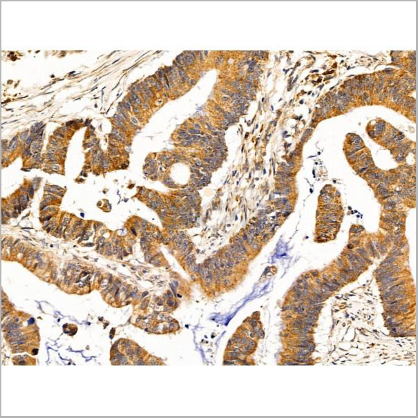
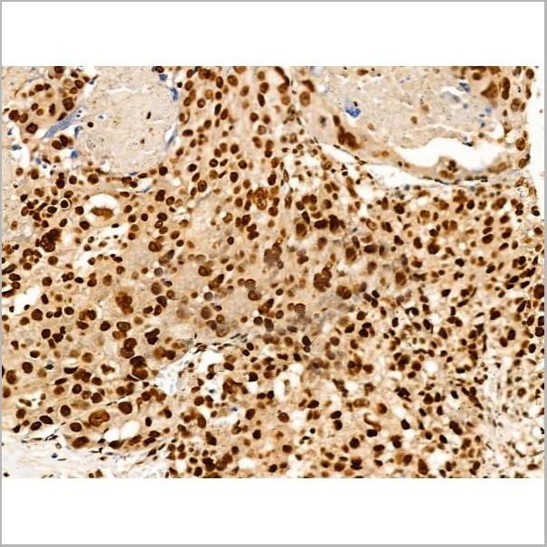
 IHC (Immunohistochemistry-Paraffin)
(AAA31245 at 1/100 staining Human kidney cancer by IHC-P. The sample was formaldehyde fixed and a heat mediated antigen retrieval step in citrate buffer was performed. The sample was then blocked and incubated with the primary antibody at 4°C overnight. An HRP conjugated anti-Rabbit antibody was used as the secondary antibody.)
IHC (Immunohistochemistry-Paraffin)
(AAA31245 at 1/100 staining Human kidney cancer by IHC-P. The sample was formaldehyde fixed and a heat mediated antigen retrieval step in citrate buffer was performed. The sample was then blocked and incubated with the primary antibody at 4°C overnight. An HRP conjugated anti-Rabbit antibody was used as the secondary antibody.)
Nesprin 1, Polyclonal Antibody (Cat# AAA31245)
Full Name
Nesprin 1 Antibody
Gene Names
SYNE1; 8B; AMCM; CPG2; ARCA1; EDMD4; KASH1; MYNE1; Nesp1; SCAR8; C6orf98; dJ45H2.2
Reactivity
Human, Mouse, Rat
Predicted Reactivity: Horse(88%), Sheep(100%), Dog(88%)
Predicted Reactivity: Horse(88%), Sheep(100%), Dog(88%)
Applications
ELISA
Purity
The antiserum was purified by peptide affinity chromatography using SulfoLink Coupling Resin (Thermo Fisher Scientific).
Pricing
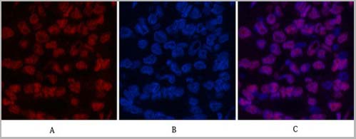
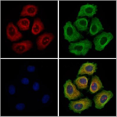
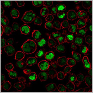
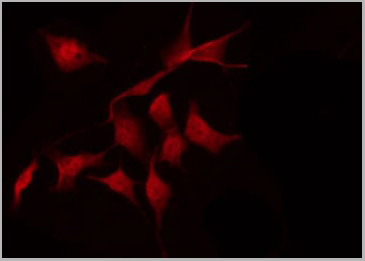
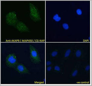 IF (Immunofluorescence)
(AAA13648 Immunofluorescence analysis of paraformaldehyde fixed U2OS cells, permeabilized with 0.15% Triton. Primary incubation 1hr (10ug/ml) followed by Alexa Fluor 488 secondary antibody (4ug/ml), showing cytoplasmic staining. The nuclear stain is DAPI (blue). Negative control: Unimmunized goat IgG (10ug/ml) followed by Alexa Fluor 488 secondary antibody (4ug/ml).)
IF (Immunofluorescence)
(AAA13648 Immunofluorescence analysis of paraformaldehyde fixed U2OS cells, permeabilized with 0.15% Triton. Primary incubation 1hr (10ug/ml) followed by Alexa Fluor 488 secondary antibody (4ug/ml), showing cytoplasmic staining. The nuclear stain is DAPI (blue). Negative control: Unimmunized goat IgG (10ug/ml) followed by Alexa Fluor 488 secondary antibody (4ug/ml).)
AKAP9/AKAP450/CG-NAP, Polyclonal Antibody (Cat# AAA13648)
Full Name
Goat anti-AKAP9/AKAP450/CG-NAP Antibody
Gene Names
AKAP9; LQT11; PRKA9; AKAP-9; CG-NAP; YOTIAO; AKAP350; AKAP450; PPP1R45; HYPERION; MU-RMS-40.16A
Reactivity
Tested: Human; Expected from sequence similarity: Human, Mouse
Applications
Peptide ELISA, Western Blot, Immunofluorescence
Purity
Purified from goat serum by ammonium sulphate precipitation followed by antigen affinity chromatography using the immunizing peptide.
Pricing
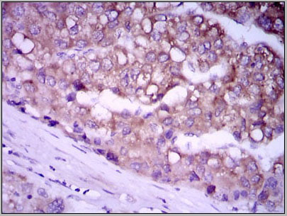
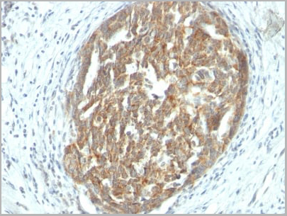 IHC (Immunohistochemistry)
(Formalin-fixed, paraffin-embedded human Ovarian Carcinoma stained with VEGF Monoclonal Antibody (SPM225).)
IHC (Immunohistochemistry)
(Formalin-fixed, paraffin-embedded human Ovarian Carcinoma stained with VEGF Monoclonal Antibody (SPM225).)
VEGF (Vascular Endothelial Growth Factor), Monoclonal Antibody (Cat# AAA13827)
Full Name
VEGF (Vascular Endothelial Growth Factor) Mouse Monoclonal Antibody
Gene Names
VEGFA; VPF; VEGF; MVCD1
Reactivity
Human, Mouse, Rat, Rabbit, Dog. Others not known.
Applications
Immunofluorescence, Immunohistochemistry
Pricing
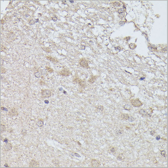
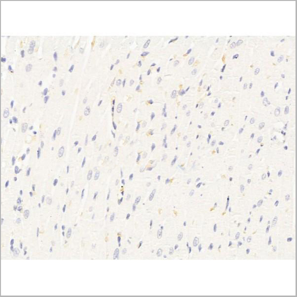
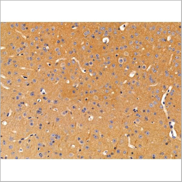
 IHC (Immunohistochemistry)
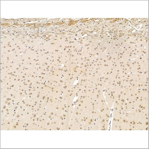
(AAA31121 at 1/100 staining Mouse brain tissue by IHC-P. The sample was formaldehyde fixed and a heat mediated antigen retrieval step in citrate buffer was performed. The sample was then blocked and incubated with the antibody for 1.5 hours at 22 degree C. An HRP conjugated goat anti-rabbit antibody was used as the secondary.)
IHC (Immunohistochemistry)
(AAA31121 at 1/100 staining Mouse brain tissue by IHC-P. The sample was formaldehyde fixed and a heat mediated antigen retrieval step in citrate buffer was performed. The sample was then blocked and incubated with the antibody for 1.5 hours at 22 degree C. An HRP conjugated goat anti-rabbit antibody was used as the secondary.)
FABP4, Polyclonal Antibody (Cat# AAA31121)
Full Name
FABP4 Antibody
Gene Names
FABP4; aP2; ALBP; AFABP; A-FABP; HEL-S-104
Reactivity
Human, Mouse, Rat
Applications
Western Blot, Immunohistochemistry
Purity
The antiserum was purified by peptide affinity chromatography using SulfoLink Coupling Resin.
Pricing
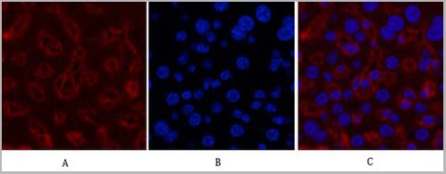
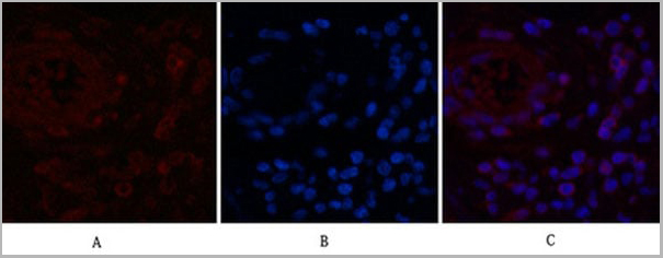
 IF (Immunofluorescence)
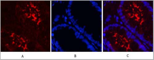
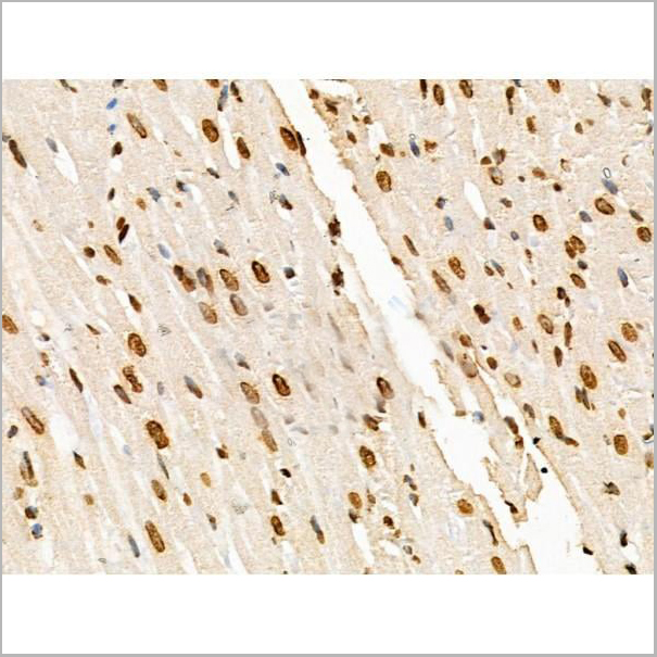
(Fig.7. Immunofluorescence analysis of rat liver tissue. 1, Histone H3 Monoclonal Antibody (2D10) (red) was diluted at 1:200 (4°C, overnight). 2, Cy3 Labeled secondary antibody was diluted at 1:300 (room temperature, 50min). 3, Picture B: DAPI (blue) 10min. Picture A: Target. Picture B: DAPI. Picture C: merge of A+B.)
IF (Immunofluorescence)
(Fig.7. Immunofluorescence analysis of rat liver tissue. 1, Histone H3 Monoclonal Antibody (2D10) (red) was diluted at 1:200 (4°C, overnight). 2, Cy3 Labeled secondary antibody was diluted at 1:300 (room temperature, 50min). 3, Picture B: DAPI (blue) 10min. Picture A: Target. Picture B: DAPI. Picture C: merge of A+B.)
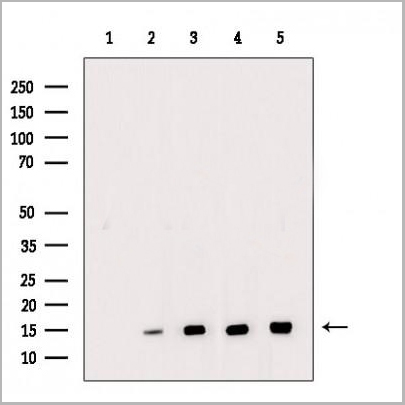
Histone H3, Monoclonal Antibody (Cat# AAA31543)
Full Name
Anti-Histone H3 Mouse Monoclonal Antibody (2D10)
Gene Names
HIST1H3A; H3/A; H3FA
Reactivity
Human, Mouse, Rat, Yeast
Applications
Western Blot, Immunohistochemistry, Immunofluorescence, Immunoprecipitation
Pricing
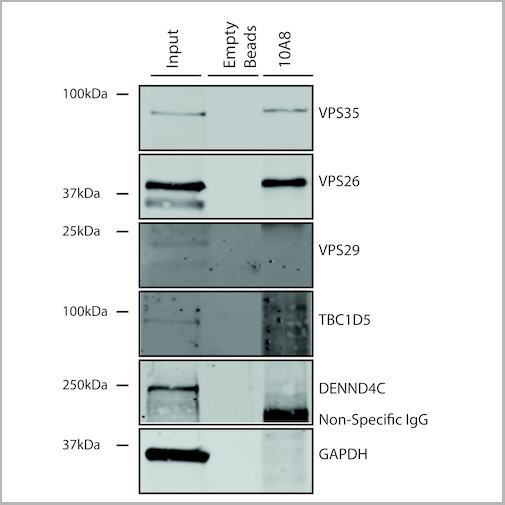
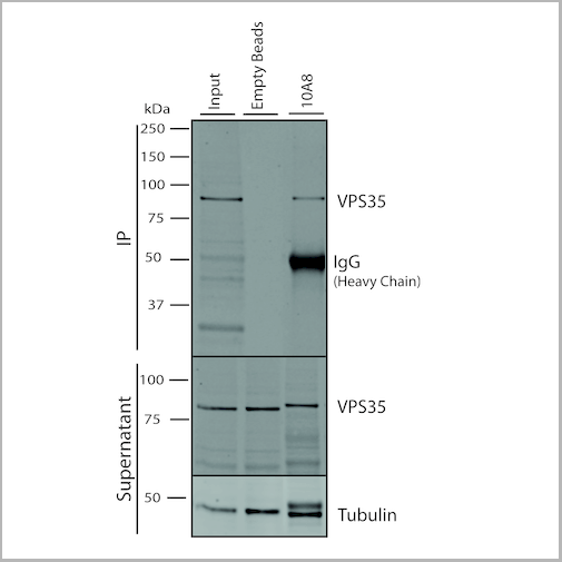
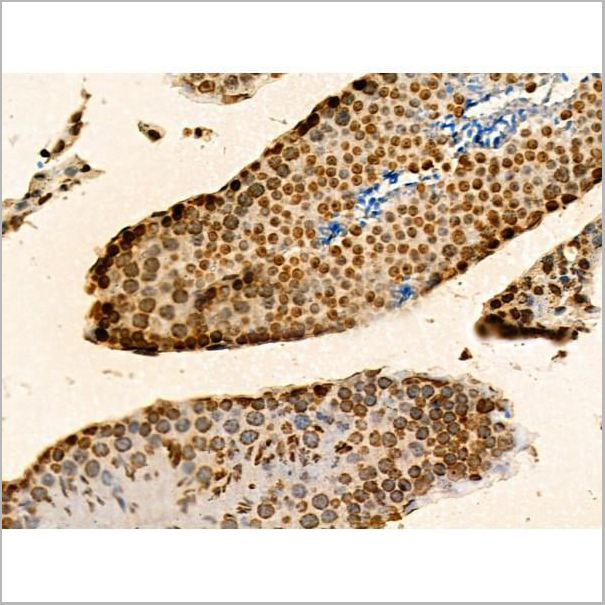
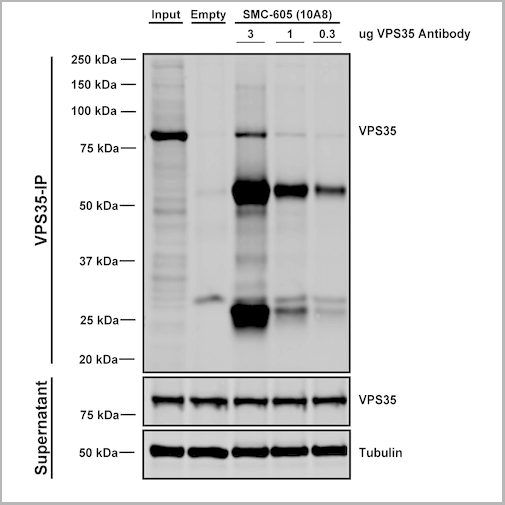 IP (Immunoprecipitation)
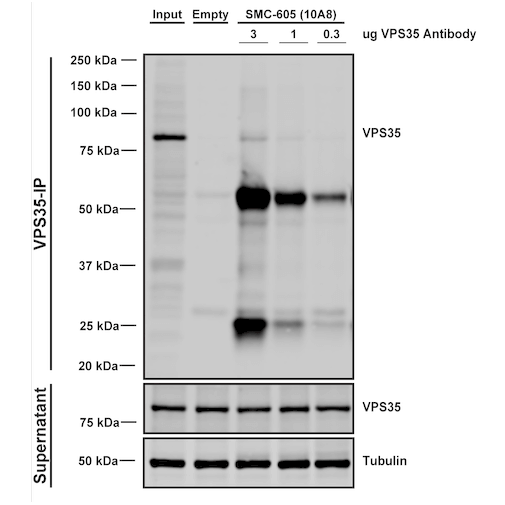
(Immunoprecipitation analysis using Mouse Anti-VPS35 Monoclonal Antibody, Clone 10A8. Tissue: embryonic fibroblast. Species: Mouse. Primary Antibody: Mouse Anti-VPS35 Monoclonal Antibody. Three amounts of (3, 1 and 0.3 ug) were non-covalently coupled to 10uL of A/G sepharose beads for 1 hour at 4 degree C and next incubated with 250ug of MEF lysate for 2 hours at 4 degree C.)
IP (Immunoprecipitation)
(Immunoprecipitation analysis using Mouse Anti-VPS35 Monoclonal Antibody, Clone 10A8. Tissue: embryonic fibroblast. Species: Mouse. Primary Antibody: Mouse Anti-VPS35 Monoclonal Antibody. Three amounts of (3, 1 and 0.3 ug) were non-covalently coupled to 10uL of A/G sepharose beads for 1 hour at 4 degree C and next incubated with 250ug of MEF lysate for 2 hours at 4 degree C.)
VPS35, Monoclonal Antibody (Cat# AAA27698)
Full Name
VPS35 Antibody, Clone 10A8: PerCP
Gene Names
VPS35; MEM3; PARK17
Reactivity
Human, Mouse, Rat
Applications
Western Blot, Immunocytochemistry, Immunofluorescence, Immunoprecipitation
Purity
Protein G Purified
Pricing
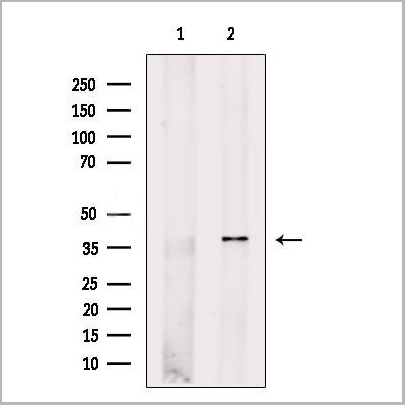
 WB (Western Blot)
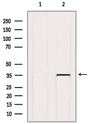
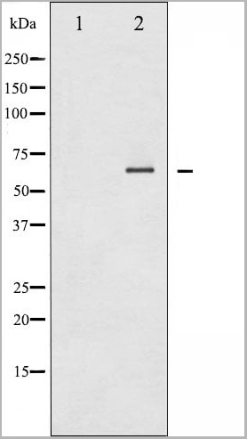
(Western blot analysis of extracts from HeLa cells, using Cytochrome P450 39A1 Antibody. The lane on the left was treated with blocking peptide.)
WB (Western Blot)
(Western blot analysis of extracts from HeLa cells, using Cytochrome P450 39A1 Antibody. The lane on the left was treated with blocking peptide.)
Cytochrome P450 39A1, Polyclonal Antibody (Cat# AAA31117)
Full Name
Cytochrome P450 39A1 Antibody
Reactivity
Human, Mouse
Applications
Western Blot, Immunohistochemistry, Immunofluorescence, Immunocytochemistry
Purity
Purified by peptide affinity chromatography using SulfoLink Coupling Resin
Pricing
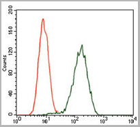
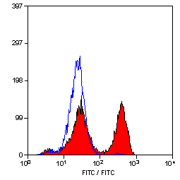
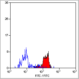
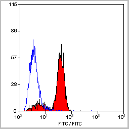
 FCM (Flow Cytometry)
(Flow Cytometric Analysis of paraformaldehyde-fixed Jurkat cells using CD31 Mouse Monoclonal Antibody (JC/70A) followed by goat anti- Mouse- IgG-CF488 (Blue); Isotype Control (Red).)
FCM (Flow Cytometry)
(Flow Cytometric Analysis of paraformaldehyde-fixed Jurkat cells using CD31 Mouse Monoclonal Antibody (JC/70A) followed by goat anti- Mouse- IgG-CF488 (Blue); Isotype Control (Red).)
CD31 / PECAM-1, Monoclonal Antibody (Cat# AAA13818)
Full Name
CD31 / PECAM-1 (Endothelial Cell Marker) Mouse Monoclonal Antibody
Gene Names
PECAM1; CD31; PECA1; GPIIA'; PECAM-1; endoCAM; CD31/EndoCAM
Reactivity
Human, Cynomolgus Monkey, Rabbit.
Does not react with Rat and Pig
Does not react with Rat and Pig
Applications
Flow Cytometry, Immunofluorescence, Western Blot, Immunohistochemistry
Pricing
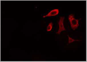
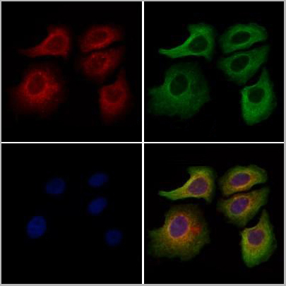
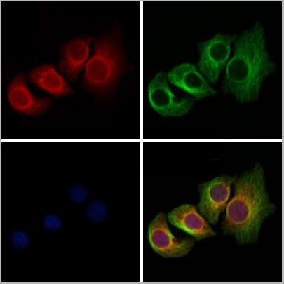
 IF (Immunofluorescence)
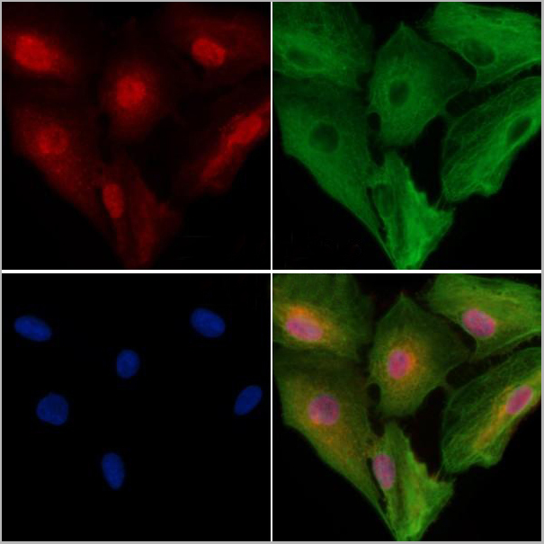
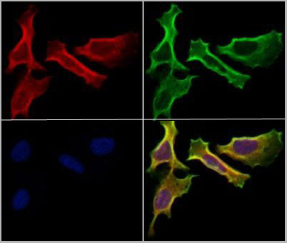
(AAA31272 staining Hela cells by IF/ICC. The samples were fixed with PFA and permeabilized in 0.1% Triton X-100,then blocked in 10% serum for 45 minutes at 25°C. Samples were then incubated with primary Ab(AAA31272 1:200) and mouse anti-beta tubulin Ab(T0023 1:200) for 1 hour at 37°C. An AlexaFluor594 conjugated goat anti-rabbit IgG(H+L) Ab(Red) and an AlexaFluor488 conjugated goat anti-mouse IgG(H+L) Ab(Green) were used as the secondary antibody.The nuclear counter stain is DAPI(blue).)
IF (Immunofluorescence)
(AAA31272 staining Hela cells by IF/ICC. The samples were fixed with PFA and permeabilized in 0.1% Triton X-100,then blocked in 10% serum for 45 minutes at 25°C. Samples were then incubated with primary Ab(AAA31272 1:200) and mouse anti-beta tubulin Ab(T0023 1:200) for 1 hour at 37°C. An AlexaFluor594 conjugated goat anti-rabbit IgG(H+L) Ab(Red) and an AlexaFluor488 conjugated goat anti-mouse IgG(H+L) Ab(Green) were used as the secondary antibody.The nuclear counter stain is DAPI(blue).)
DERL2, Polyclonal Antibody (Cat# AAA31272)
Full Name
DERL2 Antibody
Gene Names
DERL2; FLANa; F-LANa; CGI-101; F-LAN-1; derlin-2; DERtrin-2
Reactivity
Human, Mouse, Rat
Applications
Immunofluorescence, Immunocytochemistry, Western Blot, Immunohistochemistry
Purity
The antiserum was purified by peptide affinity chromatography using SulfoLink™ Coupling Resin (Thermo Fisher Scientific).
Pricing
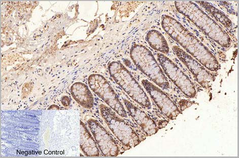
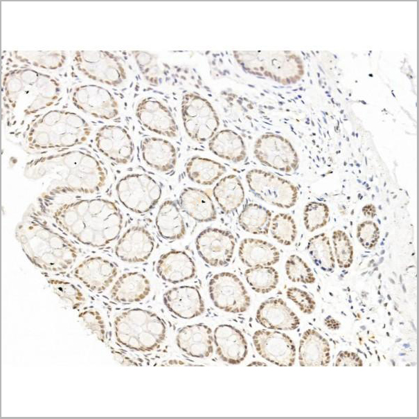
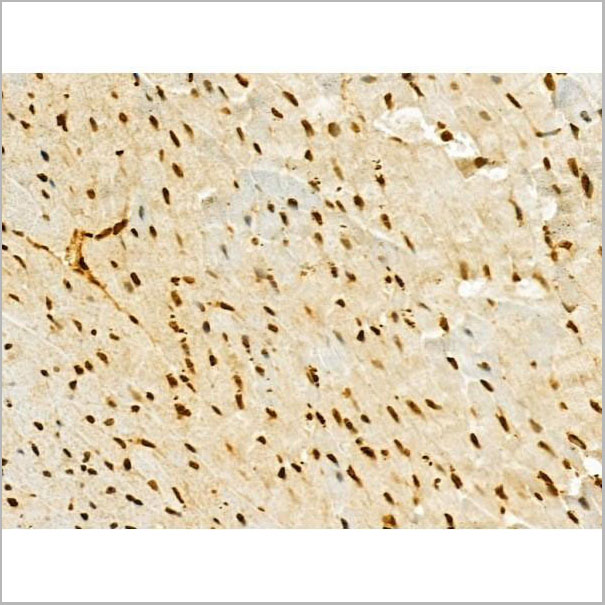
 IHC (Immunohistochemistry)
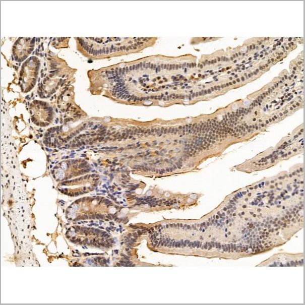
(Figure 7. IHC analysis of Flotillin 2 using anti-Flotillin 2 antibody (AAA11664).Flotillin 2 was detected in frozen section of rat small intestine tissue. Heat mediated antigen retrieval was performed in citrate buffer (pH6, epitope retrieval solution) for 20 mins. The tissue section was blocked with 10% goat serum. The tissue section was then incubated with 1ug/ml rabbit anti-Flotillin 2 Antibody (AAA11664) overnight at 4 degree C. Biotinylated goat anti-rabbit IgG was used as secondary antibody and incubated for 30 minutes at 37 degree C. The tissue section was developed using Strepavidin-Biotin-Complex (SABC) with DAB as the chromogen.)
IHC (Immunohistochemistry)
(Figure 7. IHC analysis of Flotillin 2 using anti-Flotillin 2 antibody (AAA11664).Flotillin 2 was detected in frozen section of rat small intestine tissue. Heat mediated antigen retrieval was performed in citrate buffer (pH6, epitope retrieval solution) for 20 mins. The tissue section was blocked with 10% goat serum. The tissue section was then incubated with 1ug/ml rabbit anti-Flotillin 2 Antibody (AAA11664) overnight at 4 degree C. Biotinylated goat anti-rabbit IgG was used as secondary antibody and incubated for 30 minutes at 37 degree C. The tissue section was developed using Strepavidin-Biotin-Complex (SABC) with DAB as the chromogen.)
Flotillin 2, Polyclonal Antibody (Cat# AAA11664)
Full Name
Anti-Flotillin 2 Antibody
Gene Names
FLOT2; ESA; ECS1; ESA1; ECS-1; M17S1
Reactivity
Human, Mouse, Rat
Applications
Western Blot, Immunohistochemistry
Purity
Immunogen Affinity Purified
Pricing
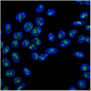
 IF (Immunofluorescence)
(AAA31246 staining A549 cells by IF/ICC. The samples were fixed with PFA and permeabilized in 0.1% Triton X-100,then blocked in 10% serum for 45 minutes at 25°C. Samples were then incubated with primary Ab(AAA31246) and mouse anti-beta tubulin Ab(T0023) for 1 hour at 37°C. An AlexaFluor594 conjugated goat anti-rabbit IgG(H+L) Ab(Red) and an AlexaFluor488 conjugated goat anti-mouse IgG(H+L) Ab(Green) were used as the secondary antibody.The nuclear counter stain is DAPI (blue).)
IF (Immunofluorescence)
(AAA31246 staining A549 cells by IF/ICC. The samples were fixed with PFA and permeabilized in 0.1% Triton X-100,then blocked in 10% serum for 45 minutes at 25°C. Samples were then incubated with primary Ab(AAA31246) and mouse anti-beta tubulin Ab(T0023) for 1 hour at 37°C. An AlexaFluor594 conjugated goat anti-rabbit IgG(H+L) Ab(Red) and an AlexaFluor488 conjugated goat anti-mouse IgG(H+L) Ab(Green) were used as the secondary antibody.The nuclear counter stain is DAPI (blue).)
TROY, Polyclonal Antibody (Cat# AAA31246)
Full Name
TROY Antibody
Gene Names
TNFRSF19; TAJ; TROY; TRADE; TAJ-alpha
Reactivity
Human, Mouse, Rat
Predicted Reactivity: Horse(88%), Sheep(86%)
Predicted Reactivity: Horse(88%), Sheep(86%)
Applications
ELISA
Purity
The antiserum was purified by peptide affinity chromatography using SulfoLink Coupling Resin (Thermo Fisher Scientific).
Pricing
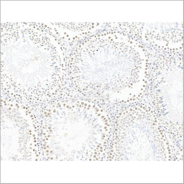
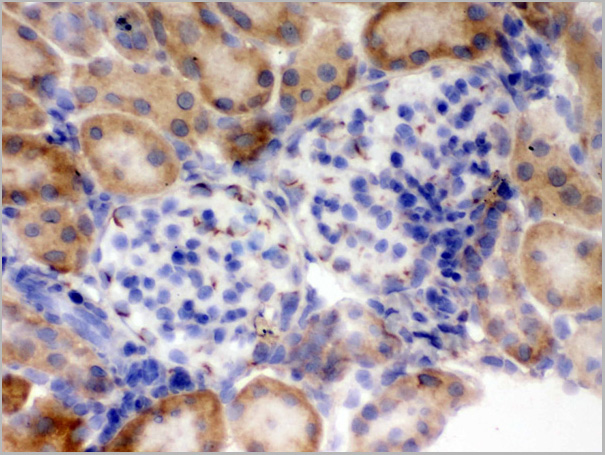
 IHC (Immunohistochemistry-Paraffin)
(AAA31254 at 1/100 staining Mouse kidney tissue by IHC-P. The sample was formaldehyde fixed and a heat mediated antigen retrieval step in citrate buffer was performed. The sample was then blocked and incubated with the primary antibody at 4°C overnight. An HRP conjugated anti-Rabbit antibody was used as the secondary antibody.)
IHC (Immunohistochemistry-Paraffin)
(AAA31254 at 1/100 staining Mouse kidney tissue by IHC-P. The sample was formaldehyde fixed and a heat mediated antigen retrieval step in citrate buffer was performed. The sample was then blocked and incubated with the primary antibody at 4°C overnight. An HRP conjugated anti-Rabbit antibody was used as the secondary antibody.)
Hhip, Polyclonal Antibody (Cat# AAA31254)
Full Name
Hhip Antibody
Gene Names
HHIP; HIP
Reactivity
Human, Mouse, Rat
Applications
Immunofluorescence, Immunocytochemistry, Western Blot, Immunohistochemistry
Purity
Peptide affinity chromatography using SulfoLink Coupling Resin
Pricing
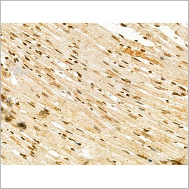
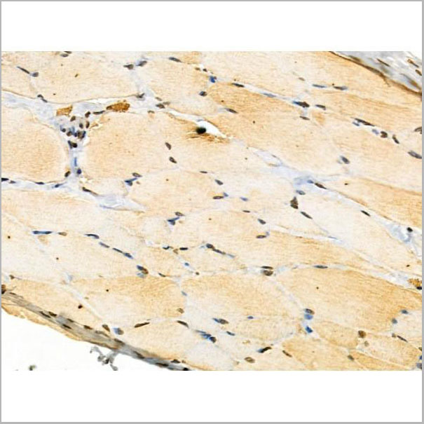
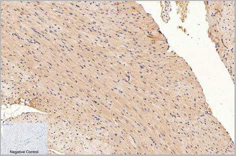
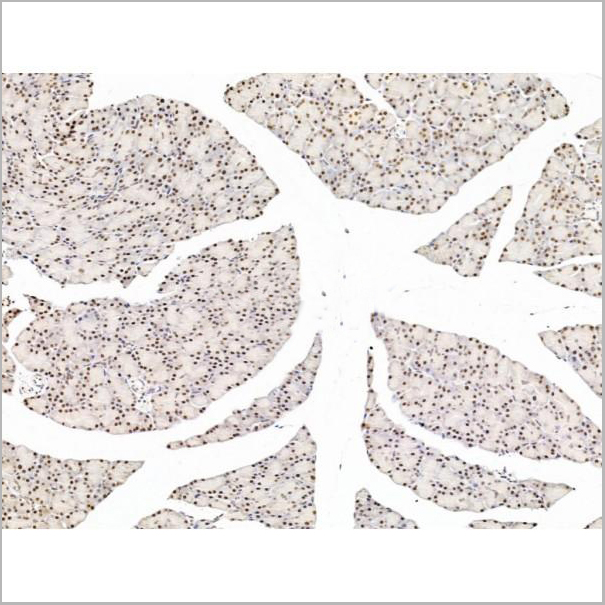
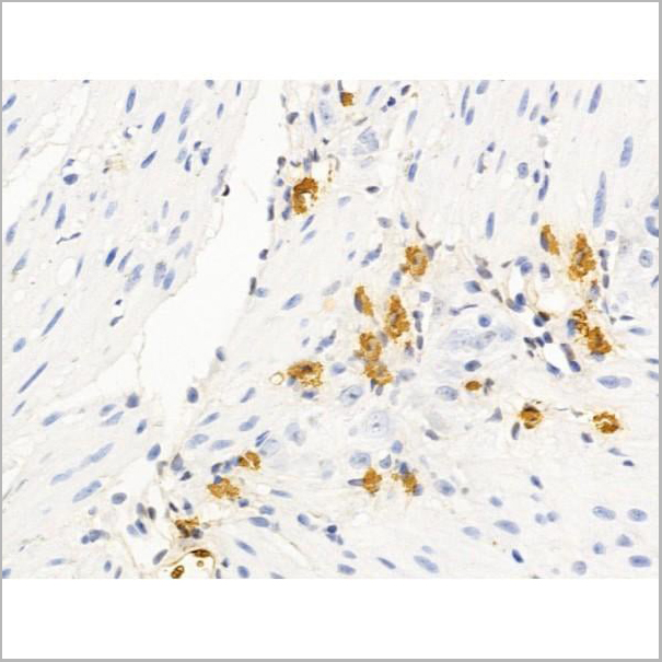
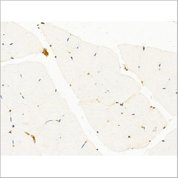
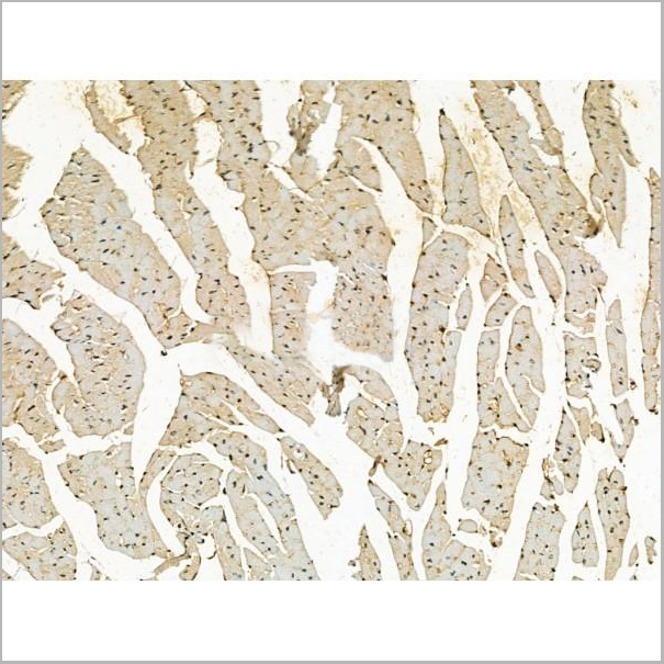
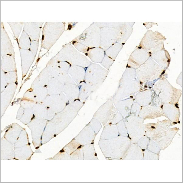
 IHC (Immunohistchemistry)
(At 1/100 staining Mouse heart tissue by IHC-P. The sample was formaldehyde fixed and a heat mediated antigen retrieval step in citrate buffer was performed. The sample was then blocked and incubated with the primary antibody at 4 degree C overnight. An HRP conjugated anti-Rabbit antibody was used as the secondary antibody.)
IHC (Immunohistchemistry)
(At 1/100 staining Mouse heart tissue by IHC-P. The sample was formaldehyde fixed and a heat mediated antigen retrieval step in citrate buffer was performed. The sample was then blocked and incubated with the primary antibody at 4 degree C overnight. An HRP conjugated anti-Rabbit antibody was used as the secondary antibody.)
Ephrin B1/B2/B3, Polyclonal Antibody (Cat# AAA31292)
Full Name
Phospho-Ephrin B1/B2/B3 (Tyr324) Antibody
Gene Names
EFNB1; CFND; CFNS; EFL3; EPLG2; Elk-L; LERK2
Reactivity
Human, Mouse, Rat
Applications
Immunohistochemistry, Peptide ELISA
Purity
Peptide affinity purification
Pricing


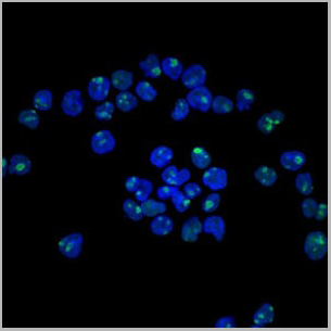
 IF (Immunofluorescence)
(AAA31213 staining Hela cells by IF/ICC. The samples were fixed with PFA and permeabilized in 0.1% Triton X-100,then blocked in 10% serum for 45 minutes at 25°C. Samples were then incubated with primary Ab(AAA31213 1:200) and mouse anti-beta tubulin Ab(T0023 1:200) for 1 hour at 37°C. An AlexaFluor594 conjugated goat anti-rabbit IgG(H+L) Ab(Red) and an AlexaFluor488 conjugated goat anti-mouse IgG(H+L) Ab(Green) were used as the secondary antibody.The nuclear counter stain is DAPI(blue).)
IF (Immunofluorescence)
(AAA31213 staining Hela cells by IF/ICC. The samples were fixed with PFA and permeabilized in 0.1% Triton X-100,then blocked in 10% serum for 45 minutes at 25°C. Samples were then incubated with primary Ab(AAA31213 1:200) and mouse anti-beta tubulin Ab(T0023 1:200) for 1 hour at 37°C. An AlexaFluor594 conjugated goat anti-rabbit IgG(H+L) Ab(Red) and an AlexaFluor488 conjugated goat anti-mouse IgG(H+L) Ab(Green) were used as the secondary antibody.The nuclear counter stain is DAPI(blue).)
Exportin-5, Polyclonal Antibody (Cat# AAA31213)
Full Name
Exportin-5 Antibody
Gene Names
XPO5; exp5
Reactivity
Human, Mouse, Rat
Predicted Reactivity: Pig(88%), Bovine(100%), Horse(100%), Sheep(100%), Rabbit(88%), Dog(100%), Chicken(100%)
Predicted Reactivity: Pig(88%), Bovine(100%), Horse(100%), Sheep(100%), Rabbit(88%), Dog(100%), Chicken(100%)
Applications
ELISA
Purity
The antiserum was purified by peptide affinity chromatography using SulfoLink Coupling Resin (Thermo Fisher Scientific).
Pricing
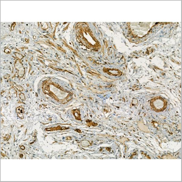

 IHC (Immunohistochemistry-Paraffin)
(AAA31179 at 1/100 staining human breast carcinoma tissue sections by IHC-P. The tissue was formaldehyde fixed and a heat mediated antigen retrieval step in citrate buffer was performed. The tissue was then blocked and incubated with the antibody for 1.5 hours at 22°C. An HRP conjugated goat anti-rabbit antibody was used as the secondary antibody.)
IHC (Immunohistochemistry-Paraffin)
(AAA31179 at 1/100 staining human breast carcinoma tissue sections by IHC-P. The tissue was formaldehyde fixed and a heat mediated antigen retrieval step in citrate buffer was performed. The tissue was then blocked and incubated with the antibody for 1.5 hours at 22°C. An HRP conjugated goat anti-rabbit antibody was used as the secondary antibody.)
CD36, Polyclonal Antibody (Cat# AAA31179)
Full Name
CD36 Antibody
Gene Names
CD36; FAT; GP4; GP3B; GPIV; CHDS7; PASIV; SCARB3; BDPLT10
Reactivity
Human, Mouse, Rat
Applications
Western Blot, Immunohistochemistry
Purity
Peptide affinity purification
Pricing

 IF (Immunofluorescence)
(AAA31230 staining Hela cells by IF/ICC. The samples were fixed with PFA and permeabilized in 0.1% Triton X-100,then blocked in 10% serum for 45 minutes at 25°C. Samples were then incubated with primary Ab(AAA31230 1:200) and mouse anti-beta tubulin Ab(T0023 1:200) for 1 hour at 37°C. An AlexaFluor594 conjugated goat anti-rabbit IgG(H+L) Ab(Red) and an AlexaFluor488 conjugated goat anti-mouse IgG(H+L) Ab(Green) were used as the secondary antibody.The nuclear counter stain is DAPI(blue).)
IF (Immunofluorescence)
(AAA31230 staining Hela cells by IF/ICC. The samples were fixed with PFA and permeabilized in 0.1% Triton X-100,then blocked in 10% serum for 45 minutes at 25°C. Samples were then incubated with primary Ab(AAA31230 1:200) and mouse anti-beta tubulin Ab(T0023 1:200) for 1 hour at 37°C. An AlexaFluor594 conjugated goat anti-rabbit IgG(H+L) Ab(Red) and an AlexaFluor488 conjugated goat anti-mouse IgG(H+L) Ab(Green) were used as the secondary antibody.The nuclear counter stain is DAPI(blue).)
ACTR1A, Polyclonal Antibody (Cat# AAA31230)
Full Name
ACTR1A Antibody
Gene Names
ACTR1A; ARP1; Arp1A; CTRN1
Reactivity
Human, Mouse, Rat
Predicted Reactivity: Pig(100%), Bovine(100%), Horse(100%), Sheep(100%), Rabbit(100%), Dog(100%)
Predicted Reactivity: Pig(100%), Bovine(100%), Horse(100%), Sheep(100%), Rabbit(100%), Dog(100%)
Applications
ELISA
Purity
The antiserum was purified by peptide affinity chromatography using SulfoLink Coupling Resin (Thermo Fisher Scientific).
Pricing
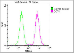
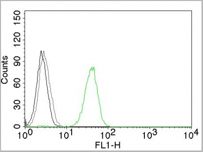
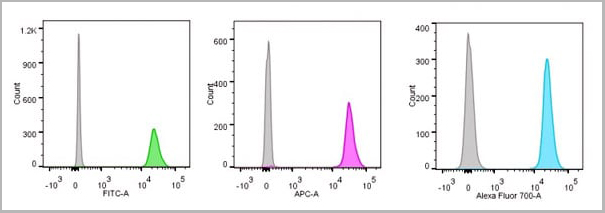
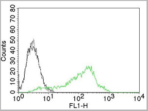
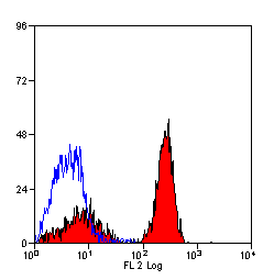
 FCM (Flow Cytometry)
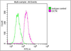
(Overlay histogram showing Ntera-2 cells stained with CSB-MA018403A0m (red line) at 1:100. The cells were incubated in 1x PBS /10% normal goat serum to block non-specific protein-protein interactions followed by primary antibody for 1 h at 4 degree C. The secondary antibody used was FITC goat anti-mouse IgG(H+L) at 1/200 dilution for 1 h at 4 degree C. Isotype control antibody (green line) was used under the same conditions. Acquisition of >10,000 events was performed.)
FCM (Flow Cytometry)
(Overlay histogram showing Ntera-2 cells stained with CSB-MA018403A0m (red line) at 1:100. The cells were incubated in 1x PBS /10% normal goat serum to block non-specific protein-protein interactions followed by primary antibody for 1 h at 4 degree C. The secondary antibody used was FITC goat anti-mouse IgG(H+L) at 1/200 dilution for 1 h at 4 degree C. Isotype control antibody (green line) was used under the same conditions. Acquisition of >10,000 events was performed.)
OCT4, Monoclonal Antibody (Cat# AAA27015)
Full Name
OCT4 Monoclonal Antibody
Gene Names
POU5F1; OCT3; OCT4; OTF3; OTF4; OTF-3; Oct-3; Oct-4
Reactivity
Human, Mouse, Rat
Applications
Western Blot, Immunohistochemistry, Immunofluorescence, Flow Cytometry
Purity
>95%
Protein G Purified
Protein G Purified
Pricing
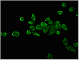
 IF (Immunofluorescence)
(AAA31177 staining Hela cells by IF/ICC. The samples were fixed with PFA and permeabilized in 0.1% Triton X-100,then blocked in 10% serum for 45 minutes at 25°C. Samples were then incubated with primary Ab(AAA31177 1:200) and mouse anti-beta tubulin Ab(T0023 1:200) for 1 hour at 37°C. An AlexaFluor594 conjugated goat anti-rabbit IgG(H+L) Ab(Red) and an AlexaFluor488 conjugated goat anti-mouse IgG(H+L) Ab(Green) were used as the secondary antibody.The nuclear counter stain is DAPI(blue).)
IF (Immunofluorescence)
(AAA31177 staining Hela cells by IF/ICC. The samples were fixed with PFA and permeabilized in 0.1% Triton X-100,then blocked in 10% serum for 45 minutes at 25°C. Samples were then incubated with primary Ab(AAA31177 1:200) and mouse anti-beta tubulin Ab(T0023 1:200) for 1 hour at 37°C. An AlexaFluor594 conjugated goat anti-rabbit IgG(H+L) Ab(Red) and an AlexaFluor488 conjugated goat anti-mouse IgG(H+L) Ab(Green) were used as the secondary antibody.The nuclear counter stain is DAPI(blue).)
Proteasome 20S LMP7, Polyclonal Antibody (Cat# AAA31177)
Full Name
Proteasome 20S LMP7 Antibody
Gene Names
PSMB8; JMP; ALDD; LMP7; NKJO; D6S216; PRAAS1; PSMB5i; RING10; D6S216E
Reactivity
Human, Mouse, Rat
Predicted Reactivity: Horse(86%), Rabbit(100%), Dog(88%)
Predicted Reactivity: Horse(86%), Rabbit(100%), Dog(88%)
Applications
Western Blot, Immunofluorescence, Immunocytochemistry
Purity
The antiserum was purified by peptide affinity chromatography using SulfoLink Coupling Resin (Thermo Fisher Scientific).
Pricing

 IF (Immunofluorescence)
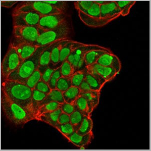
Nucleolin, Monoclonal Antibody (Cat# AAA13823)
Full Name
Nucleolin (Marker of Human Cells) Mouse Monoclonal Antibody
Gene Names
NCL; C23
Reactivity
Human.
Does not react with Mouse, Rat and Cow
Does not react with Mouse, Rat and Cow
Applications
Flow Cytometry, Immunofluorescence, Western Blot, Immunohistochemistry
Pricing
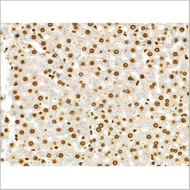
 IHC-P (Immunohistochemistry-Paraffin)
(At 1/200 staining Rat ganstric tissue sections by IHC-P. The tissue was formaldehyde fixed and a heat mediatedantigen retrieval step in citrate buffer was performed. Thetissue was then blocked and incubated with the antibody for1.5 hours at 22°C. An HRP conjugated goat anti-rabbitantibody was used as the secondary antibody.)
IHC-P (Immunohistochemistry-Paraffin)
(At 1/200 staining Rat ganstric tissue sections by IHC-P. The tissue was formaldehyde fixed and a heat mediatedantigen retrieval step in citrate buffer was performed. Thetissue was then blocked and incubated with the antibody for1.5 hours at 22°C. An HRP conjugated goat anti-rabbitantibody was used as the secondary antibody.)
c-Kit, Polyclonal Antibody (Cat# AAA31007)
Full Name
Phospho-c-Kit (Tyr721) Antibody
Gene Names
KIT; PBT; SCFR; C-Kit; CD117; MASTC
Reactivity
Human, Mouse, Rat
Applications
Western Blot, Immunohistochemistry, Immunofluorescence, Immunocytochemistry
Purity
Peptide affinity purification
Pricing
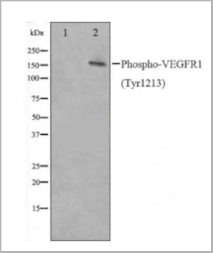
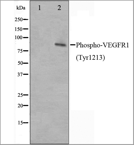
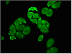
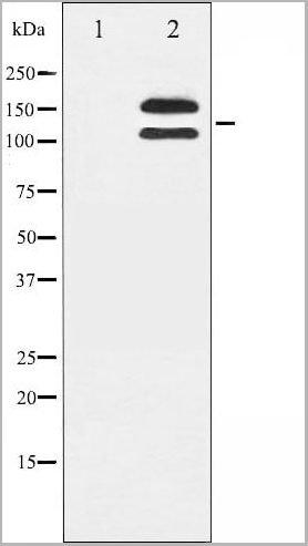
 Application Data
(AAA31020 staining HepG2 cells(30min of 4uM Forskolin treatment) by IF/ICC. The samples were fixed with PFA and permeabilized in 0.1% Triton X-100,then blocked in 10% serum for 45 minutes at 25°C. Samples were then incubated with primary Ab(AAA31020) and mouse anti-beta tubulin Ab for 1 hour at 37°C. An AlexaFluor594 conjugated goat anti-rabbit IgG(H+L) Ab(Red) and an AlexaFluor488 conjugated goat anti-mouse IgG(H+L) Ab(Green) were used as the secondary Ab. The nuclear counter stain is DAPI(blue))
Application Data
(AAA31020 staining HepG2 cells(30min of 4uM Forskolin treatment) by IF/ICC. The samples were fixed with PFA and permeabilized in 0.1% Triton X-100,then blocked in 10% serum for 45 minutes at 25°C. Samples were then incubated with primary Ab(AAA31020) and mouse anti-beta tubulin Ab for 1 hour at 37°C. An AlexaFluor594 conjugated goat anti-rabbit IgG(H+L) Ab(Red) and an AlexaFluor488 conjugated goat anti-mouse IgG(H+L) Ab(Green) were used as the secondary Ab. The nuclear counter stain is DAPI(blue))
VEGFR1, Polyclonal Antibody (Cat# AAA31020)
Full Name
Phospho-VEGFR1 (Tyr1213) Antibody
Gene Names
FLT1; FLT; FLT-1; VEGFR1; VEGFR-1
Reactivity
Human, Mouse, Rat
Applications
Western Blot, Immunofluorescence, Immunocytochemistry, Immunohistochemistry
Purity
The Ab is from purified rabbit serum by affinity purification via sequential chromatography on phospho-peptide and non-phospho-peptide affinity columns.
Pricing
 IHC (Immunohistochemistry)
(AAA31051 at 1/200 staining Human bladder cancer tissue sections by IHC-P. The tissue was formaldehyde fixed and a heat mediated antigen retrieval step in citrate buffer was performed. The tissue was then blocked and incubated with the antibody for 1.5 hours at 22 degree C. An HRP conjugated goat anti-rabbit antibody was used as the secondary.)
IHC (Immunohistochemistry)
(AAA31051 at 1/200 staining Human bladder cancer tissue sections by IHC-P. The tissue was formaldehyde fixed and a heat mediated antigen retrieval step in citrate buffer was performed. The tissue was then blocked and incubated with the antibody for 1.5 hours at 22 degree C. An HRP conjugated goat anti-rabbit antibody was used as the secondary.)
SYK, Polyclonal Antibody (Cat# AAA31051)
Full Name
Phospho-SYK (Tyr348) Antibody
Gene Names
SYK; p72-Syk
Reactivity
Human, Mouse, Rat, Monkey
Applications
Western Blot, Immunohistochemistry, Immunofluorescence, Immunocytochemistry
Purity
From purified rabbit serum by affinity purification via sequential chromatography on phospho-and non-phospho-peptide affinity columns.
Pricing
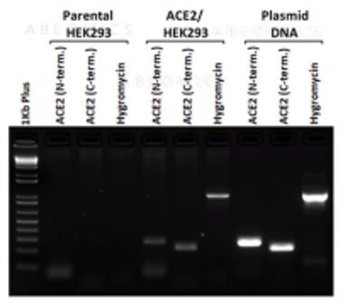
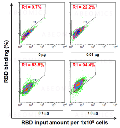
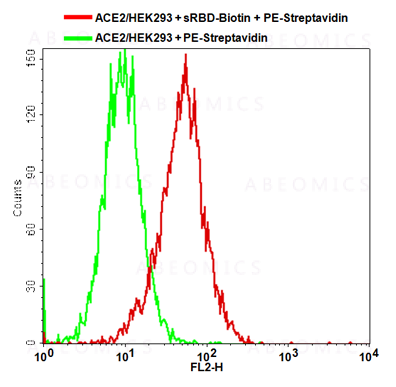
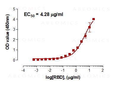
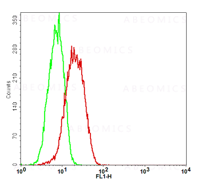 Application Data
(Fig-5: Cell Surface staining of human ACE2 in the ACE2/HEK293 stable cell line. ATTO 488-conjugated anti-hACE2 antibody (clone AC18F; was used at 1ug/ 1x10^6 cells (Red). ATTO 488-conjugated mouse IgG1 was used as isotype control at 1ug/ 1x10^6 cells (Green).)
Application Data
(Fig-5: Cell Surface staining of human ACE2 in the ACE2/HEK293 stable cell line. ATTO 488-conjugated anti-hACE2 antibody (clone AC18F; was used at 1ug/ 1x10^6 cells (Red). ATTO 488-conjugated mouse IgG1 was used as isotype control at 1ug/ 1x10^6 cells (Green).)
ACE2, Cell Line (Cat# AAA14888)
Full Name
ACE2/HEK293 Stable Cell Line
Gene Names
ACE2; ACEH
Applications
Functional Assay
Pricing
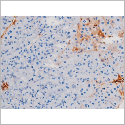
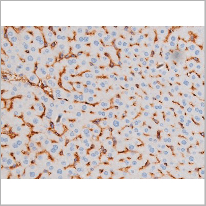
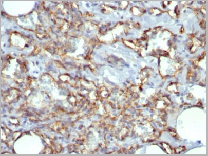
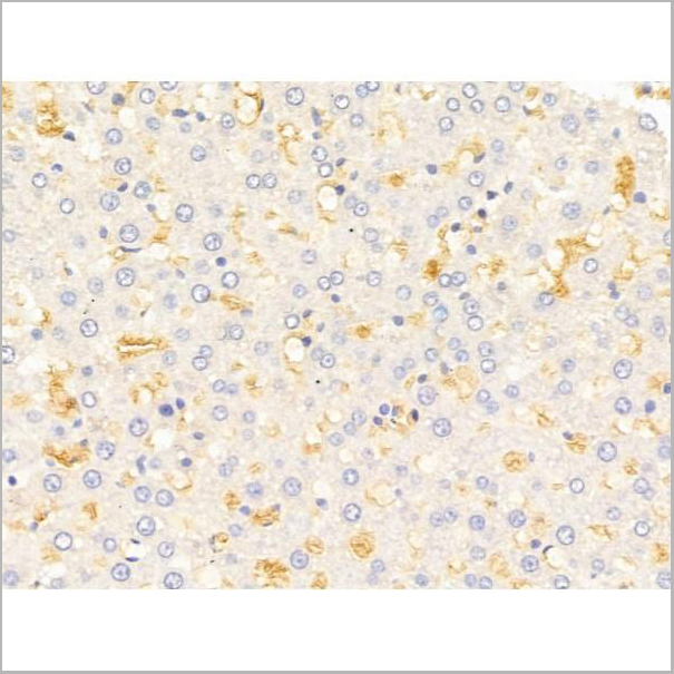
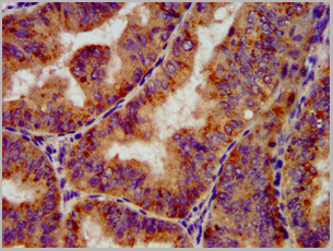
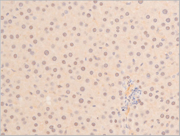
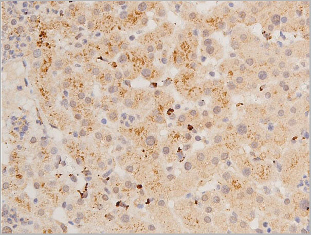
 IHC (Immunohistchemistry)
(Immunohistochemical analysis of paraffin-embedded human liver tissue using AAA28677 performed on the Leica® BOND RXm. Tissue was fixed with formaldehyde at room temperature; antigen retrieval was by heat mediation with a EDTA buffer (pH9. 0). Samples were incubated with primary antibody(1:1000) for 1 hours at room temperature. A undiluted biotinylated CRF Anti-Polyvalent HRP Polymer antibody was used as the secondary antibody.)
IHC (Immunohistchemistry)
(Immunohistochemical analysis of paraffin-embedded human liver tissue using AAA28677 performed on the Leica® BOND RXm. Tissue was fixed with formaldehyde at room temperature; antigen retrieval was by heat mediation with a EDTA buffer (pH9. 0). Samples were incubated with primary antibody(1:1000) for 1 hours at room temperature. A undiluted biotinylated CRF Anti-Polyvalent HRP Polymer antibody was used as the secondary antibody.)
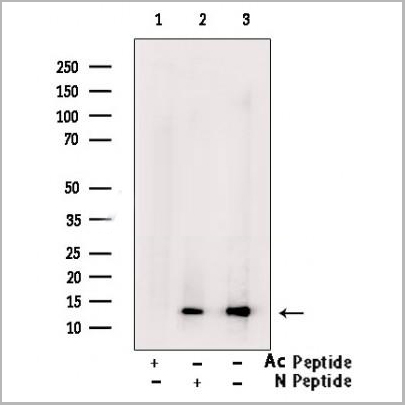
ATG7, Polyclonal Antibody (Cat# AAA28677)
Full Name
ATG7 Antibody (C-term)
Gene Names
ATG7; GSA7; APG7L; APG7-LIKE
Reactivity
Human, Mouse
Predicted reactivity: Chicken, Rat
Predicted reactivity: Chicken, Rat
Applications
Western Blot, Immunohistochemistry, Immunofluorescence
Purity
Purified Rabbit Polyclonal Antibody (Pab)
Pricing

 WB (Western Blot)
(Western blot analysis of p53 phosphorylation expression in Etoposide treated 293 whole cell lysates, The lane on the left is treated with the antigen-specific peptide.)
WB (Western Blot)
(Western blot analysis of p53 phosphorylation expression in Etoposide treated 293 whole cell lysates, The lane on the left is treated with the antigen-specific peptide.)
p53, Polyclonal Antibody (Cat# AAA30976)
Full Name
Phospho-p53 (Ser392) Antibody
Gene Names
TP53; P53; BCC7; LFS1; TRP53
Reactivity
Human, Mouse, Rat
Applications
Western Blot, Immunohistochemistry, Immunofluorescence, Immunocytochemistry
Purity
From purified rabbit serum by affinity purification via sequential chromatography on phospho-and non-phospho-peptide affinity columns.
Pricing
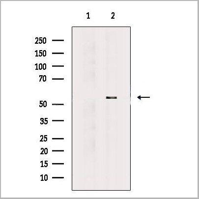
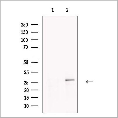
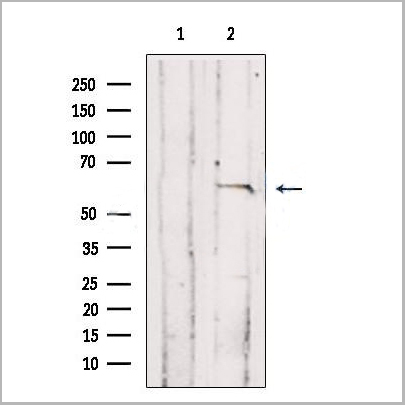
 WB (Western Blot)
(TGFBR2 Antibody western blot of mouse lung tissue lysates (35 ug/lane). The TGFBR2 antibody detected the TGFBR2 protein (arrow).)
WB (Western Blot)
(TGFBR2 Antibody western blot of mouse lung tissue lysates (35 ug/lane). The TGFBR2 antibody detected the TGFBR2 protein (arrow).)
TGFBR2, Polyclonal Antibody (Cat# AAA21269)
Full Name
Anti-TGFBR2 Antibody (aa13-40) IHC-plus
Gene Names
TGFBR2; AAT3; FAA3; LDS2; MFS2; RIIC; LDS1B; LDS2B; TAAD2; TGFR-2; TGFbeta-RII
Reactivity
Mouse, Human
Applications
Immunohistochemistry, Immunofluorescence, Western Blot
Purity
Protein A purified
Pricing
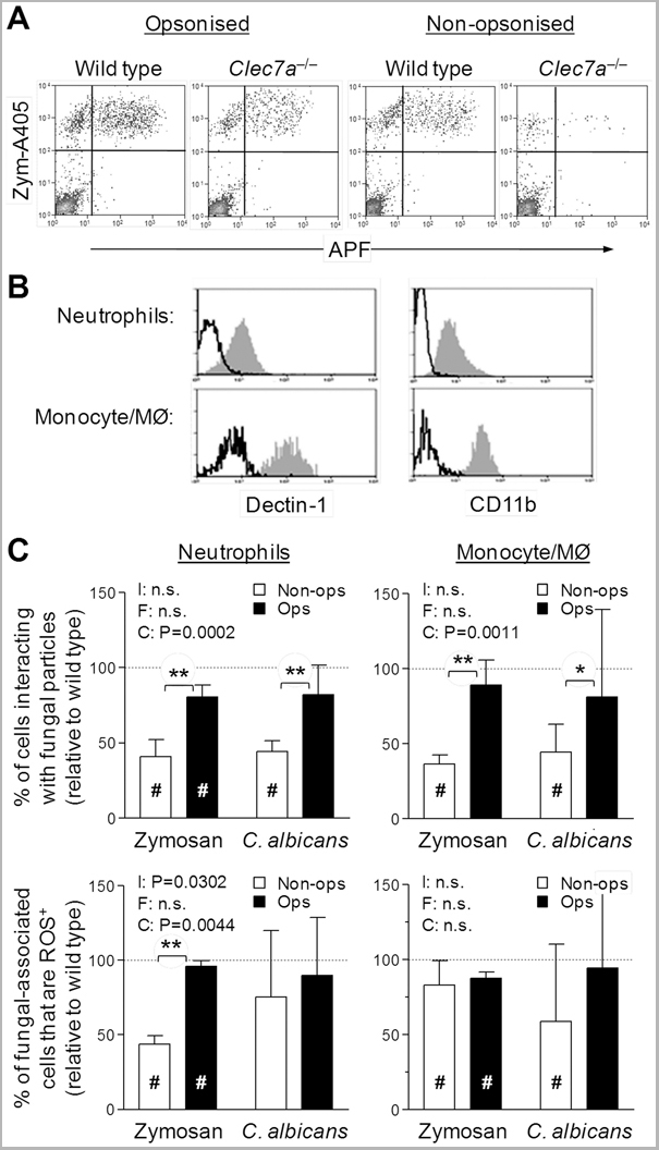
 Application Data
(Staining of mouse peritoneal macrophages with Rat anti Mouse Beta-glucan Receptor: FITC)
Application Data
(Staining of mouse peritoneal macrophages with Rat anti Mouse Beta-glucan Receptor: FITC)
DECTIN-1, Monoclonal Antibody (Cat# AAA12137)
Full Name
RAT ANTI MOUSE DECTIN-1
Gene Names
Clec7a; BGR; beta-GR; Clecsf12
Applications
Immunohistochemistry, Flow Cytometry, Immunoprecipitation
Pricing
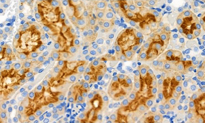
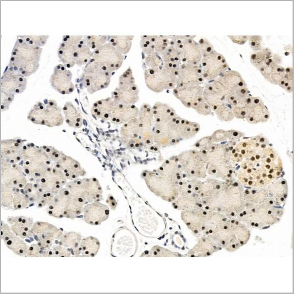
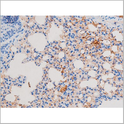
 IHC (Immunohistochemistry)
(At 1/100 staining Mouse liver tissue by IHC-P. The sample was formaldehyde fixed and a heat mediated antigen retrieval step in citrate buffer was performed. The sample was then blocked and incubated with the primary antibody at 4 degree C overnight. An HRP conjugated anti-Rabbit antibody was used as the secondary antibody.)
IHC (Immunohistochemistry)
(At 1/100 staining Mouse liver tissue by IHC-P. The sample was formaldehyde fixed and a heat mediated antigen retrieval step in citrate buffer was performed. The sample was then blocked and incubated with the primary antibody at 4 degree C overnight. An HRP conjugated anti-Rabbit antibody was used as the secondary antibody.)
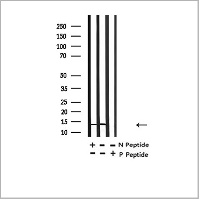
Histone H2B, Polyclonal Antibody (Cat# AAA31352)
Full Name
Acetyl-Histone H2B (Lys20) Antibody
Reactivity
Human, Mouse, Rat
Applications
Western Blot, Immunohistochemistry, Immunofluorescence, Immunocytochemistry, Peptide ELISA
Purity
The antiserum was purified by peptide affinity chromatography using SulfoLink Coupling Resin
Pricing
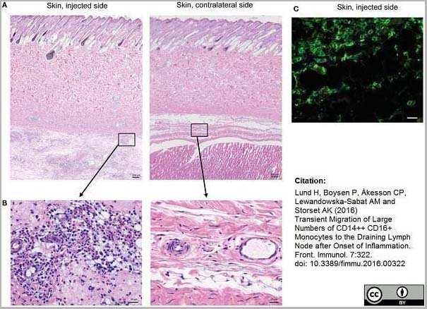 Application Data
(Mouse anti Human CD14 antibody, clone Tük4 used to identify bovine monocytes in the skin and subcutaneous tissue of adjuvant injected calves by immunofluorescence.Image caption:Cellular recruitment to skin and subcutaneous tissues. (A) HE stained sections of skin with subcutaneous tissue from the side injected with adjuvant and the contralateral side, at 24 h post-injection. Scale bars: 200 ?m. (B) Enlargement of outlined areas in A, as indicated. Scale bars: 20 ?m. (C) Immunofluorescent labeling of subcutaneous tissue on the injected side with antibody against CD14 (green). Scale bar: 20 ?m.From: Lund H, Boysen P, Åkesson CP, Lewandowska-Sabat AM and Storset AK (2016)Transient Migration of Large Numbers of CD14++ CD16+ Monocytes to the Draining Lymph Node after Onset of Inflammation.Front. Immunol. 7:322.This is from an open access article distributed under the terms of the Creative Commons Attribution License.)
Application Data
(Mouse anti Human CD14 antibody, clone Tük4 used to identify bovine monocytes in the skin and subcutaneous tissue of adjuvant injected calves by immunofluorescence.Image caption:Cellular recruitment to skin and subcutaneous tissues. (A) HE stained sections of skin with subcutaneous tissue from the side injected with adjuvant and the contralateral side, at 24 h post-injection. Scale bars: 200 ?m. (B) Enlargement of outlined areas in A, as indicated. Scale bars: 20 ?m. (C) Immunofluorescent labeling of subcutaneous tissue on the injected side with antibody against CD14 (green). Scale bar: 20 ?m.From: Lund H, Boysen P, Åkesson CP, Lewandowska-Sabat AM and Storset AK (2016)Transient Migration of Large Numbers of CD14++ CD16+ Monocytes to the Draining Lymph Node after Onset of Inflammation.Front. Immunol. 7:322.This is from an open access article distributed under the terms of the Creative Commons Attribution License.)
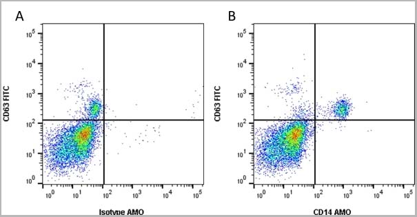
CD14, Monoclonal Antibody (Cat# AAA12266)
Full Name
Mouse Anti Human CD14: Amethyst Orange
Reactivity
Human
Applications
Flow Cytometry
Purity
Purified IgG prepared by affinity chromatography on Protein A from tissue culture supernatant.
Pricing


 Application Data
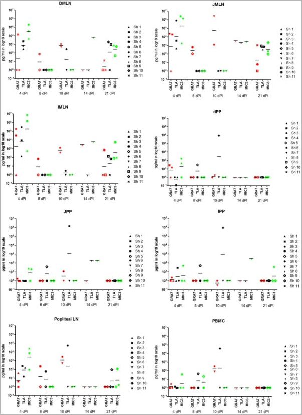
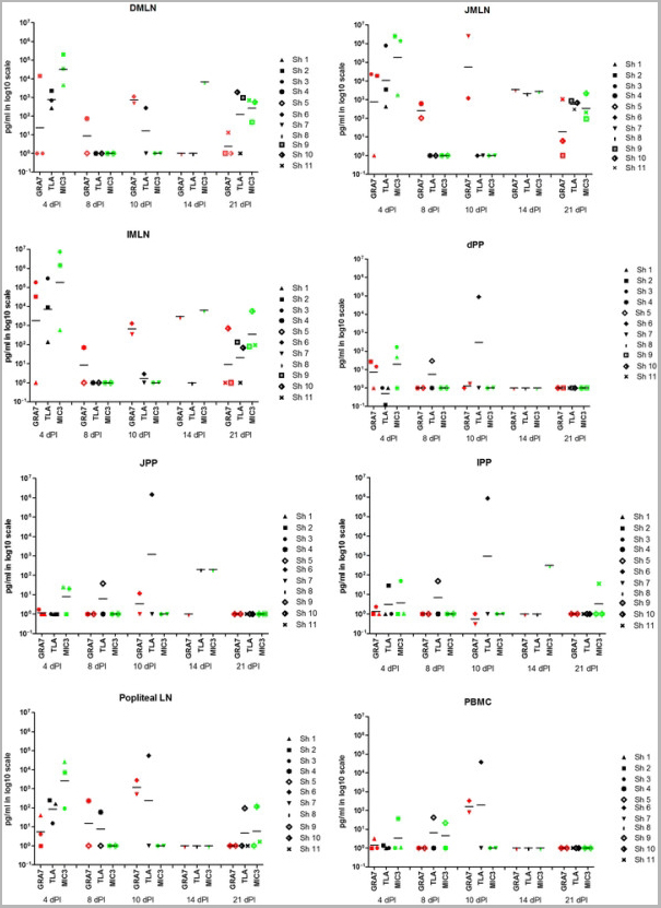
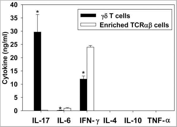
(Published customer image: gamma delta T cells are the primary source of IL-17 during B. abortus infection. C57BL/6 mice were infected i.p. with 5x10⁴ CFUs of B. abortus 2308, and two weeks later gamma delta T cells (>95% purity) and an enriched TCRalphabeta (~55% CD4+, 25% CD8+) cell fraction were isolated from the spleens of infected mice. Cells were stimulated with 500 ng/ml ionomycin and 50 ng/ml PMA for three days, and cell-free supernatants from triplicate wells were assayed for cytokine production via ELISA. The mean +/- SD is shown; * P)
Application Data
(Published customer image: gamma delta T cells are the primary source of IL-17 during B. abortus infection. C57BL/6 mice were infected i.p. with 5x10⁴ CFUs of B. abortus 2308, and two weeks later gamma delta T cells (>95% purity) and an enriched TCRalphabeta (~55% CD4+, 25% CD8+) cell fraction were isolated from the spleens of infected mice. Cells were stimulated with 500 ng/ml ionomycin and 50 ng/ml PMA for three days, and cell-free supernatants from triplicate wells were assayed for cytokine production via ELISA. The mean +/- SD is shown; * P)
IFN GAMMA, Monoclonal Antibody (Cat# AAA12095)
Full Name
MOUSE ANTI BOVINE INTERFERON GAMMA:FITC
Applications
Flow Cytometry
Pricing


 Application Data
(Published customer image: gamma delta T cells are the primary source of IL-17 during B. abortus infection. C57BL/6 mice were infected i.p. with 5x10⁴ CFUs of B. abortus 2308, and two weeks later gamma delta T cells (>95% purity) and an enriched TCRalphabeta (~55% CD4+, 25% CD8+) cell fraction were isolated from the spleens of infected mice. Cells were stimulated with 500 ng/ml ionomycin and 50 ng/ml PMA for three days, and cell-free supernatants from triplicate wells were assayed for cytokine production via ELISA. The mean +/- SD is shown; * P)
Application Data
(Published customer image: gamma delta T cells are the primary source of IL-17 during B. abortus infection. C57BL/6 mice were infected i.p. with 5x10⁴ CFUs of B. abortus 2308, and two weeks later gamma delta T cells (>95% purity) and an enriched TCRalphabeta (~55% CD4+, 25% CD8+) cell fraction were isolated from the spleens of infected mice. Cells were stimulated with 500 ng/ml ionomycin and 50 ng/ml PMA for three days, and cell-free supernatants from triplicate wells were assayed for cytokine production via ELISA. The mean +/- SD is shown; * P)
IFN GAMMA, Monoclonal Antibody (Cat# AAA12096)
Full Name
MOUSE ANTI BOVINE INTERFERON GAMMA:RPE
Applications
Flow Cytometry
Pricing


 Application Data
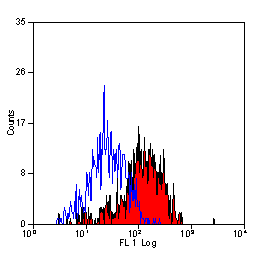
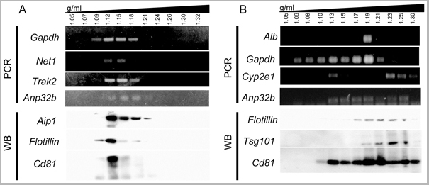
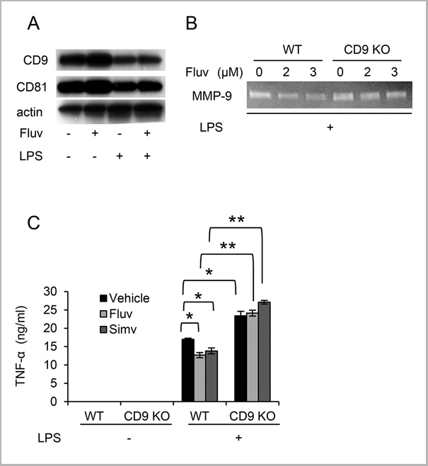
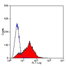
(Staining of mouse spleen with Hamster anti Mouse CD81: Alexa Fluor 488)
Application Data
(Staining of mouse spleen with Hamster anti Mouse CD81: Alexa Fluor 488)
CD81, Monoclonal Antibody (Cat# AAA12033)
Full Name
HAMSTER ANTI MOUSE CD81:RPE
Gene Names
Cd81; Tapa1; Tapa-1; Tspan28
Applications
Flow Cytometry
Pricing


 Application Data
(PE conjugatedMouse anti Human CD169 antibody, clone 7-239 used to block CD169 function on myeloid cells.Image caption:Siglec-1 mediates HIV-1 uptake into a storage compartment and enhances HIV-1 trans-infection specially in IFN?-treated monocytes and DCs. A. Uptake of HIV-1NL4–3 by different myeloid cells exposed to IFN?. Cells were cultured with HIV-1 to measure p24Gag by ELISA. Mean values and SEM from four experiments include cells from 12 donors. B. Fold change in HIV-1NL4–3 uptake of cells treated with bafilomycin A1 compared to untreated cells. Mean values and SEM include cells from three donors. C. Relative uptake of HIV-1NL4–3 by IFN?-treated myeloid cells pre-incubated with the indicated mAbs. Values are normalized to the level of HIV-1 uptake by mock-treated cells (set at 100%). Mean values and SEM from two experiments include cells from six donors. D. Confocal microscopy analysis of different IFN?-treated myeloid cells pulsed with HIV-1Cherry and stained for Siglec-1 (Alexa 488), HLA-DR (Alexa 647) and DAPI. (Top) Representative viral pattern for each kind of myeloid cell analyzed, showing maximum fluorescence intensity of four channels. (Bottom) Percentage of myeloid cells with distinct viral patterns: random distribution, polarized accumulation, and sac-like compartment formation, as illustrated in the left drawing. Mean values of 50 cells from two different donors are shown. E. HIV-1 transmission from IFN?-treated myeloid cells to a luciferase reporter CD4+ cell line. HIV-1 infection was determined by induced luciferase activity in relative light units (RLUs). Mean values and SEM from four experiments include cells from 12 donors. F. Relative HIV-1 transmission from IFN?-treated myeloid cells pre-incubated with the indicated mAbs. Values are normalized to the level of HIV-1 trans-infected by mock-treated cells. Mean values and SEM from two experiments include cells from six donors. Statistical differences were assessed with a paired t test in A and E, and with a one sample t-test in B, C and F.From: Pino M, Erkizia I, Benet S, Erikson E, Fernández-Figueras MT, Guerrero D, Dalmau J, Ouchi D, Rausell A, Ciuffi A, Keppler OT, Telenti A, Kräusslich HG, Martinez-Picado J, Izquierdo-Useros N.HIV-1 immune activation induces Siglec-1 expression and enhances viral trans-infection in blood and tissue myeloid cells.Retrovirology. 2015 May 7;12:37.This image is from an open access article distributed under the terms of the Creative Commons Attribution License.)
Application Data
(PE conjugatedMouse anti Human CD169 antibody, clone 7-239 used to block CD169 function on myeloid cells.Image caption:Siglec-1 mediates HIV-1 uptake into a storage compartment and enhances HIV-1 trans-infection specially in IFN?-treated monocytes and DCs. A. Uptake of HIV-1NL4–3 by different myeloid cells exposed to IFN?. Cells were cultured with HIV-1 to measure p24Gag by ELISA. Mean values and SEM from four experiments include cells from 12 donors. B. Fold change in HIV-1NL4–3 uptake of cells treated with bafilomycin A1 compared to untreated cells. Mean values and SEM include cells from three donors. C. Relative uptake of HIV-1NL4–3 by IFN?-treated myeloid cells pre-incubated with the indicated mAbs. Values are normalized to the level of HIV-1 uptake by mock-treated cells (set at 100%). Mean values and SEM from two experiments include cells from six donors. D. Confocal microscopy analysis of different IFN?-treated myeloid cells pulsed with HIV-1Cherry and stained for Siglec-1 (Alexa 488), HLA-DR (Alexa 647) and DAPI. (Top) Representative viral pattern for each kind of myeloid cell analyzed, showing maximum fluorescence intensity of four channels. (Bottom) Percentage of myeloid cells with distinct viral patterns: random distribution, polarized accumulation, and sac-like compartment formation, as illustrated in the left drawing. Mean values of 50 cells from two different donors are shown. E. HIV-1 transmission from IFN?-treated myeloid cells to a luciferase reporter CD4+ cell line. HIV-1 infection was determined by induced luciferase activity in relative light units (RLUs). Mean values and SEM from four experiments include cells from 12 donors. F. Relative HIV-1 transmission from IFN?-treated myeloid cells pre-incubated with the indicated mAbs. Values are normalized to the level of HIV-1 trans-infected by mock-treated cells. Mean values and SEM from two experiments include cells from six donors. Statistical differences were assessed with a paired t test in A and E, and with a one sample t-test in B, C and F.From: Pino M, Erkizia I, Benet S, Erikson E, Fernández-Figueras MT, Guerrero D, Dalmau J, Ouchi D, Rausell A, Ciuffi A, Keppler OT, Telenti A, Kräusslich HG, Martinez-Picado J, Izquierdo-Useros N.HIV-1 immune activation induces Siglec-1 expression and enhances viral trans-infection in blood and tissue myeloid cells.Retrovirology. 2015 May 7;12:37.This image is from an open access article distributed under the terms of the Creative Commons Attribution License.)
CD169, Monoclonal Antibody (Cat# AAA12265)
Full Name
Mouse Anti Human CD169: RPE
Gene Names
SIGLEC1; SN; CD169; SIGLEC-1
Reactivity
Human
Applications
Flow Cytometry
Purity
>95% by SDS PAGE
Purified IgG prepared by affinity chromatography on Protein A from tissue culture supernatant.
Purified IgG prepared by affinity chromatography on Protein A from tissue culture supernatant.
Pricing


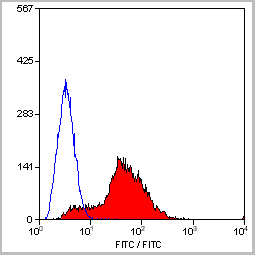
 Application Data
(Staining of mouse spleen with Hamster anti Mouse CD81: Alexa Fluor 488)
Application Data
(Staining of mouse spleen with Hamster anti Mouse CD81: Alexa Fluor 488)
CD81, Monoclonal Antibody (Cat# AAA11869)
Full Name
HAMSTER ANTI MOUSE CD81:FITC
Gene Names
Cd81; Tapa1; Tapa-1; Tspan28
Applications
Flow Cytometry
Pricing
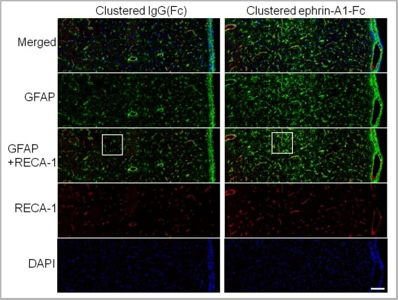 Application Data
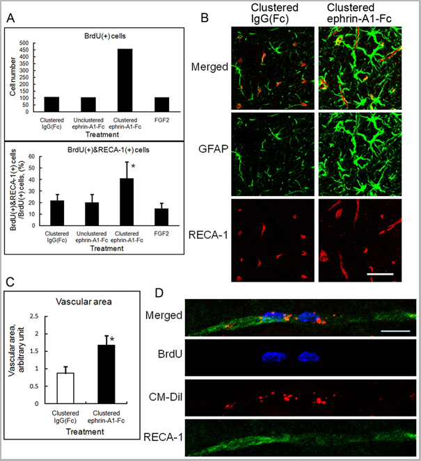
(Published customer image: Effect of clustered ephrin-A1-Fc on vascular formation in the rat striatum. Clustered ephrin-A1-Fc was injected into the lesioned side of the lateral ventricle in the unilaterally lesioned rats. Brains taken 6 weeks after injection were sectioned coronally and stained for GFAP (green) and RECA-1 (red) and with DAPI (nuclei; blue). The rectangular insets are shown in Fig. 8B. Scale bar: 100 um.From: Jing X, Miwa H, Sawada T, Nakanishi I, Kondo T, et al. (2012) Ephrin-A1-Mediated Dopaminergic Neurogenesis and Angiogenesis in a Rat Model of Parkinson's Disease. PLoS ONE 7(2): e32019.)
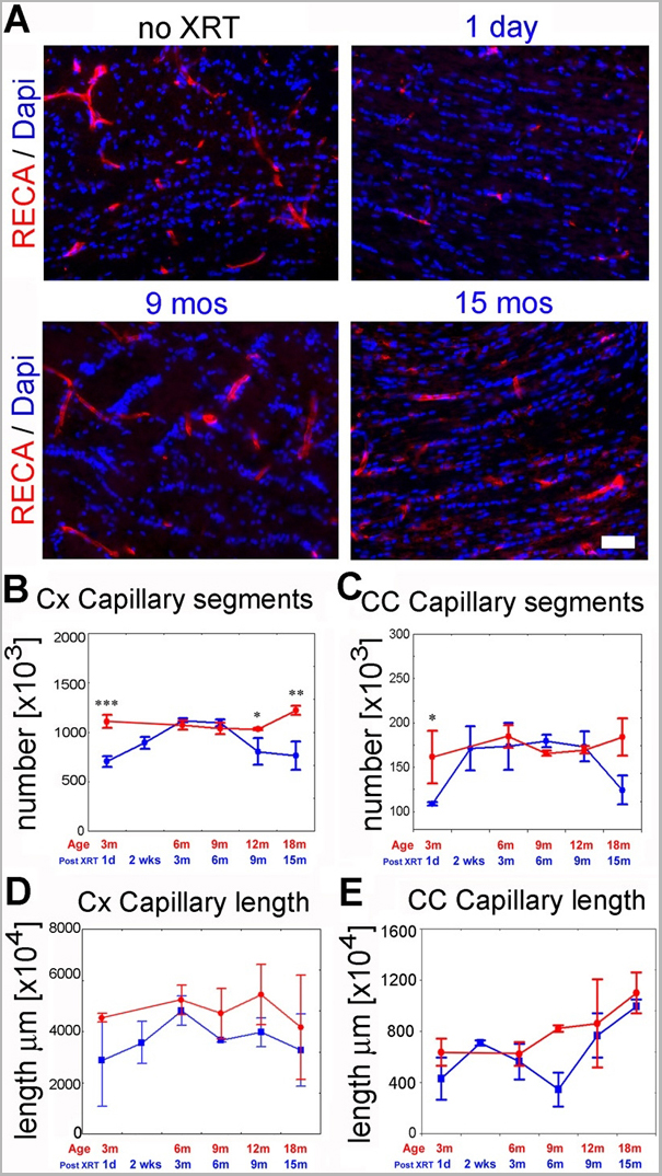
Application Data
(Published customer image: Effect of clustered ephrin-A1-Fc on vascular formation in the rat striatum. Clustered ephrin-A1-Fc was injected into the lesioned side of the lateral ventricle in the unilaterally lesioned rats. Brains taken 6 weeks after injection were sectioned coronally and stained for GFAP (green) and RECA-1 (red) and with DAPI (nuclei; blue). The rectangular insets are shown in Fig. 8B. Scale bar: 100 um.From: Jing X, Miwa H, Sawada T, Nakanishi I, Kondo T, et al. (2012) Ephrin-A1-Mediated Dopaminergic Neurogenesis and Angiogenesis in a Rat Model of Parkinson's Disease. PLoS ONE 7(2): e32019.)
RECA-1, Monoclonal Antibody (Cat# AAA12019)
Full Name
MOUSE ANTI RAT RECA-1
Applications
Immunohistochemistry, Immunofluorescence
Pricing