Filters
Clonality
Type
Reactivity
Gene Name
Isotype
Host
Application
Clone
1009 results for " Cell membrane" - showing 700-750
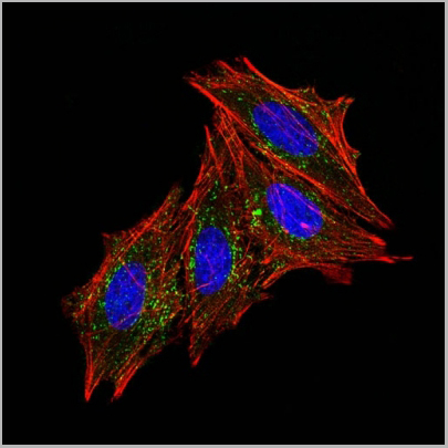
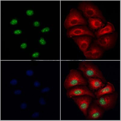
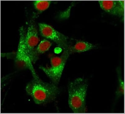 IF (Immunofluorescence)
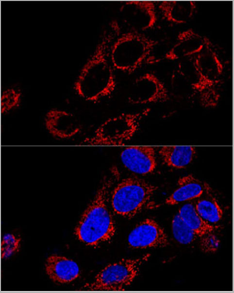
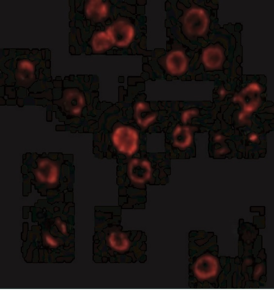
(Immunofluorescence Analysis of PFA-fixed U87MG cells stained using CD63 Mouse Monoclonal Antibody (MX-49.129.5) followed by goat anti-mouse IgG-CF488 (green). CF640R phalloidin (red).)
IF (Immunofluorescence)
(Immunofluorescence Analysis of PFA-fixed U87MG cells stained using CD63 Mouse Monoclonal Antibody (MX-49.129.5) followed by goat anti-mouse IgG-CF488 (green). CF640R phalloidin (red).)
CD63, Monoclonal Antibody (Cat# AAA13803)
Full Name
CD63 (Late Endosomes Marker) Mouse Monoclonal Antibody
Gene Names
CD63; MLA1; ME491; LAMP-3; OMA81H; TSPAN30
Reactivity
Human, Mouse
Applications
Flow Cytometry, Immunofluorescence, Western Blot, Immunohistochemistry
Pricing


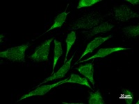
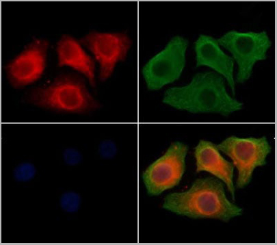
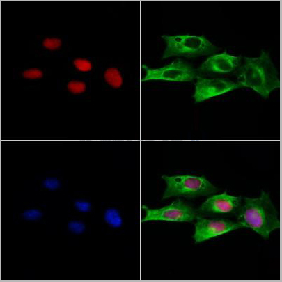
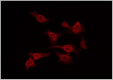
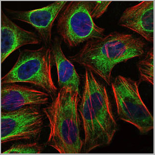
 ICC (Immunocytochemistry)
(Immunostaining analysis in HeLa cells. HeLa cells were fixed with 4% paraformaldehyde and permeabilized with 0.1% Triton X-100 in PBS. The cells were immunostained with anti-YARS mAb. [Lot No. YAR5H08-1])
ICC (Immunocytochemistry)
(Immunostaining analysis in HeLa cells. HeLa cells were fixed with 4% paraformaldehyde and permeabilized with 0.1% Triton X-100 in PBS. The cells were immunostained with anti-YARS mAb. [Lot No. YAR5H08-1])
YARS, Monoclonal Antibody (Cat# AAA10598)
Full Name
Mouse monoclonal antibody Anti-Human YARS
Gene Names
YARS; YRS; YTS; TYRRS; CMTDIC
Reactivity
Human
Applications
Immunocytochemistry, Immunoprecipitation, Western Blot, Flow Cytometry
Pricing
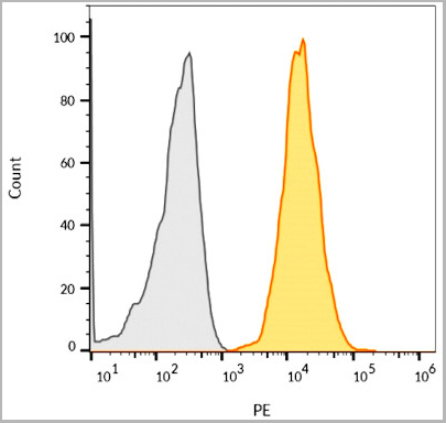
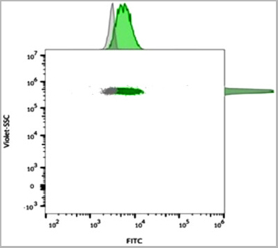
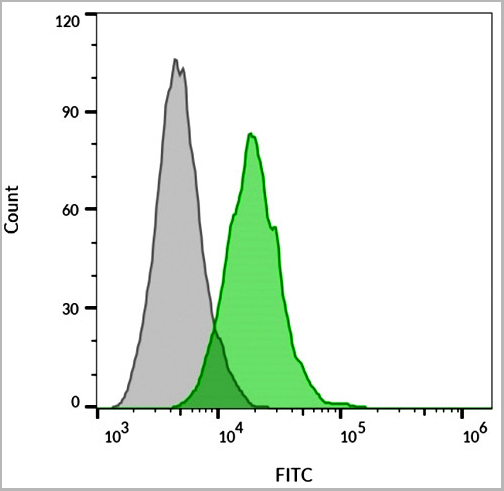
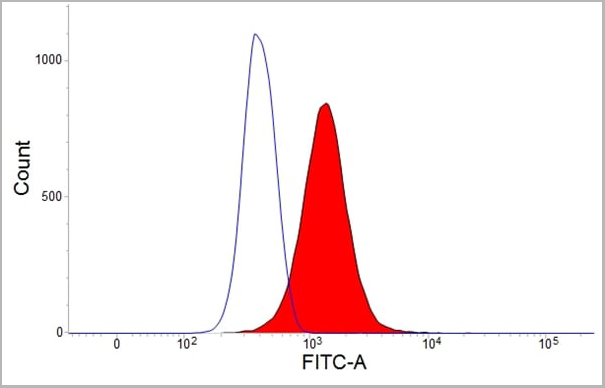
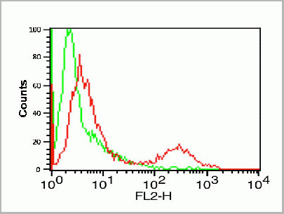
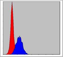
 FCM (Flow Cytometry)
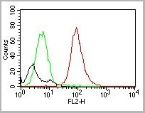
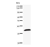
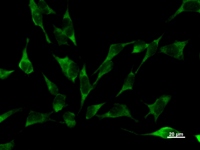
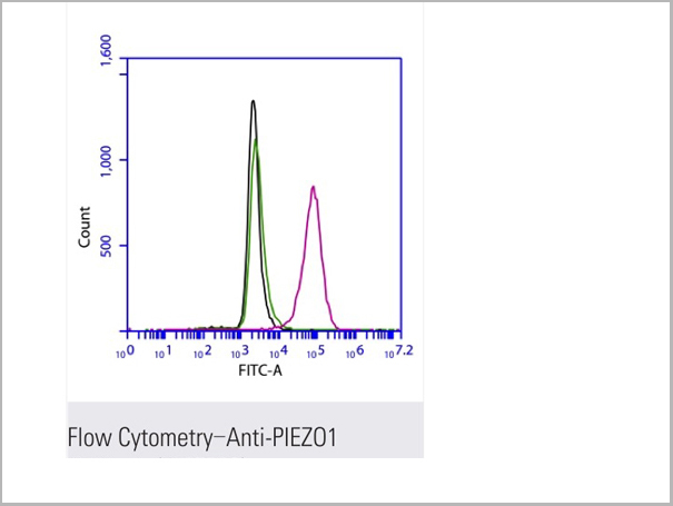
(Flow cytometric analysis of 2% paraformaldehyde-fixed THP1 (Human acute monocytic leukemia cell line) cells labeling PIEZO1 with AAA13800 at 1/200 dilution (red) compared with a mouse monoclonal IgG isotype control (black) and an unlabelled control (cells without incubation with primary antibody, green). Goat anti-mouse IgG (FITC) at 1/300 dilution was used as the secondary antibody.)
FCM (Flow Cytometry)
(Flow cytometric analysis of 2% paraformaldehyde-fixed THP1 (Human acute monocytic leukemia cell line) cells labeling PIEZO1 with AAA13800 at 1/200 dilution (red) compared with a mouse monoclonal IgG isotype control (black) and an unlabelled control (cells without incubation with primary antibody, green). Goat anti-mouse IgG (FITC) at 1/300 dilution was used as the secondary antibody.)
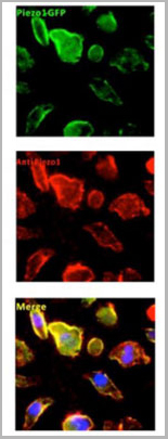
PIEZO1, Monoclonal Antibody (Cat# AAA13800)
Full Name
PIEZO1 Mouse mAb
Gene Names
PIEZO1; DHS; Mib; LMPH3; FAM38A
Reactivity
Homo Sapiens
Applications
Immunoprecipitation, Immunofluorescence, Flow Cytometry
Pricing
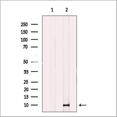
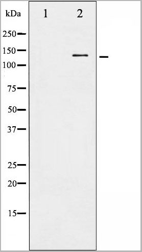
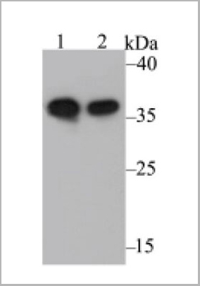 WB (Western Blot)
(Fig1: Western blot analysis of Annexin A1 on different lysates. Proteins were transferred to a PVDF membrane and blocked with 5% BSA in PBS for 1 hour at room temperature. The primary antibody was used at a 1:500 dilution in 5% BSA at room temperature for 2 hours. Goat Anti-Rabbit IgG - HRP Secondary Antibody at 1:5,000 dilution was used for 1 hour at room temperature.Positive control:Lane 1: A431 cell lysateLane 2: Rat uterus tissue lysate)
WB (Western Blot)
(Fig1: Western blot analysis of Annexin A1 on different lysates. Proteins were transferred to a PVDF membrane and blocked with 5% BSA in PBS for 1 hour at room temperature. The primary antibody was used at a 1:500 dilution in 5% BSA at room temperature for 2 hours. Goat Anti-Rabbit IgG - HRP Secondary Antibody at 1:5,000 dilution was used for 1 hour at room temperature.Positive control:Lane 1: A431 cell lysateLane 2: Rat uterus tissue lysate)
Annexin A1 Autoantibody (Anti-ANXA1), ELISA Kit (Cat# AAA10593)
Full Name
Human Annexin A1 Autoantibody (Anti-ANXA1) ELISA Kit
Reactivity
Human, Mouse, Rat
Applications
Western Blot, Immunocytochemistry, Immunohistochemistry
Pricing
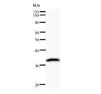
 WB (Western Blot)
TFEB, Monoclonal Antibody (Cat# AAA10615)
Full Name
Mouse monoclonal antibody Anti-Human TFEB
Gene Names
TFEB; TCFEB; BHLHE35; ALPHATFEB
Applications
Western Blot
Purity
This antibody was purified using protein A column chromatography from ascites.
Pricing


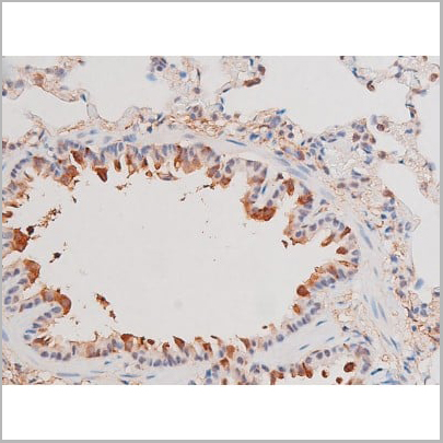
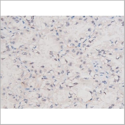
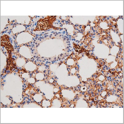
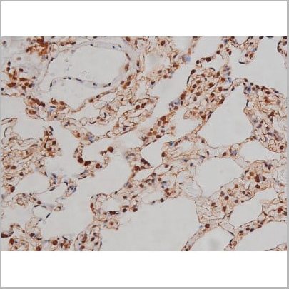
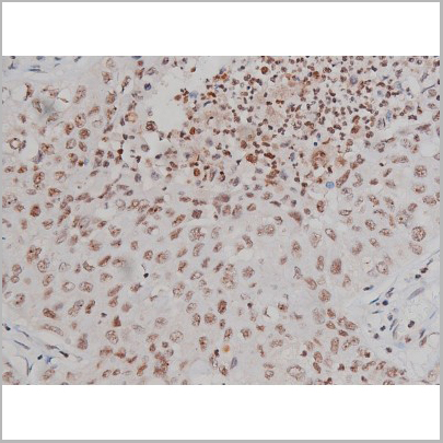
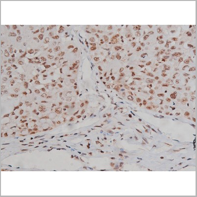
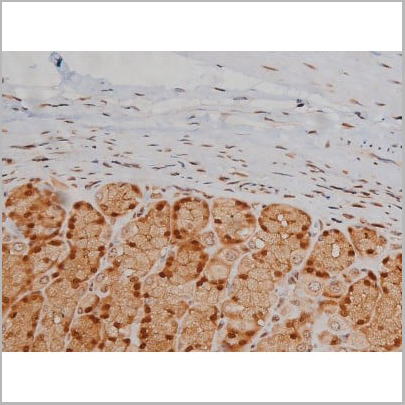
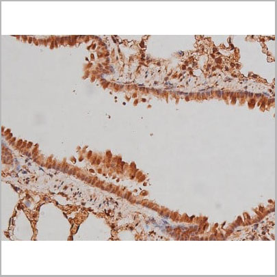
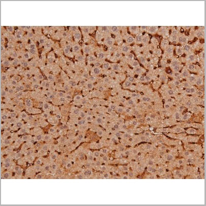
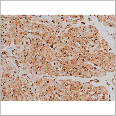
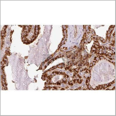
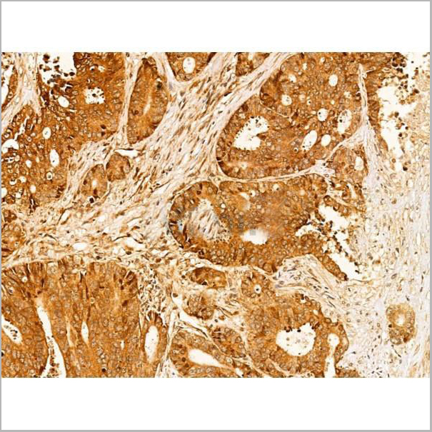
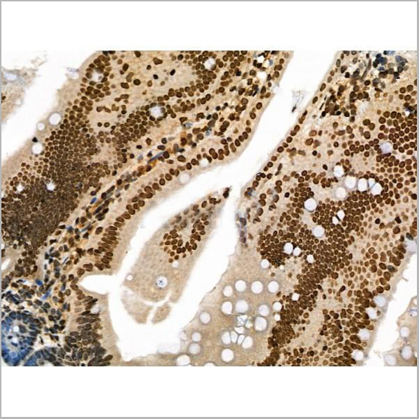
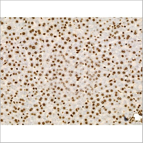
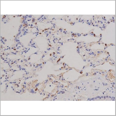
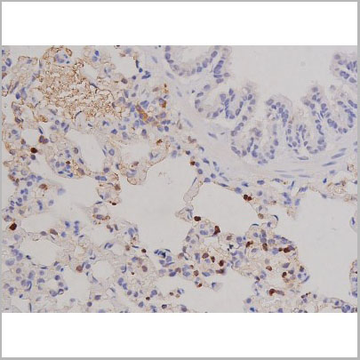
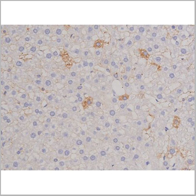
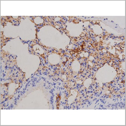
 IHC (Immunohistochemistry)
(Immunohistochemistry of paraffin-embedded human rectal cancer using MMP25 antibody at dilution of 1:200 (40x lens).)
IHC (Immunohistochemistry)
(Immunohistochemistry of paraffin-embedded human rectal cancer using MMP25 antibody at dilution of 1:200 (40x lens).)
MMP25, Polyclonal Antibody (Cat# AAA10688)
Full Name
MMP25 Polyclonal Antibody
Gene Names
MMP25; MMP20; MMPL1; MMP-25; MMP20A; MT6MMP; MTMMP6; MT-MMP6; MT6-MMP; MT-MMP 6
Reactivity
Human
Applications
Western Blot, Immunohistochemistry
Purity
Affinity Purification
Pricing
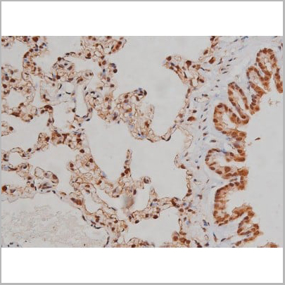
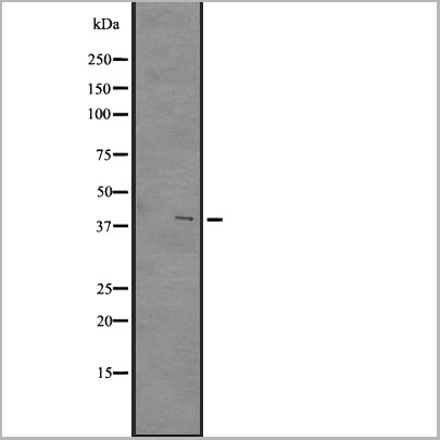 WB (Western Blot)
(Western blot analysis of GPR84 expression in A431 whole cell lysates)
WB (Western Blot)
(Western blot analysis of GPR84 expression in A431 whole cell lysates)
GPR84, Polyclonal Antibody (Cat# AAA31105)
Full Name
GPR84 Antibody
Gene Names
GPR84; EX33; GPCR4
Reactivity
Human, Mouse
Applications
Western Blot, Immunohistochemistry
Purity
The antiserum was purified by peptide affinity chromatography using SulfoLinkTM Coupling Resin (Thermo Fisher Scientific).
Pricing
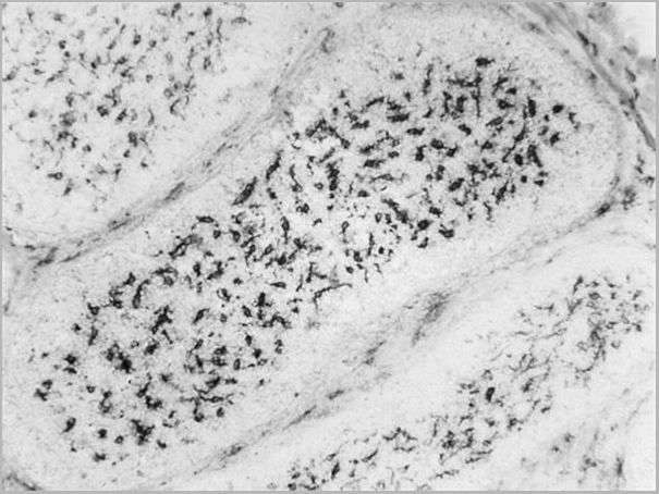
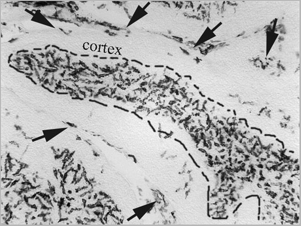
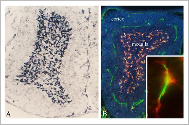
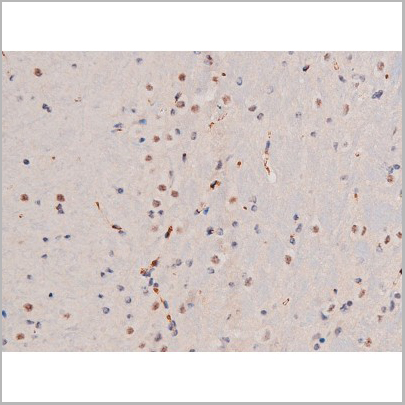
 Application Data
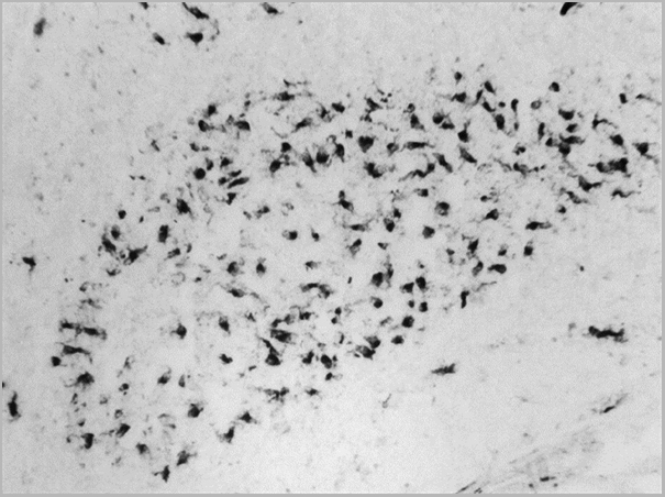
(Mouse anti Chicken CSF1R (CD115) immunohistochemical staining of guinea fowl, Agelastes meleagredisbursa of Fabricius.Clone ROS-AV170 specifically stains the bursal secretory dendritic cells (BSDC) located in the medulla. Clone ROS-AV170 also recognizes some CSF1R+ve macrophages in the interfollicular connective tissue. Image courtesy of Dr Nandor Nagy, Semmelweis University, Hungary)
Application Data
(Mouse anti Chicken CSF1R (CD115) immunohistochemical staining of guinea fowl, Agelastes meleagredisbursa of Fabricius.Clone ROS-AV170 specifically stains the bursal secretory dendritic cells (BSDC) located in the medulla. Clone ROS-AV170 also recognizes some CSF1R+ve macrophages in the interfollicular connective tissue. Image courtesy of Dr Nandor Nagy, Semmelweis University, Hungary)
CSF1R, Monoclonal Antibody (Cat# AAA12252)
Full Name
MOUSE ANTI CHICKEN CSF1R
Gene Names
CSF1R; FMS; CSFR; FIM2; HDLS; C-FMS; CD115; CSF-1R; M-CSF-R
Reactivity
Chicken
Applications
Immunohistochemistry, Flow Cytometry, Immunofluorescence
Pricing
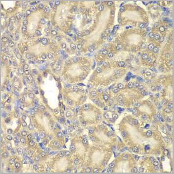
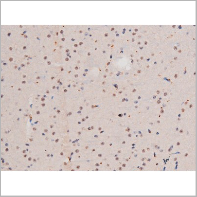
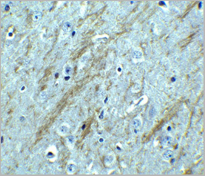
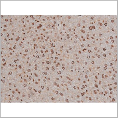
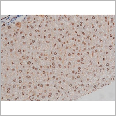
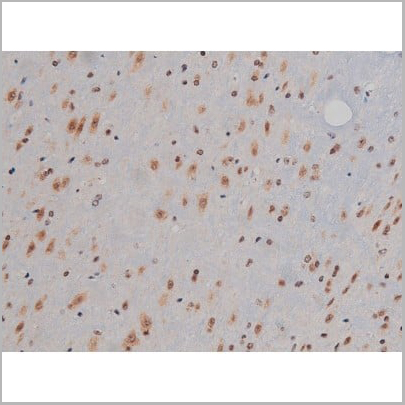
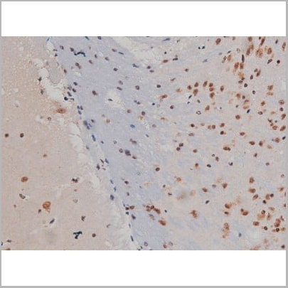
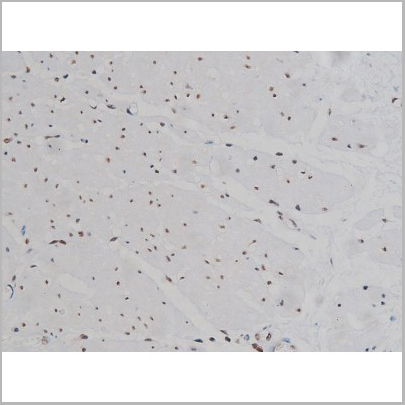
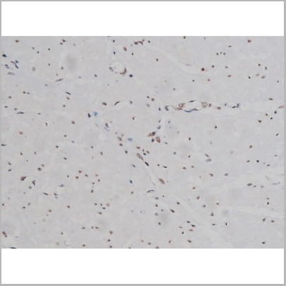
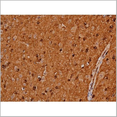
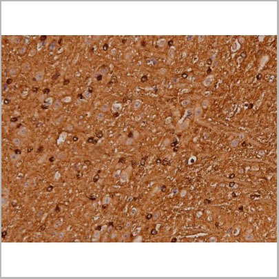
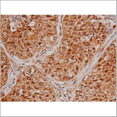
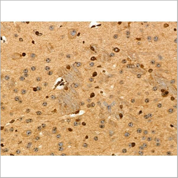
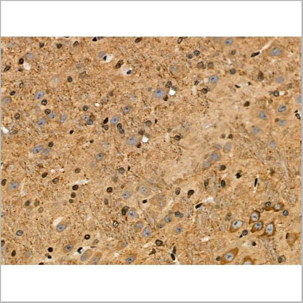
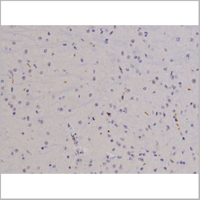
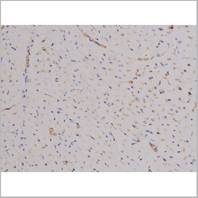
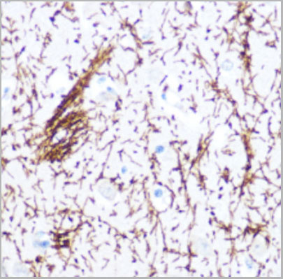 IHC (Immunohistchemistry)
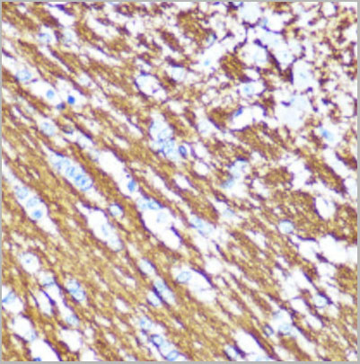
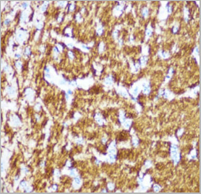
(Immunohistochemistry of paraffin-embedded human brain using Myelin Basic Protein Rabbit pAb (AAA10723) at dilution of 1:100 (40x lens).)
IHC (Immunohistchemistry)
(Immunohistochemistry of paraffin-embedded human brain using Myelin Basic Protein Rabbit pAb (AAA10723) at dilution of 1:100 (40x lens).)
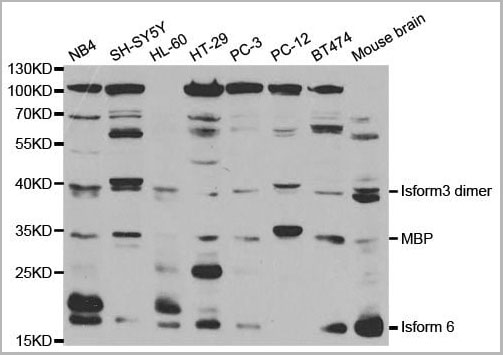
MBP, Polyclonal Antibody (Cat# AAA10723)
Full Name
MBP Polyclonal Antibody
Applications
Western Blot, Immunohistochemistry
Purity
Affinity Purification
Pricing
 IF (Immunofluorescence)
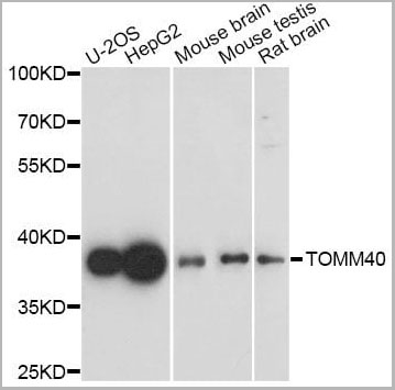
(Confocal immunofluorescence analysis of U2OS cells using TOMM40 Polyclonal Antibody at dilution of 1:200. Blue: DAPI for nuclear staining.)
IF (Immunofluorescence)
(Confocal immunofluorescence analysis of U2OS cells using TOMM40 Polyclonal Antibody at dilution of 1:200. Blue: DAPI for nuclear staining.)
TOMM40, Polyclonal Antibody (Cat# AAA10745)
Full Name
TOMM40 Polyclonal Antibody
Gene Names
TOMM40; TOM40; PEREC1; C19orf1; PER-EC1; D19S1177E
Applications
Western Blot, Immunohistochemistry, Immunofluorescence, Immunocytochemistry
Purity
Affinity Purification
Pricing
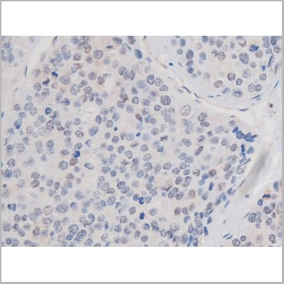
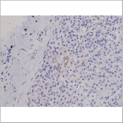
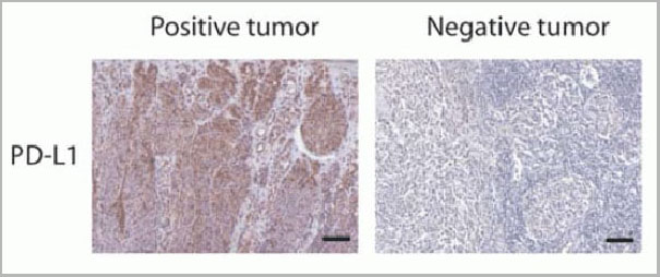 IF (Immunofluorescence)
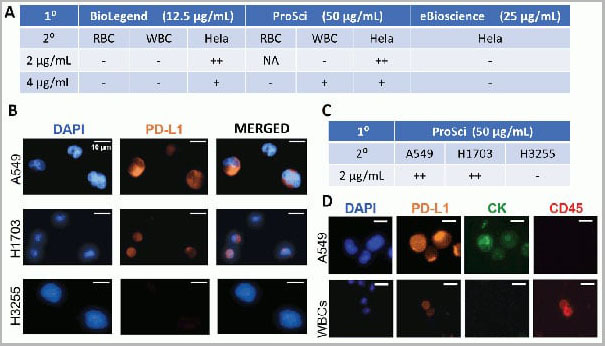
(Figure 12 Immunohistochemistry Validation of PD-L1in Human Tumors (Gadiot et al., 2011)Immunohistochemical analysis of patient tumors labeling PD-L1 with anti-PD-L1 antibodies (AAA10941). Several anti-PD-L1 antibodies were tested for staining, “Only 1 antibody gave no background staining and was competitively blocked by the addition of PD-L1Fc protein (AAA10941)”.)
IF (Immunofluorescence)
(Figure 12 Immunohistochemistry Validation of PD-L1in Human Tumors (Gadiot et al., 2011)Immunohistochemical analysis of patient tumors labeling PD-L1 with anti-PD-L1 antibodies (AAA10941). Several anti-PD-L1 antibodies were tested for staining, “Only 1 antibody gave no background staining and was competitively blocked by the addition of PD-L1Fc protein (AAA10941)”.)
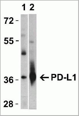
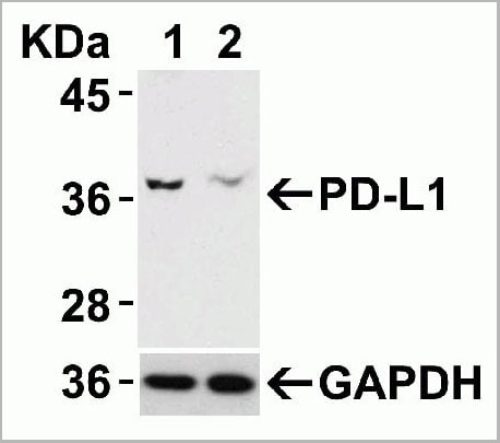
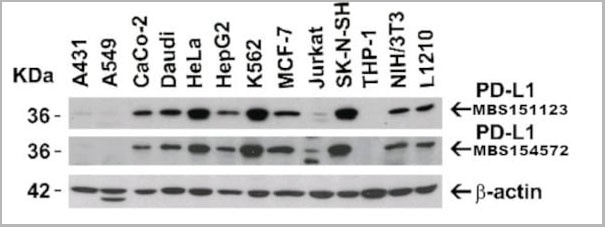
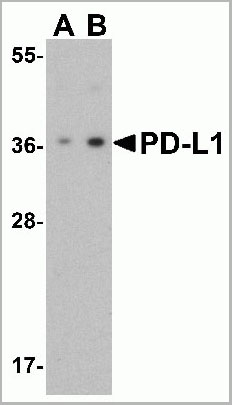
PDL-1, Polyclonal Antibody (Cat# AAA10941)
Full Name
PDL-1 Antibody
Gene Names
CD274; B7-H; B7H1; PDL1; PD-L1; PDCD1L1; PDCD1LG1
Applications
Western Blot, Immunohistochemistry, Immunofluorescence, Flow Cytometry
Purity
PD-L1 Antibody is affinity chromatography purified via peptide column.
Pricing
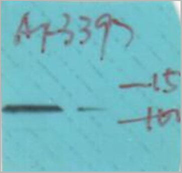
 WB (Western Blot)
(Western blot analysis of MUC2 using HT-29 whole cell lysates)
WB (Western Blot)
(Western blot analysis of MUC2 using HT-29 whole cell lysates)
MUC2, Polyclonal Antibody (Cat# AAA31137)
Full Name
MUC2 Antibody
Gene Names
MUC2; MLP; SMUC; MUC-2
Reactivity
Human, Mouse, Rat
Applications
Western Blot, Immunohistochemistry, Immunofluorescence, Immunocytochemistry
Purity
The antiserum was purified by peptide affinity chromatography using SulfoLink™ Coupling Resin (Thermo Fisher Scientific).
Pricing
RPMI 1640 Medium Modified w/o L-Glutamine, w/o Amino acids, Glucose, Culture Media (Cat# AAA14817)
Full Name
RPMI 1640 Medium Modified w/o L-Glutamine, w/o Amino acids, Glucose (Powder)
Pricing


 ICC (Immunocytochemistry)
(Immunostaining analysis in HeLa cells. HeLa cells were fixed with 4% paraformaldehyde and permeabilized with 0.1% Triton X-100 in PBS. The cells were immunostained with anti-CARD8 mAb. [Lot No. 2108C2a-1])
ICC (Immunocytochemistry)
(Immunostaining analysis in HeLa cells. HeLa cells were fixed with 4% paraformaldehyde and permeabilized with 0.1% Triton X-100 in PBS. The cells were immunostained with anti-CARD8 mAb. [Lot No. 2108C2a-1])
CARD8, Monoclonal Antibody (Cat# AAA10604)
Full Name
Mouse monoclonal antibody Anti-Human CARD8
Gene Names
CARD8; NDPP; DACAR; DAKAR; NDPP1; TUCAN; CARDINAL
Reactivity
Human
Applications
Western Blot, Immunoprecipitation, Flow Cytometry, Immunocytochemistry
Purity
This antibody was purified using protein G column chromatography from culture supernatant of hybridoma cultured in a medium containing bovine IgG-depleted (approximately 95%) fetal bovine serum.
Pricing
 IF (Immunofluorescence)
(AAA31138 staining Hela cells by IF/ICC. The samples were fixed with PFA and permeabilized in 0.1% Triton X-100, then blocked in 10% serum for 45 minutes at 25 degree C. Samples were then incubated with primary Ab(AAA31138) and mouse anti-beta tubulin Ab for 1 hour at 37 degree C. An AlexaFluor594 conjugated goat anti-rabbit IgG(H+L) Ab(Red) and an AlexaFluor488 conjugated goat anti-mouse IgG(H+L) Ab(Green) were used as the secondary Ab. The nuclear counter stain is DAPI (blue).)
IF (Immunofluorescence)
(AAA31138 staining Hela cells by IF/ICC. The samples were fixed with PFA and permeabilized in 0.1% Triton X-100, then blocked in 10% serum for 45 minutes at 25 degree C. Samples were then incubated with primary Ab(AAA31138) and mouse anti-beta tubulin Ab for 1 hour at 37 degree C. An AlexaFluor594 conjugated goat anti-rabbit IgG(H+L) Ab(Red) and an AlexaFluor488 conjugated goat anti-mouse IgG(H+L) Ab(Green) were used as the secondary Ab. The nuclear counter stain is DAPI (blue).)
Prestin, Polyclonal Antibody (Cat# AAA31138)
Full Name
Prestin Antibody
Gene Names
SLC26A5; PRES; DFNB61
Reactivity
Human, Mouse, Rat
Applications
Western Blot, Immunohistochemistry, Immunofluorescence
Purity
Peptide affinity chromatography using SulfoLink Coupling Resin (Thermo Fisher Scientific).
Pricing
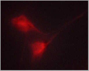
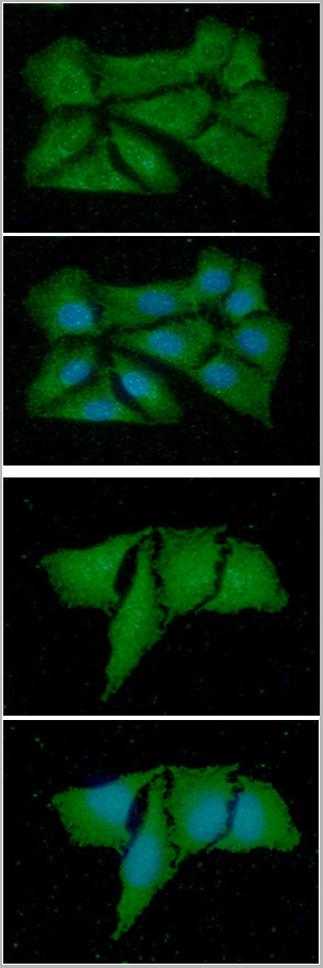 IF (Immunofluorescence)
(Figure 3 and 4: ICC/IF analysis of MIF in Balb/3T3 cells. The cell was stained with AAA11724 (1;100). The secondary antibody (green) was used Alexa Fluor 488. DAPI was stained the cell nucelus (blue).Figure 5 and 6: ICC/IF analysis of MIF in HeLa cells line. The cell was stained with AAA11724 (1:100). The secondary antibody (green) was used Alexa Fluor 488. DAPI was stained the cell nucleus (blue))
IF (Immunofluorescence)
(Figure 3 and 4: ICC/IF analysis of MIF in Balb/3T3 cells. The cell was stained with AAA11724 (1;100). The secondary antibody (green) was used Alexa Fluor 488. DAPI was stained the cell nucelus (blue).Figure 5 and 6: ICC/IF analysis of MIF in HeLa cells line. The cell was stained with AAA11724 (1:100). The secondary antibody (green) was used Alexa Fluor 488. DAPI was stained the cell nucleus (blue))
MIF, Monoclonal Antibody (Cat# AAA11724)
Full Name
MIF antibody
Gene Names
MIF; GIF; GLIF; MMIF
Reactivity
Human
Applications
Western Blot, Immunocytochemistry, Immunofluorescence
Purity
By protein-G affinity chromatography
Pricing
 IF (Immunoflouorescence)
(AAA31039 staining HeLa cells by ICC/IF. Cells were fixed with PFA and permeabilized in 0.1% saponin prior to blocking in 10% serum for 45 minutes at 37 degree C. The primary antibody was diluted 1/400 and incubated with the sample for 1 hour at 37 degree C. A Alexa Fluor 594 conjugated goat polyclonal to rabbit IgG (H+L), diluted 1/600 was used as secondary antibody.)
IF (Immunoflouorescence)
(AAA31039 staining HeLa cells by ICC/IF. Cells were fixed with PFA and permeabilized in 0.1% saponin prior to blocking in 10% serum for 45 minutes at 37 degree C. The primary antibody was diluted 1/400 and incubated with the sample for 1 hour at 37 degree C. A Alexa Fluor 594 conjugated goat polyclonal to rabbit IgG (H+L), diluted 1/600 was used as secondary antibody.)
ADD1, Polyclonal Antibody (Cat# AAA31039)
Full Name
Phospho-ADD1 (Ser726) Antibody
Gene Names
ADD1; ADDA
Reactivity
Human, Mouse, Rat
Applications
Western Blot, Immunohistochemistry, Immunofluorescence, Immunocytochemistry
Purity
From purified rabbit serum by affinity purification via sequential chromatography on phospho-and non-phospho-peptide affinity columns.
Pricing
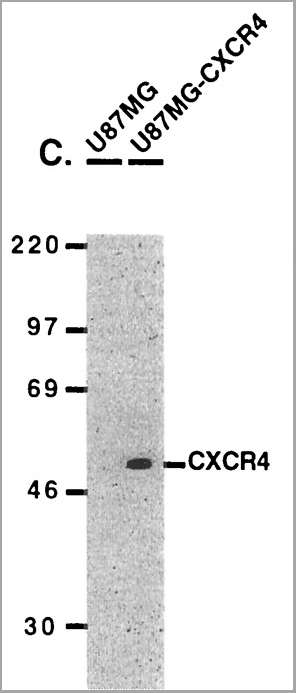

 WB (Western Blot)
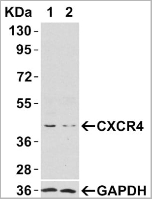
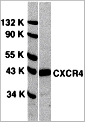
(Figure 9 WB Validation of CXCR4 in metastatic melanoma (Scala et al., 2006)CXCR4 protein was detected in the human metastatic melanoma cell lines and human melanoma cell line (colo38), but not in the human primary melanocytes (MPR1) with anti-CXCR4 antibodies.)
WB (Western Blot)
(Figure 9 WB Validation of CXCR4 in metastatic melanoma (Scala et al., 2006)CXCR4 protein was detected in the human metastatic melanoma cell lines and human melanoma cell line (colo38), but not in the human primary melanocytes (MPR1) with anti-CXCR4 antibodies.)
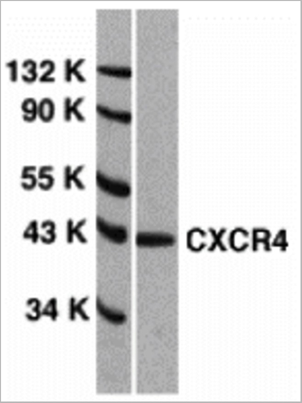
CXCR4, Polyclonal Antibody (Cat# AAA10962)
Full Name
CXCR4 Antibody
Gene Names
CXCR4; FB22; HM89; LAP3; LCR1; NPYR; WHIM; CD184; LAP-3; LESTR; NPY3R; NPYRL; HSY3RR; NPYY3R; D2S201E
Reactivity
Human, Mouse
Applications
Western Blot, Immunofluorescence
Purity
CXCR4 Antibody is Protein A purified.
Pricing
 WB (Western Blot)
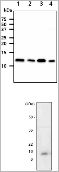
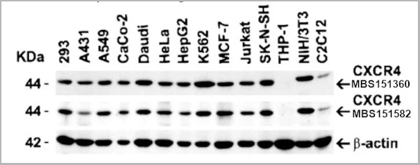
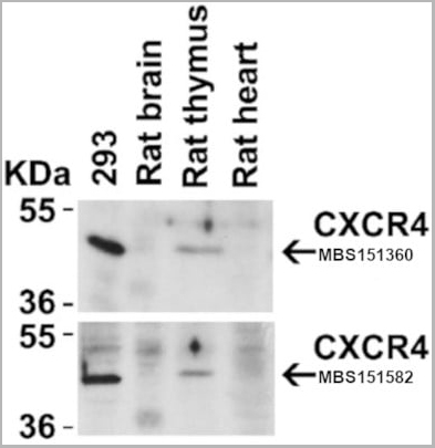
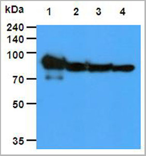
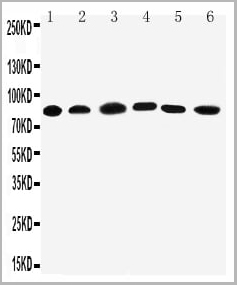
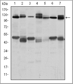
(Figure 4 Animal Species ReactivityLoading: Lysates/proteins at 20 μg per lane. Antibodies: 1009 (2 μg/mL) or 1012 (2 μg/mL). 1 h incubation at RT in 5% NFDM/TBST. Secondary: Goat anti-rabbit IgG HRP conjugate at 1:10000 dilution.)
WB (Western Blot)
(Figure 4 Animal Species ReactivityLoading: Lysates/proteins at 20 μg per lane. Antibodies: 1009 (2 μg/mL) or 1012 (2 μg/mL). 1 h incubation at RT in 5% NFDM/TBST. Secondary: Goat anti-rabbit IgG HRP conjugate at 1:10000 dilution.)
CXCR4, Polyclonal Antibody (Cat# AAA10951)
Full Name
CXCR4 Antibody
Gene Names
CXCR4; FB22; HM89; LAP3; LCR1; NPYR; WHIM; CD184; LAP-3; LESTR; NPY3R; NPYRL; HSY3RR; NPYY3R; D2S201E
Reactivity
Human, Mouse, Rat
Applications
Western Blot, Immunocytochemistry, Immunoprecipitation, Immunofluorescence, Immunohistochemistry, Flow Cytometry
Purity
CXCR4 Antibody is affinity chromatography purified via peptide column.
Pricing

 IF (Immunofluorescence)
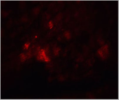
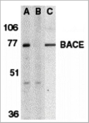
(Figure 10 KD Validation of BACE in DRG (Hyun, 2007)Decreased BACE1 expression in DRG following siRNA3 transfection. DRG neurons were transfected with 1 ug siRNA3 plasmid and incubated for 48 hours in 37°˚C. DRG neurons were stained for BACE1 us¬ing the Anti-BACE antibody (a,b) Neurons transfected with the control plas¬mid pSUPER-EGFP (green) did not display any changes in BACE1 expression (red). (c,d) DRG neurons transfected with siR¬NA3 displayed reduced BACE1 expression in the axon.)
IF (Immunofluorescence)
(Figure 10 KD Validation of BACE in DRG (Hyun, 2007)Decreased BACE1 expression in DRG following siRNA3 transfection. DRG neurons were transfected with 1 ug siRNA3 plasmid and incubated for 48 hours in 37°˚C. DRG neurons were stained for BACE1 us¬ing the Anti-BACE antibody (a,b) Neurons transfected with the control plas¬mid pSUPER-EGFP (green) did not display any changes in BACE1 expression (red). (c,d) DRG neurons transfected with siR¬NA3 displayed reduced BACE1 expression in the axon.)
BACE, Polyclonal Antibody (Cat# AAA10918)
Full Name
BACE Antibody
Gene Names
BACE1; ASP2; BACE; HSPC104
Reactivity
Human, Mouse
Applications
Immunocytochemistry, Immunofluorescence, Immunohistochemistry, Western Blot
Purity
BACE Antibody is affinity chromatography purified via peptide column.
Pricing

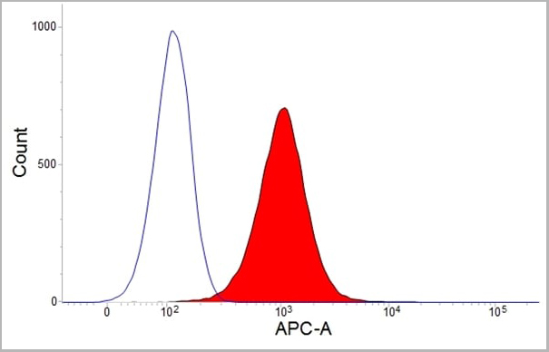
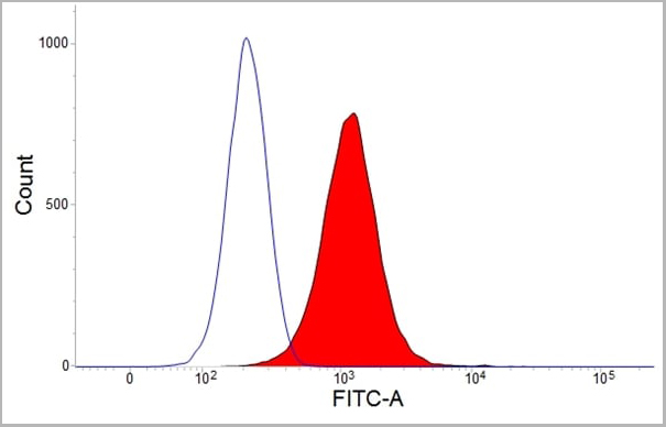
 Application Data
(Staining of human peripheral blood monocytes with Mouse anti Human CD14:RPE)
Application Data
(Staining of human peripheral blood monocytes with Mouse anti Human CD14:RPE)
CD14, Monoclonal Antibody (Cat# AAA11884)
Full Name
MOUSE ANTI HUMAN CD14:FITC
Applications
Flow Cytometry
Pricing
 WB (Western Blot)
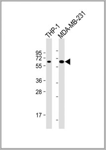
(All lanes : Anti-MB21D1 Antibody at 1:4000 dilutionLane 1: THP-1 whole cell lysateLane 2: MDA-MB-231 whole cell lysateLysates/proteins at 20 ug per lane. SecondaryGoat Anti-mouse IgG, (H+L), Peroxidase conjugated at 1/10000 dilution. Predicted band size : 59kDaBlocking/Dilution buffer: 5% NFDM/TBST.)
WB (Western Blot)
(All lanes : Anti-MB21D1 Antibody at 1:4000 dilutionLane 1: THP-1 whole cell lysateLane 2: MDA-MB-231 whole cell lysateLysates/proteins at 20 ug per lane. SecondaryGoat Anti-mouse IgG, (H+L), Peroxidase conjugated at 1/10000 dilution. Predicted band size : 59kDaBlocking/Dilution buffer: 5% NFDM/TBST.)
MB21D1, Monoclonal Antibody (Cat# AAA28800)
Full Name
MB21D1 Antibody
Gene Names
MB21D1; cGAS; h-cGAS; C6orf150
Reactivity
Human
Applications
Western Blot
Pricing
Dengue NS1, Monoclonal Antibody (Cat# AAA17926)
Full Name
Dengue NS1 antibody
Applications
Immunofluorescence, Immunohistochemistry, Western Blot
Purity
> 90% pure
Dengue NS1 antibody was purified by Protein A affinity chromatography
Dengue NS1 antibody was purified by Protein A affinity chromatography
Pricing

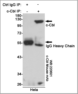
 WB (Western Blot)
(Detection of CBLB by Western blot.Samples: Whole cell lysate from human HeLa (H, 25 ug) and mouse NIH3T3 (M, 25 ug) cells. [Lot No. 246C5a-1] Predicted molecular weight: 109 kDa)
WB (Western Blot)
(Detection of CBLB by Western blot.Samples: Whole cell lysate from human HeLa (H, 25 ug) and mouse NIH3T3 (M, 25 ug) cells. [Lot No. 246C5a-1] Predicted molecular weight: 109 kDa)
CBLB, Monoclonal Antibody (Cat# AAA10613)
Full Name
Mouse monoclonal antibody Anti-Human CBLB
Gene Names
CBLB; Cbl-b; RNF56; Nbla00127
Reactivity
Human
Applications
Western Blot
Pricing
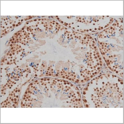

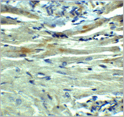
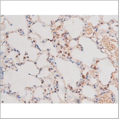
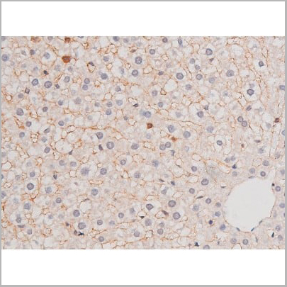
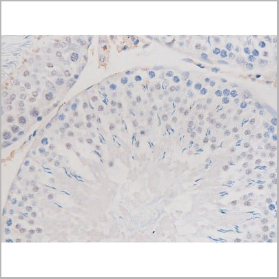
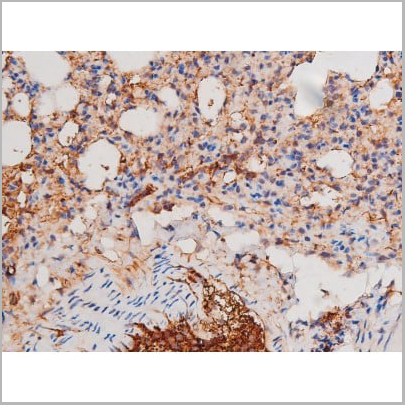
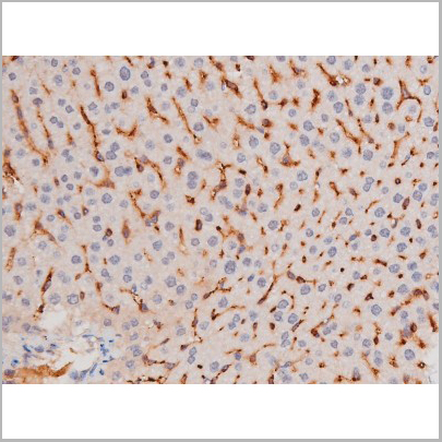
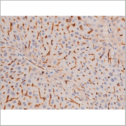
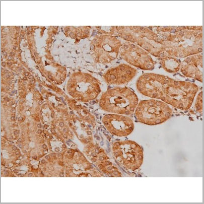
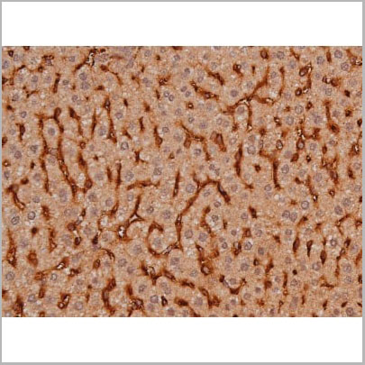
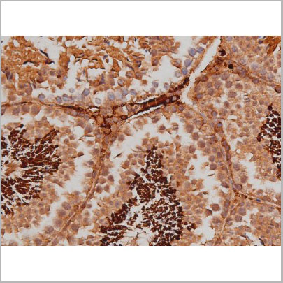
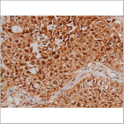
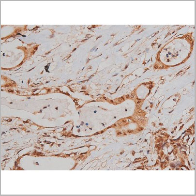
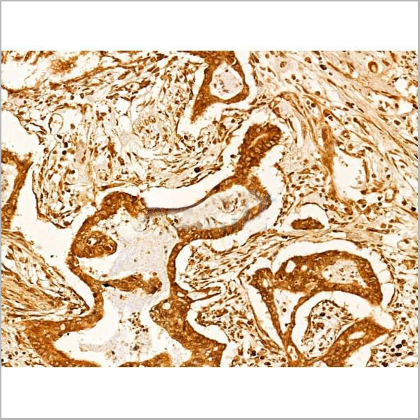
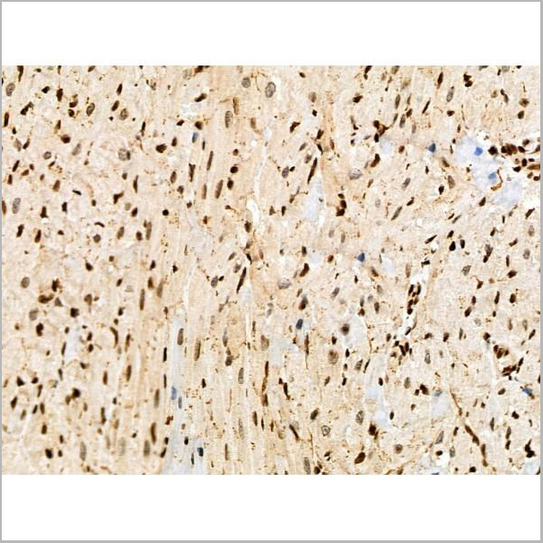
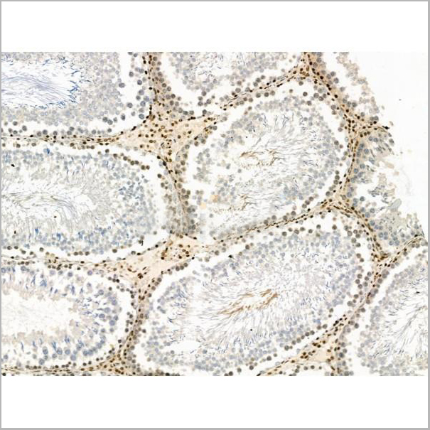
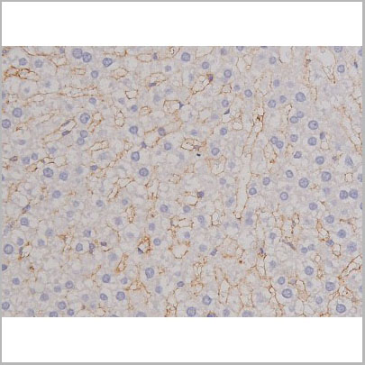
 IHC (Immunohistochemistry)
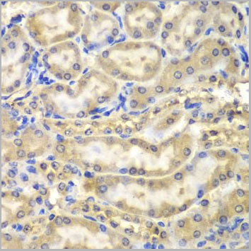
(AAA30993 at 1/200 staining Rat ganstric tissue sections by IHC-P. The tissue was formaldehyde fixed and a heat mediated antigen retrieval step in citrate buffer was performed. The tissue was then blocked and incubated with the antibody for 1.5 hours at 22 degree C. An HRP conjugated goat anti-rabbit antibody was used as the secondary.)
IHC (Immunohistochemistry)
(AAA30993 at 1/200 staining Rat ganstric tissue sections by IHC-P. The tissue was formaldehyde fixed and a heat mediated antigen retrieval step in citrate buffer was performed. The tissue was then blocked and incubated with the antibody for 1.5 hours at 22 degree C. An HRP conjugated goat anti-rabbit antibody was used as the secondary.)
IGF1R, Polyclonal Antibody (Cat# AAA30993)
Full Name
Phospho-IGF1R (Tyr1346) Antibody
Gene Names
IGF1R; IGFR; CD221; IGFIR; JTK13
Reactivity
Human, Mouse, Rat
Applications
Western Blot, Immunohistochemistry, Immunofluorescence, Immunocytochemistry
Purity
From purified rabbit serum by affinity purification via sequential chromatography on phospho-and non-phospho-peptide affinity columns.
Pricing

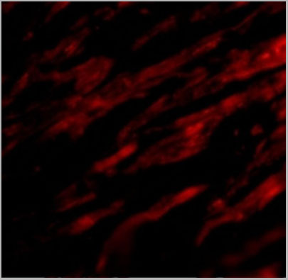
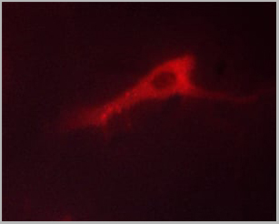
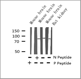
 IF (Immunofluorescence)
(Figure 10 Immunofluorescence Validation of TACE in Rat Brain (Pradillo et al, 2005)Cellular localization of TACE. Double immunofluorescence staining of brain sections from sham-operated (SHAM; A, C, E) and IPC-exposed animals (IPC; B, D, F) of TACE (red) and the cellular markers (green) NeuN (neurons; A, B), GFAP (astrocytes; C, D) and L. esculentum lectin (microglia and endothelium; E, F). White arrows indicate TACE-positive cells.)
IF (Immunofluorescence)
(Figure 10 Immunofluorescence Validation of TACE in Rat Brain (Pradillo et al, 2005)Cellular localization of TACE. Double immunofluorescence staining of brain sections from sham-operated (SHAM; A, C, E) and IPC-exposed animals (IPC; B, D, F) of TACE (red) and the cellular markers (green) NeuN (neurons; A, B), GFAP (astrocytes; C, D) and L. esculentum lectin (microglia and endothelium; E, F). White arrows indicate TACE-positive cells.)
TACE, Polyclonal Antibody (Cat# AAA10912)
Full Name
TACE Antibody
Gene Names
ADAM17; CSVP; TACE; NISBD; ADAM18; CD156B; NISBD1
Reactivity
Human, Mouse, Rat
Applications
Western Blot, Immunocytochemistry, Immunofluorescence, Flow Cytometry
Purity
TACE Antibody is affinity chromatography purified via peptide column.
Pricing

 ICC (Immunocytochemistry)
(Immunostaining analysis in HeLa cells. HeLa cells were fixed with 4% paraformaldehyde and permeabilized with 0.1% Triton X-100 in PBS. The cells were immunostained with anti-TNPO3 mAb.)
ICC (Immunocytochemistry)
(Immunostaining analysis in HeLa cells. HeLa cells were fixed with 4% paraformaldehyde and permeabilized with 0.1% Triton X-100 in PBS. The cells were immunostained with anti-TNPO3 mAb.)
TNPO3, Monoclonal Antibody (Cat# AAA10612)
Full Name
Mouse monoclonal antibody Anti-Human TNPO3
Gene Names
TNPO3; IPO12; TRNSR; LGMD1F; MTR10A; TRN-SR; TRN-SR2
Reactivity
Human
Applications
Immunocytochemistry, Western Blot
Pricing
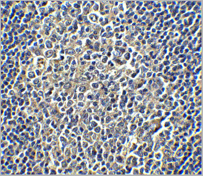
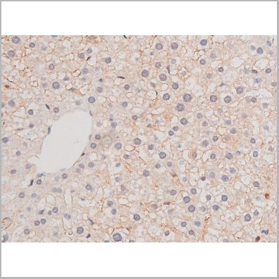
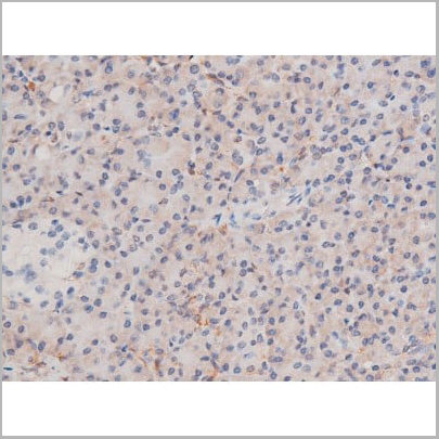
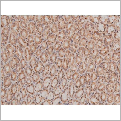
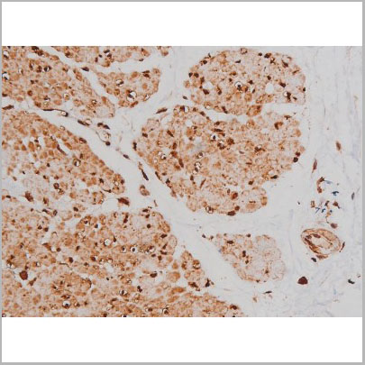
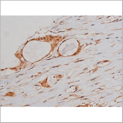
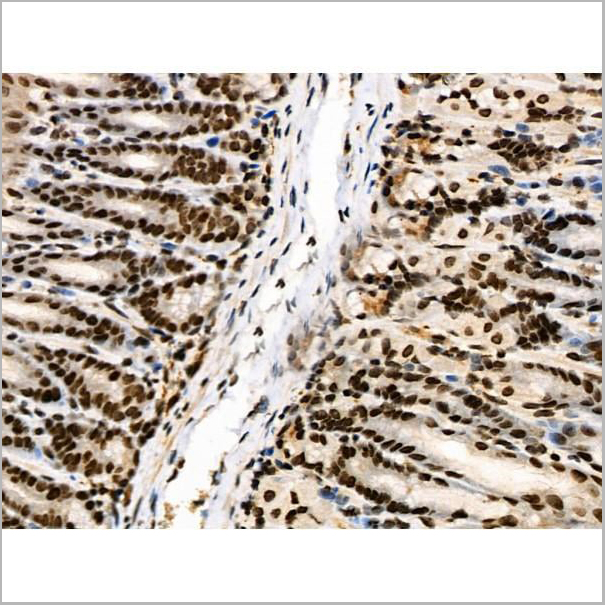
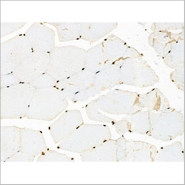
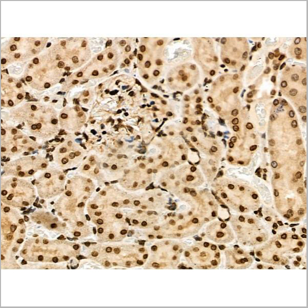
 IHC (Immunohistochemistry)
(AAA31126 at 1/100 staining Rat stomach tissue by IHC-P. The sample was formaldehyde fixed and a heat mediated antigen retrieval step in citrate buffer was performed. The sample was then blocked and incubated with the primary Ab at 4°C overnight. An HRP conjugated anti-Rabbit Ab was used as the secondary Ab.)
IHC (Immunohistochemistry)
(AAA31126 at 1/100 staining Rat stomach tissue by IHC-P. The sample was formaldehyde fixed and a heat mediated antigen retrieval step in citrate buffer was performed. The sample was then blocked and incubated with the primary Ab at 4°C overnight. An HRP conjugated anti-Rabbit Ab was used as the secondary Ab.)
MT2A, Polyclonal Antibody (Cat# AAA31126)
Full Name
MT2A Antibody
Gene Names
MT2A; MT2; MT-2; MT-II
Reactivity
Human, Mouse, Rat
Applications
Western Blot, Immunohistochemistry
Purity
peptide affinity purification
Pricing
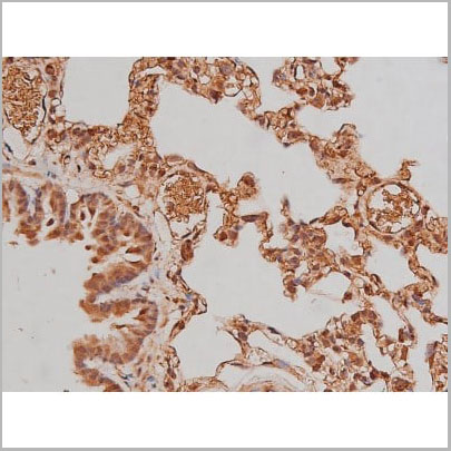
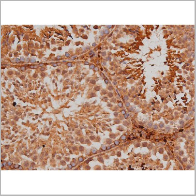
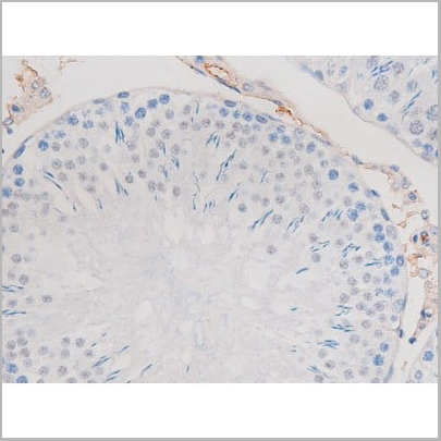
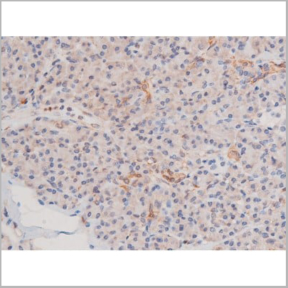
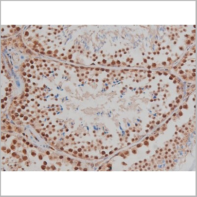
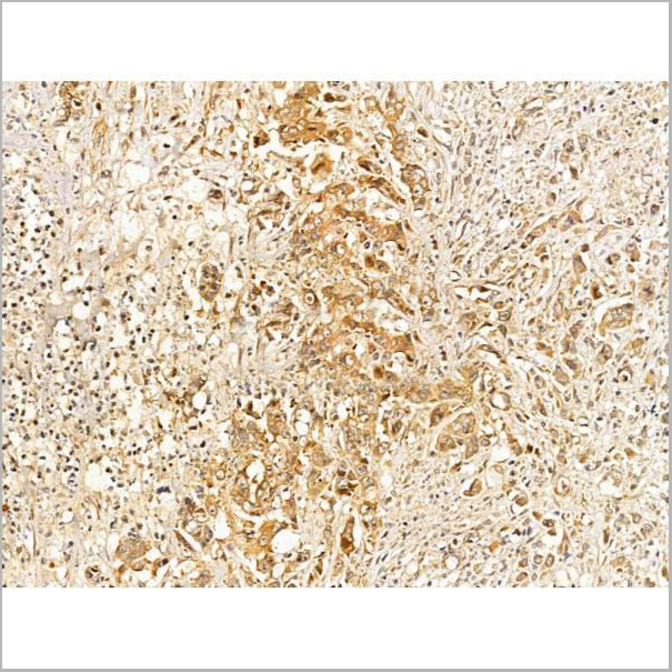
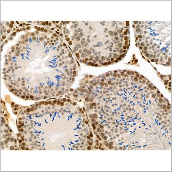
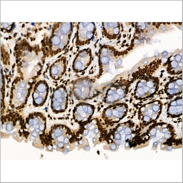
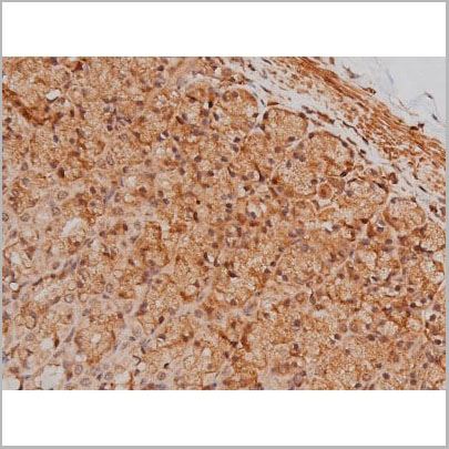 IHC (Immunohistochemistry)
(AAA31005 at 1/200 staining Rat ganstric tissue sections by IHC-P. The tissue was formaldehyde fixed and a heat mediated antigen retrieval step in citrate buffer was performed. The tissue was then blocked and incubated with the antibody for 1.5 hours at 22 degree C. An HRP conjugated goat anti-rabbit antibody was used as the secondary.)
IHC (Immunohistochemistry)
(AAA31005 at 1/200 staining Rat ganstric tissue sections by IHC-P. The tissue was formaldehyde fixed and a heat mediated antigen retrieval step in citrate buffer was performed. The tissue was then blocked and incubated with the antibody for 1.5 hours at 22 degree C. An HRP conjugated goat anti-rabbit antibody was used as the secondary.)
Tau, Polyclonal Antibody (Cat# AAA31005)
Full Name
Phospho-Tau (Ser262) Antibody
Gene Names
MAPT; TAU; MSTD; PPND; DDPAC; MAPTL; MTBT1; MTBT2; FTDP-17; PPP1R103
Reactivity
Human, Mouse, Rat
Applications
Western Blot, Immunohistochemistry
Purity
From purified rabbit serum by affinity purification via sequential chromatography on phospho-and non-phospho-peptide affinity columns.
Pricing


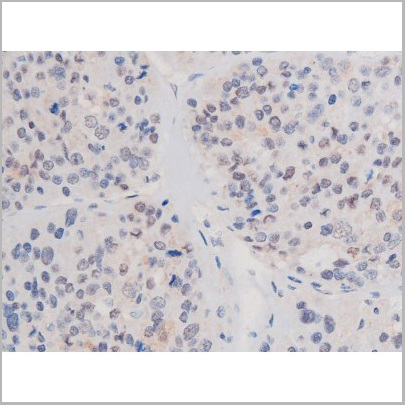
 IHC (Immunohistochemistry)
(IHC Validation of Envelope in COVID-19 Patient Skin (Magro et al., 2020) Detection of SARS-CoV-2 Envelope protein in the blood vessels of COVID-19 patients that were confirmed by PCR. The staining shows Envelope protein expression (green) detected by envelope antibodies (AAA10932, 3 ug/mL) in the endothelial cytoplasms in thrombosed and normal appearing blood vessels with hematoxylin counterstain. The staining was negative in control normal skin/lung (not shown).)
IHC (Immunohistochemistry)
(IHC Validation of Envelope in COVID-19 Patient Skin (Magro et al., 2020) Detection of SARS-CoV-2 Envelope protein in the blood vessels of COVID-19 patients that were confirmed by PCR. The staining shows Envelope protein expression (green) detected by envelope antibodies (AAA10932, 3 ug/mL) in the endothelial cytoplasms in thrombosed and normal appearing blood vessels with hematoxylin counterstain. The staining was negative in control normal skin/lung (not shown).)
COVID 19 Envelope Coronavirus, Polyclonal Antibody (Cat# AAA10932)
Full Name
SARS-CoV-2 (COVID-19, 2019-nCoV) Envelope antibody
Reactivity
Virus
Applications
Immunofluorescence, Immunohistochemistry
Purity
SARS-CoV-2 (COVID-19, 2019-nCoV) Envelope Antibody is affinity chromatography purified via peptide column.
Pricing
 Application Data
(At 25 degree C. Samples were then incubated with primary Ab(At 37 degree C. An AlexaFluor594 conjugated goat anti-mouse IgG(H+L) Ab(Red) and an AlexaFluor488 conjugated goat anti-rabbit IgG(H+L) Ab(Green) were used as the secondary antibody.The nuclear counter stain is DAPI(blue).)
Application Data
(At 25 degree C. Samples were then incubated with primary Ab(At 37 degree C. An AlexaFluor594 conjugated goat anti-mouse IgG(H+L) Ab(Red) and an AlexaFluor488 conjugated goat anti-rabbit IgG(H+L) Ab(Green) were used as the secondary antibody.The nuclear counter stain is DAPI(blue).)
Cleaved-Caspase 1, Polyclonal Antibody (Cat# AAA31336)
Full Name
Cleaved-Caspase 1 (Ala317), p10 Antibody
Gene Names
CASP1; ICE; P45; IL1BC
Reactivity
Human, Mouse, Rat
Applications
Western Blot, Immunohistochemistry, Immunofluorescence, Immunocytochemistry
Purity
The antiserum was purified by peptide affinity chromatography using SulfoLink Coupling Resin
Pricing
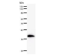
ADRM1, Monoclonal Antibody (Cat# AAA24126)
Full Name
ADRM1 (GP110, Proteasomal Ubiquitin Receptor ADRM1, 110kD Cell Membrane Glycoprotein, Adhesion-regulating Molecule 1, Proteasome Regulatory Particle Non-ATPase 13, Rpn13 Homolog, MGC29536) (AP)
Gene Names
ADRM1; ARM1; ARM-1; GP110
Reactivity
Human
Applications
Western Blot
Purity
Purified by Protein A Affinity Chromatography.
Pricing
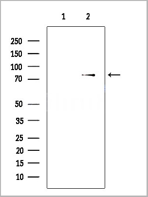
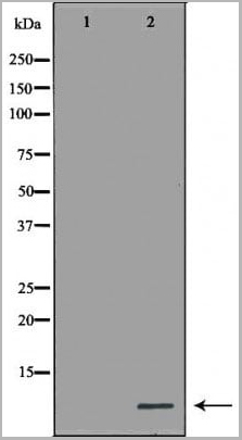
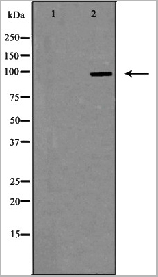
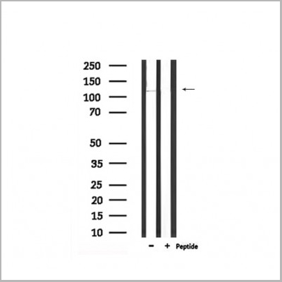
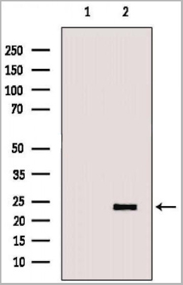
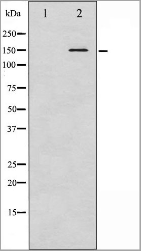
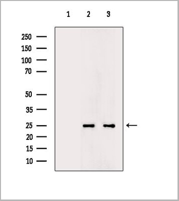
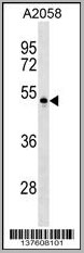 WB (Western Blot)
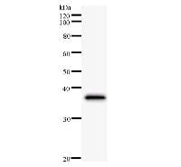
(PVRL4 Antibody (Center) (AAA28678) western blot analysis in A2058 cell line lysates (35ug/lane).This demonstrates the PVRL4 antibody detected the PVRL4 protein (arrow).)
WB (Western Blot)
(PVRL4 Antibody (Center) (AAA28678) western blot analysis in A2058 cell line lysates (35ug/lane).This demonstrates the PVRL4 antibody detected the PVRL4 protein (arrow).)
PVRL4, Polyclonal Antibody (Cat# AAA28678)
Full Name
PVRL4 Antibody (Center)
Gene Names
PVRL4; LNIR; PRR4; EDSS1; nectin-4
Reactivity
Human
Applications
Western Blot, Dot Blot, Immunohistochemistry, Immunofluorescence, Immunoprecipitation, Flow Cytometry
Purity
Peptide Affinity Purified Rabbit Polyclonal Antibody (Pab)
Pricing
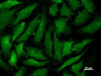
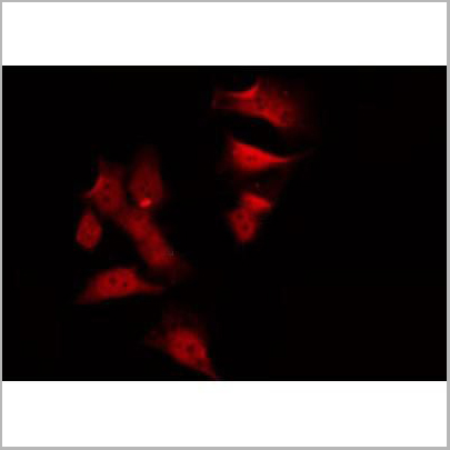
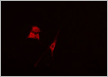
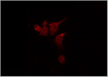 IF (Immunofluorescence)
(AAA31118 staining 293T by IF/ICC. The sample were fixed with PFA and permeabilized in 0.1% Triton X-100, then blocked in 10% serum for 45 minutes at 25 degree C. The primary antibody was diluted at 1/200 and incubated with the sample for 1 hour at 37 degree C. An Alexa Fluor 594 conjugated goat anti-rabbit IgG (H+L) Ab, diluted at 1/600, was used as the secondary antibody.)
IF (Immunofluorescence)
(AAA31118 staining 293T by IF/ICC. The sample were fixed with PFA and permeabilized in 0.1% Triton X-100, then blocked in 10% serum for 45 minutes at 25 degree C. The primary antibody was diluted at 1/200 and incubated with the sample for 1 hour at 37 degree C. An Alexa Fluor 594 conjugated goat anti-rabbit IgG (H+L) Ab, diluted at 1/600, was used as the secondary antibody.)
GPR133, Polyclonal Antibody (Cat# AAA31118)
Full Name
GPR133 Antibody
Gene Names
ADGRD1; PGR25; GPR133
Reactivity
Human, Mouse, Rat
Applications
Western Blot, Immunofluorescence, Immunocytochemistry
Purity
The antiserum was purified by peptide affinity chromatography using SulfoLink Coupling Resin (Thermo Fisher Scientific).
Pricing
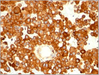
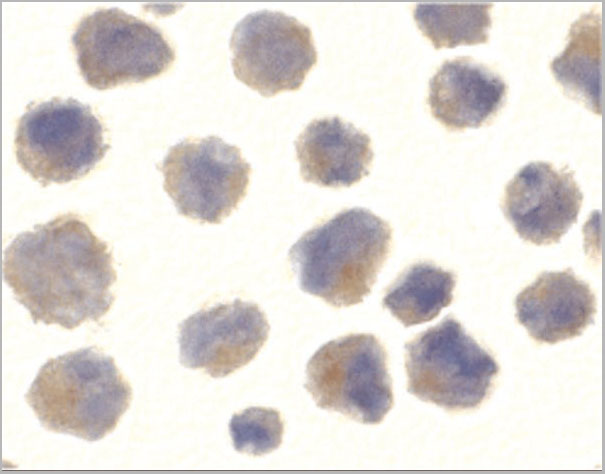
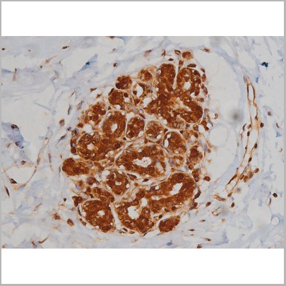
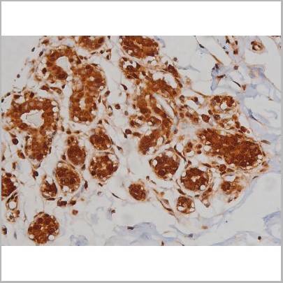
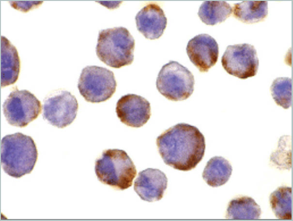
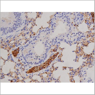
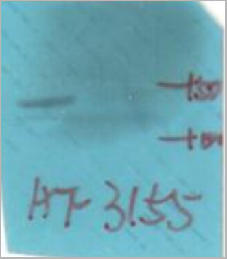
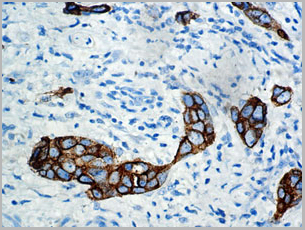 IHC (Immunohistochemistry)
(IHC of an FFPE Breast Tissue)
IHC (Immunohistochemistry)
(IHC of an FFPE Breast Tissue)
CA15-3, Monoclonal Antibody (Cat# AAA13555)
Full Name
CA15-3
Reactivity
Paraffin, frozen
Applications
Immunohistochemistry, Immunohistochemistry
Pricing
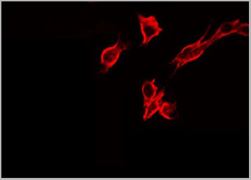
 Application Data
(At 25 degree C. The primary antibody was diluted at 1/200 and incubated with the sample for 1 hour at 37 degree C. An Alexa Fluor 594 conjugated goat anti-rabbit IgG (H+L) Ab, diluted at 1/600, was used as the secondary antibody.)
Application Data
(At 25 degree C. The primary antibody was diluted at 1/200 and incubated with the sample for 1 hour at 37 degree C. An Alexa Fluor 594 conjugated goat anti-rabbit IgG (H+L) Ab, diluted at 1/600, was used as the secondary antibody.)
FAK, Polyclonal Antibody (Cat# AAA31446)
Full Name
Phospho-FAK (Ser722) Antibody
Gene Names
PTK2; FAK; FADK; FAK1; FRNK; PPP1R71; p125FAK; pp125FAK
Reactivity
Human, Mouse, Rat
Applications
Western Blot, Immunohistochemistry, Immunofluorescence, Immunocytochemistry, Peptide ELISA
Purity
The antibody is from purified rabbit serum by affinity purification via sequential chromatography on phospho-peptide and non-phospho-peptide affinity columns.
Pricing
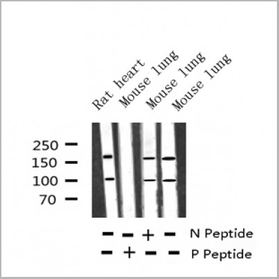
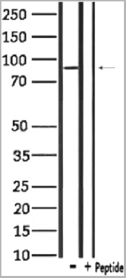
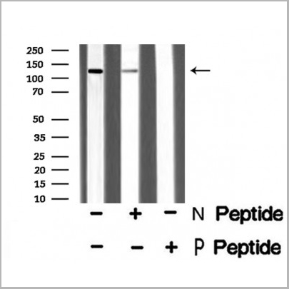
 WB (Western Blot)
(Western blot analysis of Phospho-FAK (Tyr576) Antibody expression in NIH-3T3 cells lysates.The lane on the right is treated with the antigen-specific peptide.)
WB (Western Blot)
(Western blot analysis of Phospho-FAK (Tyr576) Antibody expression in NIH-3T3 cells lysates.The lane on the right is treated with the antigen-specific peptide.)
FAK, Polyclonal Antibody (Cat# AAA31066)
Full Name
Phospho-FAK (Tyr576) Antibody
Gene Names
PTK2; FAK; FADK; FAK1; FRNK; PPP1R71; p125FAK; pp125FAK
Reactivity
Human, Mouse, Rat
Applications
Western Blot, Immunohistochemistry, Immunofluorescence, Immunocytochemistry
Purity
From purified rabbit serum by affinity purification via sequential chromatography on phospho-and non-phospho-peptide affinity columns.
Pricing
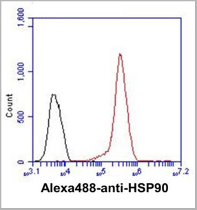
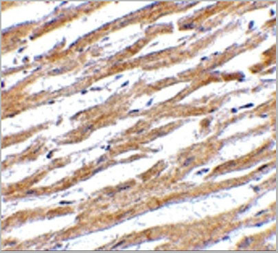
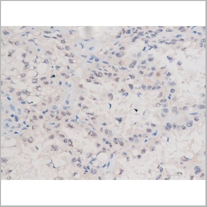
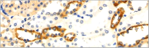
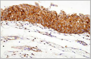 IHC (Immunohistochemistry)
(Paraffin embedded sections of human urothelium were incubated with anti-human Hsp90 (1:100)for 2 hours at room temperature. Antigen retrieval was performed in 0.1M sodium citrate buffer and detectedusing Diaminobenzidine (DAB))
IHC (Immunohistochemistry)
(Paraffin embedded sections of human urothelium were incubated with anti-human Hsp90 (1:100)for 2 hours at room temperature. Antigen retrieval was performed in 0.1M sodium citrate buffer and detectedusing Diaminobenzidine (DAB))
Hsp90, Monoclonal Antibody (Cat# AAA11726)
Full Name
Hsp90 antibody
Gene Names
HSP90AA1; EL52; HSPN; LAP2; HSP86; HSPC1; HSPCA; Hsp89; Hsp90; LAP-2; HSP89A; HSP90A; HSP90N; Hsp103; HSPCAL1; HSPCAL4; HEL-S-65p
Reactivity
Human,
Applications
Western Blot, Immunohistochemistry
Purity
By protein-G affinity chromatography
Pricing
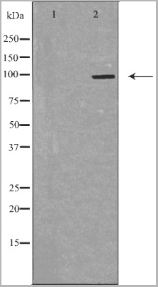
 WB (Western Blot)
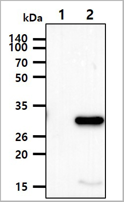
(The cell lysates(10ug) were resolved by SDS-PAGE, transferred to PVDF membrane and probed with anti-human NMNAT-1 antibody (1:2000). Proteins were visualized using a goat anti-mouse secondary antibody conjugated to HRP and an ECL detection system.Lane 1.: 293T cell lysateLane 2.: NMNAT-1 Transfected 293T cell lysate)
WB (Western Blot)
(The cell lysates(10ug) were resolved by SDS-PAGE, transferred to PVDF membrane and probed with anti-human NMNAT-1 antibody (1:2000). Proteins were visualized using a goat anti-mouse secondary antibody conjugated to HRP and an ECL detection system.Lane 1.: 293T cell lysateLane 2.: NMNAT-1 Transfected 293T cell lysate)
NMNAT-1, Monoclonal Antibody (Cat# AAA11734)
Full Name
NMNAT-1
Gene Names
NMNAT1; LCA9; NMNAT; PNAT1
Reactivity
Human
Applications
Western Blot
Purity
Protein-A affinity chromatography
Pricing


 Application Data
(Staining of human peripheral blood monocytes with Mouse anti Human CD14:RPE)
Application Data
(Staining of human peripheral blood monocytes with Mouse anti Human CD14:RPE)
CD14, Monoclonal Antibody (Cat# AAA12178)
Full Name
MOUSE ANTI HUMAN CD14:APC
Applications
Flow Cytometry
Pricing
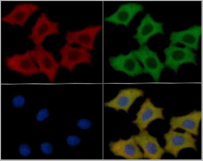
 IF (Immunofluorescence)
(AAA31122 staining A549 cells by IF/ICC. The samples were fixed with PFA and permeabilized in 0.1% Triton X-100,then blocked in 10% serum for 45 minutes at 25°C. Samples were then incubated with primary Ab(AAA31122) and mouse anti-beta tubulin Ab for 1 hour at 37°C. An AlexaFluor594 conjugated goat anti-rabbit IgG(H+L) Ab(Red) and an AlexaFluor488 conjugated goat anti-mouse IgG(H+L) Ab(Green) were used as the secondary antibody. The nuclear counter stain is DAPI (blue).)
IF (Immunofluorescence)
(AAA31122 staining A549 cells by IF/ICC. The samples were fixed with PFA and permeabilized in 0.1% Triton X-100,then blocked in 10% serum for 45 minutes at 25°C. Samples were then incubated with primary Ab(AAA31122) and mouse anti-beta tubulin Ab for 1 hour at 37°C. An AlexaFluor594 conjugated goat anti-rabbit IgG(H+L) Ab(Red) and an AlexaFluor488 conjugated goat anti-mouse IgG(H+L) Ab(Green) were used as the secondary antibody. The nuclear counter stain is DAPI (blue).)
IL-6, Polyclonal Antibody (Cat# AAA31122)
Full Name
IL-6 Antibody
Gene Names
IL6; CDF; HGF; HSF; BSF2; IL-6; BSF-2; IFNB2; IFN-beta-2
Reactivity
Human, Mouse, Rat
Applications
Western Blot, Immunohistochemistry, Immunofluorescence, Immunocytochemistry
Purity
The antiserum was purified by peptide affinity chromatography using SulfoLinkTM Coupling Resin (Thermo Fisher Scientific).
Pricing
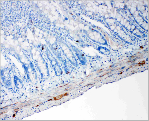
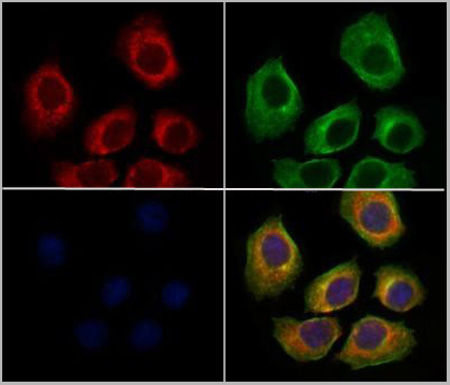
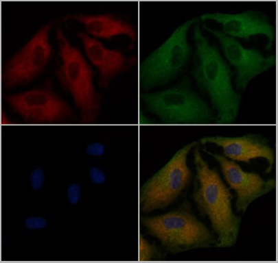
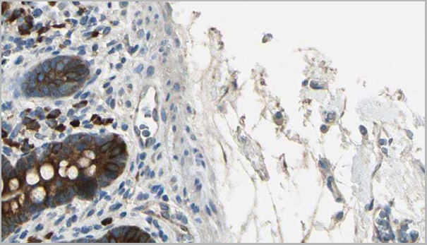
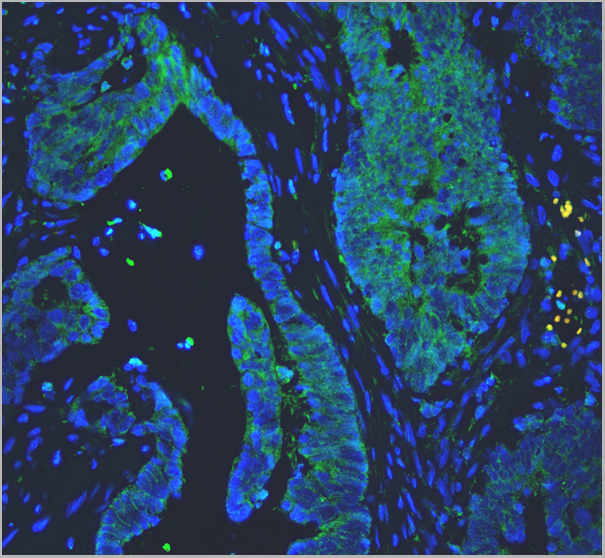 IF (Immunofluorescence)
(Figure 3. IF analysis of PC1/3/PCSK1 using anti-PC1/3/PCSK1 antibody PC1/3/PCSK1 was detected in paraffin-embedded section of human colon cancer tissues. Heat mediated antigen retrieval was performed in citrate buffer (pH6, epitope retrieval solution ) for 20 mins. The tissue section was blocked with 10% goat serum. The tissue section was then incubated with 1ug/mL rabbit anti-PC1/3/PCSK1 Antibody overnight at 4 degree C. Biotin conjugated goat anti-rabbit IgG was used as secondary antibody and incubated for 30 minutes at 37 degree C. The tissue section was developed using DyLight 488 Conjugated Avidin. The section was counterstained with DAPI. Visualize using a fluorescence microscope and filter sets appropriate for the label used.)
IF (Immunofluorescence)
(Figure 3. IF analysis of PC1/3/PCSK1 using anti-PC1/3/PCSK1 antibody PC1/3/PCSK1 was detected in paraffin-embedded section of human colon cancer tissues. Heat mediated antigen retrieval was performed in citrate buffer (pH6, epitope retrieval solution ) for 20 mins. The tissue section was blocked with 10% goat serum. The tissue section was then incubated with 1ug/mL rabbit anti-PC1/3/PCSK1 Antibody overnight at 4 degree C. Biotin conjugated goat anti-rabbit IgG was used as secondary antibody and incubated for 30 minutes at 37 degree C. The tissue section was developed using DyLight 488 Conjugated Avidin. The section was counterstained with DAPI. Visualize using a fluorescence microscope and filter sets appropriate for the label used.)
PC1/3, Polyclonal Antibody (Cat# AAA11585)
Full Name
Anti-PC1/3 antibody
Gene Names
PCSK1; PC1; PC3; NEC1; SPC3; BMIQ12
Reactivity
Reacts with: Human, rat; Predicted to work with: Mouse
Applications
Western Blot, Immunohistochemistry, Immunofluorescence
Purity
Immunogen affinity purified.
Pricing
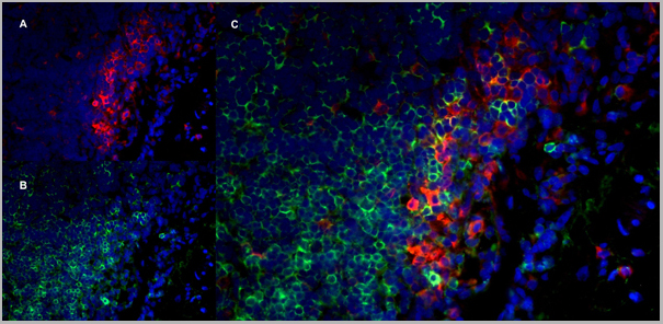
 Application Data
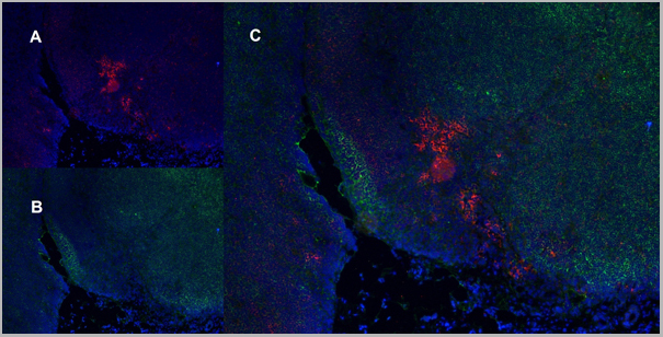
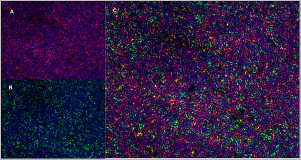
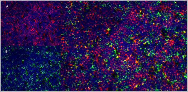
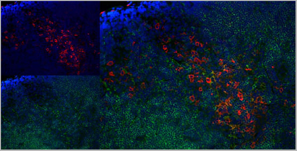
(Immunofluorescence staining of rat lymph node cryosection with Mouse anti Rat CD163 antibody , red an A and Mouse anti Rat CD8 MCA48), green in B. C is the merged image with nuclei counter-stained blue using DAPI. High power)
Application Data
(Immunofluorescence staining of rat lymph node cryosection with Mouse anti Rat CD163 antibody , red an A and Mouse anti Rat CD8 MCA48), green in B. C is the merged image with nuclei counter-stained blue using DAPI. High power)
CD8 ALPHA, Monoclonal Antibody (Cat# AAA12049)
Full Name
MOUSE ANTI RAT CD8 ALPHA:RPE
Applications
Flow Cytometry
Pricing
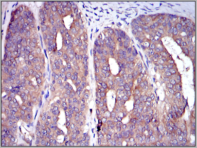
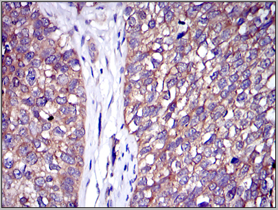 IHC (Immunohistochemistry)
(Immunohistochemical analysis of paraffin-embedded bladder cancer tissues using C-CBL mouse mAb with DAB staining.)
IHC (Immunohistochemistry)
(Immunohistochemical analysis of paraffin-embedded bladder cancer tissues using C-CBL mouse mAb with DAB staining.)
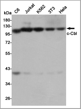
c-Cbl, Monoclonal Antibody (Cat# AAA14105)
Full Name
Anti-c-Cbl
Gene Names
CBL; CBL2; NSLL; C-CBL; RNF55; FRA11B
Applications
Western Blot, Immunoprecipitation, Immunohistochemistry, Immunocytochemistry, Flow Cytometry
Purity
Ascitic Fluid
Pricing
 IF (Immunofluorescence)
(AAA31008 staining Hela cells by IF/ICC. The sample were fixed with PFA and permeabilized in 0.1% Triton X-100, then blocked in 10% serum for 45 minutes at 25 degree C. The primary antibody was diluted at 1/200 and incubated with the sample for 1 hour at 37 degree C. An Alexa Fluor 594 conjugated goat anti-rabbit IgG (H+L) antibody, diluted at 1/600, was used as secondary antibody.)
IF (Immunofluorescence)
(AAA31008 staining Hela cells by IF/ICC. The sample were fixed with PFA and permeabilized in 0.1% Triton X-100, then blocked in 10% serum for 45 minutes at 25 degree C. The primary antibody was diluted at 1/200 and incubated with the sample for 1 hour at 37 degree C. An Alexa Fluor 594 conjugated goat anti-rabbit IgG (H+L) antibody, diluted at 1/600, was used as secondary antibody.)
Bcr, Polyclonal Antibody (Cat# AAA31008)
Full Name
Phospho-Bcr (Tyr360) Antibody
Gene Names
BCR; ALL; CML; PHL; BCR1; D22S11; D22S662
Reactivity
Human, Mouse, Rat
Applications
Western Blot, Immunohistochemistry, Immunofluorescence, Immunocytochemistry
Purity
From purified rabbit serum by affinity purification via sequential chromatography on phospho-and non-phospho-peptide affinity columns.
Pricing
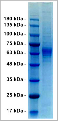 SDS-PAGE
(Purified Phlebovirus glycoprotein G2 (SFTS virus HN6))
SDS-PAGE
(Purified Phlebovirus glycoprotein G2 (SFTS virus HN6))
Membrane Glycoprotein G2 of Phlebovirus, Recombinant Protein (Cat# AAA13767)
Full Name
Membrane Glycoprotein G2 of Phlebovirus (SFTS virus HN6)
Applications
Western Blot
Purity
> 95% (SDS-PAGE gel)
Pricing
B-lymphocyte, ELISA Kit (Cat# AAA13258)
Full Name
Human B-lymphocyte antigen (CD20) ELISA Kit
Gene Names
MS4A1; B1; S7; Bp35; CD20; CVID5; MS4A2; LEU-16
Reactivity
Human
Pricing
Complement C9, Active Protein (Cat# AAA14378)
Full Name
Complement C9 protein
Gene Names
C9; C9D; ARMD15
Purity
> 95% pure
Pricing
 Application Data
(Staining of human peripheral blood lymphocytes with Mouse anti Human CD20: Pacific Blue)
Application Data
(Staining of human peripheral blood lymphocytes with Mouse anti Human CD20: Pacific Blue)
CD20, Monoclonal Antibody (Cat# AAA12032)
Full Name
MOUSE ANTI HUMAN CD20:RPE
Gene Names
MS4A1; B1; S7; Bp35; CD20; CVID5; MS4A2; LEU-16
Applications
Flow Cytometry
Pricing
 IF (Immunofluorescence)
(AAA31472 staining HepG2 cells by IF/ICC. The samples were fixed with PFA and permeabilized in 0.1% Triton X-100, then blocked in 10% serum for 45 minutes at 25°C. Samples were then incubated with primary Ab (#AAA31472) and rabbit anti-beta tubulin Ab (#) for 1 hour at 37°C. An AlexaFluor594 conjugated goat anti-mouse IgG Ab(Red) and an AlexaFluor488 conjugated goat anti-rabbit IgG Ab(Green) were used as the secondary antibody. The nuclear counter stain is DAPI (blue).)
IF (Immunofluorescence)
(AAA31472 staining HepG2 cells by IF/ICC. The samples were fixed with PFA and permeabilized in 0.1% Triton X-100, then blocked in 10% serum for 45 minutes at 25°C. Samples were then incubated with primary Ab (#AAA31472) and rabbit anti-beta tubulin Ab (#) for 1 hour at 37°C. An AlexaFluor594 conjugated goat anti-mouse IgG Ab(Red) and an AlexaFluor488 conjugated goat anti-rabbit IgG Ab(Green) were used as the secondary antibody. The nuclear counter stain is DAPI (blue).)
Claudin 18.2, Monoclonal Antibody (Cat# AAA31472)
Full Name
Claudin 18.2 Mouse monoclonal antibody
Gene Names
CLDN18; SFTA5; SFTPJ; DKFZp564B2062
Reactivity
Mouse, Rat
Applications
Western Blot, Immunofluorescence, Immunocytochemistry
Purity
Affinity-chromatography.
Pricing