Filters
Clonality
Type
Reactivity
Gene Name
Isotype
Host
Application
Clone
679 results for " Loading Antibody" - showing 600-650
TCF19, Monoclonal Antibody (Cat# AAA24385)
Full Name
TCF19 (Transcription Factor 19, TCF-19, SC1, SC1-1, Transcription Factor SC1) (AP)
Gene Names
TCF19; SC1; TCF-19
Reactivity
Human
Applications
Immunohistochemistry, Western Blot
Purity
Purified by Protein A Affinity Chromatography.
Pricing
ABL2, Monoclonal Antibody (Cat# AAA24411)
Full Name
ABL2 (Tyrosine-protein Kinase ABL2, Abelson Murine Leukemia Viral Oncogene Homolog 2, Abelson-related Gene Protein, Tyrosine Kinase ARG, ABLL, ARG) APC
Gene Names
ABL2; ARG; ABLL
Reactivity
Human
Applications
Western Blot
Purity
Purified by Protein A Affinity Chromatography.
Pricing
CBS, Monoclonal Antibody (Cat# AAA24452)
Full Name
CBS (Cystathionine beta-synthase, Serine Sulfhydrase, Beta-thionase) APC
Gene Names
CBS; HIP4
Reactivity
Human
Applications
Immunohistochemistry, Immunoprecipitation, Western Blot
Purity
Purified by Protein A Affinity Chromatography.
Pricing
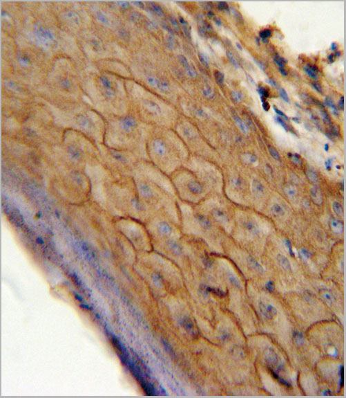
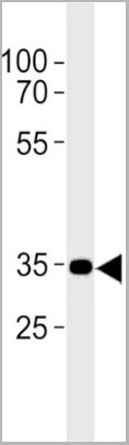
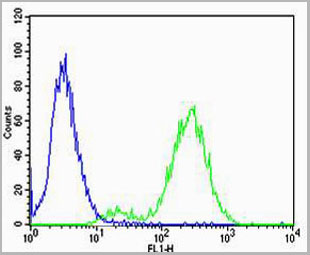
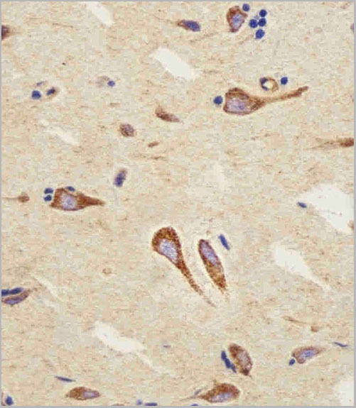
CAPNS1, Monoclonal Antibody (Cat# AAA24156)
Full Name
CAPNS1 (Calpain Small Subunit 1, CSS1, Calcium-activated Neutral Proteinase Small Subunit, CANP Small Subunit, Calcium-dependent Protease Small Subunit, CDPS, Calcium-dependent Protease Small Subunit 1, Calpain Regulatory Subunit, CAPN4, CAPNS) (AP)
Gene Names
CAPNS1; CANP; CDPS; CSS1; CANPS; CAPN4; CALPAIN4
Reactivity
Human, Rat
Applications
Immunohistochemistry, Western Blot
Purity
Purified by Protein A Affinity Chromatography.
Pricing
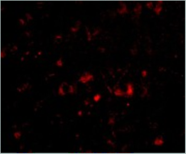
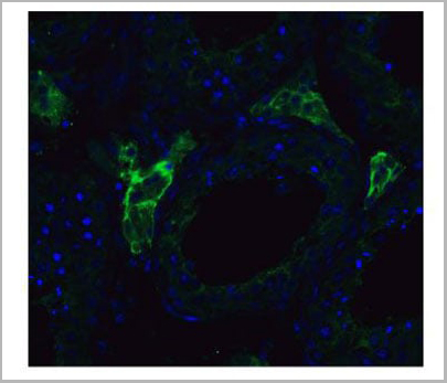
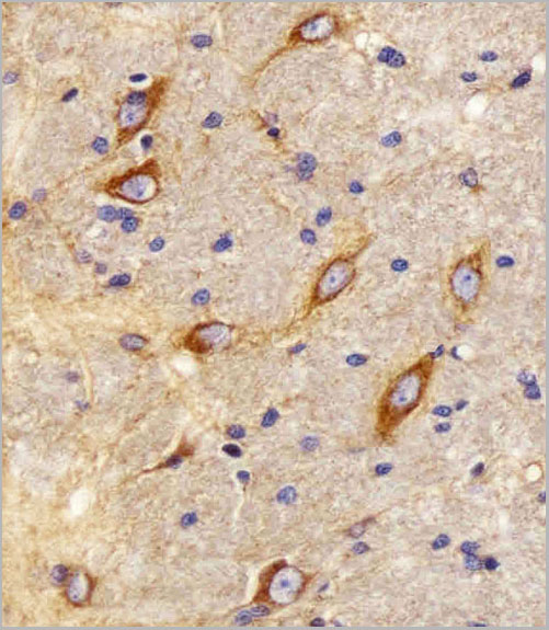
 IF (Immunofluorescence)
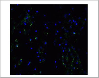
(Immunofluorescent analysis of 4% paraformaldehyde-fixed mouse brain issue labeling NPTX2 with AAA10933 at 20ug/mL, followed by goat anti-rabbit IgG secondaryantibody at 1/500 dilution (red) and DAPI staining (blue).)
IF (Immunofluorescence)
(Immunofluorescent analysis of 4% paraformaldehyde-fixed mouse brain issue labeling NPTX2 with AAA10933 at 20ug/mL, followed by goat anti-rabbit IgG secondaryantibody at 1/500 dilution (red) and DAPI staining (blue).)
NPTX2, Polyclonal Antibody (Cat# AAA10933)
Full Name
NPTX2 Antibody
Gene Names
NPTX2; NP2; NARP; NP-II
Reactivity
Human, Mouse, Rat
Applications
Western Blot, Immunohistochemistry, Immunofluorescence
Purity
NPTX2 Antibody is affinity chromatography purified via peptide column.
Pricing
CAMKIV, Monoclonal Antibody (Cat# AAA24153)
Full Name
CAMKIV (Calcium/Calmodulin-dependent Protein Kinase Type IV, CAM Kinase-GR, CaMK IV, CAMK4) (AP)
Gene Names
CAMK4; caMK; CaMKIV; CaMK IV; CaMK-GR
Reactivity
Human
Applications
Immunohistochemistry, Western Blot
Purity
Purified by Protein A Affinity Chromatography.
Pricing
ARNT, Monoclonal Antibody (Cat# AAA24140)
Full Name
ARNT (Aryl Hydrocarbon Receptor Nuclear Translocator, ARNT Protein, Class E Basic Helix-loop-helix Protein 2, bHLHe2, Dioxin Receptor, Nuclear Translocator, Hypoxia-inducible Factor 1-beta, HIF-1-beta, HIF1-beta, BHLHE2) (AP)
Gene Names
ARNT; HIF1B; TANGO; bHLHe2; HIF1BETA; HIF-1beta; HIF1-beta; HIF-1-beta
Reactivity
Human
Applications
Immunohistochemistry, Western Blot
Purity
Purified by Protein A Affinity Chromatography.
Pricing
RAD18, Monoclonal Antibody (Cat# AAA24334)
Full Name
RAD18 (E3 Ubiquitin-protein Ligase RAD18, Postreplication Repair Protein RAD18, hRAD18, hHR18, RING Finger Protein 73, RNF73) (AP)
Gene Names
RAD18; RNF73
Reactivity
Human, Rat
Applications
Immunohistochemistry, Western Blot
Purity
Purified by Protein A Affinity Chromatography.
Pricing
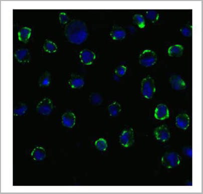 IF (Immunofluorescence)
(Figure 11 Immunofluorescence Validation of ACE2 In Caco2 CellsImmunofluorescent analysis of 4% paraformaldehyde-fixed Caco2 cells labeling ACE2 with AAA10945 at 20 ug/mL, followed by goat anti-rabbit IgG secondary antibody at 1/500 dilution (green) and DAPI staining (blue). Imageshowing membrane staining on Caco2 cells.)
IF (Immunofluorescence)
(Figure 11 Immunofluorescence Validation of ACE2 In Caco2 CellsImmunofluorescent analysis of 4% paraformaldehyde-fixed Caco2 cells labeling ACE2 with AAA10945 at 20 ug/mL, followed by goat anti-rabbit IgG secondary antibody at 1/500 dilution (green) and DAPI staining (blue). Imageshowing membrane staining on Caco2 cells.)
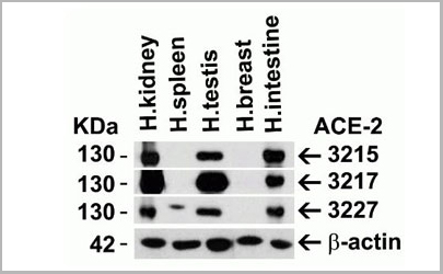
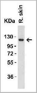
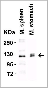
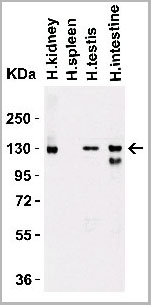
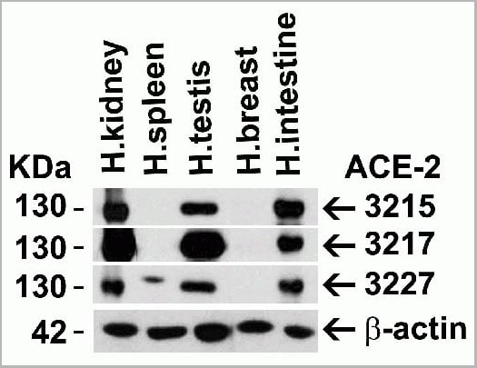
ACE2, Polyclonal Antibody (Cat# AAA10945)
Full Name
ACE2 Antibody
Gene Names
ACE2; ACEH
Reactivity
Human, Mouse, Rat
Applications
Western Blot, Immunohistochemistry, Immunofluorescence
Purity
ACE2 Antibody is affinity chromatography purified via peptide column.
Pricing
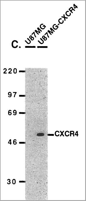

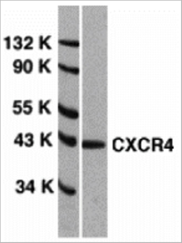
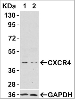
 WB (Western Blot)
(Figure 9 WB Validation of CXCR4 in metastatic melanoma (Scala et al., 2006)CXCR4 protein was detected in the human metastatic melanoma cell lines and human melanoma cell line (colo38), but not in the human primary melanocytes (MPR1) with anti-CXCR4 antibodies.)
WB (Western Blot)
(Figure 9 WB Validation of CXCR4 in metastatic melanoma (Scala et al., 2006)CXCR4 protein was detected in the human metastatic melanoma cell lines and human melanoma cell line (colo38), but not in the human primary melanocytes (MPR1) with anti-CXCR4 antibodies.)
CXCR4, Polyclonal Antibody (Cat# AAA10962)
Full Name
CXCR4 Antibody
Gene Names
CXCR4; FB22; HM89; LAP3; LCR1; NPYR; WHIM; CD184; LAP-3; LESTR; NPY3R; NPYRL; HSY3RR; NPYY3R; D2S201E
Reactivity
Human, Mouse
Applications
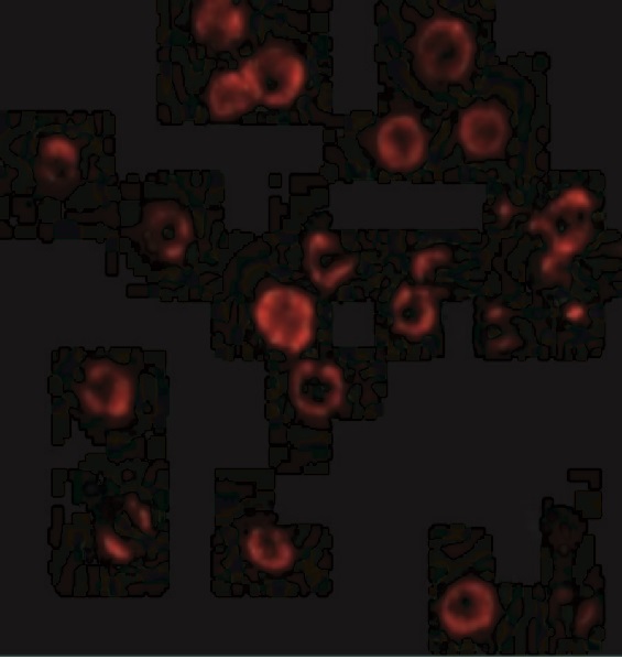
Western Blot, Immunofluorescence
Purity
CXCR4 Antibody is Protein A purified.
Pricing
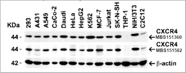
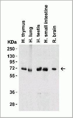
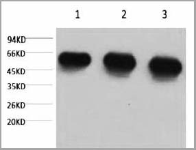
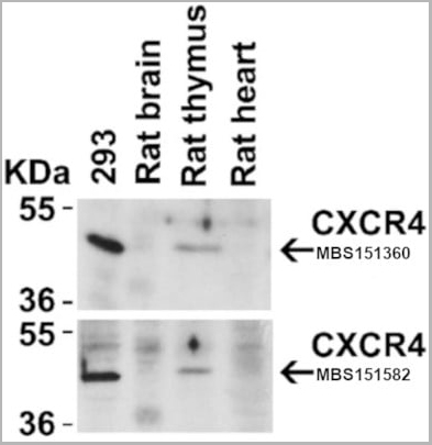 WB (Western Blot)
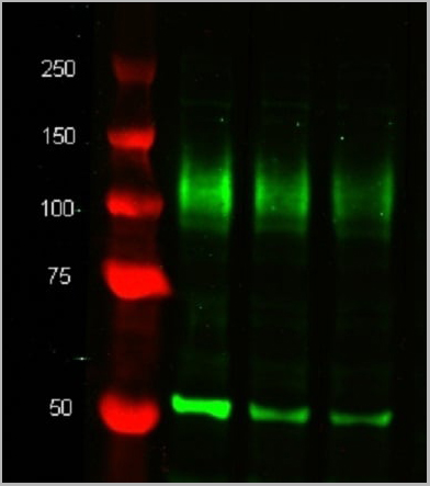
(Figure 4 Animal Species ReactivityLoading: Lysates/proteins at 20 μg per lane. Antibodies: 1009 (2 μg/mL) or 1012 (2 μg/mL). 1 h incubation at RT in 5% NFDM/TBST. Secondary: Goat anti-rabbit IgG HRP conjugate at 1:10000 dilution.)
WB (Western Blot)
(Figure 4 Animal Species ReactivityLoading: Lysates/proteins at 20 μg per lane. Antibodies: 1009 (2 μg/mL) or 1012 (2 μg/mL). 1 h incubation at RT in 5% NFDM/TBST. Secondary: Goat anti-rabbit IgG HRP conjugate at 1:10000 dilution.)
CXCR4, Polyclonal Antibody (Cat# AAA10951)
Full Name
CXCR4 Antibody
Gene Names
CXCR4; FB22; HM89; LAP3; LCR1; NPYR; WHIM; CD184; LAP-3; LESTR; NPY3R; NPYRL; HSY3RR; NPYY3R; D2S201E
Reactivity
Human, Mouse, Rat
Applications
Western Blot, Immunocytochemistry, Immunoprecipitation, Immunofluorescence, Immunohistochemistry, Flow Cytometry
Purity
CXCR4 Antibody is affinity chromatography purified via peptide column.
Pricing

 IF (Immunofluorescence)
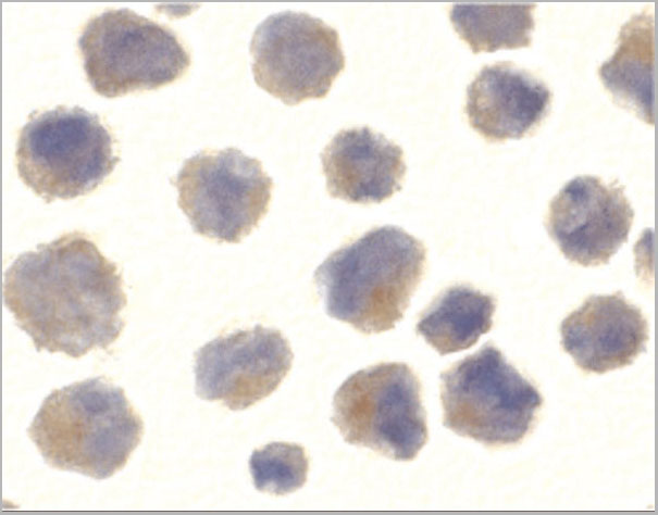
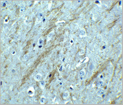
(Figure 10 KD Validation of BACE in DRG (Hyun, 2007)Decreased BACE1 expression in DRG following siRNA3 transfection. DRG neurons were transfected with 1 ug siRNA3 plasmid and incubated for 48 hours in 37°˚C. DRG neurons were stained for BACE1 us¬ing the Anti-BACE antibody (a,b) Neurons transfected with the control plas¬mid pSUPER-EGFP (green) did not display any changes in BACE1 expression (red). (c,d) DRG neurons transfected with siR¬NA3 displayed reduced BACE1 expression in the axon.)
IF (Immunofluorescence)
(Figure 10 KD Validation of BACE in DRG (Hyun, 2007)Decreased BACE1 expression in DRG following siRNA3 transfection. DRG neurons were transfected with 1 ug siRNA3 plasmid and incubated for 48 hours in 37°˚C. DRG neurons were stained for BACE1 us¬ing the Anti-BACE antibody (a,b) Neurons transfected with the control plas¬mid pSUPER-EGFP (green) did not display any changes in BACE1 expression (red). (c,d) DRG neurons transfected with siR¬NA3 displayed reduced BACE1 expression in the axon.)
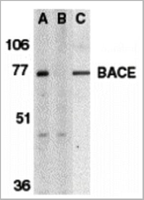
BACE, Polyclonal Antibody (Cat# AAA10918)
Full Name
BACE Antibody
Gene Names
BACE1; ASP2; BACE; HSPC104
Reactivity
Human, Mouse
Applications
Immunocytochemistry, Immunofluorescence, Immunohistochemistry, Western Blot
Purity
BACE Antibody is affinity chromatography purified via peptide column.
Pricing
ABCF2, Monoclonal Antibody (Cat# AAA24409)
Full Name
ABCF2 (ATP-binding Cassette Sub-family F Member 2, Iron-inhibited ABC Transporter 2, HUSSY-18, DKFZp586K1823, EST133090, M-ABC1) APC
Gene Names
ABCF2; ABC28; HUSSY18; HUSSY-18; EST133090
Reactivity
Human
Applications
Immunohistochemistry, Immunoprecipitation, Western Blot
Purity
Purified by Protein A Affinity Chromatography.
Pricing
TGIF2, Monoclonal Antibody (Cat# AAA24386)
Full Name
TGIF2 (TGF-beta-induced Transcription Factor 2, TGFB-induced Factor 2, Homeobox Protein TGIF2, 5'-TG-3'-interacting Factor 2) (AP)
Reactivity
Human
Applications
Western Blot
Purity
Purified by Protein A Affinity Chromatography.
Pricing

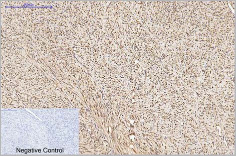
 IF (Immunofluorescence)
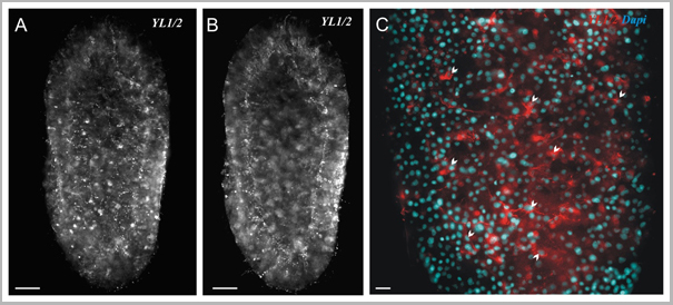
(Figure 10 Immunofluorescence Validation of TACE in Rat Brain (Pradillo et al, 2005)Cellular localization of TACE. Double immunofluorescence staining of brain sections from sham-operated (SHAM; A, C, E) and IPC-exposed animals (IPC; B, D, F) of TACE (red) and the cellular markers (green) NeuN (neurons; A, B), GFAP (astrocytes; C, D) and L. esculentum lectin (microglia and endothelium; E, F). White arrows indicate TACE-positive cells.)
IF (Immunofluorescence)
(Figure 10 Immunofluorescence Validation of TACE in Rat Brain (Pradillo et al, 2005)Cellular localization of TACE. Double immunofluorescence staining of brain sections from sham-operated (SHAM; A, C, E) and IPC-exposed animals (IPC; B, D, F) of TACE (red) and the cellular markers (green) NeuN (neurons; A, B), GFAP (astrocytes; C, D) and L. esculentum lectin (microglia and endothelium; E, F). White arrows indicate TACE-positive cells.)
TACE, Polyclonal Antibody (Cat# AAA10912)
Full Name
TACE Antibody
Gene Names
ADAM17; CSVP; TACE; NISBD; ADAM18; CD156B; NISBD1
Reactivity
Human, Mouse, Rat
Applications
Western Blot, Immunocytochemistry, Immunofluorescence, Flow Cytometry
Purity
TACE Antibody is affinity chromatography purified via peptide column.
Pricing
 Application Data
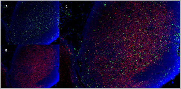
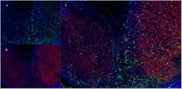
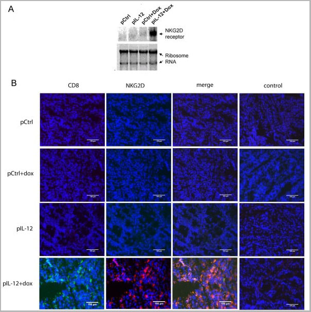
(Immunofluorescence staining of mouse lymph node cryosection with Rat anti Mouse CD11b, clone 5C6 , green in A and Rat anti Mouse CD8, clone YTS105.18 , red in B. C is the merged image with nuclei counterstained blue using DAPI. High power)
Application Data
(Immunofluorescence staining of mouse lymph node cryosection with Rat anti Mouse CD11b, clone 5C6 , green in A and Rat anti Mouse CD8, clone YTS105.18 , red in B. C is the merged image with nuclei counterstained blue using DAPI. High power)
CD8 ALPHA, Monoclonal Antibody (Cat# AAA11918)
Full Name
RAT ANTI MOUSE CD8
Gene Names
Cd8a; Ly-2; Ly-B; Ly-35; Lyt-2; BB154331
Applications
Immunohistochemistry, Flow Cytometry, Immunofluorescence
Pricing


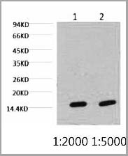
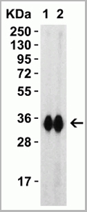
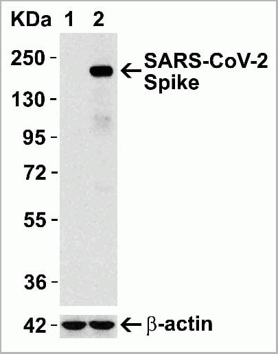 WB (Western Blot)
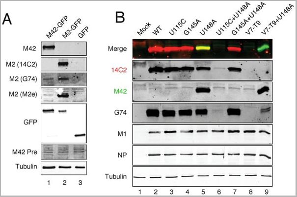
(Figure 12 Overexpression Validation in Spike Transfected 293 Cells Loading: 15 ug per lane of 293 cell lysate. Antibodies: SARS-CoV-2 (COVID-19) Spike, AAA10931 (1 ug/mL), 1h incubation at RT in 5% NFDM/TBST. Secondary: Goat anti rabbit IgG HRP conjugate at 1:10000 dilution. Lane 1: WT293 cells and Lane 2: SARS-CoV-2 Spike overexpressed 293 cells.)
WB (Western Blot)
(Figure 12 Overexpression Validation in Spike Transfected 293 Cells Loading: 15 ug per lane of 293 cell lysate. Antibodies: SARS-CoV-2 (COVID-19) Spike, AAA10931 (1 ug/mL), 1h incubation at RT in 5% NFDM/TBST. Secondary: Goat anti rabbit IgG HRP conjugate at 1:10000 dilution. Lane 1: WT293 cells and Lane 2: SARS-CoV-2 Spike overexpressed 293 cells.)
COVID 19 Spike Protein Coronavirus, Polyclonal Antibody (Cat# AAA10931)
Full Name
SARS-CoV-2 (COVID-19, 2019-nCoV) Spike Antibody
Reactivity
Virus
Applications
Immunofluorescence, Immunohistochemistry, Western Blot
Purity
SARS-CoV-2 (COVID-19, 2019-nCoV) Spike Antibody is affinity chromatography purified via peptide column.
Pricing
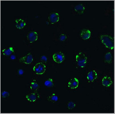 IF (Immuofluorescence)
(Figure 9 Immunofluorescence Validation of ACE2 In Caco2 Cells Immunofluorescent analysis of 4% paraformaldehyde-fixed Caco2 cells labeling ACE2 with AAA10928 at 20 ug/mL,followed by goat anti-rabbit IgG secondary antibody at1/500 dilution (green) and DAPI staining (blue). Imageshowing membrane staining on Caco2 cells.)
IF (Immuofluorescence)
(Figure 9 Immunofluorescence Validation of ACE2 In Caco2 Cells Immunofluorescent analysis of 4% paraformaldehyde-fixed Caco2 cells labeling ACE2 with AAA10928 at 20 ug/mL,followed by goat anti-rabbit IgG secondary antibody at1/500 dilution (green) and DAPI staining (blue). Imageshowing membrane staining on Caco2 cells.)
ACE2, Polyclonal Antibody (Cat# AAA10928)
Full Name
ACE2 Antibody
Gene Names
ACE2; ACEH
Reactivity
Human, Mouse, Rat
Applications
Western Blot, Immunohistochemistry, Immunofluorescence
Purity
ACE2 Antibody is affinity chromatography purified via peptide column.
Pricing
UGP2, Monoclonal Antibody (Cat# AAA24401)
Full Name
UGP2 (UTP--glucose-1-phosphate Uridylyltransferase, UDP-glucose Pyrophosphorylase, UDPGP, UGPase, UGP1) (AP)
Gene Names
UGP2; UDPG; UGP1; UDPGP; UGPP1; UGPP2; UDPGP2; pHC379
Reactivity
Human, Mouse, Rat
Applications
Western Blot
Purity
Purified by Protein A Affinity Chromatography.
Pricing
CDC25C, Monoclonal Antibody (Cat# AAA24164)
Full Name
CDC25C (Cell Division Cycle 25 Homolog C, Cdc 25C, CDC 25, Dual Specificity Phosphatase Cdc25C, M-phase Inducer Phosphatase 3, MPIP3, PPP1R60) (AP)
Gene Names
CDC25C; CDC25; PPP1R60
Reactivity
Human
Applications
Immunohistochemistry, Immunoprecipitation, Western Blot
Purity
Purified by Protein A Affinity Chromatography.
Pricing
RSK3, Monoclonal Antibody (Cat# AAA24347)
Full Name
RSK3, CT (Ribosomal S6 Kinase 3, RSK-3, pp90RSK3, 90kD Ribosomal Protein S6 Kinase 2, p90-RSK 2, p90RSK2, MAP Kinase-activated Protein Kinase 1c, MAPK-activated Protein Kinase 1c, MAPKAP Kinase 1c, MAPKAPK1C, MAPKAPK-1c, Ribosomal Protein S6 Kinase alpha-
Gene Names
RPS6KA2; RSK; HU-2; RSK3; p90RSK2; p90-RSK3; pp90RSK3; MAPKAPK1C; S6K-alpha; S6K-alpha2
Reactivity
Human
Applications
Immunohistochemistry, Western Blot
Purity
Purified by Protein A Affinity Chromatography.
Pricing


 IF (Immunofluorescence)
(Figure 8 Immunofluorescence Validation of JMJD3 in K562 Cells Immunofluorescent analysis of 4% paraformaldehyde fixed K562 cells labeling JMJD3 with AAA10925 at 20 ug/mL, followed by goat anti-rabbit IgG secondary antibody at 1/500 dilution (red).)
IF (Immunofluorescence)
(Figure 8 Immunofluorescence Validation of JMJD3 in K562 Cells Immunofluorescent analysis of 4% paraformaldehyde fixed K562 cells labeling JMJD3 with AAA10925 at 20 ug/mL, followed by goat anti-rabbit IgG secondary antibody at 1/500 dilution (red).)
JMJD3, Antibody (Cat# AAA10925)
Full Name
JMJD3 Antibody
Gene Names
KDM6B; JMJD3
Reactivity
Human, Mouse, Rat
Applications
Immunocytochemistry, Immunofluorescence, Western Blot
Purity
JMJD3 Antibody is affinity chromatography purified via peptide column.
Pricing
 Application Data
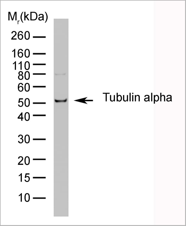
(Western blot analysis of CD107b on J774 cell lysate probed with Rat anti Mouse CD107b at 1/500, 1/1000, 1/2000. Rat anti tubulin alpha is included as a loading control. Detection is with Goat anti Rat IgG:Dylight®)
Application Data
(Western blot analysis of CD107b on J774 cell lysate probed with Rat anti Mouse CD107b at 1/500, 1/1000, 1/2000. Rat anti tubulin alpha is included as a loading control. Detection is with Goat anti Rat IgG:Dylight®)
TUBULIN ALPHA, Monoclonal Antibody (Cat# AAA12010)
Full Name
RAT ANTI TUBULIN ALPHA
Applications
Immunohistochemistry, Immunofluorescence, Radioimmunoassay, Western Blot
Pricing


 Application Data
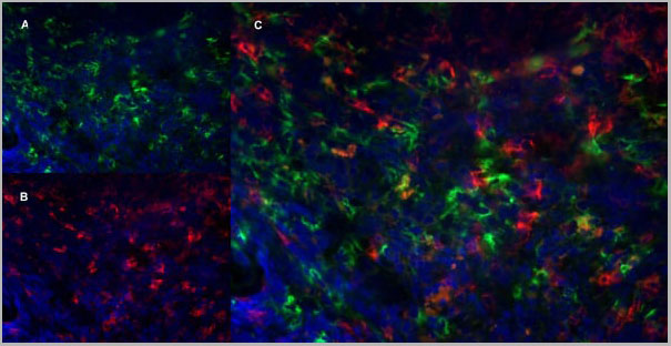
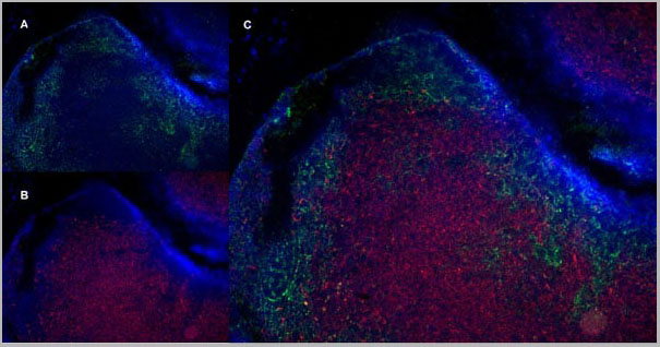
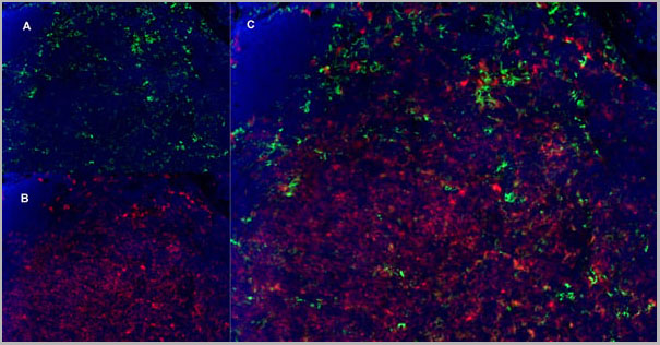
(Immunofluorescence staining of mouse lymph node cryosection with Rat anti Mouse CD11b, clone 5C6 , green in A and Rat anti Mouse CD8, clone YTS105.18 , red in B. C is the merged image with nuclei counterstained blue using DAPI. High power)
Application Data
(Immunofluorescence staining of mouse lymph node cryosection with Rat anti Mouse CD11b, clone 5C6 , green in A and Rat anti Mouse CD8, clone YTS105.18 , red in B. C is the merged image with nuclei counterstained blue using DAPI. High power)
CD8 ALPHA, Monoclonal Antibody (Cat# AAA12069)
Full Name
RAT ANTI MOUSE CD8
Gene Names
Cd8a; Ly-2; Ly-B; Ly-35; Lyt-2; BB154331
Reactivity
Mouse
Applications
Immunohistochemistry, Flow Cytometry, Immunofluorescence
Pricing
 Application Data
(The cancer cell lines were stably expressing DYKDDDDK-tagged RIP3 and RIP expression was detected by anti-RIP3 antibodies (AAA10922) in RIP3-overexpressed cells.)
Application Data
(The cancer cell lines were stably expressing DYKDDDDK-tagged RIP3 and RIP expression was detected by anti-RIP3 antibodies (AAA10922) in RIP3-overexpressed cells.)
RIP3, Polyclonal Antibody (Cat# AAA10922)
Full Name
RIP3 Antibody
Gene Names
Ripk3; Rip3; AW107945; 2610528K09Rik
Reactivity
Human, Mouse, Rat
Applications
Immunofluorescence, Immunohistochemistry, Immunoprecipitation, Western Blot
Purity
RIP3 Antibody is affinity chromatography purified via peptide column.
Pricing
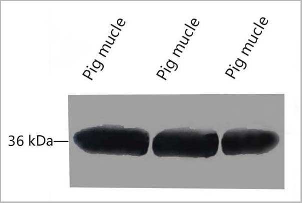
ENO3, Monoclonal Antibody (Cat# AAA24202)
Full Name
ENO3 (Enolase 3, 2-phospho-D-glycerate Hydro-lyase, beta-Enolase, Muscle-specific Enolase, MSE, Skeletal Muscle Enolase) (AP)
Gene Names
ENO3; MSE; GSD13
Reactivity
Human
Applications
EIA, IHC, WB
Purity
Purified by Protein A Affinity Chromatography.
Pricing
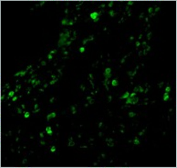
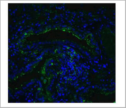
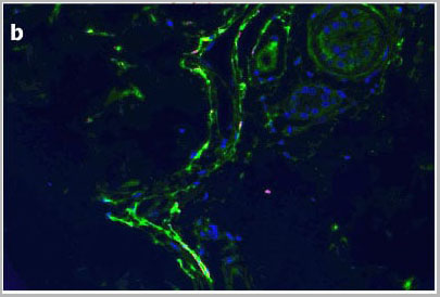
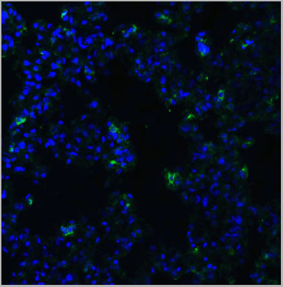
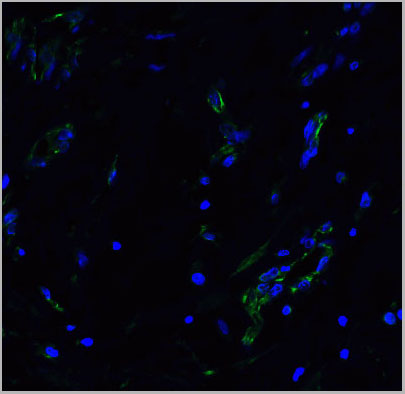
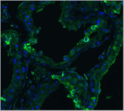
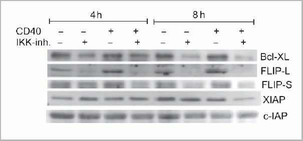 IF (Immunofluorescence)
(Immunofluorescence Validation of cIAP in Human Lung Tissue Immunofluorescent analysis of 4% paraformaldehydefixed human lung tissue abeling cIAP with 3325 at 20 ug/mL, followed by goat anti-rabbit IgG secondary antibody at 1/500 dilution (green).)
IF (Immunofluorescence)
(Immunofluorescence Validation of cIAP in Human Lung Tissue Immunofluorescent analysis of 4% paraformaldehydefixed human lung tissue abeling cIAP with 3325 at 20 ug/mL, followed by goat anti-rabbit IgG secondary antibody at 1/500 dilution (green).)
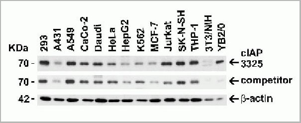
cIAP, Polyclonal Antibody (Cat# AAA10954)
Full Name
cIAP Antibody
Gene Names
BIRC2; API1; MIHB; HIAP2; RNF48; cIAP1; Hiap-2; c-IAP1
Reactivity
Human, Mouse
Applications
Western Blot, Immunohistochemistry, Immunofluorescence
Purity
cIAP Antibody is affinity chromatography purified via peptide column.
Pricing
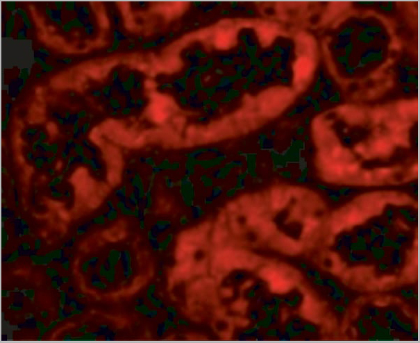
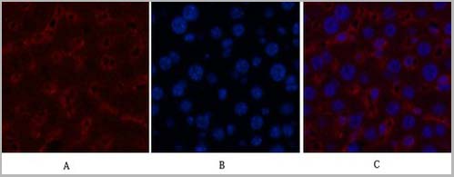
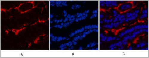
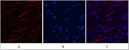
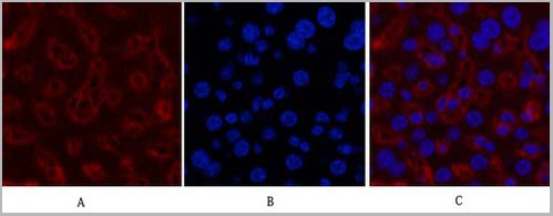
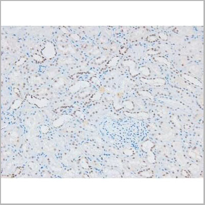
 IF (Immunofluorescence)
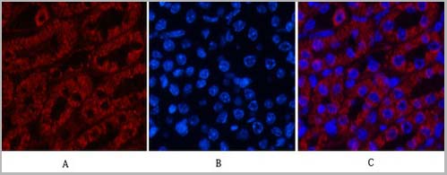
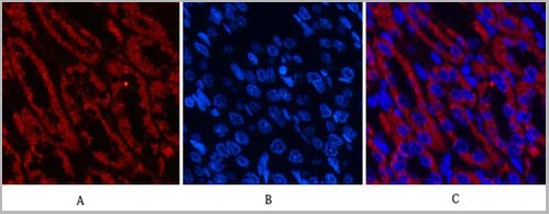
(Fig.6. Immunofluorescence analysis of rat kidney tissue. 1, COX IV Monoclonal Antibody (14Y2) (red) was diluted at 1:200 (4°C, overnight). 2, Cy3 Labeled secondary antibody was diluted at 1:300 (room temperature, 50min). 3, Picture B: DAPI (blue) 10min. Picture A: Target. Picture B: DAPI. Picture C: merge of A+B.)
IF (Immunofluorescence)
(Fig.6. Immunofluorescence analysis of rat kidney tissue. 1, COX IV Monoclonal Antibody (14Y2) (red) was diluted at 1:200 (4°C, overnight). 2, Cy3 Labeled secondary antibody was diluted at 1:300 (room temperature, 50min). 3, Picture B: DAPI (blue) 10min. Picture A: Target. Picture B: DAPI. Picture C: merge of A+B.)
COX IV, Monoclonal Antibody (Cat# AAA31542)
Full Name
Anti-COX IV Mouse Monoclonal Antibody (14Y2)
Gene Names
COX4I1; COX4; COXIV; COX4-1
Reactivity
Human, Mouse, Rat
Applications
Western Blot, Immunohistochemistry, Immunofluorescence
Purity
The antibody was affinity-purified from mouse ascites by affinity-chromatography using specific immunogen
Pricing
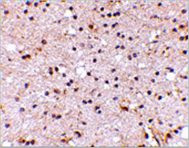
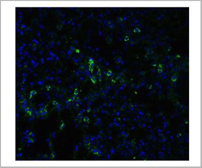
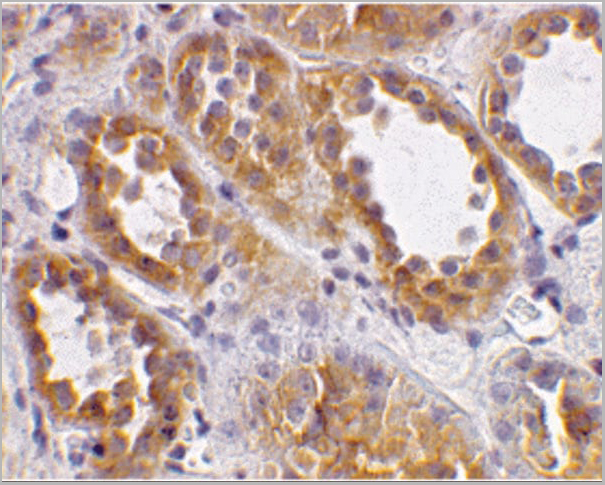
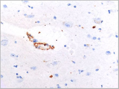
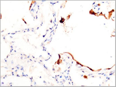
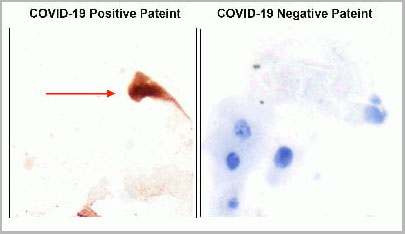
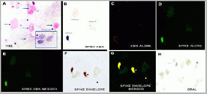
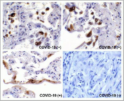
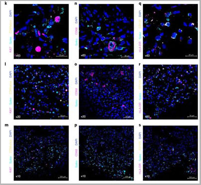
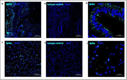
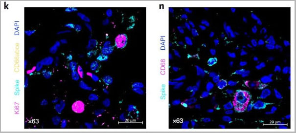
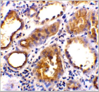
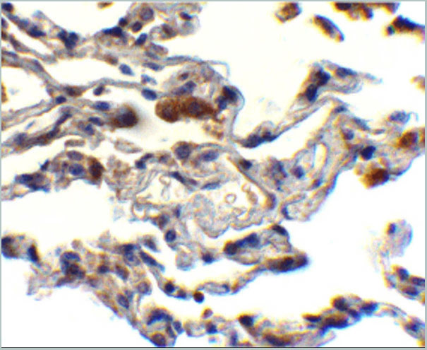
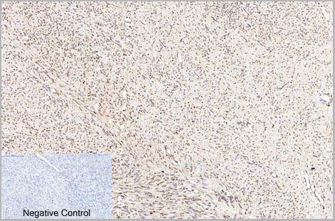
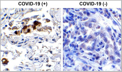 IHC (Immunohistochemistry)
(Immunohistochemistry Validation of SARSCoV-2 (COVID-19) Spike RBD in COVID-19 Patient Lung Immunohistochemical analysis of paraffin-embedded COVID-19 patient lung tissue using anti- SARS-CoV-2 (COVID-19) Spike RBD antibody (AAA11030, 0.5 µg/mL). Tissue was fixed with formaldehyde and blocked with 10% serum for 1 h at RT; antigen retrieval was by heat mediation with a citrate buffer (pH6). Samples were incubated with primary antibody overnight at 4°C. A goat anti-rabbit IgG H&L (HRP) at 1/250 was used as secondary. Counter stained with Hematoxylin. Strong signal of SARS-COV-2 Spike RBD protein was observed in macrophage of COVID-19 patient lung, but not in non-COVID-19 patient lung.)
IHC (Immunohistochemistry)
(Immunohistochemistry Validation of SARSCoV-2 (COVID-19) Spike RBD in COVID-19 Patient Lung Immunohistochemical analysis of paraffin-embedded COVID-19 patient lung tissue using anti- SARS-CoV-2 (COVID-19) Spike RBD antibody (AAA11030, 0.5 µg/mL). Tissue was fixed with formaldehyde and blocked with 10% serum for 1 h at RT; antigen retrieval was by heat mediation with a citrate buffer (pH6). Samples were incubated with primary antibody overnight at 4°C. A goat anti-rabbit IgG H&L (HRP) at 1/250 was used as secondary. Counter stained with Hematoxylin. Strong signal of SARS-COV-2 Spike RBD protein was observed in macrophage of COVID-19 patient lung, but not in non-COVID-19 patient lung.)
COVID 19 Spike RBD Coronavirus, Polyclonal Antibody (Cat# AAA11030)
Full Name
SARS-CoV-2 (COVID-19) Spike RBD Antibody
Gene Names
S; spike glycoprotein
Reactivity
Virus
Applications
Immunofluorescence, Immunohistochemistry, Western Blot
Purity
SARS-CoV-2 (COVID-19) Spike RBD antibody is affinity chromatography purified via peptide column.
Pricing
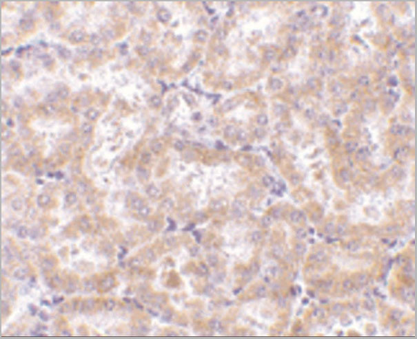
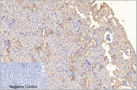
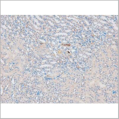
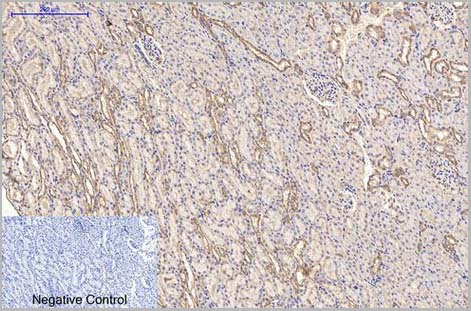 IHC (Immunohistchemistry)
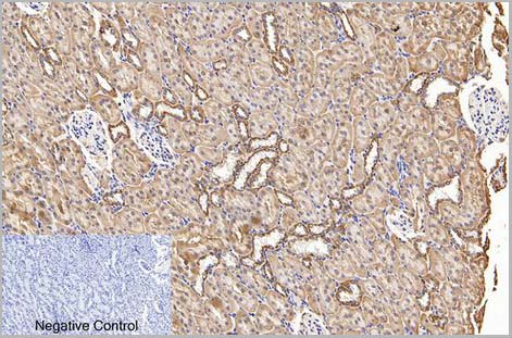
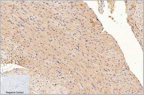
(Fig.6. Immunohistochemical analysis of paraffin-embedded rat kidney tissue. 1, alpha-tubulin Monoclonal Antibody (3G5) was diluted at 1:200 (4°C, overnight). 2, Sodium citrate pH 6.0 was used for antibody retrieval (>98°C, 20min). 3, secondary antibody was diluted at 1:200 (room temperature, 30min). Negative control was used by secondary antibody only.)
IHC (Immunohistchemistry)
(Fig.6. Immunohistochemical analysis of paraffin-embedded rat kidney tissue. 1, alpha-tubulin Monoclonal Antibody (3G5) was diluted at 1:200 (4°C, overnight). 2, Sodium citrate pH 6.0 was used for antibody retrieval (>98°C, 20min). 3, secondary antibody was diluted at 1:200 (room temperature, 30min). Negative control was used by secondary antibody only.)
alpha-Tubulin, Monoclonal Antibody (Cat# AAA31544)
Full Name
Anti-alpha-Tubulin Monoclonal Antibody (3G5)
Gene Names
TUBA1A; LIS3; TUBA3; B-ALPHA-1
Reactivity
Human, Mouse, Rat
Applications
Western Blot, Immunohistochemistry, Immunofluorescence, Immunoprecipitation
Purity
The antibody was affinity-purified from mouse ascites by affinity-chromatography using specific immunogen
Pricing
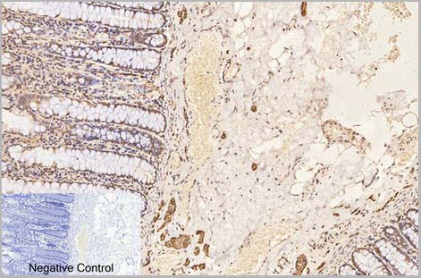
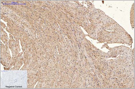
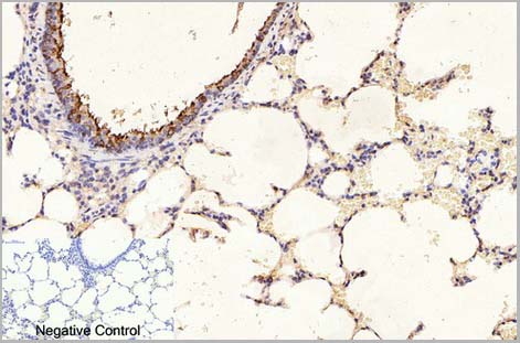 IHC (Immunohistchemistry)
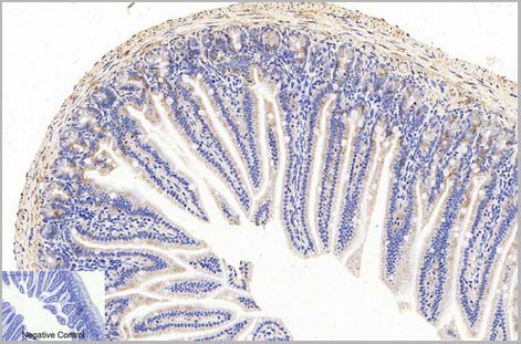
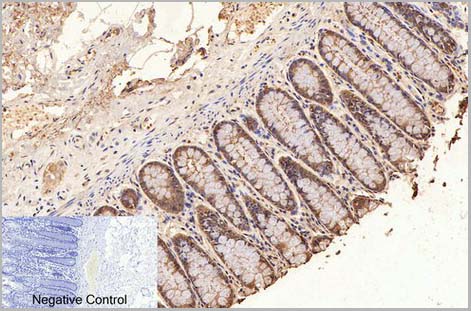
(Fig.6. Immunohistochemical analysis of paraffin-embedded rat lung tissue. 1, beta-Tubulin Monoclonal Antibody (3G6) was diluted at 1:400 (4°C, overnight). Negative control was used by secondary antibody only.)
IHC (Immunohistchemistry)
(Fig.6. Immunohistochemical analysis of paraffin-embedded rat lung tissue. 1, beta-Tubulin Monoclonal Antibody (3G6) was diluted at 1:400 (4°C, overnight). Negative control was used by secondary antibody only.)
beta-Tubulin, Monoclonal Antibody (Cat# AAA31540)
Full Name
Anti-beta-Tubulin Mouse Monoclonal Antibody (3G6)
Gene Names
TUBB3; CDCBM; TUBB4; beta-4; CFEOM3A
Reactivity
Human, Mouse, Rat, Monkey, Dog, Chicken, Rabbit, Sheep, Insect, Yeast, Hamster
Applications
Western Blot, Immunohistochemistry, Immunofluorescence
Purity
The antibody was affinity-purified from mouse ascites by affinity-chromatography using specific immunogen
Pricing
 WB (Western Blot)
(Western blot analysis of ZO-1 using various lysatesLanes 1 - 2: Merged signal (red and green). Green - AAA31075 observed at 195 kDa. Red - loading control, , observed at 55 kDa. Blots were developed with Goat Anti- Rabbit IgG(H+L) FITC–conjugated and Goat Anti-Mouse IgG(H+L) Alexa Fluor 594–conjugated secondary antibodies)
WB (Western Blot)
(Western blot analysis of ZO-1 using various lysatesLanes 1 - 2: Merged signal (red and green). Green - AAA31075 observed at 195 kDa. Red - loading control, , observed at 55 kDa. Blots were developed with Goat Anti- Rabbit IgG(H+L) FITC–conjugated and Goat Anti-Mouse IgG(H+L) Alexa Fluor 594–conjugated secondary antibodies)
ZO 1, Polyclonal Antibody (Cat# AAA31075)
Full Name
ZO 1 Antibody
Gene Names
TJP1; ZO-1
Reactivity
Human, Mouse, Rat, Pig, Monkey
Applications
Western Blot, Immunohistochemistry, Immunofluorescence, Immunocytochemistry
Purity
The antiserum was purified by peptide affinity chromatography using SulfoLink Coupling Resin
Pricing


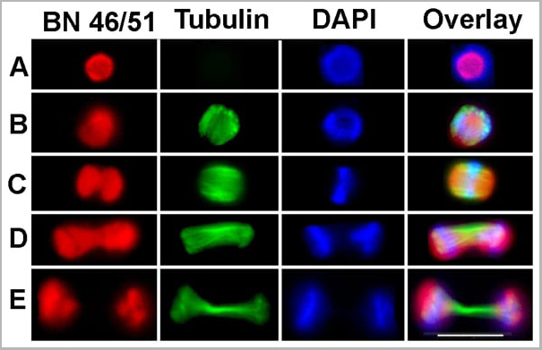
 Application Data
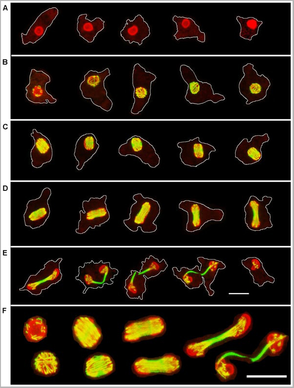
(Published customer image: Spindle abnormalities in embryos derived from imp-a2D14/imp-betaKetRE34 and imp-a2D14/imp-betac02473; NLSB-/+ females. (A -D) Wild-type and mutant embryos stained for a-tubulin (green) and DNA (blue). (A) Mitotic spindles in wild-type embryos at metaphase and anaphase. (B, C) Categories of spindle abnormalities found in embryos derived from (B) imp-a2D14/imp-betaKetRE34 and (C) imp-a2D14/imp-betac02743; NLSB-/+ females. (D) Formation of aster networks found in both genotypes. Scale bar: 10 um. (E) Frequency of spindle defects in embryos from both types of mutant females. Female genotypes are displayed at the upper right corner. At least 200 spindles were scored for both genotypes.From: Specific Cooperation Between Imp-a2 and Imp-beta/Ketel in Spindle Assembly During Drosophila Early Nuclear Divisions Erika Vir¡gh, M¡ty¡s Gorj¡n¡cz, Istv¡n T¶r¶k, Tolga Eichhorn, Sowjanya Kallakuri, Tam¡s Szlanka, Istv¡n Kiss, and Bernard M. Mechler G3 January 2012 2:1-14.)
Application Data
(Published customer image: Spindle abnormalities in embryos derived from imp-a2D14/imp-betaKetRE34 and imp-a2D14/imp-betac02473; NLSB-/+ females. (A -D) Wild-type and mutant embryos stained for a-tubulin (green) and DNA (blue). (A) Mitotic spindles in wild-type embryos at metaphase and anaphase. (B, C) Categories of spindle abnormalities found in embryos derived from (B) imp-a2D14/imp-betaKetRE34 and (C) imp-a2D14/imp-betac02743; NLSB-/+ females. (D) Formation of aster networks found in both genotypes. Scale bar: 10 um. (E) Frequency of spindle defects in embryos from both types of mutant females. Female genotypes are displayed at the upper right corner. At least 200 spindles were scored for both genotypes.From: Specific Cooperation Between Imp-a2 and Imp-beta/Ketel in Spindle Assembly During Drosophila Early Nuclear Divisions Erika Vir¡gh, M¡ty¡s Gorj¡n¡cz, Istv¡n T¶r¶k, Tolga Eichhorn, Sowjanya Kallakuri, Tam¡s Szlanka, Istv¡n Kiss, and Bernard M. Mechler G3 January 2012 2:1-14.)
TUBULIN ALPHA, Monoclonal Antibody (Cat# AAA12232)
Full Name
RAT ANTI TUBULIN ALPHA:HRP
Applications
Immunohistochemistry, Western Blot
Pricing


 Application Data
(Published customer image: Spindle abnormalities in embryos derived from imp-a2D14/imp-betaKetRE34 and imp-a2D14/imp-betac02473; NLSB-/+ females. (A -D) Wild-type and mutant embryos stained for a-tubulin (green) and DNA (blue). (A) Mitotic spindles in wild-type embryos at metaphase and anaphase. (B, C) Categories of spindle abnormalities found in embryos derived from (B) imp-a2D14/imp-betaKetRE34 and (C) imp-a2D14/imp-betac02743; NLSB-/+ females. (D) Formation of aster networks found in both genotypes. Scale bar: 10 um. (E) Frequency of spindle defects in embryos from both types of mutant females. Female genotypes are displayed at the upper right corner. At least 200 spindles were scored for both genotypes.From: Specific Cooperation Between Imp-a2 and Imp-beta/Ketel in Spindle Assembly During Drosophila Early Nuclear Divisions Erika Vir¡gh, M¡ty¡s Gorj¡n¡cz, Istv¡n T¶r¶k, Tolga Eichhorn, Sowjanya Kallakuri, Tam¡s Szlanka, Istv¡n Kiss, and Bernard M. Mechler G3 January 2012 2:1-14.)
Application Data
(Published customer image: Spindle abnormalities in embryos derived from imp-a2D14/imp-betaKetRE34 and imp-a2D14/imp-betac02473; NLSB-/+ females. (A -D) Wild-type and mutant embryos stained for a-tubulin (green) and DNA (blue). (A) Mitotic spindles in wild-type embryos at metaphase and anaphase. (B, C) Categories of spindle abnormalities found in embryos derived from (B) imp-a2D14/imp-betaKetRE34 and (C) imp-a2D14/imp-betac02743; NLSB-/+ females. (D) Formation of aster networks found in both genotypes. Scale bar: 10 um. (E) Frequency of spindle defects in embryos from both types of mutant females. Female genotypes are displayed at the upper right corner. At least 200 spindles were scored for both genotypes.From: Specific Cooperation Between Imp-a2 and Imp-beta/Ketel in Spindle Assembly During Drosophila Early Nuclear Divisions Erika Vir¡gh, M¡ty¡s Gorj¡n¡cz, Istv¡n T¶r¶k, Tolga Eichhorn, Sowjanya Kallakuri, Tam¡s Szlanka, Istv¡n Kiss, and Bernard M. Mechler G3 January 2012 2:1-14.)
TUBULIN ALPHA, Monoclonal Antibody (Cat# AAA12009)
Full Name
RAT ANTI TUBULIN ALPHA
Applications
Immunohistochemistry, Immunofluorescence, Immunoprecipitation, Radioimmunoassay, Western Blot
Pricing


 Application Data
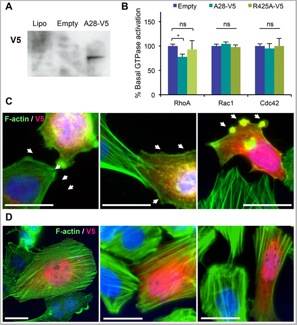
(Published customer image: Mouse anti V5 tag antibody, clone SV5-Pk1 used for the detection of V5 tagged WEEV_nsP3 protein by western blotting and immunofluorescenceImage caption: WEEV nsP3 interaction with host IKKbeta. A) U87MGs were transfected in a 6-well plate with 5 ug of pUC19 and WEEV_nsP3_HA for 24 hours. Cell lysates were resolved using SDS-PAGE and subsequently immunoblotted with V5 antibody and beta-actin served as a loading control. B) U87MGs were transfected with WEEV_nsP3_V5; cells were fixed after 24 hours and stained with antibodies against the endogenous IKKbeta and the V5 tag. Cells were incubated with appropriate secondary Alexa Fluor antibodies and the nuclei stained with DAPI. Co-localization of IKKbeta with WEEV_nsP3_V5 (yellow) was observed as shown by the arrows. B) Panels E -H serve as an example of transfected cells in a given field of view that show co-localization of IKKbeta and WEEV_nsP3_V5 24 hours post transfection. Panels I-L represent magnified images of other cells showing co-localization of IKKbeta and WEEV_nsP3_V5. Panel M is a magnified image of panel L. The co-localization was confirmed by Z-stack analysis. Co-localization was calculated to be approximately in 61% of cells (163 cells were counted of which 44% demonstrated expression of nsP3. Of those cells that expressed nsP3, 61% showed co-localization of both proteins). Images were taken using Nikon Eclipse TE2000-U at 60x magnification and are representative of 2 independent experiments.From: Amaya M, Voss K, Sampey G, Senina S, de la Fuente C, et al. (2014) The Role of IKKbeta in Venezuelan Equine Encephalitis Virus Infection. PLoS ONE 9(2): e86745.)
Application Data
(Published customer image: Mouse anti V5 tag antibody, clone SV5-Pk1 used for the detection of V5 tagged WEEV_nsP3 protein by western blotting and immunofluorescenceImage caption: WEEV nsP3 interaction with host IKKbeta. A) U87MGs were transfected in a 6-well plate with 5 ug of pUC19 and WEEV_nsP3_HA for 24 hours. Cell lysates were resolved using SDS-PAGE and subsequently immunoblotted with V5 antibody and beta-actin served as a loading control. B) U87MGs were transfected with WEEV_nsP3_V5; cells were fixed after 24 hours and stained with antibodies against the endogenous IKKbeta and the V5 tag. Cells were incubated with appropriate secondary Alexa Fluor antibodies and the nuclei stained with DAPI. Co-localization of IKKbeta with WEEV_nsP3_V5 (yellow) was observed as shown by the arrows. B) Panels E -H serve as an example of transfected cells in a given field of view that show co-localization of IKKbeta and WEEV_nsP3_V5 24 hours post transfection. Panels I-L represent magnified images of other cells showing co-localization of IKKbeta and WEEV_nsP3_V5. Panel M is a magnified image of panel L. The co-localization was confirmed by Z-stack analysis. Co-localization was calculated to be approximately in 61% of cells (163 cells were counted of which 44% demonstrated expression of nsP3. Of those cells that expressed nsP3, 61% showed co-localization of both proteins). Images were taken using Nikon Eclipse TE2000-U at 60x magnification and are representative of 2 independent experiments.From: Amaya M, Voss K, Sampey G, Senina S, de la Fuente C, et al. (2014) The Role of IKKbeta in Venezuelan Equine Encephalitis Virus Infection. PLoS ONE 9(2): e86745.)
V5-TAG, Monoclonal Antibody (Cat# AAA12081)
Full Name
MOUSE ANTI V5-TAG:HRP
Applications
Western Blot
Pricing
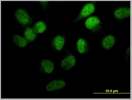
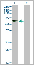
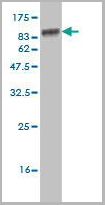
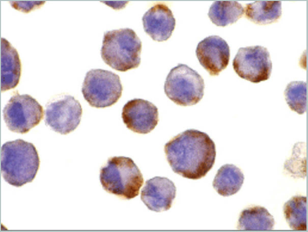
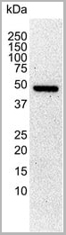
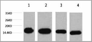
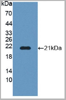
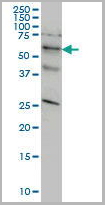 WB (Western Blot)
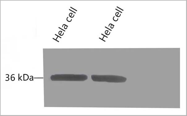
(STK33 monoclonal antibody, Western Blot analysis of STK33 expression in HeLa.)
WB (Western Blot)
(STK33 monoclonal antibody, Western Blot analysis of STK33 expression in HeLa.)
STK33, Monoclonal Antibody (Cat# AAA25856)
Full Name
STK33 (Serine/Threonine Kinase 33) (PE)
Reactivity
Human
Applications
Immunofluorescence, Immunoprecipitation, Western Blot
Purity
Purified by Protein A Affinity Chromatography.
Pricing
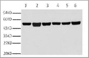
 WB (Western Blot)
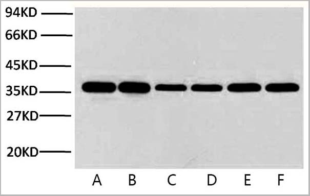
(Fig.8 Western blot analysis of Hela, GAPDH Monoclonal Antibody (2B5) was diluted at 1:10000 (25°C, 3h).)
WB (Western Blot)
(Fig.8 Western blot analysis of Hela, GAPDH Monoclonal Antibody (2B5) was diluted at 1:10000 (25°C, 3h).)
GAPDH, Monoclonal Antibody (Cat# AAA31539)
Full Name
Anti-GAPDH Mouse Monoclonal Antibody (2B5)
Gene Names
GAPDH; G3PD; GAPD
Reactivity
Human, Mouse, Rat, Monkey, Dog, Chicken, Rabbit, Pig, Sheep, Yeast, Hamster
Applications
Western Blot, Immunohistochemistry, Immunofluorescence
Purity
The antibody was affinity-purified from mouse ascites by affinity-chromatography using specific immunogen
Pricing
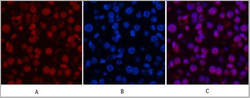
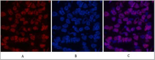
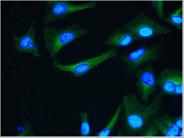
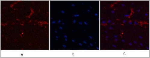
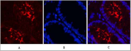
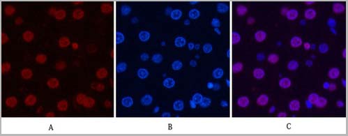 IF (Immunofluorescence)
(Fig.7. Immunofluorescence analysis of rat liver tissue. 1, Histone H3 Monoclonal Antibody (2D10) (red) was diluted at 1:200 (4°C, overnight). 2, Cy3 Labeled secondary antibody was diluted at 1:300 (room temperature, 50min). 3, Picture B: DAPI (blue) 10min. Picture A: Target. Picture B: DAPI. Picture C: merge of A+B.)
IF (Immunofluorescence)
(Fig.7. Immunofluorescence analysis of rat liver tissue. 1, Histone H3 Monoclonal Antibody (2D10) (red) was diluted at 1:200 (4°C, overnight). 2, Cy3 Labeled secondary antibody was diluted at 1:300 (room temperature, 50min). 3, Picture B: DAPI (blue) 10min. Picture A: Target. Picture B: DAPI. Picture C: merge of A+B.)
Histone H3, Monoclonal Antibody (Cat# AAA31543)
Full Name
Anti-Histone H3 Mouse Monoclonal Antibody (2D10)
Gene Names
HIST1H3A; H3/A; H3FA
Reactivity
Human, Mouse, Rat, Yeast
Applications
Western Blot, Immunohistochemistry, Immunofluorescence, Immunoprecipitation
Pricing


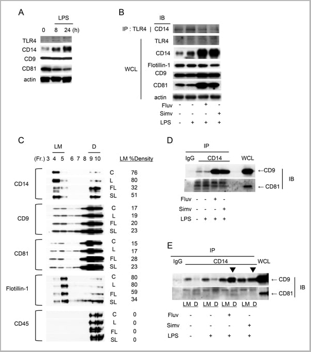


 Application Data
(Staining of mouse spleen with Hamster anti Mouse CD81: Alexa Fluor 488)
Application Data
(Staining of mouse spleen with Hamster anti Mouse CD81: Alexa Fluor 488)
CD81, Monoclonal Antibody (Cat# AAA11869)
Full Name
HAMSTER ANTI MOUSE CD81:FITC
Gene Names
Cd81; Tapa1; Tapa-1; Tspan28
Applications
Flow Cytometry
Pricing


 Application Data
(Staining of mouse spleen with Hamster anti Mouse CD81: Alexa Fluor 488)
Application Data
(Staining of mouse spleen with Hamster anti Mouse CD81: Alexa Fluor 488)
CD81, Monoclonal Antibody (Cat# AAA12033)
Full Name
HAMSTER ANTI MOUSE CD81:RPE
Gene Names
Cd81; Tapa1; Tapa-1; Tspan28
Applications
Flow Cytometry
Pricing
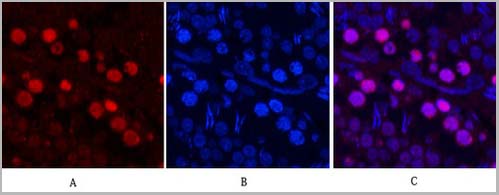
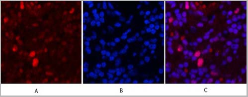
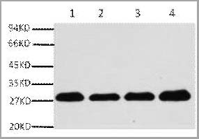
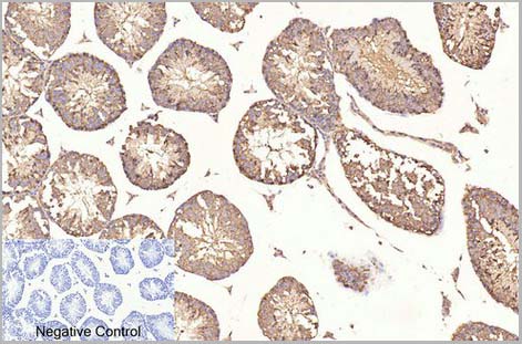
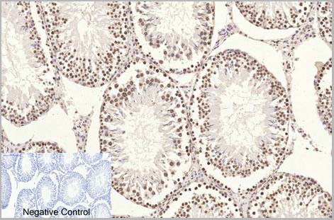
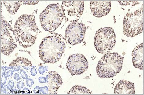
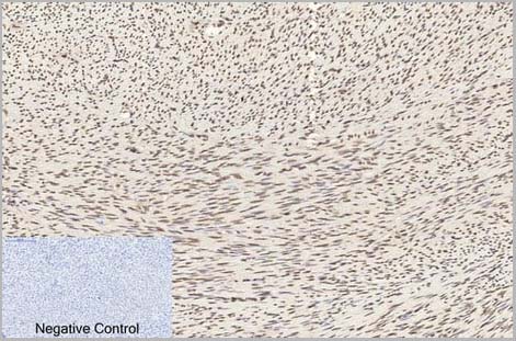
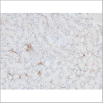
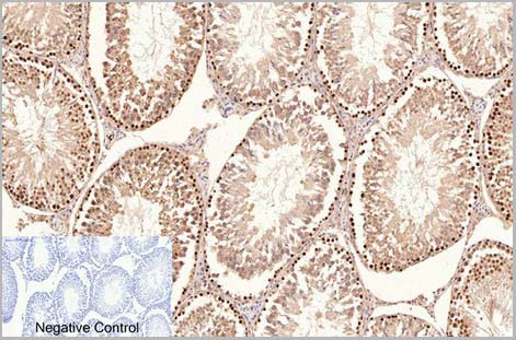 IHC (Immunohistchemistry)
(Fig.6. Immunohistochemical analysis of paraffin-embedded rat testis tissue. 1, PCNA Monoclonal Antibod y(1D7) was diluted at 1:200 (4°C, overnight). 2, Sodium citrate pH 6.0 was used for antibody retrieval(>98°C, 20min). 3, secondary antibody was diluted at 1:200 (room temperature, 30min). Negative control was used by secondary antibody only.)
IHC (Immunohistchemistry)
(Fig.6. Immunohistochemical analysis of paraffin-embedded rat testis tissue. 1, PCNA Monoclonal Antibod y(1D7) was diluted at 1:200 (4°C, overnight). 2, Sodium citrate pH 6.0 was used for antibody retrieval(>98°C, 20min). 3, secondary antibody was diluted at 1:200 (room temperature, 30min). Negative control was used by secondary antibody only.)
PCNA, Monoclonal Antibody (Cat# AAA31541)
Full Name
Anti-PCNA Mouse Monoclonal Antibody (1D7)
Reactivity
Human, Mouse, Rat
Applications
Western Blot, Immunohistochemistry, Immunofluorescence
Purity
The antibody was affinity-purified from mouse ascites by affinity-chromatography using specific immunogen
Pricing
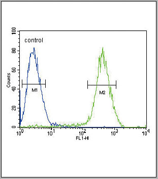 FCM (Flow Cytometry)
(CB2 Antibody (C-term) flow cytometric analysis of Jurkat cells (right histogram) compared to a negative control cell (left histogram).FITC-conjugated goat-anti-rabbit secondary antibodies were used for the analysis.)
FCM (Flow Cytometry)
(CB2 Antibody (C-term) flow cytometric analysis of Jurkat cells (right histogram) compared to a negative control cell (left histogram).FITC-conjugated goat-anti-rabbit secondary antibodies were used for the analysis.)
CB2, Polyclonal Antibody (Cat# AAA28672)
Full Name
CB2 Antibody (C-term)
Gene Names
CNR2; CB2; CX5; CB-2
Reactivity
Human
Applications
Immunohistochemistry, Flow Cytometry, Western Blot
Purity
This antibody is purified through a protein A column, followed by peptide affinity purification.
Pricing
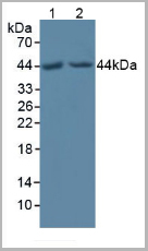
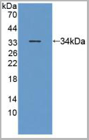
 Knockout Validation
(Knockout Validation: Lane 1: Wild-type Hela cell lysate; Lane 2: POFUT1 knockout Hela cell lysate; Predicted MW: 44,22kd Observed MW: 44kd Primary Ab: 1ug/ml Rabbit Anti-Human POFUT1 Antibody Second Ab: 0.2ug/mL HRP-Linked Caprine Anti-Rabbit IgG Polyclonal Antibody ( Human.)
Knockout Validation
(Knockout Validation: Lane 1: Wild-type Hela cell lysate; Lane 2: POFUT1 knockout Hela cell lysate; Predicted MW: 44,22kd Observed MW: 44kd Primary Ab: 1ug/ml Rabbit Anti-Human POFUT1 Antibody Second Ab: 0.2ug/mL HRP-Linked Caprine Anti-Rabbit IgG Polyclonal Antibody ( Human.)
Protein O-Fucosyltransferase 1, Polyclonal Antibody (Cat# AAA21025)
Full Name
Protein O-Fucosyltransferase 1 (POFUT1) Polyclonal Antibody
Gene Names
POFUT1; DDD2; FUT12; O-FUT; OFUCT1; O-Fuc-T; O-FucT-1
Reactivity
Human
Applications
Western Blot, Immunocytochemistry, Immunohistochemistry
Purity
Affinity Chromatography
Pricing
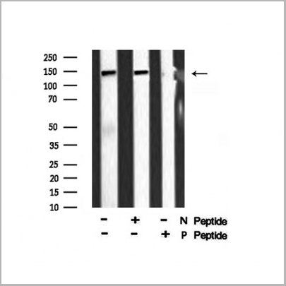
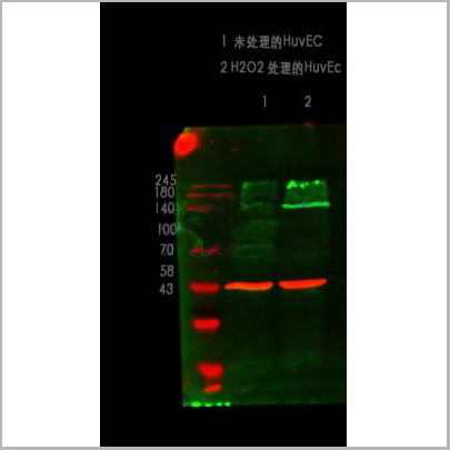
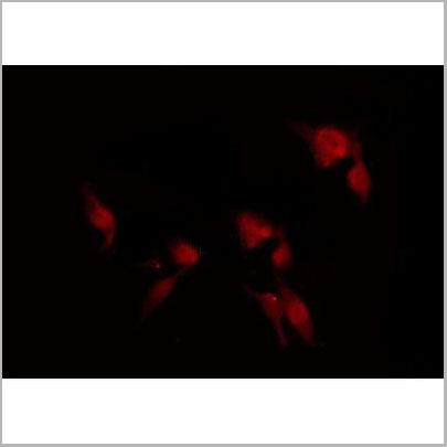 Application Data
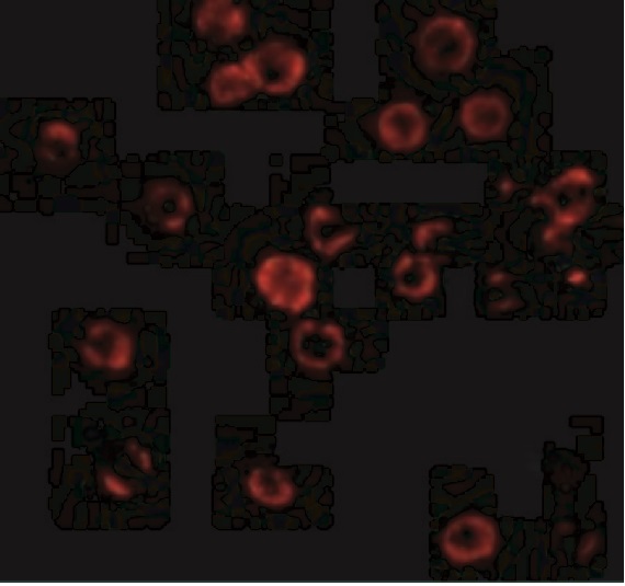
(At 25 degree C. The primary antibody was diluted at 1/200 and incubated with the sample for 1 hour at 37 degree C. An Alexa Fluor 594 conjugated goat anti-rabbit IgG (H+L) Ab, diluted at 1/600, was used as the secondary antibody.)
Application Data
(At 25 degree C. The primary antibody was diluted at 1/200 and incubated with the sample for 1 hour at 37 degree C. An Alexa Fluor 594 conjugated goat anti-rabbit IgG (H+L) Ab, diluted at 1/600, was used as the secondary antibody.)
eNOS, Polyclonal Antibody (Cat# AAA31394)
Full Name
Phospho-eNOS (Ser1179) Antibody
Gene Names
NOS3; eNOS; ECNOS
Reactivity
Human, Mouse, Rat
Predicted Reactivity: Pig (100%), Bovine (100%), Rabbit (100%), Dog (100%)
Predicted Reactivity: Pig (100%), Bovine (100%), Rabbit (100%), Dog (100%)
Applications
Western Blot, Immunohistochemistry, Immunofluorescence, Immunocytochemistry, Peptide ELISA
Purity
The antibody is from purified rabbit serum by affinity purification via sequential chromatography on phospho-peptide and non-phospho-peptide affinity columns.
Pricing


 Application Data
(Published customer image: Mouse anti V5 tag antibody, clone SV5-Pk1 used for the detection of V5 tagged WEEV_nsP3 protein by western blotting and immunofluorescenceImage caption: WEEV nsP3 interaction with host IKKbeta. A) U87MGs were transfected in a 6-well plate with 5 ug of pUC19 and WEEV_nsP3_HA for 24 hours. Cell lysates were resolved using SDS-PAGE and subsequently immunoblotted with V5 antibody and beta-actin served as a loading control. B) U87MGs were transfected with WEEV_nsP3_V5; cells were fixed after 24 hours and stained with antibodies against the endogenous IKKbeta and the V5 tag. Cells were incubated with appropriate secondary Alexa Fluor antibodies and the nuclei stained with DAPI. Co-localization of IKKbeta with WEEV_nsP3_V5 (yellow) was observed as shown by the arrows. B) Panels E -H serve as an example of transfected cells in a given field of view that show co-localization of IKKbeta and WEEV_nsP3_V5 24 hours post transfection. Panels I-L represent magnified images of other cells showing co-localization of IKKbeta and WEEV_nsP3_V5. Panel M is a magnified image of panel L. The co-localization was confirmed by Z-stack analysis. Co-localization was calculated to be approximately in 61% of cells (163 cells were counted of which 44% demonstrated expression of nsP3. Of those cells that expressed nsP3, 61% showed co-localization of both proteins). Images were taken using Nikon Eclipse TE2000-U at 60x magnification and are representative of 2 independent experiments.From: Amaya M, Voss K, Sampey G, Senina S, de la Fuente C, et al. (2014) The Role of IKKbeta in Venezuelan Equine Encephalitis Virus Infection. PLoS ONE 9(2): e86745.)
Application Data
(Published customer image: Mouse anti V5 tag antibody, clone SV5-Pk1 used for the detection of V5 tagged WEEV_nsP3 protein by western blotting and immunofluorescenceImage caption: WEEV nsP3 interaction with host IKKbeta. A) U87MGs were transfected in a 6-well plate with 5 ug of pUC19 and WEEV_nsP3_HA for 24 hours. Cell lysates were resolved using SDS-PAGE and subsequently immunoblotted with V5 antibody and beta-actin served as a loading control. B) U87MGs were transfected with WEEV_nsP3_V5; cells were fixed after 24 hours and stained with antibodies against the endogenous IKKbeta and the V5 tag. Cells were incubated with appropriate secondary Alexa Fluor antibodies and the nuclei stained with DAPI. Co-localization of IKKbeta with WEEV_nsP3_V5 (yellow) was observed as shown by the arrows. B) Panels E -H serve as an example of transfected cells in a given field of view that show co-localization of IKKbeta and WEEV_nsP3_V5 24 hours post transfection. Panels I-L represent magnified images of other cells showing co-localization of IKKbeta and WEEV_nsP3_V5. Panel M is a magnified image of panel L. The co-localization was confirmed by Z-stack analysis. Co-localization was calculated to be approximately in 61% of cells (163 cells were counted of which 44% demonstrated expression of nsP3. Of those cells that expressed nsP3, 61% showed co-localization of both proteins). Images were taken using Nikon Eclipse TE2000-U at 60x magnification and are representative of 2 independent experiments.From: Amaya M, Voss K, Sampey G, Senina S, de la Fuente C, et al. (2014) The Role of IKKbeta in Venezuelan Equine Encephalitis Virus Infection. PLoS ONE 9(2): e86745.)
V5-TAG, Monoclonal Antibody (Cat# AAA11930)
Full Name
MOUSE ANTI V5-TAG
Applications
Immunohistochemistry, Flow Cytometry, Immunofluorescence, Immunoprecipitation, Western Blot, Radioimmunoassay
Pricing


 Application Data
(Published customer image: Mouse anti V5 tag antibody, clone SV5-Pk1 used for the detection of V5 tagged WEEV_nsP3 protein by western blotting and immunofluorescenceImage caption: WEEV nsP3 interaction with host IKKbeta. A) U87MGs were transfected in a 6-well plate with 5 ug of pUC19 and WEEV_nsP3_HA for 24 hours. Cell lysates were resolved using SDS-PAGE and subsequently immunoblotted with V5 antibody and beta-actin served as a loading control. B) U87MGs were transfected with WEEV_nsP3_V5; cells were fixed after 24 hours and stained with antibodies against the endogenous IKKbeta and the V5 tag. Cells were incubated with appropriate secondary Alexa Fluor antibodies and the nuclei stained with DAPI. Co-localization of IKKbeta with WEEV_nsP3_V5 (yellow) was observed as shown by the arrows. B) Panels E -H serve as an example of transfected cells in a given field of view that show co-localization of IKKbeta and WEEV_nsP3_V5 24 hours post transfection. Panels I-L represent magnified images of other cells showing co-localization of IKKbeta and WEEV_nsP3_V5. Panel M is a magnified image of panel L. The co-localization was confirmed by Z-stack analysis. Co-localization was calculated to be approximately in 61% of cells (163 cells were counted of which 44% demonstrated expression of nsP3. Of those cells that expressed nsP3, 61% showed co-localization of both proteins). Images were taken using Nikon Eclipse TE2000-U at 60x magnification and are representative of 2 independent experiments.From: Amaya M, Voss K, Sampey G, Senina S, de la Fuente C, et al. (2014) The Role of IKKbeta in Venezuelan Equine Encephalitis Virus Infection. PLoS ONE 9(2): e86745.)
Application Data
(Published customer image: Mouse anti V5 tag antibody, clone SV5-Pk1 used for the detection of V5 tagged WEEV_nsP3 protein by western blotting and immunofluorescenceImage caption: WEEV nsP3 interaction with host IKKbeta. A) U87MGs were transfected in a 6-well plate with 5 ug of pUC19 and WEEV_nsP3_HA for 24 hours. Cell lysates were resolved using SDS-PAGE and subsequently immunoblotted with V5 antibody and beta-actin served as a loading control. B) U87MGs were transfected with WEEV_nsP3_V5; cells were fixed after 24 hours and stained with antibodies against the endogenous IKKbeta and the V5 tag. Cells were incubated with appropriate secondary Alexa Fluor antibodies and the nuclei stained with DAPI. Co-localization of IKKbeta with WEEV_nsP3_V5 (yellow) was observed as shown by the arrows. B) Panels E -H serve as an example of transfected cells in a given field of view that show co-localization of IKKbeta and WEEV_nsP3_V5 24 hours post transfection. Panels I-L represent magnified images of other cells showing co-localization of IKKbeta and WEEV_nsP3_V5. Panel M is a magnified image of panel L. The co-localization was confirmed by Z-stack analysis. Co-localization was calculated to be approximately in 61% of cells (163 cells were counted of which 44% demonstrated expression of nsP3. Of those cells that expressed nsP3, 61% showed co-localization of both proteins). Images were taken using Nikon Eclipse TE2000-U at 60x magnification and are representative of 2 independent experiments.From: Amaya M, Voss K, Sampey G, Senina S, de la Fuente C, et al. (2014) The Role of IKKbeta in Venezuelan Equine Encephalitis Virus Infection. PLoS ONE 9(2): e86745.)
V5-TAG, Monoclonal Antibody (Cat# AAA11864)
Full Name
MOUSE ANTI V5-TAG:FITC
Applications
Immunofluorescence
Pricing
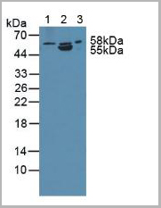
 Knockout Validation
(Knockout Validation: Lane 1: Wild-type PC3 cell lysate; Lane 2: ANTXR2 knockout PC3 cell lysate; Predicted MW: 54,53,43,35kd Observed MW: 58kd Primary Ab: 1ug/ml Rabbit Anti-Human ANTXR2 Antibody Second Ab: 0.2ug/mL HRP-Linked Caprine Anti-Rabbit IgG Polyclonal Antibody ( Human.)
Knockout Validation
(Knockout Validation: Lane 1: Wild-type PC3 cell lysate; Lane 2: ANTXR2 knockout PC3 cell lysate; Predicted MW: 54,53,43,35kd Observed MW: 58kd Primary Ab: 1ug/ml Rabbit Anti-Human ANTXR2 Antibody Second Ab: 0.2ug/mL HRP-Linked Caprine Anti-Rabbit IgG Polyclonal Antibody ( Human.)
Anthrax Toxin Receptor 2, Polyclonal Antibody (Cat# AAA21007)
Full Name
Anthrax Toxin Receptor 2 (ANTXR2) Polyclonal Antibody
Gene Names
ANTXR2; HFS; ISH; JHF; CMG2; CMG-2
Reactivity
Human, Rat, Pig
Applications
Western Blot, Immunocytochemistry, Immunohistochemistry
Purity
Affinity Chromatography
Pricing


 Application Data
(Staining of mouse spleen with Hamster anti Mouse CD81: Alexa Fluor 488)
Application Data
(Staining of mouse spleen with Hamster anti Mouse CD81: Alexa Fluor 488)
CD81, Monoclonal Antibody (Cat# AAA11942)
Full Name
HAMSTER ANTI MOUSE CD81
Gene Names
Cd81; Tapa1; Tapa-1; Tspan28
Reactivity
Rat
Applications
Immunohistochemistry, Flow Cytometry, Immunoprecipitation, Western Blot
Pricing


 Application Data
(Published customer image: Mouse anti V5 tag antibody, clone SV5-Pk1 used for the detection of V5 tagged WEEV_nsP3 protein by western blotting and immunofluorescenceImage caption: WEEV nsP3 interaction with host IKKbeta. A) U87MGs were transfected in a 6-well plate with 5 ug of pUC19 and WEEV_nsP3_HA for 24 hours. Cell lysates were resolved using SDS-PAGE and subsequently immunoblotted with V5 antibody and beta-actin served as a loading control. B) U87MGs were transfected with WEEV_nsP3_V5; cells were fixed after 24 hours and stained with antibodies against the endogenous IKKbeta and the V5 tag. Cells were incubated with appropriate secondary Alexa Fluor antibodies and the nuclei stained with DAPI. Co-localization of IKKbeta with WEEV_nsP3_V5 (yellow) was observed as shown by the arrows. B) Panels E -H serve as an example of transfected cells in a given field of view that show co-localization of IKKbeta and WEEV_nsP3_V5 24 hours post transfection. Panels I-L represent magnified images of other cells showing co-localization of IKKbeta and WEEV_nsP3_V5. Panel M is a magnified image of panel L. The co-localization was confirmed by Z-stack analysis. Co-localization was calculated to be approximately in 61% of cells (163 cells were counted of which 44% demonstrated expression of nsP3. Of those cells that expressed nsP3, 61% showed co-localization of both proteins). Images were taken using Nikon Eclipse TE2000-U at 60x magnification and are representative of 2 independent experiments.From: Amaya M, Voss K, Sampey G, Senina S, de la Fuente C, et al. (2014) The Role of IKKbeta in Venezuelan Equine Encephalitis Virus Infection. PLoS ONE 9(2): e86745.)
Application Data
(Published customer image: Mouse anti V5 tag antibody, clone SV5-Pk1 used for the detection of V5 tagged WEEV_nsP3 protein by western blotting and immunofluorescenceImage caption: WEEV nsP3 interaction with host IKKbeta. A) U87MGs were transfected in a 6-well plate with 5 ug of pUC19 and WEEV_nsP3_HA for 24 hours. Cell lysates were resolved using SDS-PAGE and subsequently immunoblotted with V5 antibody and beta-actin served as a loading control. B) U87MGs were transfected with WEEV_nsP3_V5; cells were fixed after 24 hours and stained with antibodies against the endogenous IKKbeta and the V5 tag. Cells were incubated with appropriate secondary Alexa Fluor antibodies and the nuclei stained with DAPI. Co-localization of IKKbeta with WEEV_nsP3_V5 (yellow) was observed as shown by the arrows. B) Panels E -H serve as an example of transfected cells in a given field of view that show co-localization of IKKbeta and WEEV_nsP3_V5 24 hours post transfection. Panels I-L represent magnified images of other cells showing co-localization of IKKbeta and WEEV_nsP3_V5. Panel M is a magnified image of panel L. The co-localization was confirmed by Z-stack analysis. Co-localization was calculated to be approximately in 61% of cells (163 cells were counted of which 44% demonstrated expression of nsP3. Of those cells that expressed nsP3, 61% showed co-localization of both proteins). Images were taken using Nikon Eclipse TE2000-U at 60x magnification and are representative of 2 independent experiments.From: Amaya M, Voss K, Sampey G, Senina S, de la Fuente C, et al. (2014) The Role of IKKbeta in Venezuelan Equine Encephalitis Virus Infection. PLoS ONE 9(2): e86745.)
V5-TAG, Monoclonal Antibody (Cat# AAA12211)
Full Name
MOUSE ANTI V5-TAG
Applications
Immunohistochemistry, Flow Cytometry, Immunofluorescence, Immunoprecipitation, Western Blot, Radioimmunoassay
Pricing
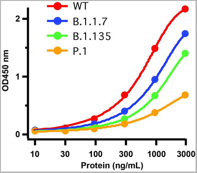
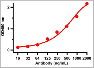
 ELISA
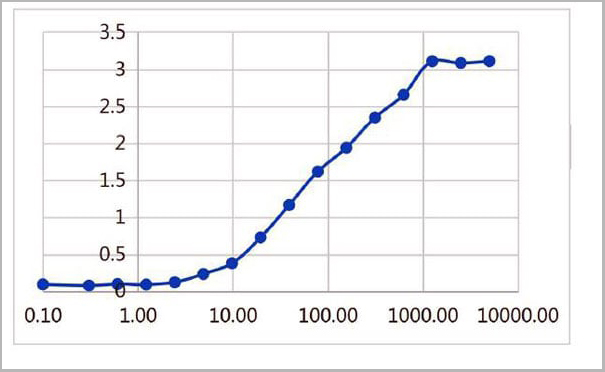
(ELISA plate was coated by the 2019-nCoV S1 protein, 100 uL/cell at 5ug/ml. The indirect ELISA analysis was performed by loading 100 uL per well of the anti-2019-nCoV S1 mAb (S1) at various concentrations. The plate was incubated for 1 hours at 37 degree C, then washed 5 times. An anti hFc HRP conjugated mAb at a concentration of 1:2000, 100uL/well was used as the secondary antibody. Again, the plate was incubated for 1 hours at 37 degree C, then washed 5 times. Detection was performed using TMB substrate for 10 minutes at room temperature in the dark. The plate was stopped with 2M sulfuric acid. Signal was read on a spectrophotometer at 450 nm.)
ELISA
(ELISA plate was coated by the 2019-nCoV S1 protein, 100 uL/cell at 5ug/ml. The indirect ELISA analysis was performed by loading 100 uL per well of the anti-2019-nCoV S1 mAb (S1) at various concentrations. The plate was incubated for 1 hours at 37 degree C, then washed 5 times. An anti hFc HRP conjugated mAb at a concentration of 1:2000, 100uL/well was used as the secondary antibody. Again, the plate was incubated for 1 hours at 37 degree C, then washed 5 times. Detection was performed using TMB substrate for 10 minutes at room temperature in the dark. The plate was stopped with 2M sulfuric acid. Signal was read on a spectrophotometer at 450 nm.)
COVID 19 Spike S1 Protein Coronavirus, Monoclonal Antibody (Cat# AAA27959)
Full Name
human anti-2019-nCoV Spike S1 mAb (S1)
Applications
ELISA
Purity
Purity: ≥ 95% as determined by SDS-PAGE
Purification: Protein A
Purification: Protein A
Pricing