Filters
Clonality
Type
Reactivity
Gene Name
Isotype
Host
Application
Clone
2279 results for "Protein C" - showing 450-500


 IHC (Immunohistchemistry)
(At 1/100 staining Mouse brain tissue by IHC-P. The sample was formaldehyde fixed and a heat mediated antigen retrieval step in citrate buffer was performed. The sample was then blocked and incubated with the primary antibody at 4 degree C overnight. An HRP conjugated anti-Rabbit antibody was used as the secondary antibody.)
IHC (Immunohistchemistry)
(At 1/100 staining Mouse brain tissue by IHC-P. The sample was formaldehyde fixed and a heat mediated antigen retrieval step in citrate buffer was performed. The sample was then blocked and incubated with the primary antibody at 4 degree C overnight. An HRP conjugated anti-Rabbit antibody was used as the secondary antibody.)
USF1, Polyclonal Antibody (Cat# AAA31437)
Full Name
Phospho-USF1 (Thr153) Antibody
Gene Names
USF1; UEF; FCHL; MLTF; FCHL1; MLTFI; HYPLIP1; bHLHb11
Reactivity
Human, Mouse, Rat
Predicted Reactivity: Pig (100%), Bovine (100%), Rabbit (100%), Dog (100%)
Predicted Reactivity: Pig (100%), Bovine (100%), Rabbit (100%), Dog (100%)
Applications
Western Blot, Immunohistochemistry, Peptide ELISA
Purity
The antibody is from purified rabbit serum by affinity purification via sequential chromatography on phospho-peptide and non-phospho-peptide affinity columns.
Pricing


 IHC (Immunohistchemistry)
((3.75ug/ml) staining of paraffin embedded Human Liver. Steamed antigen retrieval with citrate buffer pH 6, AP-staining.)
IHC (Immunohistchemistry)
((3.75ug/ml) staining of paraffin embedded Human Liver. Steamed antigen retrieval with citrate buffer pH 6, AP-staining.)
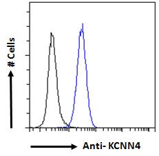
KCNN4/KCa3.1, Antibody (Cat# AAA13678)
Full Name
Goat Anti-KCNN4/KCa3.1 Antibody
Gene Names
KCNN4; IK; IK1; SK4; DHS2; KCA4; hSK4; IKCA1; hKCa4; KCa3.1; hIKCa1
Reactivity
Human, Mouse
Expected from sequence similarity: Human, Mouse, Rat, Dog
Expected from sequence similarity: Human, Mouse, Rat, Dog
Applications
Peptide ELISA, Western Blot, Immunofluorescence, Flow Cytometry, Immunohistochemistry
Purity
Purified from goat serum by ammonium sulphate precipitation followed by antigen affinity chromatography using the immunizing peptide.
Pricing


 WB (Western Blot)
(Anti-HA Tag mouse monoclonal antibody at 1:1000 dilutionLane A: HA-GST (Recombinant protein)(30ng)Lane B: GST-HA (Recombinant protein)(10ng)Lane C: HA-ARG1-myc transfected 293 cell lysate (2ug)Lane D: HA-mFABP4-myc transfected 293 cell lysate (2ug)Lane E: myc-mFABP4-HA transfected 293 cell lysate (0.5ug)Lane F: myc-ARG1-HA transfected 293 cell lysate (2ug)SecondaryGoat Anti-Mouse IgG H&L (Dylight800) at 1/15000 dilution.Developed using the Odyssey technique. Performed under reducing conditions.)
WB (Western Blot)
(Anti-HA Tag mouse monoclonal antibody at 1:1000 dilutionLane A: HA-GST (Recombinant protein)(30ng)Lane B: GST-HA (Recombinant protein)(10ng)Lane C: HA-ARG1-myc transfected 293 cell lysate (2ug)Lane D: HA-mFABP4-myc transfected 293 cell lysate (2ug)Lane E: myc-mFABP4-HA transfected 293 cell lysate (0.5ug)Lane F: myc-ARG1-HA transfected 293 cell lysate (2ug)SecondaryGoat Anti-Mouse IgG H&L (Dylight800) at 1/15000 dilution.Developed using the Odyssey technique. Performed under reducing conditions.)
HA, Monoclonal Antibody (Cat# AAA27712)
Full Name
Anti-HA tag Antibody, Mouse Monoclonal
Reactivity
HA tag
Applications
Western Blot, Flow Cytometry, Immunocytochemistry, Immunofluorescence, Immunoprecipitation
Purity
Protein A
Pricing


 FCM (Flow Cytometry)
(Figure-6: Epitope binding study by flow cytometric analysis. MCF-7 cells expressing HER2 antigen were treated with Herceptin and Samceptin (2 ug/10^6 Cells). Surface staining was done using FITC conjugated antibodies.)
FCM (Flow Cytometry)
(Figure-6: Epitope binding study by flow cytometric analysis. MCF-7 cells expressing HER2 antigen were treated with Herceptin and Samceptin (2 ug/10^6 Cells). Surface staining was done using FITC conjugated antibodies.)
ErbB2/HER2, Monoclonal Recombinant Antibody (Cat# AAA14887)
Full Name
Recombinant anti-human ErbB2/HER2 Antibody (Trastuzumab) FITC Conjugated
Gene Names
ERBB2; NEU; NGL; HER2; TKR1; CD340; HER-2; MLN 19; HER-2/neu
Reactivity
Human
Applications
Functional Assay, Flow Cytometry, Antibody-Dependent Cellular Cytotoxicity
Purity
Purity: >98.0% as determined by SEC-HPLC & SDS-PAGE
Pricing


 FCM (Flow Cytometry)
(Figure 7. Flow Cytometry analysis of HL-60 cells using anti-Sumo 1/SUMO1 antibody (AAA19224).Overlay histogram showing HL-60 cells stained with AAA19224 (Blue line). The cells were blocked with 10% normal goat serum. And then incubated with rabbit anti-Sumo 1/SUMO1 Antibody (AAA19224, 1μg/1x106 cells) for 30 min at 20 degree C. DyLight®488 conjugated goat anti-rabbit IgG (5-10μg/1x106 cells) was used as secondary antibody for 30 minutes at 20 degree C. Isotype control antibody (Green line) was rabbit IgG (1μg/1x106) used under the same conditions. Unlabelled sample (Red line) was also used as a control.)
FCM (Flow Cytometry)
(Figure 7. Flow Cytometry analysis of HL-60 cells using anti-Sumo 1/SUMO1 antibody (AAA19224).Overlay histogram showing HL-60 cells stained with AAA19224 (Blue line). The cells were blocked with 10% normal goat serum. And then incubated with rabbit anti-Sumo 1/SUMO1 Antibody (AAA19224, 1μg/1x106 cells) for 30 min at 20 degree C. DyLight®488 conjugated goat anti-rabbit IgG (5-10μg/1x106 cells) was used as secondary antibody for 30 minutes at 20 degree C. Isotype control antibody (Green line) was rabbit IgG (1μg/1x106) used under the same conditions. Unlabelled sample (Red line) was also used as a control.)
Sumo 1/SUMO1, Polyclonal Antibody (Cat# AAA19224)
Full Name
Anti-Sumo 1/SUMO1 Antibody
Gene Names
SUMO1; DAP1; GMP1; PIC1; SMT3; UBL1; OFC10; SENP2; SMT3C; SMT3H3
Reactivity
Human, Mouse, Rat
Applications
Immunohistochemistry, Immunocytochemistry, Immunofluorescence, Flow Cytometry
Purity
Immunogen affinity purified.
Pricing


 IF (Immunofluorescence)
(Immunofluorescence analysis of MCF-7 cells using PRKCA antibody.)
IF (Immunofluorescence)
(Immunofluorescence analysis of MCF-7 cells using PRKCA antibody.)
PRKCA , Monoclonal Antibody (Cat# AAA28235)
Full Name
PRKCA Monoclonal Antibody
Gene Names
PRKCA; AAG6; PKCA; PRKACA; PKC-alpha
Reactivity
Human, Mouse, Rat
Applications
Western Blot, Immunohistochemistry, Immunofluorescence, Immunoprecipitation
Purity
Affinity Purification
Pricing


 IF (Immunofluorescence)
(Confocal immunofluorescent analysis of C9JLR9 Antibody (C-term) (AAA28790) with HepG2 cell followed by Alexa Fluor 488-conjugated goat anti-rabbit lgG (green). Actin filaments have been labeled with Alexa Fluor555 phalloidin (red). DAPI was used to stain the cell nuclear (blue).)
IF (Immunofluorescence)
(Confocal immunofluorescent analysis of C9JLR9 Antibody (C-term) (AAA28790) with HepG2 cell followed by Alexa Fluor 488-conjugated goat anti-rabbit lgG (green). Actin filaments have been labeled with Alexa Fluor555 phalloidin (red). DAPI was used to stain the cell nuclear (blue).)
C9JLR9, Polyclonal Antibody (Cat# AAA28790)
Full Name
C9JLR9 Antibody (C-term)
Reactivity
Human
Predicted Reactivity: Mouse
Predicted Reactivity: Mouse
Applications
Western Blot, Immunohistochemistry, Immunofluorescence
Purity
This antibody is purified through a protein A column, followed by peptide affinity purification.
Pricing


 ICC (Immunocytochemistry)
(ICC staining Histone H3 in Hela cells (green). The nuclear counter stain is DAPI (blue). Cells were fixed in paraformaldehyde, permeabilised with 0.25% Triton X100/PBS.)
ICC (Immunocytochemistry)
(ICC staining Histone H3 in Hela cells (green). The nuclear counter stain is DAPI (blue). Cells were fixed in paraformaldehyde, permeabilised with 0.25% Triton X100/PBS.)
Histone H3, Monoclonal Antibody (Cat# AAA30242)
Full Name
Histone H3 Antibody
Gene Names
HIST1H3A; H3/A; H3FA
Reactivity
Human, Mouse, Rat
Applications
Western Blot, Immunocytochemistry, Immunofluorescence, Immunohistochemistry, Chromatin Immunoprecipitation
Purity
ProA affinity purified
Pricing
 Standard Curve (Sample)
Dapper Homolog 1 (DACT1), ELISA Kit (Cat# AAA31500)
Full Name
Human Dapper Homolog 1 (DACT1) ELISA Kit
Gene Names
DACT1; DPR1; TBS2; FRODO; HDPR1; DAPPER; THYEX3; DAPPER1
Reactivity
Human
Pricing


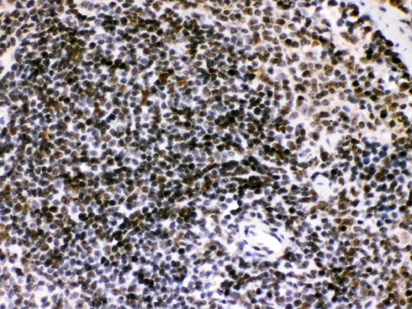
 IHC (Immunohistochemistry)
(Immunochemical staining MSH6 in Ferret spleen with rabbit monoclonal antibody at 1:200 dilution, formalin-fixed paraffin embedded sections.)
IHC (Immunohistochemistry)
(Immunochemical staining MSH6 in Ferret spleen with rabbit monoclonal antibody at 1:200 dilution, formalin-fixed paraffin embedded sections.)
MSH6, Monoclonal Antibody (Cat# AAA27721)
Full Name
Recombinant Anti-MSH6 Antibody, Rabbit Monoclonal
Gene Names
MSH6; GTBP; HSAP; p160; GTMBP; HNPCC5
Reactivity
Human, Cynomolgus, Mouse, Rat, Ferret
Applications
Immunohistochemistry, Immunocytochemistry, Immunofluorescence
Purity
Protein A
Pricing


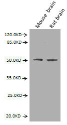
 WB (Western Blot)
(Western blot analysis of extracts of various celllines, using GSK3 alpha/beta Antibody.)
WB (Western Blot)
(Western blot analysis of extracts of various celllines, using GSK3 alpha/beta Antibody.)
GSK3 alpha/beta, Polyclonal Antibody (Cat# AAA31094)
Full Name
GSK3 alpha/beta Antibody
Reactivity
Human, Mouse, Rat
Applications
Western Blot, Immunohistochemistry
Purity
The antiserum was purified by peptide affinity chromatography using SulfoLink Coupling Resin.
Pricing


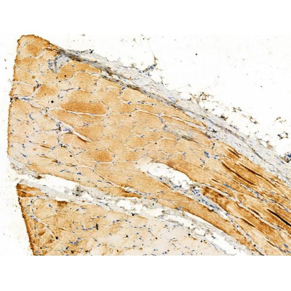
 IHC (Immunohistchemistry)
(At 1/100 staining Mouse heart tissue by IHC-P. The sample was formaldehyde fixed and a heat mediated antigen retrieval step in citrate buffer was performed. The sample was then blocked and incubated with the primary antibody at 4 degree C overnight. An HRP conjugated anti-Rabbit antibody was used as the secondary antibody.)
IHC (Immunohistchemistry)
(At 1/100 staining Mouse heart tissue by IHC-P. The sample was formaldehyde fixed and a heat mediated antigen retrieval step in citrate buffer was performed. The sample was then blocked and incubated with the primary antibody at 4 degree C overnight. An HRP conjugated anti-Rabbit antibody was used as the secondary antibody.)
OTUB1, Polyclonal Antibody (Cat# AAA31284)
Full Name
Phospho-OTUB1 (Ser16) Antibody
Gene Names
OTUB1; OTB1; OTU1; HSPC263
Reactivity
Human, Mouse, Rat
Applications
Immunohistochemistry, Peptide ELISA
Purity
The antibody is from purified rabbit serum by affinity purification via sequential chromatography on phospho-peptide and non-phospho-peptide affinity columns.
Pricing


 FCM (Flow Cytometry)
(Overlay Peak curve showing 293F cells transfected with GST stained with AAA28065 (red line) at 1:189. The cells were fixed in 4% formaldehyde and permeated by 0.2% TritonX-100. Then 10% normal goat serum was Incubated to block non-specific protein-protein interactions followed by the antibody (1?g/1*106cells) for 1 h at 4 degree C. The secondary antibody used was FITC-conjugated Goat Anti-Mouse IgG(H+L) at 1/100 dilution for 30min at 4 degree C. Isotype control antibody (green line) was mouse IgG2b (1?g/1*106cells) used under the same conditions. Acquisition of >10,000 events was performed.)
FCM (Flow Cytometry)
(Overlay Peak curve showing 293F cells transfected with GST stained with AAA28065 (red line) at 1:189. The cells were fixed in 4% formaldehyde and permeated by 0.2% TritonX-100. Then 10% normal goat serum was Incubated to block non-specific protein-protein interactions followed by the antibody (1?g/1*106cells) for 1 h at 4 degree C. The secondary antibody used was FITC-conjugated Goat Anti-Mouse IgG(H+L) at 1/100 dilution for 30min at 4 degree C. Isotype control antibody (green line) was mouse IgG2b (1?g/1*106cells) used under the same conditions. Acquisition of >10,000 events was performed.)
GST, Monoclonal Antibody (Cat# AAA28065)
Full Name
GST Monoclonal Antibody
Reactivity
All
Applications
Western Blot, Immunofluorescence, Flow Cytometry, Immunoprecipitation
Purity
>95%, Protein A purified
Pricing


 FCM (Flow Cytometry)
(Figure 7. Flow Cytometry analysis of A431 cells using anti-BIK antibody (AAA11666).Overlay histogram showing A431 cells stained with AAA11666 (Blue line).The cells were blocked with 10% normal goat serum. And then incubated with rabbit anti-BIK Antibody (AAA11666,1ug/1x10^6 cells) for 30 min at 20 degree C. DyLight®488 conjugated goat anti-rabbit IgG (5-10ug/1x10^6 cells) was used as secondary antibody for 30 minutes at 20 degree C. Isotype control antibody (Green line) was rabbit IgG (1ug/1x106) used under the same conditions. Unlabelled sample (Red line) was also used as a control.)
FCM (Flow Cytometry)
(Figure 7. Flow Cytometry analysis of A431 cells using anti-BIK antibody (AAA11666).Overlay histogram showing A431 cells stained with AAA11666 (Blue line).The cells were blocked with 10% normal goat serum. And then incubated with rabbit anti-BIK Antibody (AAA11666,1ug/1x10^6 cells) for 30 min at 20 degree C. DyLight®488 conjugated goat anti-rabbit IgG (5-10ug/1x10^6 cells) was used as secondary antibody for 30 minutes at 20 degree C. Isotype control antibody (Green line) was rabbit IgG (1ug/1x106) used under the same conditions. Unlabelled sample (Red line) was also used as a control.)
Bik, Polyclonal Antibody (Cat# AAA11666)
Full Name
Anti-Bik Antibody
Gene Names
BIK; BP4; NBK; BIP1
Reactivity
Human, Mouse, Rat
Applications
Western Blot, Immunohistochemistry
Purity
Immunogen Affinity Purified
Pricing


 IHC (Immunohistochemistry)
(At 1/100 staining Mouse heart tissue by IHC-P. The sample was formaldehyde fixed and a heat mediated antigen retrieval step in citrate buffer was performed. The sample was then blocked and incubated with the primary antibody at 4 degree C overnight. An HRP conjugated anti-Rabbit antibody was used as the secondary antibody.)
IHC (Immunohistochemistry)
(At 1/100 staining Mouse heart tissue by IHC-P. The sample was formaldehyde fixed and a heat mediated antigen retrieval step in citrate buffer was performed. The sample was then blocked and incubated with the primary antibody at 4 degree C overnight. An HRP conjugated anti-Rabbit antibody was used as the secondary antibody.)
AR, Polyclonal Antibody (Cat# AAA31420)
Full Name
Phospho-AR (Tyr535) Antibody
Gene Names
AR; KD; AIS; TFM; DHTR; SBMA; HYSP1; NR3C4; SMAX1; HUMARA
Reactivity
Human, Mouse, Rat, Monkey
Predicted Reactivity: Pig (92%), Bovine (85%), Sheep (85%), Rabbit (92%), Dog (92%)
Predicted Reactivity: Pig (92%), Bovine (85%), Sheep (85%), Rabbit (92%), Dog (92%)
Applications
Western Blot, Immunohistochemistry, Peptide ELISA
Purity
The antibody is from purified rabbit serum by affinity purification via sequential chromatography on phospho-peptide and non-phospho-peptide affinity columns.
Pricing
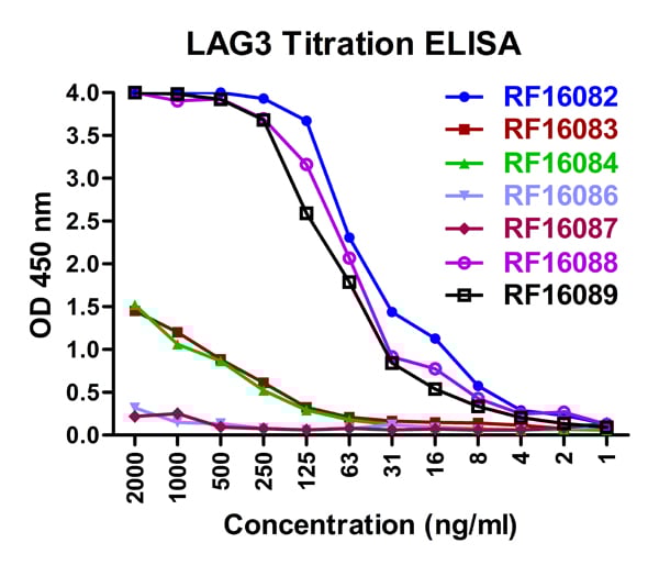


 ELISA
(A sandwich ELISA was performed using the anti-LAG3 mAbs as the capture antibodies for the LAG3 extracellular domain antigen with biotin-labeled Risk-Free anti-LAG3 mAbs as the detection antibodies.)
ELISA
(A sandwich ELISA was performed using the anti-LAG3 mAbs as the capture antibodies for the LAG3 extracellular domain antigen with biotin-labeled Risk-Free anti-LAG3 mAbs as the detection antibodies.)
LAG3, Monoclonal Antibody (Cat# AAA11021)
Full Name
LAG3 Antibody [1G4]
Gene Names
LAG3; CD223
Reactivity
Human
Applications
Western Blot, Immunohistochemistry, Immunocytochemistry, Immunofluorescence, Flow Cytometry
Purity
Protein A purified
Pricing


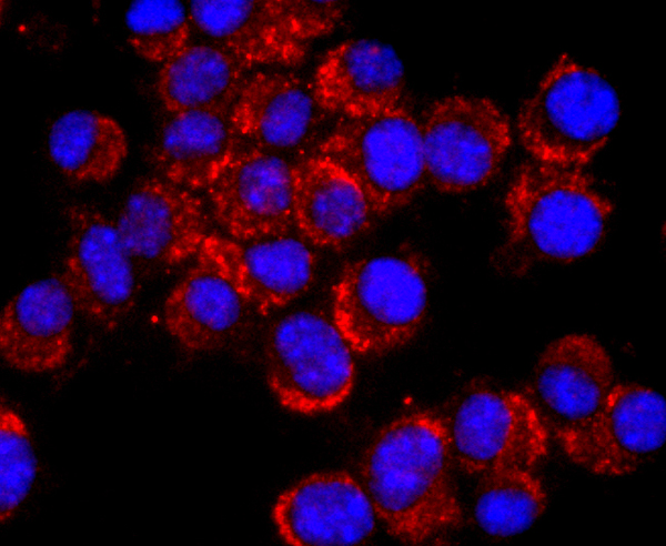
 Application Data
(At 25 degree C. The primary antibody was diluted at 1/200 and incubated with the sample for 1 hour at 37 degree C. An Alexa Fluor 594 conjugated goat anti-rabbit IgG (H+L) Ab, diluted at 1/600, was used as the secondary antibody.)
Application Data
(At 25 degree C. The primary antibody was diluted at 1/200 and incubated with the sample for 1 hour at 37 degree C. An Alexa Fluor 594 conjugated goat anti-rabbit IgG (H+L) Ab, diluted at 1/600, was used as the secondary antibody.)
LIMK1, Polyclonal Antibody (Cat# AAA31425)
Full Name
Phospho-LIMK1 (Ser310) Antibody
Gene Names
LIMK1; LIMK; LIMK-1
Reactivity
Human, Mouse, Rat
Predicted Reactivity: Pig (100%), Bovine (100%), Horse (100%), Sheep (100%), Rabbit (100%), Dog (88%), Chicken (100%)
Predicted Reactivity: Pig (100%), Bovine (100%), Horse (100%), Sheep (100%), Rabbit (100%), Dog (88%), Chicken (100%)
Applications
Western Blot, Immunohistochemistry, Immunofluorescence, Immunocytochemistry, Peptide ELISA
Purity
The antibody is from purified rabbit serum by affinity purification via sequential chromatography on phospho-peptide and non-phospho-peptide affinity columns.
Pricing


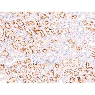 IHC (Immunohistochemistry)
(At 1/200 staining Human kidney tissue sections by IHC-P. The tissue was formaldehyde fixed and a heat mediated antigen retrieval step in citrate buffer was performed. The tissue was then blocked and incubated with the antibody for 1.5 hours at 22 degree C. An HRP conjugated goat anti-rabbit antibody was used as the secondary antibody.)
IHC (Immunohistochemistry)
(At 1/200 staining Human kidney tissue sections by IHC-P. The tissue was formaldehyde fixed and a heat mediated antigen retrieval step in citrate buffer was performed. The tissue was then blocked and incubated with the antibody for 1.5 hours at 22 degree C. An HRP conjugated goat anti-rabbit antibody was used as the secondary antibody.)
RhoA, Polyclonal Antibody (Cat# AAA31383)
Full Name
Phospho-RhoA (Ser188) Antibody
Gene Names
RHOA; ARHA; ARH12; RHO12; RHOH12
Reactivity
Human, Mouse, Rat
Predicted Reactivity: Pig (100%), Bovine (100%), Sheep (100%), Dog (100%)
Predicted Reactivity: Pig (100%), Bovine (100%), Sheep (100%), Dog (100%)
Applications
Western Blot, Immunohistochemistry, Peptide ELISA
Purity
The antibody is from purified rabbit serum by affinity purification via sequential chromatography on phospho-peptide and non-phospho-peptide affinity columns.
Pricing
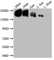
 WB (Western Blot)
(Western blot analysis of MMP26 expression in HEK293T (A), Raw264.7 (B), H9C2 (C) whole cell lysates.)
WB (Western Blot)
(Western blot analysis of MMP26 expression in HEK293T (A), Raw264.7 (B), H9C2 (C) whole cell lysates.)
MMP26, Polyclonal Antibody (Cat# AAA27797)
Full Name
Anti-MMP26 Antibody
Reactivity
Human, Mouse, Rat, Monkey
Applications
Western Blot
Purity
The antibody was purified by immunogen affinity chromatography.
Pricing


 Application Data
(Staining of human peripheral blood lymphocytes with MOUSE ANTI HUMAN CD45RA:FITC (MCA88F))
Application Data
(Staining of human peripheral blood lymphocytes with MOUSE ANTI HUMAN CD45RA:FITC (MCA88F))
CD45RA, Monoclonal Antibody (Cat# AAA26783)
Full Name
CD45RA (CD45 Antigen, B220, GP180, Leukocyte Common Antigen, LCA, L-CA, LY5, LY-5, Protein Tyrosine Phosphatase Receptor Type C Polypeptide, PTPRC, T200, T200 Glycoprotein) (MaxLight 405)
Reactivity
Human, Monkey
Applications
Flow Cytometry, Immunohistochemistry
Purity
Purified by protein G affinity chromatography from tissue culture supernatant.
Pricing


 Gene Sequencing (extract)
Apolipoprotein C2 (APOC2), Recombinant Protein (Cat# AAA20227)
Full Name
Recombinant Apolipoprotein C2 (APOC2)
Reactivity
Mus musculus (Mouse)
Applications
Western Blot
Purity
> 80%
Pricing


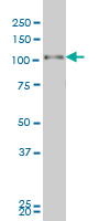
 IHC (Immunohistchemistry)
(Immunoperoxidase of monoclonal antibody to USF2 on formalin-fixed paraffin-embedded human esophagus. [antibody concentration 1 ug/ml])
IHC (Immunohistchemistry)
(Immunoperoxidase of monoclonal antibody to USF2 on formalin-fixed paraffin-embedded human esophagus. [antibody concentration 1 ug/ml])
USF2, Monoclonal Antibody (Cat# AAA25962)
Full Name
USF2 (Upstream Transcription Factor 2, c-fos Interacting, FIP, bHLHb12) (AP)
Gene Names
USF2; FIP; bHLHb12
Applications
Immunohistochemistry, Western Blot
Purity
Purified
Pricing


 Application Data
(Detection limit for recombinant GST tagged PGR is ~0.3ng/ml as a capture antibody.)
Application Data
(Detection limit for recombinant GST tagged PGR is ~0.3ng/ml as a capture antibody.)
Progesterone Receptor, Monoclonal Antibody (Cat# AAA25805)
Full Name
Progesterone Receptor (PgR, PR, Nuclear Receptor Subfamily 3 Group C Member 3, NR3C3) (PE)
Gene Names
PGR; PR; NR3C3
Reactivity
Human, Rat
Applications
EIA, IF, IHC, WB
Purity
Purified by Protein A Affinity Chromatography.
Pricing


 WB (Western Blot)
(Western blot analysis of RAD51C over-expressed 293 cell line, cotransfected with RAD51C Validated Chimera RNAi (Lane 2) or non-transfected control (Lane 1). Blot probed with RAD51C monoclonal antibody. GAPDH (36.1kD) used as specificity and loading control.)
WB (Western Blot)
(Western blot analysis of RAD51C over-expressed 293 cell line, cotransfected with RAD51C Validated Chimera RNAi (Lane 2) or non-transfected control (Lane 1). Blot probed with RAD51C monoclonal antibody. GAPDH (36.1kD) used as specificity and loading control.)
RAD51C, Monoclonal Antibody (Cat# AAA24335)
Full Name
RAD51C (DNA Repair Protein RAD51 Homolog 3, R51H3, RAD51 Homolog C, RAD51-like Protein 2, MGC104277, RAD51L2) (AP)
Gene Names
RAD51C; FANCO; R51H3; BROVCA3; RAD51L2
Reactivity
Human
Applications
Western Blot
Purity
Purified by Protein A Affinity Chromatography.
Pricing


 ELISA
(Red: Control Antigen (100ng); Purple: Antigen (10ng); Green: Antigen (50ng); Blue: Antigen (100ng);)
ELISA
(Red: Control Antigen (100ng); Purple: Antigen (10ng); Green: Antigen (50ng); Blue: Antigen (100ng);)
c-CBL, Monoclonal Antibody (Cat# AAA12378)
Full Name
Anti-c-CBL Antibody (clone 3B12) IHC-plus
Gene Names
CBL; CBL2; NSLL; C-CBL; RNF55; FRA11B
Reactivity
Mouse, Rat, Human
Applications
Immunohistochemistry, Immunocytochemistry, Immunofluorescence, Western Blot, Flow Cytometry
Purity
Ascites
Pricing


 FCM (Flow Cytometry)
(Flow cytometric analysis of SH-SY-5Y cells with NCAM antibody at 1/50 dilution (red) compared with an unlabelled control (cells without incubation with primary antibody; black). Alexa Fluor 488-conjugated goat anti rabbit IgG was used as the secondary antibody.)
FCM (Flow Cytometry)
(Flow cytometric analysis of SH-SY-5Y cells with NCAM antibody at 1/50 dilution (red) compared with an unlabelled control (cells without incubation with primary antibody; black). Alexa Fluor 488-conjugated goat anti rabbit IgG was used as the secondary antibody.)
NCAM, Monoclonal Antibody (Cat# AAA30279)
Full Name
NCAM Antibody
Gene Names
NCAM1; CD56; NCAM; MSK39
Reactivity
Human, Mouse, Zebrafish
Applications
Western Blot, Immunocytochemistry, Immunofluorescence, Immunohistochemistry, Immunoprecipitation, Flow Cytometry
Purity
ProA affinity purified
Pricing

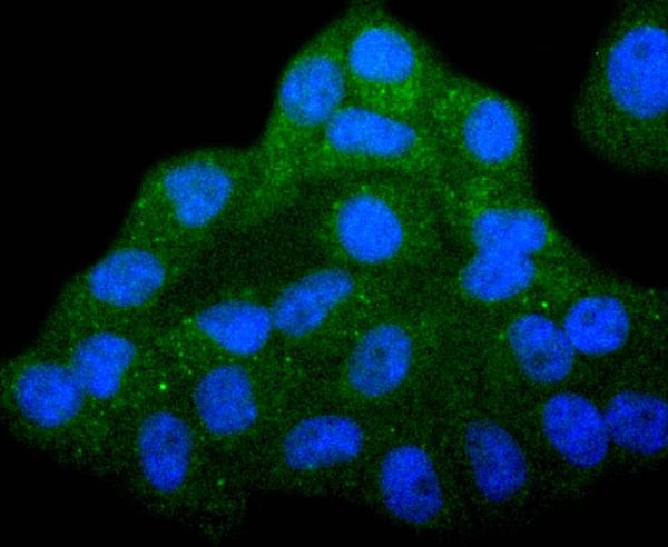


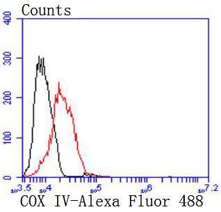 FCM (Flow Cytometry)
(Flow cytometric analysis of MCF-7 cells with COX IV antibody at 1/50 dilution (red) compared with an unlabelled control (cells without incubation with primary antibody; black). Alexa Fluor 488-conjugated goat anti rabbit IgG was used as the secondary antibody.)
FCM (Flow Cytometry)
(Flow cytometric analysis of MCF-7 cells with COX IV antibody at 1/50 dilution (red) compared with an unlabelled control (cells without incubation with primary antibody; black). Alexa Fluor 488-conjugated goat anti rabbit IgG was used as the secondary antibody.)
COX IV, Monoclonal Antibody (Cat# AAA30241)
Full Name
COX IV Antibody
Gene Names
COX4I1; COX4; COXIV; COX4-1
Reactivity
Human, Mouse, Rat
Applications
Western Blot, Immunocytochemistry, Immunofluorescence, Immunohistochemistry, Immunoprecipitation, Flow Cytometry
Purity
ProA affinity purified
Pricing


 FCM (Flow Cytometry)
(Flow cytometric analysis of Jurkat cells with TCF7L2 antibody at 1/50 dilution (red) compared with an unlabelled control (cells without incubation with primary antibody; black). Alexa Fluor 488-conjugated goat anti rabbit IgG was used as the secondary antibody.)
FCM (Flow Cytometry)
(Flow cytometric analysis of Jurkat cells with TCF7L2 antibody at 1/50 dilution (red) compared with an unlabelled control (cells without incubation with primary antibody; black). Alexa Fluor 488-conjugated goat anti rabbit IgG was used as the secondary antibody.)
TCF7L2, Monoclonal Antibody (Cat# AAA30140)
Full Name
TCF7L2 Antibody
Gene Names
TCF7L2; TCF4; TCF-4
Reactivity
Human, Mouse, Rat
Applications
Western Blot, Immunocytochemistry, Immunofluorescence, Immunohistochemistry, Immunoprecipitation, Flow Cytometry
Purity
ProA affinity purified
Pricing


 Application Data
(Staining of human peripheral blood granulocytes with Mouse anti Human CD45: Pacific Blue)
Application Data
(Staining of human peripheral blood granulocytes with Mouse anti Human CD45: Pacific Blue)
CD45, Monoclonal Antibody (Cat# AAA12203)
Full Name
MOUSE ANTI HUMAN CD45
Gene Names
PTPRC; LCA; LY5; B220; CD45; L-CA; T200; CD45R; GP180
Applications
Immunohistochemistry, Flow Cytometry, Immunoprecipitation, Immunohistochemistry
Pricing


 Application Data
(Proximity Ligation Analysis (PLA) of protein-protein interactions between BAD and MAPK8 Mahlavu cells were stained with BAD rabbit purified polyclonal 1:1200 and anti-MAPK8 mouse monoclonal antibody 1:50. Signals were detected 30 Detection Kit 613 (red), and nuclei were counterstained with DAPI (blue). Each red dot represents the detection of protein-protein interaction complex.)
Application Data
(Proximity Ligation Analysis (PLA) of protein-protein interactions between BAD and MAPK8 Mahlavu cells were stained with BAD rabbit purified polyclonal 1:1200 and anti-MAPK8 mouse monoclonal antibody 1:50. Signals were detected 30 Detection Kit 613 (red), and nuclei were counterstained with DAPI (blue). Each red dot represents the detection of protein-protein interaction complex.)
MAPK8, Monoclonal Antibody (Cat# AAA25743)
Full Name
MAPK8 (JNK1, PRKM8, SAPK1, SAPK1C, Mitogen-activated Protein Kinase 8, MAP Kinase 8, MAPK 8, JNK-46, Stress-activated Protein Kinase 1c, Stress-activated Protein Kinase JNK1, c-Jun N-terminal Kinase 1) (PE)
Gene Names
MAPK8; JNK; JNK1; PRKM8; SAPK1; JNK-46; JNK1A2; SAPK1c; JNK21B1/2
Reactivity
Human
Applications
Western Blot
Purity
Purified by Protein A Affinity Chromatography.
Pricing


 FCM (Flow Cytometry)
(Overlay histogram showing Raji cells stained with AAA27027 (red line) at 1:100. The cells were incubated in 1x PBS /10% normal goat serum to block non-specific protein-protein interactions followed by primary antibody for 1 h at 4 degree C. The secondary antibody used was FITC goat anti-mouse IgG(H+L) at 1/200 dilution for 1 h at 4 degree C. Isotype control antibody (green line) was used under the same conditions. Acquisition of >10,000 events was performed.)
FCM (Flow Cytometry)
(Overlay histogram showing Raji cells stained with AAA27027 (red line) at 1:100. The cells were incubated in 1x PBS /10% normal goat serum to block non-specific protein-protein interactions followed by primary antibody for 1 h at 4 degree C. The secondary antibody used was FITC goat anti-mouse IgG(H+L) at 1/200 dilution for 1 h at 4 degree C. Isotype control antibody (green line) was used under the same conditions. Acquisition of >10,000 events was performed.)
CD19, Monoclonal Antibody (Cat# AAA27027)
Full Name
CD19 Monoclonal Antibody
Gene Names
CD19; B4; CVID3
Reactivity
Human
Applications
Western Blot, Immunohistochemistry, Immunofluorescence, Flow Cytometry
Purity
>95%, Protein A Purified
Pricing


 IHC (Immunohistochemistry)
(At 1/100 staining Human gastric cancer by IHC-P. The sample was formaldehyde fixed and a heat mediated antigen retrieval step in citrate buffer was performed. The sample was then blocked and incubated with the primary antibody at 4 degree C overnight. An HRP conjugated anti-Rabbit antibody was used as the secondary antibody.)
IHC (Immunohistochemistry)
(At 1/100 staining Human gastric cancer by IHC-P. The sample was formaldehyde fixed and a heat mediated antigen retrieval step in citrate buffer was performed. The sample was then blocked and incubated with the primary antibody at 4 degree C overnight. An HRP conjugated anti-Rabbit antibody was used as the secondary antibody.)
Abi 1, Polyclonal Antibody (Cat# AAA31442)
Full Name
Phospho-Abi 1 (Tyr213) Antibody
Gene Names
ABI1; E3B1; ABI-1; ABLBP4; NAP1BP; SSH3BP; SSH3BP1
Reactivity
Human, Mouse, Rat
Predicted Reactivity: Pig (100%), Bovine (100%), Horse (100%), Sheep (100%), Rabbit (100%), Dog (100%), Chicken (100%), Xenopus (100%)
Predicted Reactivity: Pig (100%), Bovine (100%), Horse (100%), Sheep (100%), Rabbit (100%), Dog (100%), Chicken (100%), Xenopus (100%)
Applications
Western Blot, Immunohistochemistry, Peptide ELISA
Purity
The antibody is from purified rabbit serum by affinity purification via sequential chromatography on phospho-peptide and non-phospho-peptide affinity columns.
Pricing


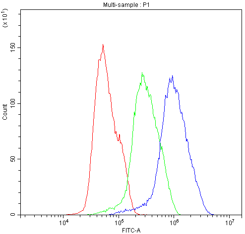
 FCM (Flow Cytometry)
(Figure 12. Flow Cytometry analysis of A549 cells using anti-Transketolase/TKT antibody (AAA19367).Overlay histogram showing A549 cells stained with AAA19367 (Blue line). The cells were blocked with 10% normal goat serum. And then incubated with mouse anti- Transketolase/TKT Antibody (AAA19367, 1μg/1x106 cells) for 30 min at 20 degree C. DyLight®488 conjugated goat anti-mouse IgG (BA1126, 5-10μg/1x106 cells) was used as secondary antibody for 30 minutes at 20 degree C. Isotype control antibody (Green line) was mouse IgG (1μg/1x106) used under the same conditions. Unlabelled sample (Red line) was also used as a control.)
FCM (Flow Cytometry)
(Figure 12. Flow Cytometry analysis of A549 cells using anti-Transketolase/TKT antibody (AAA19367).Overlay histogram showing A549 cells stained with AAA19367 (Blue line). The cells were blocked with 10% normal goat serum. And then incubated with mouse anti- Transketolase/TKT Antibody (AAA19367, 1μg/1x106 cells) for 30 min at 20 degree C. DyLight®488 conjugated goat anti-mouse IgG (BA1126, 5-10μg/1x106 cells) was used as secondary antibody for 30 minutes at 20 degree C. Isotype control antibody (Green line) was mouse IgG (1μg/1x106) used under the same conditions. Unlabelled sample (Red line) was also used as a control.)
Transketolase/TKT, Monoclonal Antibody (Cat# AAA19367)
Full Name
Anti-Transketolase/TKT Antibody (monoclonal, 3E5)
Gene Names
TKT; TK; TKT1; HEL107
Reactivity
Human, Mouse, Rat
Applications
WB, IHC-P, ICC, IF, FC/FACS/FCM
Purity
Immunogen affinity purified.
Pricing


 IHC (Immunohistochemistry)
(At 1/100 staining Human colorectal cancer and adjacent normal tissues by IHC-P. The sample was formaldehyde fixed and a heat mediated antigen retrieval step in citrate buffer was performed. The sample was then blocked and incubated with the primary antibody at 4 degree C overnight. An HRP conjugated anti-Rabbit antibody was used as the secondary antibody.)
IHC (Immunohistochemistry)
(At 1/100 staining Human colorectal cancer and adjacent normal tissues by IHC-P. The sample was formaldehyde fixed and a heat mediated antigen retrieval step in citrate buffer was performed. The sample was then blocked and incubated with the primary antibody at 4 degree C overnight. An HRP conjugated anti-Rabbit antibody was used as the secondary antibody.)
Bamacan, Polyclonal Antibody (Cat# AAA31455)
Full Name
Phospho-Bamacan (Ser1083) Antibody
Gene Names
SMC3; BAM; BMH; HCAP; CDLS3; CSPG6; SMC3L1
Reactivity
Human, Mouse, Rat
Predicted Reactivity: Pig (100%), Horse (100%), Sheep (100%), Rabbit (100%), Dog (100%), Chicken (100%)
Predicted Reactivity: Pig (100%), Horse (100%), Sheep (100%), Rabbit (100%), Dog (100%), Chicken (100%)
Applications
Western Blot, Immunohistochemistry, Peptide ELISA
Purity
The antibody is from purified rabbit serum by affinity purification via sequential chromatography on phospho-peptide and non-phospho-peptide affinity columns.
Pricing


 IHC (Immunohistchemistry)
(Immunoperoxidase of monoclonal antibody to USF2 on formalin-fixed paraffin-embedded human esophagus. [antibody concentration 1 ug/ml])
IHC (Immunohistchemistry)
(Immunoperoxidase of monoclonal antibody to USF2 on formalin-fixed paraffin-embedded human esophagus. [antibody concentration 1 ug/ml])
USF2, Monoclonal Antibody (Cat# AAA26626)
Full Name
USF2 (Upstream Transcription Factor 2, c-fos Interacting, FIP, bHLHb12) (PE)
Gene Names
USF2; FIP; bHLHb12
Applications
Immunofluorescence, Immunohistochemistry, Western Blot
Purity
Purified
Pricing


 Application Data
(Detection limit for recombinant GST tagged PGR is approximately 0.1ng/ml as a capture antibody.)
Application Data
(Detection limit for recombinant GST tagged PGR is approximately 0.1ng/ml as a capture antibody.)
PGR, Monoclonal Antibody (Cat# AAA26457)
Full Name
PGR (Progesterone Receptor, NR3C3, PR) (HRP)
Gene Names
PGR; PR; NR3C3
Applications
Immunofluorescence, Immunohistochemistry, Western Blot
Purity
Purified
Pricing


 ICC (Immunocytochemistry)
(ICC staining Chk2 in 293 cells (green). The nuclear counter stain is DAPI (blue). Cells were fixed in paraformaldehyde, permeabilised with 0.25% Triton X100/PBS.)
ICC (Immunocytochemistry)
(ICC staining Chk2 in 293 cells (green). The nuclear counter stain is DAPI (blue). Cells were fixed in paraformaldehyde, permeabilised with 0.25% Triton X100/PBS.)
Chk2, Monoclonal Antibody (Cat# AAA30126)
Full Name
Chk2 Antibody
Gene Names
CHEK2; CDS1; CHK2; LFS2; RAD53; hCds1; HuCds1; PP1425
Reactivity
Human
Applications
Western Blot, Immunocytochemistry, Immunofluorescence, Immunohistochemistry, Immunoprecipitation
Purity
ProA affinity purified
Pricing


 FCM (Flow Cytometry)
(Flow cytometric analysis of Daudi cells with IMPDH2 antibody at 1/100 dilution (purple) compared with an unlabelled control (cells without incubation with primary antibody; yellow). Alexa Fluor 488-conjugated goat anti-rabbit IgG was used as the secondary antibody.)
FCM (Flow Cytometry)
(Flow cytometric analysis of Daudi cells with IMPDH2 antibody at 1/100 dilution (purple) compared with an unlabelled control (cells without incubation with primary antibody; yellow). Alexa Fluor 488-conjugated goat anti-rabbit IgG was used as the secondary antibody.)
IMPDH2, Monoclonal Antibody (Cat# AAA30527)
Full Name
IMPDH2 Antibody
Gene Names
IMPDH2; IMPD2; IMPDH-II
Reactivity
Human, Mouse, Rat
Applications
Western Blot, Immunocytochemistry, Immunofluorescence, Immunohistochemistry, Flow Cytometry, Immunoprecipitation
Purity
ProA affinity purified
Pricing


 IHC (Immunohistchemistry)
(Immunohistochemistry Analysis: Representative lot data. (Fig. 1 and 2) Paraffin-embedded mouse and human brain tissue was prepared using heat-induced epitope retrieval in citrate buffer, pH 6.0. Immunostaining was performed using a 1:100 dilution. Reactivity was detected using the IHC-Select Detection Kit. Staining pattern appears as cytoplasmic. (Fig. 3 and 4) Paraffin-embedded mouse and mouse olfactory lobe and cerebellum brain tissue was prepared using heat-induced epitope retrieval in citrate buffer, pH 6.0. Immunostaining was performed using a Chicken IgY Antibody 1:100 dilution of Cat. No. AB15894, anti-Tbr2. Reactivity was detected using the IHC-Select Detection Kit. Immunoreactivity seen here is mostly nuclear.)
IHC (Immunohistchemistry)
(Immunohistochemistry Analysis: Representative lot data. (Fig. 1 and 2) Paraffin-embedded mouse and human brain tissue was prepared using heat-induced epitope retrieval in citrate buffer, pH 6.0. Immunostaining was performed using a 1:100 dilution. Reactivity was detected using the IHC-Select Detection Kit. Staining pattern appears as cytoplasmic. (Fig. 3 and 4) Paraffin-embedded mouse and mouse olfactory lobe and cerebellum brain tissue was prepared using heat-induced epitope retrieval in citrate buffer, pH 6.0. Immunostaining was performed using a Chicken IgY Antibody 1:100 dilution of Cat. No. AB15894, anti-Tbr2. Reactivity was detected using the IHC-Select Detection Kit. Immunoreactivity seen here is mostly nuclear.)
EOMES, Polyclonal Antibody (Cat# AAA26889)
Full Name
EOMES (Eomesodermin Homolog, T-box Brain Protein 2, TBR2, T-brain-2, TBR-2)
Gene Names
Eomes; Tbr2; TBR-2; C77258
Reactivity
Mouse, Human, Rat
Applications
Immunohistochemistry, Western Blot
Purity
Purified by affinity chromatography.
Pricing


 IF (Immunofluorescence)
(Confocal immunofluorescent analysis of CTNNB1 Antibody (C-term) with T47D cell followed by Alexa Fluor 488-conjugated goat anti-rabbit lgG (green). Actin filaments have been labeled with Alexa Fluor 555 phalloidin (red). DAPI was used to stain the cell nuclear (blue).)
IF (Immunofluorescence)
(Confocal immunofluorescent analysis of CTNNB1 Antibody (C-term) with T47D cell followed by Alexa Fluor 488-conjugated goat anti-rabbit lgG (green). Actin filaments have been labeled with Alexa Fluor 555 phalloidin (red). DAPI was used to stain the cell nuclear (blue).)
CTNNB1, Polyclonal Antibody (Cat# AAA28720)
Full Name
CTNNB1 Antibody (C-term)
Gene Names
CTNNB1; CTNNB; MRD19; armadillo
Reactivity
Human (Predicted Reactivity: Bovine, Mouse, Rat)
Applications
Western Blot, Immunohistochemistry, Flow Cytometry, Immunofluorescence
Purity
Peptide Affinity Purified Rabbit Polyclonal Antibody (Pab)
Pricing


 IF (Immunofluorescence)
(Confocal immunofluorescent analysis of C2C12 cells, either LY294002-treated (left) or insulin-treated (right), using AAA14695 (green). Actin filaments have been labeled with DY554 phalloidin (red). Blue pseudocolor = DRAQ5™ (fluorescent DNA dye).)
IF (Immunofluorescence)
(Confocal immunofluorescent analysis of C2C12 cells, either LY294002-treated (left) or insulin-treated (right), using AAA14695 (green). Actin filaments have been labeled with DY554 phalloidin (red). Blue pseudocolor = DRAQ5™ (fluorescent DNA dye).)
Akt, pan, Monoclonal Antibody (Cat# AAA14695)
Full Name
Akt, pan (Rac PKa, PKBa)
Gene Names
Akt1; akt; Akt; AKT; Akt/PKB; AKT/PKB; akt1; AKT1; Akt1|PKB; CG4006; D-Akt; dakt; dAkt; dAKT; Dakt; DAkt; dAKT/dPKB; dAkt/PKB; dakt1; dAkt1; dAKT1; Dakt1; DAkt1; DAKT1; DAKT1/PKB; DmelCG4006; dPKB; Dpkb; DPKB; DRAC-PK; DRAC-PK66; DRAC-PK85; l(3)04226; l(3)89Bq;
Reactivity
Human, Monkey, Mouse, Rat
Applications
Western Blot, Immunoprecipitation, Immunohistochemistry, Flow Cytometry, Immunofluorescence
Purity
Ascites
Ascites
Ascites
Pricing


 FCM (Flow Cytometry)
(Overlay histogram showing Raji cells stained with AAA28060 (red line) at 1:500. The cells were incubated in 10% normal goat serum to block non-specific protein-protein interactions followed by the antibody (1ug/1*106cells) for 1 h at 4 degree C. The secondary antibody used was FITC-conjugated Goat Anti-Mouse IgG(H+L) at 1/100 dilution for 30min at 4 degree C. Isotype control antibody (green line) was mouse IgG2b (1ug/1*106cells) used under the same conditions. Acquisition of >10,000 events was performed.)
FCM (Flow Cytometry)
(Overlay histogram showing Raji cells stained with AAA28060 (red line) at 1:500. The cells were incubated in 10% normal goat serum to block non-specific protein-protein interactions followed by the antibody (1ug/1*106cells) for 1 h at 4 degree C. The secondary antibody used was FITC-conjugated Goat Anti-Mouse IgG(H+L) at 1/100 dilution for 30min at 4 degree C. Isotype control antibody (green line) was mouse IgG2b (1ug/1*106cells) used under the same conditions. Acquisition of >10,000 events was performed.)
CD45, Monoclonal Antibody (Cat# AAA28060)
Full Name
CD45 Monoclonal Antibody
Gene Names
PTPRC; LCA; LY5; B220; CD45; L-CA; T200; CD45R; GP180
Reactivity
Human
Applications
Western Blot, Immunohistochemistry, Immunofluorescence, Flow Cytometry
Purity
>95%, Protein A purified
Pricing


 Application Data
(Overlay histogram showing SY5Y cells stained with AAA27008 (red line). The cells were fixed with 70% Ethylalcohol (18h) and then permeabilized with 0.3% Triton X-100 for 2 min. The cells were then incubated in 1x PBS /10% normal goat serum to block non-specific protein-protein interactions followed by the antibody (10ug/1x10^6cells) for 1 h at 4 degree C. The secondary antibody used was FITC goat anti-mouse IgG (H+L) at 1/200 dilution for 1 h at 4 degree C. Isotype control antibody (green line) was mouse IgG2b (10ug/1x10^6cells) used under the same conditions. Acquisition of >10, 000 events was performed.)
Application Data
(Overlay histogram showing SY5Y cells stained with AAA27008 (red line). The cells were fixed with 70% Ethylalcohol (18h) and then permeabilized with 0.3% Triton X-100 for 2 min. The cells were then incubated in 1x PBS /10% normal goat serum to block non-specific protein-protein interactions followed by the antibody (10ug/1x10^6cells) for 1 h at 4 degree C. The secondary antibody used was FITC goat anti-mouse IgG (H+L) at 1/200 dilution for 1 h at 4 degree C. Isotype control antibody (green line) was mouse IgG2b (10ug/1x10^6cells) used under the same conditions. Acquisition of >10, 000 events was performed.)
GFAP, Monoclonal Antibody (Cat# AAA27008)
Full Name
GFAP Monoclonal Antibody
Gene Names
GFAP; ALXDRD
Reactivity
Human, Mouse, Rat
Applications
Western Blot, Immunohistochemistry, Immunofluorescence, Flow Cytometry
Purity
>95%, Protein G purified
Pricing
 Standard Curve (Sample)
v-myc myelocytomatosis viral oncogene homolog (avian), ELISA Kit (Cat# AAA15533)
Full Name
Human c-myc Oncogene product, c-myc ELISA Kit
Gene Names
MYC; MRTL; MYCC; c-Myc; bHLHe39
Reactivity
Human
Pricing


 Application Data
(Staining of mouse spleen cells with Rat anti Mouse CD45 (Ly-5))
Application Data
(Staining of mouse spleen cells with Rat anti Mouse CD45 (Ly-5))
CD45, Monoclonal Antibody (Cat# AAA11915)
Full Name
RAT ANTI MOUSE CD45
Gene Names
Ptprc; loc; B220; Cd45; L-CA; Ly-5; T200; CD45R; Lyt-4
Applications
Immunohistochemistry, Flow Cytometry, Immunofluorescence, Immunoprecipitation
Pricing
 SDS-PAGE
(Fig.SDS-PAGE analysis of Recombinant SARS-CoV-2 Spike RBD Protein, His tag)
SDS-PAGE
(Fig.SDS-PAGE analysis of Recombinant SARS-CoV-2 Spike RBD Protein, His tag)
COVID 19 S1 Spike RBD Coronavirus, Recombinant Protein (Cat# AAA31529)
Full Name
Recombinant SARS-CoV-2 Spike RBD Protein, His tag (Animal-Free)
Purity
>95% as determined by SDS-PAGE
Pricing


 IF (Immunofluorescence)
(Immunofluorescence of IRE1p in rat small intestine tissue with IRE1p antibody at 20 μg/ml.Green: IRE1p Antibody (3655)Blue: DAPI staining)
IF (Immunofluorescence)
(Immunofluorescence of IRE1p in rat small intestine tissue with IRE1p antibody at 20 μg/ml.Green: IRE1p Antibody (3655)Blue: DAPI staining)
IRE1p, Polyclonal Antibody (Cat# AAA10921)
Full Name
IRE1p Antibody
Gene Names
ERN1; IRE1; IRE1P; IRE1a; hIRE1p
Reactivity
Human, Mouse, Rat
Applications
Western Blot, Immunocytochemistry, Immunofluorescence
Purity
IRE1p Antibody is affinity chromatography purified via peptide column.
Pricing


 Application Data
(Staining of human peripheral blood monocytes with Mouse anti Human CD284: Low Endotoxin)
Application Data
(Staining of human peripheral blood monocytes with Mouse anti Human CD284: Low Endotoxin)
CD284, Monoclonal Antibody (Cat# AAA11871)
Full Name
MOUSE ANTI HUMAN CD284:FITC
Gene Names
TLR4; TOLL; CD284; TLR-4; ARMD10
Applications
Flow Cytometry
Pricing


 Application Data
(Published customer image: Leukocyte infiltration in COX-2-M/-M and COX-2+/+ mice. MPO enzymatic activity (panel A) was statistically similar in COX-2-M/-M and COX-2+/+ livers at 6 h and 24 h post-IRI. Ly-6G+ neutrophil (panel B) and granulocyte (panel C) infiltration were also comparable in COX-2-M/-M and COX-2+/+ livers after IRI. Mac-1+ (panel D) and CD68 (panel E) infiltrating macrophages were significantly reduced in COX-2-M/-M livers at 24 h post-reperfusion, but were statistically indistinguishable in COX-2-M/-M and COX-2+/+ livers at 6 h after IRI. No statistical differences in MMP-9 expression (panel F) could be demonstrated in livers of COX-2-M/-M and COX-2+/+ mice post-IRI. Representative immunostaining (panel G) of infiltrating Ly-6G+ (a,b,e,f) and Mac-1+ (c,d,g,h) leukocytes in livers of COX-2+/+ (a,c,e,g) and COX-2-M/-M (b,d,f,h) mice at 6 h (a to d) and 24 h (e to h) post IRI; (n = 5 -6/group; * indicates p)
Application Data
(Published customer image: Leukocyte infiltration in COX-2-M/-M and COX-2+/+ mice. MPO enzymatic activity (panel A) was statistically similar in COX-2-M/-M and COX-2+/+ livers at 6 h and 24 h post-IRI. Ly-6G+ neutrophil (panel B) and granulocyte (panel C) infiltration were also comparable in COX-2-M/-M and COX-2+/+ livers after IRI. Mac-1+ (panel D) and CD68 (panel E) infiltrating macrophages were significantly reduced in COX-2-M/-M livers at 24 h post-reperfusion, but were statistically indistinguishable in COX-2-M/-M and COX-2+/+ livers at 6 h after IRI. No statistical differences in MMP-9 expression (panel F) could be demonstrated in livers of COX-2-M/-M and COX-2+/+ mice post-IRI. Representative immunostaining (panel G) of infiltrating Ly-6G+ (a,b,e,f) and Mac-1+ (c,d,g,h) leukocytes in livers of COX-2+/+ (a,c,e,g) and COX-2-M/-M (b,d,f,h) mice at 6 h (a to d) and 24 h (e to h) post IRI; (n = 5 -6/group; * indicates p)
CD68, Monoclonal Antibody (Cat# AAA12103)
Full Name
RAT ANTI MOUSE CD68:Biotin
Gene Names
Cd68; Lamp4; gp110; Scard1
Applications
Flow Cytometry
Pricing


 Application Data
(Immunoperoxidase staining of rat lymph node cryosection with Mouse anti Rat CD86 antibody, clone 42F followed by horseradish peroxidase conjugated Goat anti Mouse IgG . Low power)
Application Data
(Immunoperoxidase staining of rat lymph node cryosection with Mouse anti Rat CD86 antibody, clone 42F followed by horseradish peroxidase conjugated Goat anti Mouse IgG . Low power)
CD86, Monoclonal Antibody (Cat# AAA12229)
Full Name
MOUSE ANTI RAT CD86:FITC
Gene Names
CD86; B70; B7-2; B7.2; LAB72; CD28LG2
Applications
Flow Cytometry
Pricing