Filters
Clonality
Type
Reactivity
Gene Name
Isotype
Host
Application
Clone
103 results for "Primary Antibodies Mouse Monoclonal Antibodies" - showing 1-50


 Application Data
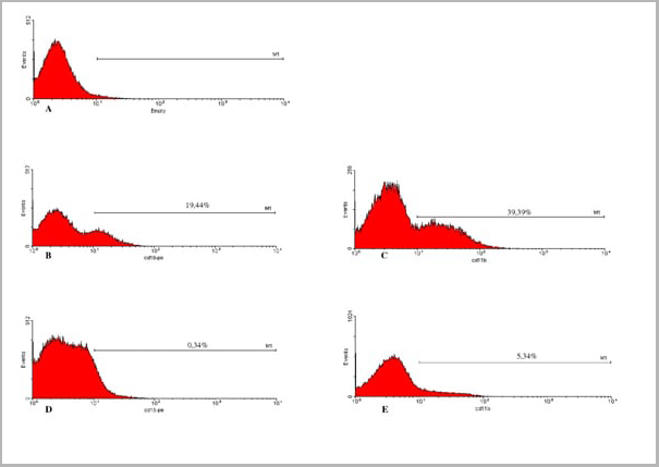
(Published clone specific image: Flow cytometric analysis of AM from NO2-exposed and control rats. Rats were exposed to NO2 for the indicated times and BAL cells were stained with antibodies to ED7, ED9, RM-4, and OX-6. To overcome autofluorescence signals, primary antibodies were detected using a biotin-PE/streptavidin-anti-streptavidin enhancing system and labeling of AM was analyzed by flow cytometry following gating by help of forward and sideward scatter properties. Shown are representative results of at least six animals per group.From: Garn H, Siese A, Stumpf S, Wensing A, Renz H, Gemsa D. Phenotypical and functional characterization of alveolar macrophage subpopulations in the lungs of NO2-exposed rats. Respir Res. 2006 Jan 6;7:4.)
Application Data
(Published clone specific image: Flow cytometric analysis of AM from NO2-exposed and control rats. Rats were exposed to NO2 for the indicated times and BAL cells were stained with antibodies to ED7, ED9, RM-4, and OX-6. To overcome autofluorescence signals, primary antibodies were detected using a biotin-PE/streptavidin-anti-streptavidin enhancing system and labeling of AM was analyzed by flow cytometry following gating by help of forward and sideward scatter properties. Shown are representative results of at least six animals per group.From: Garn H, Siese A, Stumpf S, Wensing A, Renz H, Gemsa D. Phenotypical and functional characterization of alveolar macrophage subpopulations in the lungs of NO2-exposed rats. Respir Res. 2006 Jan 6;7:4.)
CD172a, Monoclonal Antibody (Cat# AAA12056)
Full Name
MOUSE ANTI RAT CD172a:RPE
Gene Names
Sirpa; Bit; Ptpns1; SHPS-1
Applications
Flow Cytometry
Pricing


 Application Data
(Published customer image: Increased accumulation of repair-associated macrophages surrounding collaterals in ischemic hind limbs is PAR2-dependent. (A) Stainings of CD206-positive macrophages (green) and SMA-positive vessels (red) in non-ischemic (control) and ischemic (ligated) hind limbs of WT, PAR1-/- and PAR2-/- mice are shown. Nuclei were visualized with DAPI (blue). Arrows indicate single macrophages in the non-ischemic adductor. Quantification of the average number of repair-associated macrophages per vessel is indicated on the right. (B) Correlation between the number of CD206-positive macrophages in the ischemic tissues and the expression of CD11b and (C) CD115 on monocytes. ** p)
Application Data
(Published customer image: Increased accumulation of repair-associated macrophages surrounding collaterals in ischemic hind limbs is PAR2-dependent. (A) Stainings of CD206-positive macrophages (green) and SMA-positive vessels (red) in non-ischemic (control) and ischemic (ligated) hind limbs of WT, PAR1-/- and PAR2-/- mice are shown. Nuclei were visualized with DAPI (blue). Arrows indicate single macrophages in the non-ischemic adductor. Quantification of the average number of repair-associated macrophages per vessel is indicated on the right. (B) Correlation between the number of CD206-positive macrophages in the ischemic tissues and the expression of CD11b and (C) CD115 on monocytes. ** p)
CD206, Monoclonal Antibody (Cat# AAA12117)
Full Name
RAT ANTI MOUSE CD206:Biotin
Gene Names
Mrc1; MR; CD206; AW259686
Applications
Flow Cytometry
Pricing


 Application Data
(Published customer image: Increased accumulation of repair-associated macrophages surrounding collaterals in ischemic hind limbs is PAR2-dependent. (A) Stainings of CD206-positive macrophages (green) and SMA-positive vessels (red) in non-ischemic (control) and ischemic (ligated) hind limbs of WT, PAR1-/- and PAR2-/- mice are shown. Nuclei were visualized with DAPI (blue). Arrows indicate single macrophages in the non-ischemic adductor. Quantification of the average number of repair-associated macrophages per vessel is indicated on the right. (B) Correlation between the number of CD206-positive macrophages in the ischemic tissues and the expression of CD11b and (C) CD115 on monocytes. ** p)
Application Data
(Published customer image: Increased accumulation of repair-associated macrophages surrounding collaterals in ischemic hind limbs is PAR2-dependent. (A) Stainings of CD206-positive macrophages (green) and SMA-positive vessels (red) in non-ischemic (control) and ischemic (ligated) hind limbs of WT, PAR1-/- and PAR2-/- mice are shown. Nuclei were visualized with DAPI (blue). Arrows indicate single macrophages in the non-ischemic adductor. Quantification of the average number of repair-associated macrophages per vessel is indicated on the right. (B) Correlation between the number of CD206-positive macrophages in the ischemic tissues and the expression of CD11b and (C) CD115 on monocytes. ** p)
CD206, Monoclonal Antibody (Cat# AAA12122)
Full Name
RAT ANTI MOUSE CD206:FITC
Gene Names
Mrc1; MR; CD206; AW259686
Applications
Flow Cytometry
Pricing


 Application Data
(Published customer image: Increased accumulation of repair-associated macrophages surrounding collaterals in ischemic hind limbs is PAR2-dependent. (A) Stainings of CD206-positive macrophages (green) and SMA-positive vessels (red) in non-ischemic (control) and ischemic (ligated) hind limbs of WT, PAR1-/- and PAR2-/- mice are shown. Nuclei were visualized with DAPI (blue). Arrows indicate single macrophages in the non-ischemic adductor. Quantification of the average number of repair-associated macrophages per vessel is indicated on the right. (B) Correlation between the number of CD206-positive macrophages in the ischemic tissues and the expression of CD11b and (C) CD115 on monocytes. ** p)
Application Data
(Published customer image: Increased accumulation of repair-associated macrophages surrounding collaterals in ischemic hind limbs is PAR2-dependent. (A) Stainings of CD206-positive macrophages (green) and SMA-positive vessels (red) in non-ischemic (control) and ischemic (ligated) hind limbs of WT, PAR1-/- and PAR2-/- mice are shown. Nuclei were visualized with DAPI (blue). Arrows indicate single macrophages in the non-ischemic adductor. Quantification of the average number of repair-associated macrophages per vessel is indicated on the right. (B) Correlation between the number of CD206-positive macrophages in the ischemic tissues and the expression of CD11b and (C) CD115 on monocytes. ** p)
CD206, Monoclonal Antibody (Cat# AAA12125)
Full Name
RAT ANTI MOUSE CD206:RPE
Gene Names
Mrc1; MR; CD206; AW259686
Applications
Flow Cytometry
Pricing


 Application Data
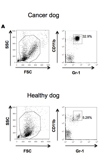
(Published customer image: Effects of EDTA versus media storage of patient blood samples on flow cytometric results. Peripheral blood cells were (a) stained immediate following collection with CD11b and CADO48A or maintained in EDTA collection tubes for (b) 24 or (c) 48 hours or in media for (d) 24 or (e) 48 hours prior to staining with CD11b and CADO48A. Both EDTA and media samples were kept refrigerated. These findings show a decrease over time of population distinction in EDTA with minimal changes when cells are kept in media up to 48 hours. All cells were gated on P1.From: Sherger et al. BMC Veterinary Research 2012 8:209)
Application Data
(Published customer image: Effects of EDTA versus media storage of patient blood samples on flow cytometric results. Peripheral blood cells were (a) stained immediate following collection with CD11b and CADO48A or maintained in EDTA collection tubes for (b) 24 or (c) 48 hours or in media for (d) 24 or (e) 48 hours prior to staining with CD11b and CADO48A. Both EDTA and media samples were kept refrigerated. These findings show a decrease over time of population distinction in EDTA with minimal changes when cells are kept in media up to 48 hours. All cells were gated on P1.From: Sherger et al. BMC Veterinary Research 2012 8:209)
CD11b, Monoclonal Antibody (Cat# AAA12092)
Full Name
MOUSE ANTI DOG CD11b
Gene Names
ITGAM; CD11B
Applications
Immunohistochemistry, Flow Cytometry, Immunoprecipitation
Pricing


 ICC (Immunocytochemistry)
(Figure 10 Immunocytochemistry Validation of PD-L1Immunocytochemical analysis of 4% paraformaldehyde-fixed PD-L1 transfected 293 cells labeling PD-L1 with at 1 μg/ml, followed by Goat anti-mouse IgG secondary antibody at 1/250 dilution (red). Lower left: Use mouse IgG antibody at 1 μg/ml as control.)
ICC (Immunocytochemistry)
(Figure 10 Immunocytochemistry Validation of PD-L1Immunocytochemical analysis of 4% paraformaldehyde-fixed PD-L1 transfected 293 cells labeling PD-L1 with at 1 μg/ml, followed by Goat anti-mouse IgG secondary antibody at 1/250 dilution (red). Lower left: Use mouse IgG antibody at 1 μg/ml as control.)
PDL1, Monoclonal Antibody (Cat# AAA10992)
Full Name
PDL1 Antibody [6H10]
Gene Names
CD274; B7-H; B7H1; PDL1; PD-L1; PDCD1L1; PDCD1LG1
Reactivity
Human
Applications
Western Blot, Immunohistochemistry, Immunocytochemistry, Immunofluorescence
Purity
Protein A purified
Pricing


 Application Data
(Published customer image: Increased accumulation of repair-associated macrophages surrounding collaterals in ischemic hind limbs is PAR2-dependent. (A) Stainings of CD206-positive macrophages (green) and SMA-positive vessels (red) in non-ischemic (control) and ischemic (ligated) hind limbs of WT, PAR1-/- and PAR2-/- mice are shown. Nuclei were visualized with DAPI (blue). Arrows indicate single macrophages in the non-ischemic adductor. Quantification of the average number of repair-associated macrophages per vessel is indicated on the right. (B) Correlation between the number of CD206-positive macrophages in the ischemic tissues and the expression of CD11b and (C) CD115 on monocytes. ** p)
Application Data
(Published customer image: Increased accumulation of repair-associated macrophages surrounding collaterals in ischemic hind limbs is PAR2-dependent. (A) Stainings of CD206-positive macrophages (green) and SMA-positive vessels (red) in non-ischemic (control) and ischemic (ligated) hind limbs of WT, PAR1-/- and PAR2-/- mice are shown. Nuclei were visualized with DAPI (blue). Arrows indicate single macrophages in the non-ischemic adductor. Quantification of the average number of repair-associated macrophages per vessel is indicated on the right. (B) Correlation between the number of CD206-positive macrophages in the ischemic tissues and the expression of CD11b and (C) CD115 on monocytes. ** p)
CD206, Monoclonal Antibody (Cat# AAA12120)
Full Name
RAT ANTI MOUSE CD206:FITC
Gene Names
Mrc1; MR; CD206; AW259686
Applications
Flow Cytometry
Pricing


 Application Data
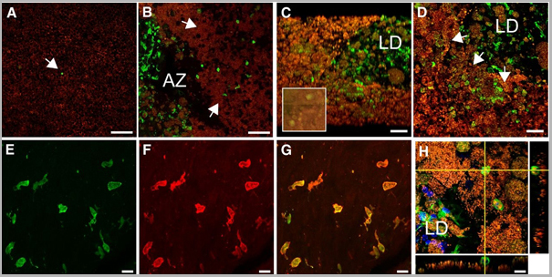
(Published customer image Infiltration of GFP+ BM-cells in infarct and peri-infarct regions. (A-B) Dot plots of viable macrophages/granulocytes (CD11b+CD45high, top right quadrants) and microglia (CD11b+CD45dim, bottom right quadrants) in cortex from BM-chimeric unmanipulated mice and mice exposed to pMCAO. (C) Bar graph showing mean numbers of CD11b+CD45dim microglia and CD11b+CD45high macrophages/granulocytes in BM-chimeric mice 24 hours after pMCAO, subdivided based on expression of GFP (n = 5). Approximately 92% of of the CD45high population were GFP+. (D) Estimation and comparison of mean numbers of CD11b+CD45dim microglia in non-chimeric (n = 10) versus BM-chimeric mice (n = 5) 24 hours after of pMCAO shows significantly fewer CD11b+CD45dim microglial cells in irradiated mice. (E) Overview, showing distribution of infiltrating GFP+ BM-derived cells into infarct (IF) and peri-infarct (P-IF) regions 24 hours after pMCAO. (E-G) By 24 hours, GFP+ single cells (F) and vessel-associated aggregates of GFP+ cells (arrows in G) were observed in infarct and peri-infarct regions. Some of the vessel-associated cells were round, leukocyte-like cells (arrows) while others were elongated cells lining the vasculature (arrow heads in G and in insert). (H) Bar graph showing mean numbers of single GFP+ cells and vessel-associated aggregates of GFP+ cells in ipsi- and contralateral cortex 24 hours after surgery (n = 10). (I-P) Immunohistochemical staining of CD45.1 (I, K), CD45.2 (J, L), IgG2a (M, O) and CD45 (N, P) in ischemic tissue in BM-chimeric (I, J, M, N) and non-chimeric mice (K, L, O, P) 24 hours after pMCAO. N.D, none detected. Scale bars: 200 um (A), 10 um (B, C). 50 um (I-P) *P < 0.05, **P < 0.01, and ***P < 0.001.From: Clausen BH, Lambertsen KL, Babcock AA, Holm TH, Dagnaes-Hansen F, Finsen B. Interleukin-1beta and tumor necrosis factor-alpha are expressed by different subsets of microglia and macrophages after ischemic stroke in mice. J Neuroinflammation. 2008 Oct 23;5:46.)
Application Data
(Published customer image Infiltration of GFP+ BM-cells in infarct and peri-infarct regions. (A-B) Dot plots of viable macrophages/granulocytes (CD11b+CD45high, top right quadrants) and microglia (CD11b+CD45dim, bottom right quadrants) in cortex from BM-chimeric unmanipulated mice and mice exposed to pMCAO. (C) Bar graph showing mean numbers of CD11b+CD45dim microglia and CD11b+CD45high macrophages/granulocytes in BM-chimeric mice 24 hours after pMCAO, subdivided based on expression of GFP (n = 5). Approximately 92% of of the CD45high population were GFP+. (D) Estimation and comparison of mean numbers of CD11b+CD45dim microglia in non-chimeric (n = 10) versus BM-chimeric mice (n = 5) 24 hours after of pMCAO shows significantly fewer CD11b+CD45dim microglial cells in irradiated mice. (E) Overview, showing distribution of infiltrating GFP+ BM-derived cells into infarct (IF) and peri-infarct (P-IF) regions 24 hours after pMCAO. (E-G) By 24 hours, GFP+ single cells (F) and vessel-associated aggregates of GFP+ cells (arrows in G) were observed in infarct and peri-infarct regions. Some of the vessel-associated cells were round, leukocyte-like cells (arrows) while others were elongated cells lining the vasculature (arrow heads in G and in insert). (H) Bar graph showing mean numbers of single GFP+ cells and vessel-associated aggregates of GFP+ cells in ipsi- and contralateral cortex 24 hours after surgery (n = 10). (I-P) Immunohistochemical staining of CD45.1 (I, K), CD45.2 (J, L), IgG2a (M, O) and CD45 (N, P) in ischemic tissue in BM-chimeric (I, J, M, N) and non-chimeric mice (K, L, O, P) 24 hours after pMCAO. N.D, none detected. Scale bars: 200 um (A), 10 um (B, C). 50 um (I-P) *P < 0.05, **P < 0.01, and ***P < 0.001.From: Clausen BH, Lambertsen KL, Babcock AA, Holm TH, Dagnaes-Hansen F, Finsen B. Interleukin-1beta and tumor necrosis factor-alpha are expressed by different subsets of microglia and macrophages after ischemic stroke in mice. J Neuroinflammation. 2008 Oct 23;5:46.)
CD11b, Monoclonal Antibody (Cat# AAA12182)
Full Name
RAT ANTI MOUSE CD11b:FITC
Gene Names
Itgam; CR3; CR3A; MAC1; Cd11b; Ly-40; Mac-1; Mac-1a; CD11b/CD18; F730045J24Rik
Applications
Flow Cytometry
Pricing


 IP (Immunoprecipitation)
(Immunoprecipitating GAPDH in Hela whole cell lysateLane 1: Mouse control IgG instead of in Hela whole cell lysate. Lane 2: (5ul) + Hela whole cell lysate (500ug)Lane 3: Hela whole cell lysate (10ug)For western blotting, the blot was detected at 1:5000, and a HRP-conjugated Protein G antibody was used as the secondary antibody at 1:2000)
IP (Immunoprecipitation)
(Immunoprecipitating GAPDH in Hela whole cell lysateLane 1: Mouse control IgG instead of in Hela whole cell lysate. Lane 2: (5ul) + Hela whole cell lysate (500ug)Lane 3: Hela whole cell lysate (10ug)For western blotting, the blot was detected at 1:5000, and a HRP-conjugated Protein G antibody was used as the secondary antibody at 1:2000)
GAPDH, Monoclonal Antibody (Cat# AAA27042)
Full Name
GAPDH Monoclonal Antibody
Reactivity
Human, Mouse, Rabbit
Applications
Western Blot, Immunohistochemistry, Immunoprecipitation, Immunofluorescence
Purity
>95%, Protein G purified
Pricing


 FCM (Flow Cytometry)
(Overlay Peak curve showing 293F cells transfected with GST stained with AAA28065 (red line) at 1:189. The cells were fixed in 4% formaldehyde and permeated by 0.2% TritonX-100. Then 10% normal goat serum was Incubated to block non-specific protein-protein interactions followed by the antibody (1?g/1*106cells) for 1 h at 4 degree C. The secondary antibody used was FITC-conjugated Goat Anti-Mouse IgG(H+L) at 1/100 dilution for 30min at 4 degree C. Isotype control antibody (green line) was mouse IgG2b (1?g/1*106cells) used under the same conditions. Acquisition of >10,000 events was performed.)
FCM (Flow Cytometry)
(Overlay Peak curve showing 293F cells transfected with GST stained with AAA28065 (red line) at 1:189. The cells were fixed in 4% formaldehyde and permeated by 0.2% TritonX-100. Then 10% normal goat serum was Incubated to block non-specific protein-protein interactions followed by the antibody (1?g/1*106cells) for 1 h at 4 degree C. The secondary antibody used was FITC-conjugated Goat Anti-Mouse IgG(H+L) at 1/100 dilution for 30min at 4 degree C. Isotype control antibody (green line) was mouse IgG2b (1?g/1*106cells) used under the same conditions. Acquisition of >10,000 events was performed.)
GST, Monoclonal Antibody (Cat# AAA28065)
Full Name
GST Monoclonal Antibody
Reactivity
All
Applications
Western Blot, Immunofluorescence, Flow Cytometry, Immunoprecipitation
Purity
>95%, Protein A purified
Pricing


 Application Data
(Published customer imageSLAMF3 blockade in human hepatocytes is associated with lower susceptibility to HCV. (A) SLAMF3 was stained in primary human hepatocytes (PHHs) and cells from the Huh-7 human hepatoma cell line with a specific antibody (HLy9.1.25 clone; grey) and an isotype-matched control (empty). One of four independent experiments is shown. Huh-7 cells were transfected with scrambled control (sc) siRNA or three specific siRNAs (#1, #2 and #3) targeting SLAMF3, prior to infection with HCVcc; siRNA efficiency was checked by quantifying SLAMF3 mRNA (B) and the CD81 expression level (C) by flow cytometry analysis at 48 h post-transfection. Results are presented as the mean +/-SD (n = 3). Intracellular viral RNA was quantified at 72 h p.i. (D) and the infection was measured at 72 h p.i. by focus-forming units FFUs counting (E) (as inhibition percent; mean of three independent experiments; error bars: SD. **p)
Application Data
(Published customer imageSLAMF3 blockade in human hepatocytes is associated with lower susceptibility to HCV. (A) SLAMF3 was stained in primary human hepatocytes (PHHs) and cells from the Huh-7 human hepatoma cell line with a specific antibody (HLy9.1.25 clone; grey) and an isotype-matched control (empty). One of four independent experiments is shown. Huh-7 cells were transfected with scrambled control (sc) siRNA or three specific siRNAs (#1, #2 and #3) targeting SLAMF3, prior to infection with HCVcc; siRNA efficiency was checked by quantifying SLAMF3 mRNA (B) and the CD81 expression level (C) by flow cytometry analysis at 48 h post-transfection. Results are presented as the mean +/-SD (n = 3). Intracellular viral RNA was quantified at 72 h p.i. (D) and the infection was measured at 72 h p.i. by focus-forming units FFUs counting (E) (as inhibition percent; mean of three independent experiments; error bars: SD. **p)
CD229, Monoclonal Antibody (Cat# AAA11951)
Full Name
MOUSE ANTI HUMAN CD229
Gene Names
LY9; hly9; mLY9; CD229; SLAMF3
Applications
Flow Cytometry, Immunoprecipitation
Pricing


 Application Data
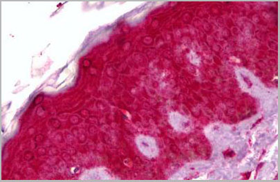
(Formalin fixed, paraffin embedded human breast cancer biopsy stained with Mouse anti Human estrogen receptor beta5 antibody followed by HRP polymer detection and DAB substrate development following heat mediated antigen retrieval using citrate buffer at pH6.2 (low power))
Application Data
(Formalin fixed, paraffin embedded human breast cancer biopsy stained with Mouse anti Human estrogen receptor beta5 antibody followed by HRP polymer detection and DAB substrate development following heat mediated antigen retrieval using citrate buffer at pH6.2 (low power))
ESTROGEN RECEPTOR BETA 5, Monoclonal Antibody (Cat# AAA12216)
Full Name
MOUSE ANTI HUMAN ESTROGEN RECEPTOR BETA 5
Gene Names
ESR2; Erb; ESRB; ESTRB; NR3A2; ER-BETA; ESR-BETA
Applications
Immunohistochemistry, Western Blot
Pricing


 Application Data
(Published customer image Infiltration of GFP+ BM-cells in infarct and peri-infarct regions. (A-B) Dot plots of viable macrophages/granulocytes (CD11b+CD45high, top right quadrants) and microglia (CD11b+CD45dim, bottom right quadrants) in cortex from BM-chimeric unmanipulated mice and mice exposed to pMCAO. (C) Bar graph showing mean numbers of CD11b+CD45dim microglia and CD11b+CD45high macrophages/granulocytes in BM-chimeric mice 24 hours after pMCAO, subdivided based on expression of GFP (n = 5). Approximately 92% of of the CD45high population were GFP+. (D) Estimation and comparison of mean numbers of CD11b+CD45dim microglia in non-chimeric (n = 10) versus BM-chimeric mice (n = 5) 24 hours after of pMCAO shows significantly fewer CD11b+CD45dim microglial cells in irradiated mice. (E) Overview, showing distribution of infiltrating GFP+ BM-derived cells into infarct (IF) and peri-infarct (P-IF) regions 24 hours after pMCAO. (E-G) By 24 hours, GFP+ single cells (F) and vessel-associated aggregates of GFP+ cells (arrows in G) were observed in infarct and peri-infarct regions. Some of the vessel-associated cells were round, leukocyte-like cells (arrows) while others were elongated cells lining the vasculature (arrow heads in G and in insert). (H) Bar graph showing mean numbers of single GFP+ cells and vessel-associated aggregates of GFP+ cells in ipsi- and contralateral cortex 24 hours after surgery (n = 10). (I-P) Immunohistochemical staining of CD45.1 (I, K), CD45.2 (J, L), IgG2a (M, O) and CD45 (N, P) in ischemic tissue in BM-chimeric (I, J, M, N) and non-chimeric mice (K, L, O, P) 24 hours after pMCAO. N.D, none detected. Scale bars: 200 um (A), 10 um (B, C). 50 um (I-P) *P < 0.05, **P < 0.01, and ***P < 0.001.From: Clausen BH, Lambertsen KL, Babcock AA, Holm TH, Dagnaes-Hansen F, Finsen B. Interleukin-1beta and tumor necrosis factor-alpha are expressed by different subsets of microglia and macrophages after ischemic stroke in mice. J Neuroinflammation. 2008 Oct 23;5:46.)
Application Data
(Published customer image Infiltration of GFP+ BM-cells in infarct and peri-infarct regions. (A-B) Dot plots of viable macrophages/granulocytes (CD11b+CD45high, top right quadrants) and microglia (CD11b+CD45dim, bottom right quadrants) in cortex from BM-chimeric unmanipulated mice and mice exposed to pMCAO. (C) Bar graph showing mean numbers of CD11b+CD45dim microglia and CD11b+CD45high macrophages/granulocytes in BM-chimeric mice 24 hours after pMCAO, subdivided based on expression of GFP (n = 5). Approximately 92% of of the CD45high population were GFP+. (D) Estimation and comparison of mean numbers of CD11b+CD45dim microglia in non-chimeric (n = 10) versus BM-chimeric mice (n = 5) 24 hours after of pMCAO shows significantly fewer CD11b+CD45dim microglial cells in irradiated mice. (E) Overview, showing distribution of infiltrating GFP+ BM-derived cells into infarct (IF) and peri-infarct (P-IF) regions 24 hours after pMCAO. (E-G) By 24 hours, GFP+ single cells (F) and vessel-associated aggregates of GFP+ cells (arrows in G) were observed in infarct and peri-infarct regions. Some of the vessel-associated cells were round, leukocyte-like cells (arrows) while others were elongated cells lining the vasculature (arrow heads in G and in insert). (H) Bar graph showing mean numbers of single GFP+ cells and vessel-associated aggregates of GFP+ cells in ipsi- and contralateral cortex 24 hours after surgery (n = 10). (I-P) Immunohistochemical staining of CD45.1 (I, K), CD45.2 (J, L), IgG2a (M, O) and CD45 (N, P) in ischemic tissue in BM-chimeric (I, J, M, N) and non-chimeric mice (K, L, O, P) 24 hours after pMCAO. N.D, none detected. Scale bars: 200 um (A), 10 um (B, C). 50 um (I-P) *P < 0.05, **P < 0.01, and ***P < 0.001.From: Clausen BH, Lambertsen KL, Babcock AA, Holm TH, Dagnaes-Hansen F, Finsen B. Interleukin-1beta and tumor necrosis factor-alpha are expressed by different subsets of microglia and macrophages after ischemic stroke in mice. J Neuroinflammation. 2008 Oct 23;5:46.)
CD11b, Monoclonal Antibody (Cat# AAA12183)
Full Name
RAT ANTI MOUSE CD11b:FITC
Gene Names
Itgam; CR3; CR3A; MAC1; Cd11b; Ly-40; Mac-1; Mac-1a; CD11b/CD18; F730045J24Rik
Applications
Flow Cytometry
Pricing


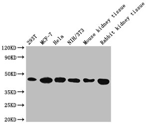
 IP (Immunoprecipitation)
(Immunoprecipitating GAPDH in Hela whole cell lysate Lane 1: Mouse control IgG instead of AAA27043 in Hela whole cell lysate. Lane 2: AAA27043 (5ul) + Hela whole cell lysate (500ug) Lane 3: Hela whole cell lysate (10ug) For western blotting, the blot was detected with AAA27043 at 1:5000, and a HRPconjugated Protein G antibody was used as the secondary antibody at 1:2000)
IP (Immunoprecipitation)
(Immunoprecipitating GAPDH in Hela whole cell lysate Lane 1: Mouse control IgG instead of AAA27043 in Hela whole cell lysate. Lane 2: AAA27043 (5ul) + Hela whole cell lysate (500ug) Lane 3: Hela whole cell lysate (10ug) For western blotting, the blot was detected with AAA27043 at 1:5000, and a HRPconjugated Protein G antibody was used as the secondary antibody at 1:2000)
GAPDH, Monoclonal Antibody (Cat# AAA27043)
Full Name
GAPDH Monoclonal Antibody
Reactivity
Human, Rat, Rabbit, Mouse
Applications
Western Blot, Immunohistochemistry, Immunoprecipitation, Immunofluorescence
Purity
>95%, Protein G purified
Pricing


 Application Data
(Published customer image: Increased accumulation of repair-associated macrophages surrounding collaterals in ischemic hind limbs is PAR2-dependent. (A) Stainings of CD206-positive macrophages (green) and SMA-positive vessels (red) in non-ischemic (control) and ischemic (ligated) hind limbs of WT, PAR1-/- and PAR2-/- mice are shown. Nuclei were visualized with DAPI (blue). Arrows indicate single macrophages in the non-ischemic adductor. Quantification of the average number of repair-associated macrophages per vessel is indicated on the right. (B) Correlation between the number of CD206-positive macrophages in the ischemic tissues and the expression of CD11b and (C) CD115 on monocytes. ** p)
Application Data
(Published customer image: Increased accumulation of repair-associated macrophages surrounding collaterals in ischemic hind limbs is PAR2-dependent. (A) Stainings of CD206-positive macrophages (green) and SMA-positive vessels (red) in non-ischemic (control) and ischemic (ligated) hind limbs of WT, PAR1-/- and PAR2-/- mice are shown. Nuclei were visualized with DAPI (blue). Arrows indicate single macrophages in the non-ischemic adductor. Quantification of the average number of repair-associated macrophages per vessel is indicated on the right. (B) Correlation between the number of CD206-positive macrophages in the ischemic tissues and the expression of CD11b and (C) CD115 on monocytes. ** p)
CD206, Monoclonal Antibody (Cat# AAA12118)
Full Name
RAT ANTI MOUSE CD206:Biotin
Gene Names
Mrc1; MR; CD206; AW259686
Applications
Flow Cytometry
Pricing


 FCM (Flow Cytometry)
(Overlay Peak curve showing Hela cells stained with AAA28066 (red line) at 1:200. The cells were fixed in 4% formaldehyde and permeated by 0.2% TritonX-100. Then 10% normal goat serum was Incubated to block non-specific protein-protein interactions followed by the antibody (1?g/1*106cells) for 1 h at 4 degree C. The secondary antibody used was FITC-conjugated Goat Anti-Mouse IgG(H+L) at 1/100 dilution for 30min at 4 degree C. Isotype control antibody (green line) was mouse IgG2b (1?g/1*106cells) used under the same conditions. Acquisition of >10,000 events was performed.)
FCM (Flow Cytometry)
(Overlay Peak curve showing Hela cells stained with AAA28066 (red line) at 1:200. The cells were fixed in 4% formaldehyde and permeated by 0.2% TritonX-100. Then 10% normal goat serum was Incubated to block non-specific protein-protein interactions followed by the antibody (1?g/1*106cells) for 1 h at 4 degree C. The secondary antibody used was FITC-conjugated Goat Anti-Mouse IgG(H+L) at 1/100 dilution for 30min at 4 degree C. Isotype control antibody (green line) was mouse IgG2b (1?g/1*106cells) used under the same conditions. Acquisition of >10,000 events was performed.)
ACTB, Monoclonal Antibody (Cat# AAA28066)
Full Name
ACTB Monoclonal Antibody
Reactivity
Human, Mouse, Rat, Rabbit
Applications
Western Blot, Immunohistochemistry, Immunofluorescence, Flow Cytometry, Immunoprecipitation
Purity
>95%, Protein A purified
Pricing


 Application Data
(Published customer image: Increased accumulation of repair-associated macrophages surrounding collaterals in ischemic hind limbs is PAR2-dependent. (A) Stainings of CD206-positive macrophages (green) and SMA-positive vessels (red) in non-ischemic (control) and ischemic (ligated) hind limbs of WT, PAR1-/- and PAR2-/- mice are shown. Nuclei were visualized with DAPI (blue). Arrows indicate single macrophages in the non-ischemic adductor. Quantification of the average number of repair-associated macrophages per vessel is indicated on the right. (B) Correlation between the number of CD206-positive macrophages in the ischemic tissues and the expression of CD11b and (C) CD115 on monocytes. ** p)
Application Data
(Published customer image: Increased accumulation of repair-associated macrophages surrounding collaterals in ischemic hind limbs is PAR2-dependent. (A) Stainings of CD206-positive macrophages (green) and SMA-positive vessels (red) in non-ischemic (control) and ischemic (ligated) hind limbs of WT, PAR1-/- and PAR2-/- mice are shown. Nuclei were visualized with DAPI (blue). Arrows indicate single macrophages in the non-ischemic adductor. Quantification of the average number of repair-associated macrophages per vessel is indicated on the right. (B) Correlation between the number of CD206-positive macrophages in the ischemic tissues and the expression of CD11b and (C) CD115 on monocytes. ** p)
CD206, Monoclonal Antibody (Cat# AAA12124)
Full Name
RAT ANTI MOUSE CD206:RPE
Gene Names
Mrc1; MR; CD206; AW259686
Applications
Flow Cytometry
Pricing


 Application Data
(Published customer image Infiltration of GFP+ BM-cells in infarct and peri-infarct regions. (A-B) Dot plots of viable macrophages/granulocytes (CD11b+CD45high, top right quadrants) and microglia (CD11b+CD45dim, bottom right quadrants) in cortex from BM-chimeric unmanipulated mice and mice exposed to pMCAO. (C) Bar graph showing mean numbers of CD11b+CD45dim microglia and CD11b+CD45high macrophages/granulocytes in BM-chimeric mice 24 hours after pMCAO, subdivided based on expression of GFP (n = 5). Approximately 92% of of the CD45high population were GFP+. (D) Estimation and comparison of mean numbers of CD11b+CD45dim microglia in non-chimeric (n = 10) versus BM-chimeric mice (n = 5) 24 hours after of pMCAO shows significantly fewer CD11b+CD45dim microglial cells in irradiated mice. (E) Overview, showing distribution of infiltrating GFP+ BM-derived cells into infarct (IF) and peri-infarct (P-IF) regions 24 hours after pMCAO. (E-G) By 24 hours, GFP+ single cells (F) and vessel-associated aggregates of GFP+ cells (arrows in G) were observed in infarct and peri-infarct regions. Some of the vessel-associated cells were round, leukocyte-like cells (arrows) while others were elongated cells lining the vasculature (arrow heads in G and in insert). (H) Bar graph showing mean numbers of single GFP+ cells and vessel-associated aggregates of GFP+ cells in ipsi- and contralateral cortex 24 hours after surgery (n = 10). (I-P) Immunohistochemical staining of CD45.1 (I, K), CD45.2 (J, L), IgG2a (M, O) and CD45 (N, P) in ischemic tissue in BM-chimeric (I, J, M, N) and non-chimeric mice (K, L, O, P) 24 hours after pMCAO. N.D, none detected. Scale bars: 200 um (A), 10 um (B, C). 50 um (I-P) *P < 0.05, **P < 0.01, and ***P < 0.001.From: Clausen BH, Lambertsen KL, Babcock AA, Holm TH, Dagnaes-Hansen F, Finsen B. Interleukin-1beta and tumor necrosis factor-alpha are expressed by different subsets of microglia and macrophages after ischemic stroke in mice. J Neuroinflammation. 2008 Oct 23;5:46.)
Application Data
(Published customer image Infiltration of GFP+ BM-cells in infarct and peri-infarct regions. (A-B) Dot plots of viable macrophages/granulocytes (CD11b+CD45high, top right quadrants) and microglia (CD11b+CD45dim, bottom right quadrants) in cortex from BM-chimeric unmanipulated mice and mice exposed to pMCAO. (C) Bar graph showing mean numbers of CD11b+CD45dim microglia and CD11b+CD45high macrophages/granulocytes in BM-chimeric mice 24 hours after pMCAO, subdivided based on expression of GFP (n = 5). Approximately 92% of of the CD45high population were GFP+. (D) Estimation and comparison of mean numbers of CD11b+CD45dim microglia in non-chimeric (n = 10) versus BM-chimeric mice (n = 5) 24 hours after of pMCAO shows significantly fewer CD11b+CD45dim microglial cells in irradiated mice. (E) Overview, showing distribution of infiltrating GFP+ BM-derived cells into infarct (IF) and peri-infarct (P-IF) regions 24 hours after pMCAO. (E-G) By 24 hours, GFP+ single cells (F) and vessel-associated aggregates of GFP+ cells (arrows in G) were observed in infarct and peri-infarct regions. Some of the vessel-associated cells were round, leukocyte-like cells (arrows) while others were elongated cells lining the vasculature (arrow heads in G and in insert). (H) Bar graph showing mean numbers of single GFP+ cells and vessel-associated aggregates of GFP+ cells in ipsi- and contralateral cortex 24 hours after surgery (n = 10). (I-P) Immunohistochemical staining of CD45.1 (I, K), CD45.2 (J, L), IgG2a (M, O) and CD45 (N, P) in ischemic tissue in BM-chimeric (I, J, M, N) and non-chimeric mice (K, L, O, P) 24 hours after pMCAO. N.D, none detected. Scale bars: 200 um (A), 10 um (B, C). 50 um (I-P) *P < 0.05, **P < 0.01, and ***P < 0.001.From: Clausen BH, Lambertsen KL, Babcock AA, Holm TH, Dagnaes-Hansen F, Finsen B. Interleukin-1beta and tumor necrosis factor-alpha are expressed by different subsets of microglia and macrophages after ischemic stroke in mice. J Neuroinflammation. 2008 Oct 23;5:46.)
CD11b, Monoclonal Antibody (Cat# AAA12186)
Full Name
RAT ANTI MOUSE CD11b:RPE
Gene Names
Itgam; CR3; CR3A; MAC1; Cd11b; Ly-40; Mac-1; Mac-1a; CD11b/CD18; F730045J24Rik
Applications
Flow Cytometry
Pricing
 Application Data
HIV-1 p24, Monoclonal Antibody (Cat# AAA13685)
Full Name
Anti-HIV-1 p24
Reactivity
Reacts with P24(HIC-1) proteins
Applications
Western Blot
Purity
Ion exchange column
Pricing


 Application Data
(Published clone specific image: Flow cytometric analysis of AM from NO2-exposed and control rats. Rats were exposed to NO2 for the indicated times and BAL cells were stained with antibodies to ED7, ED9, RM-4, and OX-6. To overcome autofluorescence signals, primary antibodies were detected using a biotin-PE/streptavidin-anti-streptavidin enhancing system and labeling of AM was analyzed by flow cytometry following gating by help of forward and sideward scatter properties. Shown are representative results of at least six animals per group.From: Garn H, Siese A, Stumpf S, Wensing A, Renz H, Gemsa D. Phenotypical and functional characterization of alveolar macrophage subpopulations in the lungs of NO2-exposed rats. Respir Res. 2006 Jan 6;7:4.)
Application Data
(Published clone specific image: Flow cytometric analysis of AM from NO2-exposed and control rats. Rats were exposed to NO2 for the indicated times and BAL cells were stained with antibodies to ED7, ED9, RM-4, and OX-6. To overcome autofluorescence signals, primary antibodies were detected using a biotin-PE/streptavidin-anti-streptavidin enhancing system and labeling of AM was analyzed by flow cytometry following gating by help of forward and sideward scatter properties. Shown are representative results of at least six animals per group.From: Garn H, Siese A, Stumpf S, Wensing A, Renz H, Gemsa D. Phenotypical and functional characterization of alveolar macrophage subpopulations in the lungs of NO2-exposed rats. Respir Res. 2006 Jan 6;7:4.)
CD172a, Monoclonal Antibody (Cat# AAA12001)
Full Name
MOUSE ANTI RAT CD172a
Gene Names
Sirpa; Bit; Ptpns1; SHPS-1
Applications
Immunohistochemistry, Flow Cytometry, Immunoprecipitation, Western Blot
Pricing


 Application Data
(Published customer image: Increased accumulation of repair-associated macrophages surrounding collaterals in ischemic hind limbs is PAR2-dependent. (A) Stainings of CD206-positive macrophages (green) and SMA-positive vessels (red) in non-ischemic (control) and ischemic (ligated) hind limbs of WT, PAR1-/- and PAR2-/- mice are shown. Nuclei were visualized with DAPI (blue). Arrows indicate single macrophages in the non-ischemic adductor. Quantification of the average number of repair-associated macrophages per vessel is indicated on the right. (B) Correlation between the number of CD206-positive macrophages in the ischemic tissues and the expression of CD11b and (C) CD115 on monocytes. ** p)
Application Data
(Published customer image: Increased accumulation of repair-associated macrophages surrounding collaterals in ischemic hind limbs is PAR2-dependent. (A) Stainings of CD206-positive macrophages (green) and SMA-positive vessels (red) in non-ischemic (control) and ischemic (ligated) hind limbs of WT, PAR1-/- and PAR2-/- mice are shown. Nuclei were visualized with DAPI (blue). Arrows indicate single macrophages in the non-ischemic adductor. Quantification of the average number of repair-associated macrophages per vessel is indicated on the right. (B) Correlation between the number of CD206-positive macrophages in the ischemic tissues and the expression of CD11b and (C) CD115 on monocytes. ** p)
CD206, Monoclonal Antibody (Cat# AAA12119)
Full Name
RAT ANTI MOUSE CD206:FITC
Gene Names
Mrc1; MR; CD206; AW259686
Applications
Flow Cytometry
Pricing


 Application Data
(Published customer image: Increased accumulation of repair-associated macrophages surrounding collaterals in ischemic hind limbs is PAR2-dependent. (A) Stainings of CD206-positive macrophages (green) and SMA-positive vessels (red) in non-ischemic (control) and ischemic (ligated) hind limbs of WT, PAR1-/- and PAR2-/- mice are shown. Nuclei were visualized with DAPI (blue). Arrows indicate single macrophages in the non-ischemic adductor. Quantification of the average number of repair-associated macrophages per vessel is indicated on the right. (B) Correlation between the number of CD206-positive macrophages in the ischemic tissues and the expression of CD11b and (C) CD115 on monocytes. ** p)
Application Data
(Published customer image: Increased accumulation of repair-associated macrophages surrounding collaterals in ischemic hind limbs is PAR2-dependent. (A) Stainings of CD206-positive macrophages (green) and SMA-positive vessels (red) in non-ischemic (control) and ischemic (ligated) hind limbs of WT, PAR1-/- and PAR2-/- mice are shown. Nuclei were visualized with DAPI (blue). Arrows indicate single macrophages in the non-ischemic adductor. Quantification of the average number of repair-associated macrophages per vessel is indicated on the right. (B) Correlation between the number of CD206-positive macrophages in the ischemic tissues and the expression of CD11b and (C) CD115 on monocytes. ** p)
CD206, Monoclonal Antibody (Cat# AAA12121)
Full Name
RAT ANTI MOUSE CD206:FITC
Gene Names
Mrc1; MR; CD206; AW259686
Applications
Flow Cytometry
Pricing

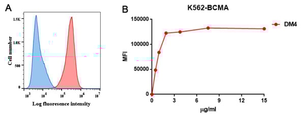


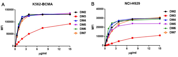
 IP (Immunoprecipitation)
(Figure 6. Immunoprecipitation analysis. Cellular overexpression lysates (made from HEK293F cells transfected with DYKDDDDK tagged human BCMA full length gene) were pre-incubated with 6 different rabbit DimAb clones and negative control IgG. The immunocomplexes were further pulled down by protein A beads, fractionated, and blotted with mouse anti-DYKDDDDK monoclonal antibody.)
IP (Immunoprecipitation)
(Figure 6. Immunoprecipitation analysis. Cellular overexpression lysates (made from HEK293F cells transfected with DYKDDDDK tagged human BCMA full length gene) were pre-incubated with 6 different rabbit DimAb clones and negative control IgG. The immunocomplexes were further pulled down by protein A beads, fractionated, and blotted with mouse anti-DYKDDDDK monoclonal antibody.)
BCMA, Monoclonal Antibody (Cat# AAA11703)
Full Name
Anti-BCMA antibody (DM4), Rabbit mAb
Reactivity
Human
Applications
Flow Cytometry, Immunoprecipitation, Immunofluorescence
Purity
Purified from cell culture supernatant by affinity chromatography
Pricing


 Application Data
(Published clone specific image: Flow cytometric analysis of AM from NO2-exposed and control rats. Rats were exposed to NO2 for the indicated times and BAL cells were stained with antibodies to ED7, ED9, RM-4, and OX-6. To overcome autofluorescence signals, primary antibodies were detected using a biotin-PE/streptavidin-anti-streptavidin enhancing system and labeling of AM was analyzed by flow cytometry following gating by help of forward and sideward scatter properties. Shown are representative results of at least six animals per group.From: Garn H, Siese A, Stumpf S, Wensing A, Renz H, Gemsa D. Phenotypical and functional characterization of alveolar macrophage subpopulations in the lungs of NO2-exposed rats. Respir Res. 2006 Jan 6;7:4.)
Application Data
(Published clone specific image: Flow cytometric analysis of AM from NO2-exposed and control rats. Rats were exposed to NO2 for the indicated times and BAL cells were stained with antibodies to ED7, ED9, RM-4, and OX-6. To overcome autofluorescence signals, primary antibodies were detected using a biotin-PE/streptavidin-anti-streptavidin enhancing system and labeling of AM was analyzed by flow cytometry following gating by help of forward and sideward scatter properties. Shown are representative results of at least six animals per group.From: Garn H, Siese A, Stumpf S, Wensing A, Renz H, Gemsa D. Phenotypical and functional characterization of alveolar macrophage subpopulations in the lungs of NO2-exposed rats. Respir Res. 2006 Jan 6;7:4.)
CD172a, Monoclonal Antibody (Cat# AAA11888)
Full Name
MOUSE ANTI RAT CD172a:FITC
Gene Names
Sirpa; Bit; Ptpns1; SHPS-1
Applications
Flow Cytometry
Pricing
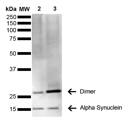

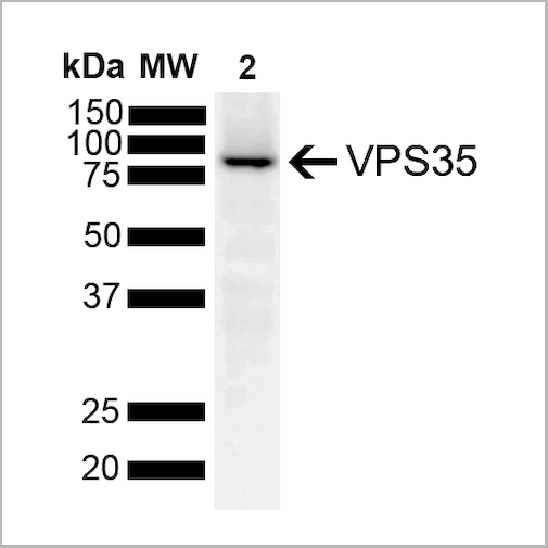
 WB (Western Blot)
(Western Blot analysis of Mouse, Rat Brain showing detection of 14 kDa Alpha Synuclein protein using Mouse Anti-Alpha Synuclein Monoclonal Antibody, Clone 3C11. Lane 1: Molecular Weight Ladder (MW). Lane 2: Mouse brain cell lysate. Lane 3: Rat brain cell lysate. Load: 15 ug. Block: 5% Skim Milk in 1X TBST. Primary Antibody: Mouse Anti-Alpha Synuclein Monoclonal Antibody at 1:1000 for 2 hours at RT. Secondary Antibody: Goat Anti-Mouse HRP:IgG at 1:3000 for 1 hour at RT. Color Development: ECL solution (Super Signal West Pico) for 5 min in RT. Predicted/Observed Size: 14 kDa. Other Band(s): ~30 kDa (dimer).)
WB (Western Blot)
(Western Blot analysis of Mouse, Rat Brain showing detection of 14 kDa Alpha Synuclein protein using Mouse Anti-Alpha Synuclein Monoclonal Antibody, Clone 3C11. Lane 1: Molecular Weight Ladder (MW). Lane 2: Mouse brain cell lysate. Lane 3: Rat brain cell lysate. Load: 15 ug. Block: 5% Skim Milk in 1X TBST. Primary Antibody: Mouse Anti-Alpha Synuclein Monoclonal Antibody at 1:1000 for 2 hours at RT. Secondary Antibody: Goat Anti-Mouse HRP:IgG at 1:3000 for 1 hour at RT. Color Development: ECL solution (Super Signal West Pico) for 5 min in RT. Predicted/Observed Size: 14 kDa. Other Band(s): ~30 kDa (dimer).)
Alpha Synuclein, Monoclonal Antibody (Cat# AAA17810)
Full Name
Alpha Synuclein Antibody, Clone 3C11
Gene Names
SNCA; PD1; NACP; PARK1; PARK4
Reactivity
Human; Mouse; Rat
Applications
WB, DB, ICC, IF, EIA
Purity
Protein G Purified
Pricing


 IP (Immunoprecipitation)
(Figure 6. Immunoprecipitation analysis. Cellular overexpression lysates (made from HEK293F cells transfected with DYKDDDDK tagged human BCMA full length gene) were pre-incubated with 6 different rabbit DimAb clones and negative control IgG. The immunocomplexes were further pulled down by protein A beads, fractionated, and blotted with mouse anti-DYKDDDDK monoclonal antibody.)
IP (Immunoprecipitation)
(Figure 6. Immunoprecipitation analysis. Cellular overexpression lysates (made from HEK293F cells transfected with DYKDDDDK tagged human BCMA full length gene) were pre-incubated with 6 different rabbit DimAb clones and negative control IgG. The immunocomplexes were further pulled down by protein A beads, fractionated, and blotted with mouse anti-DYKDDDDK monoclonal antibody.)
BCMA, Monoclonal Antibody (Cat# AAA11704)
Full Name
Anti-BCMA antibody (DM6), Rabbit mAb
Reactivity
Human
Applications
Flow Cytometry, Immunofluorescence
Purity
Purified from cell culture supernatant by affinity chromatography
Pricing


 WB (Western Blot)
(FG Pancreatic Carcinoma Cell Lines stably expressing vector along (FG-V) the b3 integrin subunit (FG-b3) or a b3 truncation mutant (FG-759x). Src Mab (AAA28639) was diluted 1:500 in 1% BSA/TBST and incubated Overnight at 4 degree C. After washing 3x 5 min. with TBST the blots were incubated with 1:5000 Goat anti-mouse or Goat anti-rabbit secondary antibody for 1 hr at Room temperature. The blots were again washed 3x 5 min. with TBST and developed using ECL reagent.Data and protocol kindly provided by Dr. Weis of Cheresh Lab, UCSD.)
WB (Western Blot)
(FG Pancreatic Carcinoma Cell Lines stably expressing vector along (FG-V) the b3 integrin subunit (FG-b3) or a b3 truncation mutant (FG-759x). Src Mab (AAA28639) was diluted 1:500 in 1% BSA/TBST and incubated Overnight at 4 degree C. After washing 3x 5 min. with TBST the blots were incubated with 1:5000 Goat anti-mouse or Goat anti-rabbit secondary antibody for 1 hr at Room temperature. The blots were again washed 3x 5 min. with TBST and developed using ECL reagent.Data and protocol kindly provided by Dr. Weis of Cheresh Lab, UCSD.)
SRC, Monoclonal Antibody (Cat# AAA28639)
Full Name
SRC Antibody
Gene Names
SRC; ASV; SRC1; c-SRC; p60-Src
Reactivity
Human, mouse
Applications
WB, EIA, IF
Purity
This antibody is purified through a protein G column, followed by dialysis against PBS.
Pricing
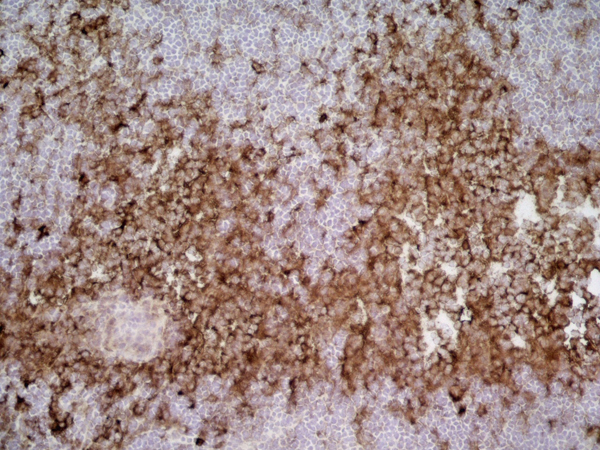
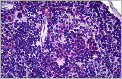 IHC (Immunohistochemistry)
(Anti-TCR Gamma/Delta antibody IHC of mouse spleen. Immunohistochemistry of formalin-fixed, paraffin-embedded tissue after heat-induced antigen retrieval. Antibody dilution 1:50.)
IHC (Immunohistochemistry)
(Anti-TCR Gamma/Delta antibody IHC of mouse spleen. Immunohistochemistry of formalin-fixed, paraffin-embedded tissue after heat-induced antigen retrieval. Antibody dilution 1:50.)
TCR Gamma+TCR Delta, Monoclonal Antibody (Cat# AAA12360)
Full Name
Armenian Hamster Monoclonal [clone GL-3] (IgG) to Mouse TCR Gamma+TCR Delta
Reactivity
Mouse
Applications
Immunohistochemistry, Flow Cytometry
Purity
Protein G Purified
Pricing
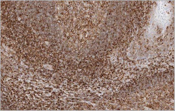 Application Data
(Figure 1. Immunostaining of human tonsil tissue in paraffin section with HC10 (dilution 1:20.000).)
Application Data
(Figure 1. Immunostaining of human tonsil tissue in paraffin section with HC10 (dilution 1:20.000).)
HLA Class I Heavy Chain, Monoclonal Antibody (Cat# AAA14567)
Full Name
Mouse anti Human HLA Class I Heavy Chain (Restricted expression)
Reactivity
Human
Applications
Flow Cytometry, Immunocytochemistry, Immunohistochemistry, Immunoprecipitation, Western Blot
Pricing
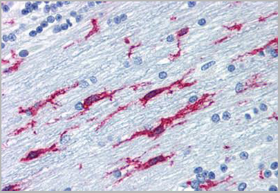 IHC (Immunohistochemistry)
(Anti-ITGB2 / CD18 antibody IHC of human brain, cerebellum. Immunohistochemistry of formalin-fixed, paraffin-embedded tissue after heat-induced antigen retrieval. Antibody concentration 10 ug/ml.)
IHC (Immunohistochemistry)
(Anti-ITGB2 / CD18 antibody IHC of human brain, cerebellum. Immunohistochemistry of formalin-fixed, paraffin-embedded tissue after heat-induced antigen retrieval. Antibody concentration 10 ug/ml.)
ITGB2 / MAC-1 / CD18, Monoclonal Antibody (Cat# AAA12341)
Full Name
Mouse Monoclonal [clone MEM-48] (IgG1) to Human ITGB2 / MAC-1 / CD18
Gene Names
ITGB2; LAD; CD18; MF17; MFI7; LCAMB; LFA-1; MAC-1
Reactivity
Human
Applications
Immunohistochemistry, Western Blot, Immunoprecipitation, Flow Cytometry, Functional Assay
Purity
Purified by Protein A affinity chromatography.
>95% by SDS-PAGE.
>95% by SDS-PAGE.
Pricing
 WB (Western Blot)
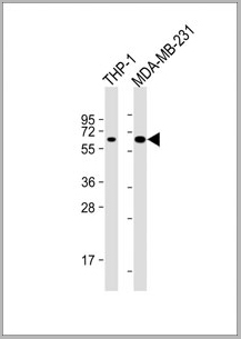
(All lanes : Anti-MB21D1 Antibody at 1:4000 dilutionLane 1: THP-1 whole cell lysateLane 2: MDA-MB-231 whole cell lysateLysates/proteins at 20 ug per lane. SecondaryGoat Anti-mouse IgG, (H+L), Peroxidase conjugated at 1/10000 dilution. Predicted band size : 59kDaBlocking/Dilution buffer: 5% NFDM/TBST.)
WB (Western Blot)
(All lanes : Anti-MB21D1 Antibody at 1:4000 dilutionLane 1: THP-1 whole cell lysateLane 2: MDA-MB-231 whole cell lysateLysates/proteins at 20 ug per lane. SecondaryGoat Anti-mouse IgG, (H+L), Peroxidase conjugated at 1/10000 dilution. Predicted band size : 59kDaBlocking/Dilution buffer: 5% NFDM/TBST.)
MB21D1, Monoclonal Antibody (Cat# AAA28800)
Full Name
MB21D1 Antibody
Gene Names
MB21D1; cGAS; h-cGAS; C6orf150
Reactivity
Human
Applications
Western Blot
Pricing
 IHC (Immunohistochemistry)
(Anti-GPNMB antibody IHC of human skin. Immunohistochemistry of formalin-fixed, paraffin-embedded tissue after heat-induced antigen retrieval. Antibody concentration 5 ug/ml.)
IHC (Immunohistochemistry)
(Anti-GPNMB antibody IHC of human skin. Immunohistochemistry of formalin-fixed, paraffin-embedded tissue after heat-induced antigen retrieval. Antibody concentration 5 ug/ml.)
GPNMB, Monoclonal Antibody (Cat# AAA12347)
Full Name
Anti-GPNMB/Osteoactivin Antibody; GPNMB / Osteoactivin
Gene Names
GPNMB; NMB; HGFIN
Reactivity
Human
Applications
Immunohistochemistry, Western Blot
Purity
Purified from ascites by Protein A
Pricing


 Application Data
(Staining of human peripheral blood granulocytes with CD18: Alexa Fluor 647)
Application Data
(Staining of human peripheral blood granulocytes with CD18: Alexa Fluor 647)
CD18, Monoclonal Antibody (Cat# AAA12051)
Full Name
RAT ANTI HUMAN CD18:RPE
Gene Names
ITGB2; LAD; CD18; MF17; MFI7; LCAMB; LFA-1; MAC-1
Applications
Flow Cytometry
Pricing


 Application Data
(Staining of human peripheral blood granulocytes with CD18: Alexa Fluor 647 (AAA11986A647))
Application Data
(Staining of human peripheral blood granulocytes with CD18: Alexa Fluor 647 (AAA11986A647))
CD18, Monoclonal Antibody (Cat# AAA11986)
Full Name
RAT ANTI HUMAN CD18
Gene Names
ITGB2; LAD; CD18; MF17; MFI7; LCAMB; LFA-1; MAC-1
Reactivity
Dog, Guinea Pig
Applications
Immunohistochemistry, Flow Cytometry, Immunoprecipitation
Pricing
 IHC (Immunohistochemistry)
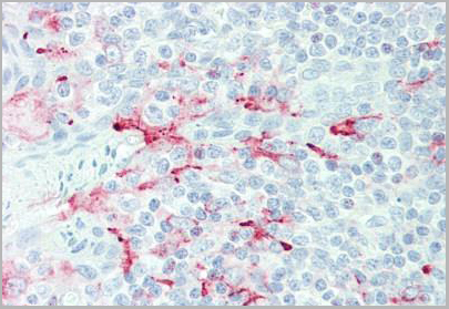
(Anti-GDF15 antibody IHC of human placenta. Immunohistochemistry of formalin-fixed, paraffin-embedded tissue after heat-induced antigen retrieval. Antibody dilution 10 ug/ml.)
IHC (Immunohistochemistry)
(Anti-GDF15 antibody IHC of human placenta. Immunohistochemistry of formalin-fixed, paraffin-embedded tissue after heat-induced antigen retrieval. Antibody dilution 10 ug/ml.)
GDF15, Monoclonal Antibody (Cat# AAA12357)
Full Name
Rat Monoclonal [clone 6D12.H10.E4] (IgG) to Human GDF15
Gene Names
GDF15; PDF; MIC1; PLAB; MIC-1; NAG-1; PTGFB; GDF-15
Reactivity
Chimpanzee, Macaque
Applications
Immunohistochemistry, Immunohistochemistry, Western Blot
Purity
Protein G Purified chromatography
Pricing
 IHC (Immunohistochemistry)
(Anti-ACTH antibody IHC of human pituitary. Immunohistochemistry of formalin-fixed, paraffin-embedded tissue after heat-induced antigen retrieval. Antibody concentration 5 ug/ml.)
IHC (Immunohistochemistry)
(Anti-ACTH antibody IHC of human pituitary. Immunohistochemistry of formalin-fixed, paraffin-embedded tissue after heat-induced antigen retrieval. Antibody concentration 5 ug/ml.)
ACTH, Monoclonal Antibody (Cat# AAA12344)
Full Name
Mouse Monoclonal [clone 57] (IgG1) to Human ACTH
Reactivity
Human, Rat
Applications
Immunohistochemistry
Purity
Protein A Purified
Pricing
 IHC (Immunohistochemistry)
(Anti-c-Met antibody IHC of human uterus. Immunohistochemistry of formalin-fixed, paraffin-embedded tissue after heat-induced antigen retrieval. Antibody concentration 10 ug/ml.)
IHC (Immunohistochemistry)
(Anti-c-Met antibody IHC of human uterus. Immunohistochemistry of formalin-fixed, paraffin-embedded tissue after heat-induced antigen retrieval. Antibody concentration 10 ug/ml.)
c-Met, Monoclonal Antibody (Cat# AAA12343)
Full Name
Mouse Monoclonal (IgG2a) to Human c-Met
Gene Names
MET; HGFR; AUTS9; RCCP2; c-Met
Reactivity
Human
Applications
Immunohistochemistry
Purity
Purified
Pricing


 IP (Immunoprecipitation)
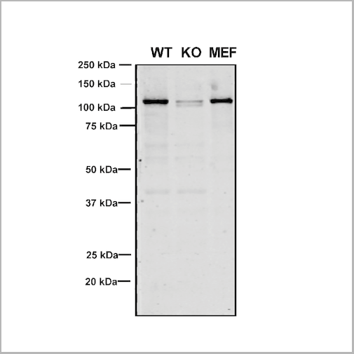
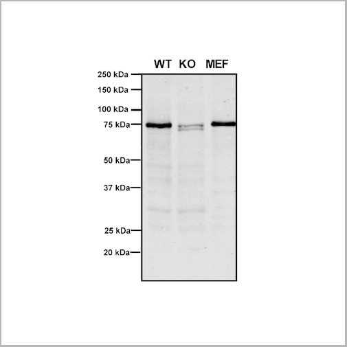
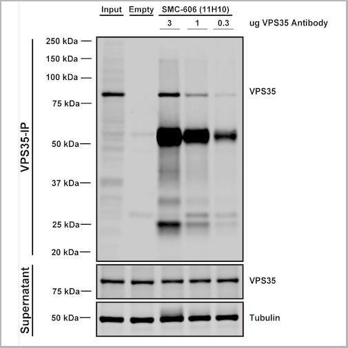
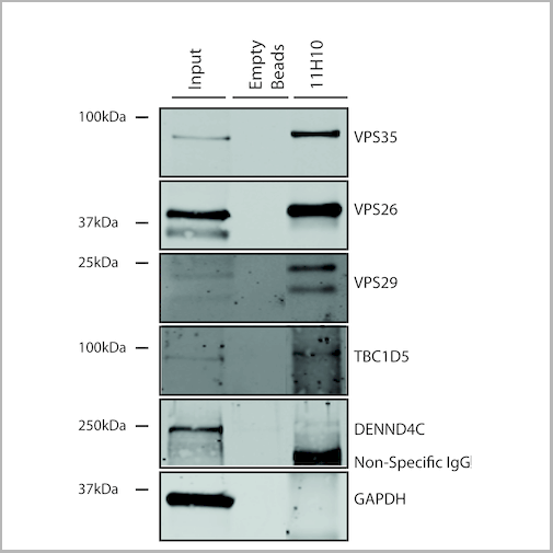
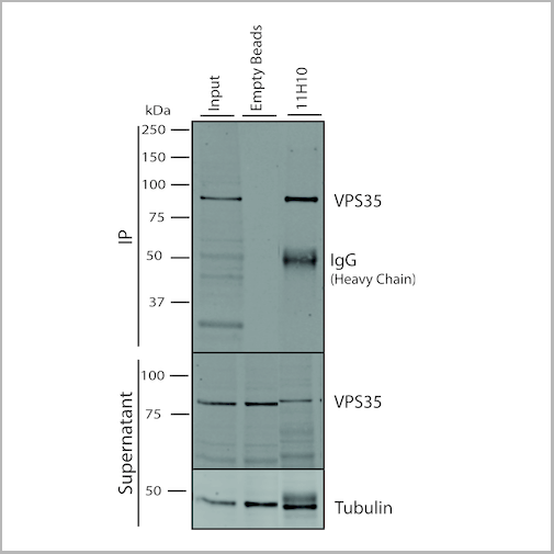
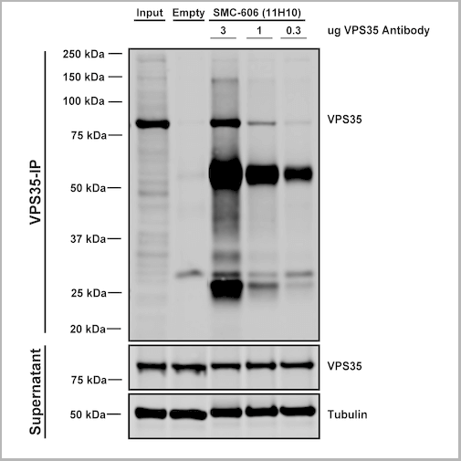
(Immunoprecipitation analysis using Mouse Anti-VPS35 Monoclonal Antibody, Clone 11H10. Tissue: embryonic fibroblast. Species: Mouse. Primary Antibody: Mouse Anti-VPS35 Monoclonal Antibody. Three amounts of (3, 1 and 0.3 ug) were non-covalently coupled to 10uL of A/G sepharose beads for 1 hour at 4 degree C and next incubated with 250ug of MEF lysate for 2 hours at 4 degree C.)
IP (Immunoprecipitation)
(Immunoprecipitation analysis using Mouse Anti-VPS35 Monoclonal Antibody, Clone 11H10. Tissue: embryonic fibroblast. Species: Mouse. Primary Antibody: Mouse Anti-VPS35 Monoclonal Antibody. Three amounts of (3, 1 and 0.3 ug) were non-covalently coupled to 10uL of A/G sepharose beads for 1 hour at 4 degree C and next incubated with 250ug of MEF lysate for 2 hours at 4 degree C.)
VPS35, Monoclonal Antibody (Cat# AAA27707)
Full Name
VPS35 Antibody, Clone 11H10: PerCP
Gene Names
VPS35; MEM3; PARK17
Reactivity
Human, Mouse, Rat
Applications
Western Blot, Immunoprecipitation
Purity
Protein G Purified
Pricing


 IP (Immunoprecipitation)
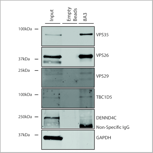
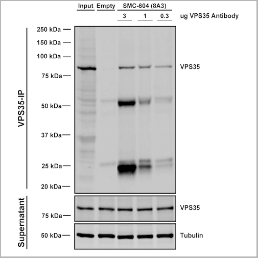
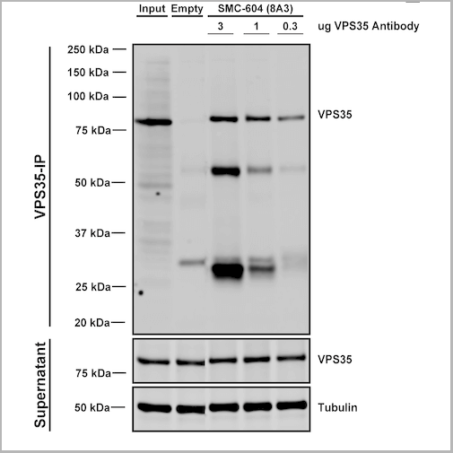
(Immunoprecipitation analysis using Mouse Anti-VPS35 Monoclonal Antibody, Clone 8A3. Tissue: embryonic fibroblast. Species: Mouse. Primary Antibody: Mouse Anti-VPS35 Monoclonal Antibody. Three amounts of (3, 1 and 0.3 ug) were non-covalently coupled to 10uL of A/G sepharose beads for 1 hour at 4 degree C and next incubated with 250ug of MEF lysate for 2 hours at 4 degree C.)
IP (Immunoprecipitation)
(Immunoprecipitation analysis using Mouse Anti-VPS35 Monoclonal Antibody, Clone 8A3. Tissue: embryonic fibroblast. Species: Mouse. Primary Antibody: Mouse Anti-VPS35 Monoclonal Antibody. Three amounts of (3, 1 and 0.3 ug) were non-covalently coupled to 10uL of A/G sepharose beads for 1 hour at 4 degree C and next incubated with 250ug of MEF lysate for 2 hours at 4 degree C.)
VPS35, Monoclonal Antibody (Cat# AAA27689)
Full Name
VPS35 Antibody, Clone 8A3: PerCP
Gene Names
VPS35; MEM3; PARK17
Reactivity
Human, Mouse, Rat
Applications
Western Blot, Immunocytochemistry, Immunofluorescence, Immunoprecipitation
Purity
Protein G Purified
Pricing


 Application Data
(Staining of human peripheral blood granulocytes with CD18: Alexa Fluor 647)
Application Data
(Staining of human peripheral blood granulocytes with CD18: Alexa Fluor 647)
CD18, Monoclonal Antibody (Cat# AAA11881)
Full Name
RAT ANTI HUMAN CD18:FITC
Gene Names
ITGB2; LAD; CD18; MF17; MFI7; LCAMB; LFA-1; MAC-1
Applications
Flow Cytometry
Pricing


 Application Data
(Staining of human peripheral blood granulocytes with CD18: Alexa Fluor 647)
Application Data
(Staining of human peripheral blood granulocytes with CD18: Alexa Fluor 647)
CD18, Monoclonal Antibody (Cat# AAA11987)
Full Name
RAT ANTI HUMAN CD18
Gene Names
ITGB2; LAD; CD18; MF17; MFI7; LCAMB; LFA-1; MAC-1
Applications
Immunohistochemistry, Flow Cytometry, Immunoprecipitation
Pricing


 IP (Immunoprecipitation)
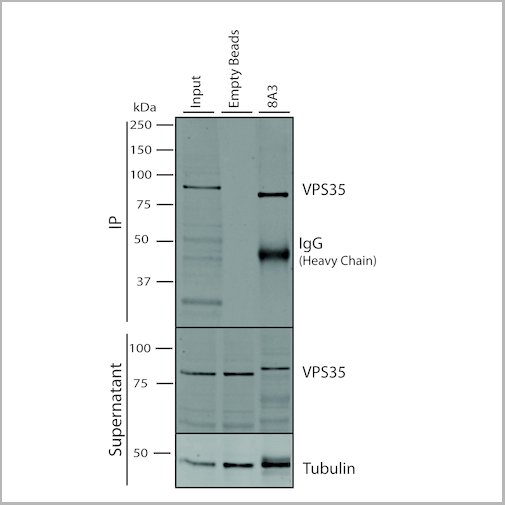
(Immunoprecipitation analysis using Mouse Anti-VPS35 Monoclonal Antibody, Clone 8A3. Tissue: embryonic fibroblast. Species: Mouse. Primary Antibody: Mouse Anti-VPS35 Monoclonal Antibody. Three amounts of (3, 1 and 0.3 ug) were non-covalently coupled to 10uL of A/G sepharose beads for 1 hour at 4 degree C and next incubated with 250ug of MEF lysate for 2 hours at 4 degree C.)
IP (Immunoprecipitation)
(Immunoprecipitation analysis using Mouse Anti-VPS35 Monoclonal Antibody, Clone 8A3. Tissue: embryonic fibroblast. Species: Mouse. Primary Antibody: Mouse Anti-VPS35 Monoclonal Antibody. Three amounts of (3, 1 and 0.3 ug) were non-covalently coupled to 10uL of A/G sepharose beads for 1 hour at 4 degree C and next incubated with 250ug of MEF lysate for 2 hours at 4 degree C.)
VPS35, Monoclonal Antibody (Cat# AAA27682)
Full Name
VPS35 Antibody, Clone 8A3
Gene Names
VPS35; MEM3; PARK17
Reactivity
Human, Mouse, Rat
Applications
Western Blot, Immunocytochemistry, Immunofluorescence, Immunoprecipitation
Purity
Protein G Purified
Pricing
 FCM (Flow Cytometry)
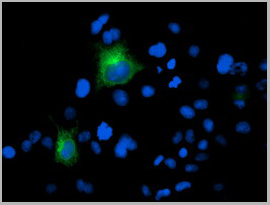
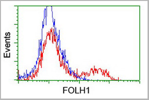
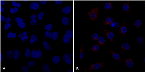
(HEK293T cells transfected with either overexpress plasmid (Red) or empty vector control plasmid (Blue) were immunostained by anti-FOLH1 antibody, and then analyzed by flow cytometry.)
FCM (Flow Cytometry)
(HEK293T cells transfected with either overexpress plasmid (Red) or empty vector control plasmid (Blue) were immunostained by anti-FOLH1 antibody, and then analyzed by flow cytometry.)
FOLH1 / PSMA, Monoclonal Antibody (Cat# AAA12376)
Full Name
Anti-FOLH1 / PSMA Antibody (clone 3H5) IHC-plus
Gene Names
FOLH1; PSM; FGCP; FOLH; GCP2; PSMA; mGCP; GCPII; NAALAD1; NAALAdase
Reactivity
Mouse, Human
Applications
Immunohistochemistry, Immunofluorescence, Western Blot, Flow Cytometry
Purity
Protein A/G purified
Pricing
HBsAg, Monoclonal Antibody (Cat# AAA14624)
Full Name
mAb anti-HBsAg, RM-1
Reactivity
Human hepatitis B virus. Others not tested.
Applications
ELISA
Purity
Protein G affinity purified
Pricing
 IP (Immunoprecipitation)
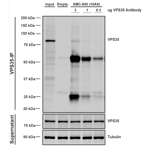
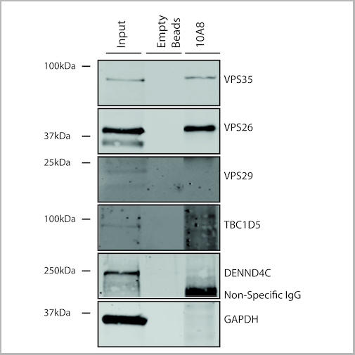
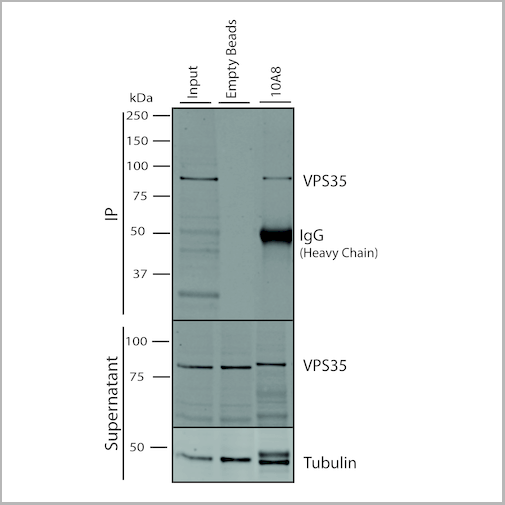
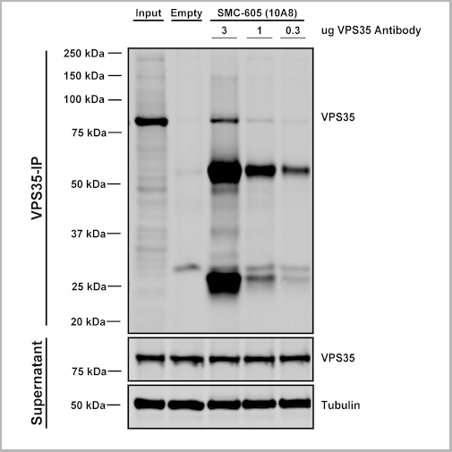
(Immunoprecipitation analysis using Mouse Anti-VPS35 Monoclonal Antibody, Clone 10A8. Tissue: embryonic fibroblast. Species: Mouse. Primary Antibody: Mouse Anti-VPS35 Monoclonal Antibody. Three amounts of (3, 1 and 0.3 ug) were non-covalently coupled to 10uL of A/G sepharose beads for 1 hour at 4 degree C and next incubated with 250ug of MEF lysate for 2 hours at 4 degree C.)
IP (Immunoprecipitation)
(Immunoprecipitation analysis using Mouse Anti-VPS35 Monoclonal Antibody, Clone 10A8. Tissue: embryonic fibroblast. Species: Mouse. Primary Antibody: Mouse Anti-VPS35 Monoclonal Antibody. Three amounts of (3, 1 and 0.3 ug) were non-covalently coupled to 10uL of A/G sepharose beads for 1 hour at 4 degree C and next incubated with 250ug of MEF lysate for 2 hours at 4 degree C.)
VPS35, Monoclonal Antibody (Cat# AAA27694)
Full Name
VPS35 Antibody, Clone 10A8: ATTO 594
Gene Names
VPS35; MEM3; PARK17
Reactivity
Human, Mouse, Rat
Applications
Western Blot, Immunocytochemistry, Immunofluorescence, Immunoprecipitation
Purity
Protein G Purified
Pricing
 WB (Western Blot)
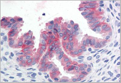
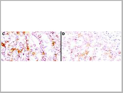
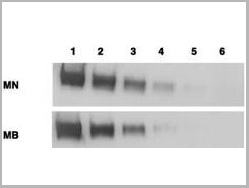
(Anti-Mesothelin Antibodies - Immunohistochemistry. Immunohistochemistry using anti-mesothelin antibodies to detect mesothelin in PEFF human tissue sections treated by antigen retrieval methods. Anti-mesothelin primary antibodies were used to label these sections as follows: C, MAb MB; and D, MAb MN. Reprinted with permission fromClin. Cancer Res. 11(16):5840-6.)
WB (Western Blot)
(Anti-Mesothelin Antibodies - Immunohistochemistry. Immunohistochemistry using anti-mesothelin antibodies to detect mesothelin in PEFF human tissue sections treated by antigen retrieval methods. Anti-mesothelin primary antibodies were used to label these sections as follows: C, MAb MB; and D, MAb MN. Reprinted with permission fromClin. Cancer Res. 11(16):5840-6.)
MSLN / Mesothelin, Monoclonal Antibody (Cat# AAA12374)
Full Name
Mouse Monoclonal [clone MN-1] (IgG) to Human MSLN / Mesothelin
Gene Names
MSLN; MPF; SMRP
Reactivity
Human
Applications
Immunohistochemistry, Western Blot
Purity
Protein A Purified
Pricing
 Application Data
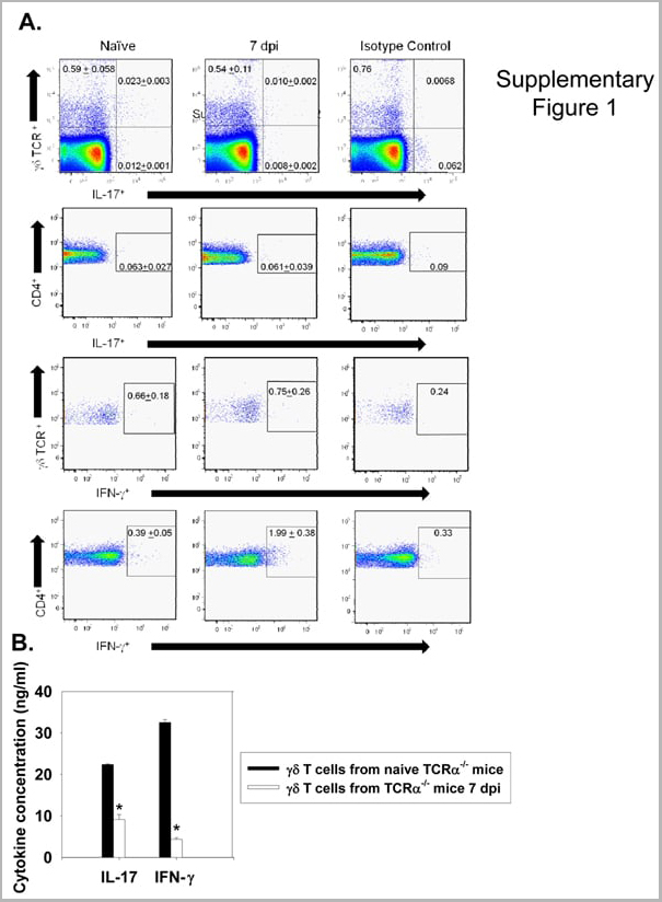
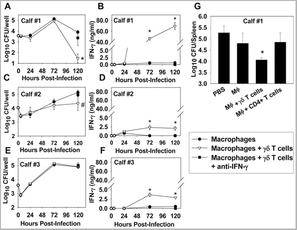
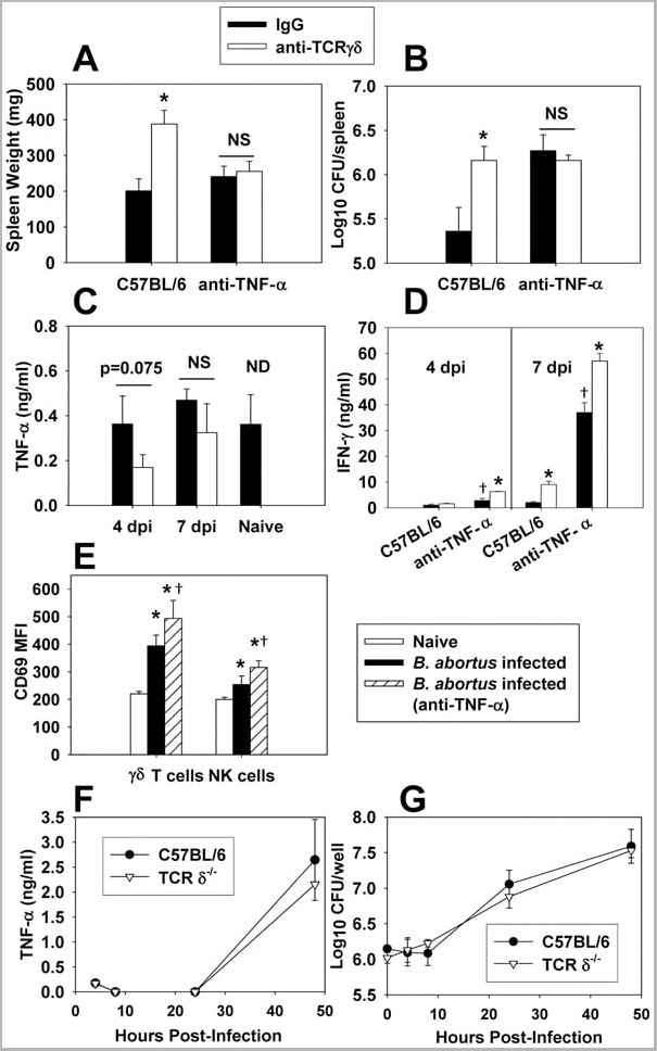
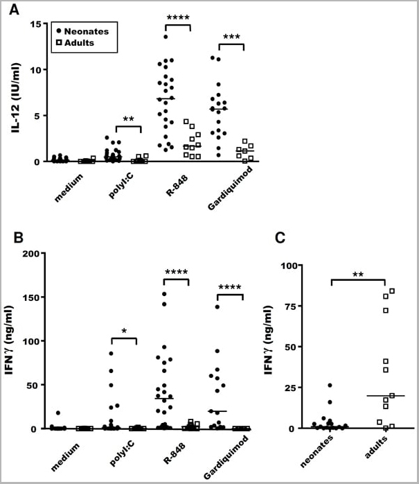
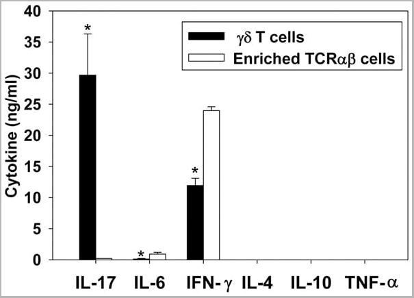
(Published customer image: gamma delta T cells are the primary source of IL-17 during B. abortus infection. C57BL/6 mice were infected i.p. with 5x10⁴ CFUs of B. abortus 2308, and two weeks later gamma delta T cells (>95% purity) and an enriched TCRalphabeta (~55% CD4+, 25% CD8+) cell fraction were isolated from the spleens of infected mice. Cells were stimulated with 500 ng/ml ionomycin and 50 ng/ml PMA for three days, and cell-free supernatants from triplicate wells were assayed for cytokine production via ELISA. The mean +/- SD is shown; * P)
Application Data
(Published customer image: gamma delta T cells are the primary source of IL-17 during B. abortus infection. C57BL/6 mice were infected i.p. with 5x10⁴ CFUs of B. abortus 2308, and two weeks later gamma delta T cells (>95% purity) and an enriched TCRalphabeta (~55% CD4+, 25% CD8+) cell fraction were isolated from the spleens of infected mice. Cells were stimulated with 500 ng/ml ionomycin and 50 ng/ml PMA for three days, and cell-free supernatants from triplicate wells were assayed for cytokine production via ELISA. The mean +/- SD is shown; * P)
IFN GAMMA, Monoclonal Antibody (Cat# AAA12093)
Full Name
MOUSE ANTI BOVINE INTERFERON GAMMA
Reactivity
Dog, Dolphin, Ferret, Fin Whale, Goat, Horse, Human, Mink, Rabbit, Pig, Sheep.
Based on sequence similarity, is expected to react with: Mustelid
N.B. Antibody reactivity and working conditions may vary between species.
Based on sequence similarity, is expected to react with: Mustelid
N.B. Antibody reactivity and working conditions may vary between species.
Applications
Flow Cytometry
Pricing
 WB (Western Blot)
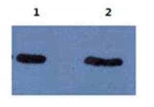
(The following image is the result of using mAb 5B8 ascites at 1:5000 dilution to detect Abeta40 and Abeta42 on Western blot. Lane 1: Abeta42, Lane 2: Abeta40.)
WB (Western Blot)
(The following image is the result of using mAb 5B8 ascites at 1:5000 dilution to detect Abeta40 and Abeta42 on Western blot. Lane 1: Abeta42, Lane 2: Abeta40.)
amyloid beta peptide N-terminal, Monoclonal Antibody (Cat# AAA14629)
Full Name
mAb anti-human amyloid beta peptide N-terminus
Reactivity
Human
Applications
Western Blot
Purity
Protein G affinity purified
Pricing
 IP (Immunoprecipitation)
(Immunoprecipitation analysis using Mouse Anti-VPS35 Monoclonal Antibody, Clone 11H10. Tissue: embryonic fibroblast. Species: Mouse. Primary Antibody: Mouse Anti-VPS35 Monoclonal Antibody. Three amounts of (3, 1 and 0.3 ug) were non-covalently coupled to 10uL of A/G sepharose beads for 1 hour at 4 degree C and next incubated with 250ug of MEF lysate for 2 hours at 4 degree C.)
IP (Immunoprecipitation)
(Immunoprecipitation analysis using Mouse Anti-VPS35 Monoclonal Antibody, Clone 11H10. Tissue: embryonic fibroblast. Species: Mouse. Primary Antibody: Mouse Anti-VPS35 Monoclonal Antibody. Three amounts of (3, 1 and 0.3 ug) were non-covalently coupled to 10uL of A/G sepharose beads for 1 hour at 4 degree C and next incubated with 250ug of MEF lysate for 2 hours at 4 degree C.)
VPS35, Monoclonal Antibody (Cat# AAA27705)
Full Name
VPS35 Antibody, Clone 11H10: FITC
Gene Names
VPS35; MEM3; PARK17
Reactivity
Human, Mouse, Rat
Applications
Western Blot, Immunoprecipitation
Purity
Protein G Purified
Pricing


 IP (Immunoprecipitation)
(Immunoprecipitation analysis using Mouse Anti-VPS35 Monoclonal Antibody, Clone 8A3. Tissue: embryonic fibroblast. Species: Mouse. Primary Antibody: Mouse Anti-VPS35 Monoclonal Antibody. Three amounts of (3, 1 and 0.3 ug) were non-covalently coupled to 10uL of A/G sepharose beads for 1 hour at 4 degree C and next incubated with 250ug of MEF lysate for 2 hours at 4 degree C.)
IP (Immunoprecipitation)
(Immunoprecipitation analysis using Mouse Anti-VPS35 Monoclonal Antibody, Clone 8A3. Tissue: embryonic fibroblast. Species: Mouse. Primary Antibody: Mouse Anti-VPS35 Monoclonal Antibody. Three amounts of (3, 1 and 0.3 ug) were non-covalently coupled to 10uL of A/G sepharose beads for 1 hour at 4 degree C and next incubated with 250ug of MEF lysate for 2 hours at 4 degree C.)
VPS35, Monoclonal Antibody (Cat# AAA27685)
Full Name
VPS35 Antibody, Clone 8A3: ATTO 594
Gene Names
VPS35; MEM3; PARK17
Reactivity
Human, Mouse, Rat
Applications
Western Blot, Immunocytochemistry, Immunofluorescence, Immunoprecipitation
Purity
Protein G Purified
Pricing