Filters
Clonality
Type
Reactivity
Gene Name
Isotype
Host
Application
Clone
407 results for " Interleukin" - showing 1-50
 Standard Curve (Sample)
IL-6, ELISA Kit (Cat# AAA17871)
Full Name
Mouse IL-6 ELISA Kit
Gene Names
Il6; Il-6
Reactivity
Mouse
Applications
SE
Pricing


 IHC (Immunohistchemistry)
(Anti-CXCR2 antibody IHC of human Skin, Melanoma. Immunohistochemistry of formalin-fixed, paraffin-embedded tissue after heat-induced antigen retrieval.)
IHC (Immunohistchemistry)
(Anti-CXCR2 antibody IHC of human Skin, Melanoma. Immunohistochemistry of formalin-fixed, paraffin-embedded tissue after heat-induced antigen retrieval.)
CXCR2, Polyclonal Antibody (Cat# AAA12364)
Full Name
Rabbit Polyclonal to Human CXCR2
Gene Names
CXCR2; CD182; IL8R2; IL8RA; IL8RB; CMKAR2; CDw128b
Reactivity
Chimpanzee, Human, Monkey
Applications
Immunohistochemistry
Purity
Immunoaffinity Purified
Pricing
 Standard Curve (Sample)
Interleukin-2 Receptor-alpha, ELISA Kit (Cat# AAA17436)
Full Name
Rat Interleukin-2 Receptor-alpha ELISA Kit
Gene Names
IL2RA; p55; CD25; IL2R; IMD41; TCGFR; IDDM10
Reactivity
Rat
Pricing
 Standard Curve (Sample)
interleukin 1, alpha, ELISA Kit (Cat# AAA15554)
Full Name
Pig Interleukin 1alpha, IL-1alpha ELISA Kit
Gene Names
IL1A; IL-1alpha
Reactivity
Pig
Pricing
IL1RA, Polyclonal Antibody (Cat# AAA14336)
Full Name
IL1RA antibody
Gene Names
IL1RN; DIRA; IRAP; IL1F3; IL1RA; MVCD4; IL-1RN; IL-1ra; IL-1ra3; ICIL-1RA
Reactivity
Not determined
Applications
Western Blot
Pricing
 Standard Curve (Sample)
interleukin 1, beta, ELISA Kit (Cat# AAA15076)
Full Name
Pig Interleukin 1beta, IL-1beta ELISA Kit
Reactivity
Pig
Pricing
 Standard Curve (Sample)
IL-4, ELISA Kit (Cat# AAA13519)
Full Name
IL-4 Rat ELISA Kit
Reactivity
Rat
Applications
ELISA
Pricing


 IHC (Immunohistochemistry)
(DAB staining on IHC-P;Samples: RabbitCardiac Muscle Tissue;Primary Ab: 40ug/ml Mouse Anti-Rabbit IL6 AntibodySecond Ab: 2ug/mL HRP-Linked Caprine Anti-Mouse IgG Polyclonal Antibody (Catalog: ))
IHC (Immunohistochemistry)
(DAB staining on IHC-P;Samples: RabbitCardiac Muscle Tissue;Primary Ab: 40ug/ml Mouse Anti-Rabbit IL6 AntibodySecond Ab: 2ug/mL HRP-Linked Caprine Anti-Mouse IgG Polyclonal Antibody (Catalog: ))
Interleukin 6, Monoclonal Antibody (Cat# AAA21003)
Full Name
Interleukin 6 (IL6) Monoclonal Antibody
Gene Names
IL6; IL-6
Reactivity
Rabbit, Rat
Applications
Westen Blot, Immunohistochemistry, Immunocytochemistry, Immunoprecipitation
Purity
Protein A + Protein G affinity chromatography
Pricing


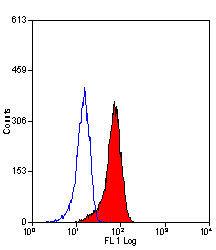
 FCM (Flow Cytometry)
(Overlay histogram showing Hela cells stained with CSB-MA878942A1m (red line) at 1:300. The cells were incubated in 1x PBS /10% normal goat serum to block non-specific protein-protein interactions followed by primary antibody for 1 h at 4 degree C. The secondary antibody used was FITC goat anti-mouse IgG(H+L) at 1/200 dilution for 1 h at 4 degree C. Isotype control antibody (green line) was used under the same conditions. Acquisition of >10,000 events was performed.)
FCM (Flow Cytometry)
(Overlay histogram showing Hela cells stained with CSB-MA878942A1m (red line) at 1:300. The cells were incubated in 1x PBS /10% normal goat serum to block non-specific protein-protein interactions followed by primary antibody for 1 h at 4 degree C. The secondary antibody used was FITC goat anti-mouse IgG(H+L) at 1/200 dilution for 1 h at 4 degree C. Isotype control antibody (green line) was used under the same conditions. Acquisition of >10,000 events was performed.)
PD-L1, Monoclonal Antibody (Cat# AAA27017)
Full Name
PD-L1 Monoclonal Antibody
Gene Names
CD274; B7-H; B7H1; PDL1; PD-L1; PDCD1L1; PDCD1LG1
Reactivity
Human
Applications
Western Blot, Immunohistochemistry, Immunofluorescence, Flow Cytometry
Purity
>95%
Protein G Purified
Protein G Purified
Pricing


 Application Data
(Published customer image: Increased accumulation of repair-associated macrophages surrounding collaterals in ischemic hind limbs is PAR2-dependent. (A) Stainings of CD206-positive macrophages (green) and SMA-positive vessels (red) in non-ischemic (control) and ischemic (ligated) hind limbs of WT, PAR1-/- and PAR2-/- mice are shown. Nuclei were visualized with DAPI (blue). Arrows indicate single macrophages in the non-ischemic adductor. Quantification of the average number of repair-associated macrophages per vessel is indicated on the right. (B) Correlation between the number of CD206-positive macrophages in the ischemic tissues and the expression of CD11b and (C) CD115 on monocytes. ** p)
Application Data
(Published customer image: Increased accumulation of repair-associated macrophages surrounding collaterals in ischemic hind limbs is PAR2-dependent. (A) Stainings of CD206-positive macrophages (green) and SMA-positive vessels (red) in non-ischemic (control) and ischemic (ligated) hind limbs of WT, PAR1-/- and PAR2-/- mice are shown. Nuclei were visualized with DAPI (blue). Arrows indicate single macrophages in the non-ischemic adductor. Quantification of the average number of repair-associated macrophages per vessel is indicated on the right. (B) Correlation between the number of CD206-positive macrophages in the ischemic tissues and the expression of CD11b and (C) CD115 on monocytes. ** p)
CD206, Monoclonal Antibody (Cat# AAA12117)
Full Name
RAT ANTI MOUSE CD206:Biotin
Gene Names
Mrc1; MR; CD206; AW259686
Applications
Flow Cytometry
Pricing


 Application Data
(Published customer image: Increased accumulation of repair-associated macrophages surrounding collaterals in ischemic hind limbs is PAR2-dependent. (A) Stainings of CD206-positive macrophages (green) and SMA-positive vessels (red) in non-ischemic (control) and ischemic (ligated) hind limbs of WT, PAR1-/- and PAR2-/- mice are shown. Nuclei were visualized with DAPI (blue). Arrows indicate single macrophages in the non-ischemic adductor. Quantification of the average number of repair-associated macrophages per vessel is indicated on the right. (B) Correlation between the number of CD206-positive macrophages in the ischemic tissues and the expression of CD11b and (C) CD115 on monocytes. ** p)
Application Data
(Published customer image: Increased accumulation of repair-associated macrophages surrounding collaterals in ischemic hind limbs is PAR2-dependent. (A) Stainings of CD206-positive macrophages (green) and SMA-positive vessels (red) in non-ischemic (control) and ischemic (ligated) hind limbs of WT, PAR1-/- and PAR2-/- mice are shown. Nuclei were visualized with DAPI (blue). Arrows indicate single macrophages in the non-ischemic adductor. Quantification of the average number of repair-associated macrophages per vessel is indicated on the right. (B) Correlation between the number of CD206-positive macrophages in the ischemic tissues and the expression of CD11b and (C) CD115 on monocytes. ** p)
CD206, Monoclonal Antibody (Cat# AAA12122)
Full Name
RAT ANTI MOUSE CD206:FITC
Gene Names
Mrc1; MR; CD206; AW259686
Applications
Flow Cytometry
Pricing


 IF (Immunofluorescence)
IL-1RL2, Polyclonal Antibody (Cat# AAA10970)
Full Name
IL-1RL2 Antibody
Gene Names
IL1RL2; IL-36R; IL1RRP2; IL-1Rrp2; IL1R-rp2
Reactivity
Human, Mouse, Rat
Applications
Immunofluorescence, Immunohistochemistry, Western Blot
Purity
Affinity chromatography purified via peptide column.
Pricing
 Standard Curve (Sample)
IL-8, ELISA Kit (Cat# AAA13498)
Full Name
IL-8 Human ELISA Kit
Gene Names
IL8; CXCL8; AMCF-I
Reactivity
Human
Applications
ELISA
Pricing


 Application Data
(Published customer image: Increased accumulation of repair-associated macrophages surrounding collaterals in ischemic hind limbs is PAR2-dependent. (A) Stainings of CD206-positive macrophages (green) and SMA-positive vessels (red) in non-ischemic (control) and ischemic (ligated) hind limbs of WT, PAR1-/- and PAR2-/- mice are shown. Nuclei were visualized with DAPI (blue). Arrows indicate single macrophages in the non-ischemic adductor. Quantification of the average number of repair-associated macrophages per vessel is indicated on the right. (B) Correlation between the number of CD206-positive macrophages in the ischemic tissues and the expression of CD11b and (C) CD115 on monocytes. ** p)
Application Data
(Published customer image: Increased accumulation of repair-associated macrophages surrounding collaterals in ischemic hind limbs is PAR2-dependent. (A) Stainings of CD206-positive macrophages (green) and SMA-positive vessels (red) in non-ischemic (control) and ischemic (ligated) hind limbs of WT, PAR1-/- and PAR2-/- mice are shown. Nuclei were visualized with DAPI (blue). Arrows indicate single macrophages in the non-ischemic adductor. Quantification of the average number of repair-associated macrophages per vessel is indicated on the right. (B) Correlation between the number of CD206-positive macrophages in the ischemic tissues and the expression of CD11b and (C) CD115 on monocytes. ** p)
CD206, Monoclonal Antibody (Cat# AAA12125)
Full Name
RAT ANTI MOUSE CD206:RPE
Gene Names
Mrc1; MR; CD206; AW259686
Applications
Flow Cytometry
Pricing


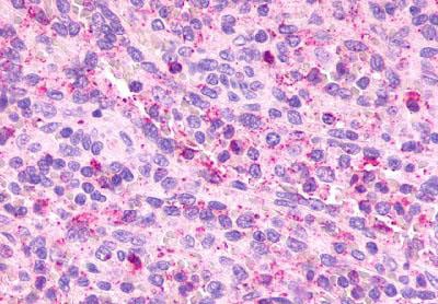
 IHC (Immunohistchemistry)
(Immunohistochemistry of paraffin-embedded mouse spleen using IL2 antibody at dilution of 1:100 (40x lens).)
IHC (Immunohistchemistry)
(Immunohistochemistry of paraffin-embedded mouse spleen using IL2 antibody at dilution of 1:100 (40x lens).)
IL2, Polyclonal Antibody (Cat# AAA28327)
Full Name
IL2 Rabbit pAb
Gene Names
IL2; IL-2; TCGF; lymphokine
Reactivity
Human, Mouse, Rat
Applications
Western Blot, Immunohistochemistry
Purity
Affinity purification
Pricing


 FCM (Flow Cytometry)
(SOCS1 Antibody (N-term) flow cytometric analysis of WiDr cells (right histogram) compared to a negative control cell (left histogram).FITC-conjugated goat-anti-rabbit secondary antibodies were used for the analysis.)
FCM (Flow Cytometry)
(SOCS1 Antibody (N-term) flow cytometric analysis of WiDr cells (right histogram) compared to a negative control cell (left histogram).FITC-conjugated goat-anti-rabbit secondary antibodies were used for the analysis.)
SOCS1, Polyclonal Antibody (Cat# AAA28752)
Full Name
SOCS1 Antibody (N-term)
Gene Names
SOCS1; JAB; CIS1; SSI1; TIP3; CISH1; SSI-1; SOCS-1
Reactivity
Mouse, rat
Applications
Western Blot, Immunohistochemistry, Immunofluorescence, Flow Cytometry
Purity
Purified Rabbit Polyclonal Antibody (Pab)
Pricing


 Application Data
(Published customer image: Increased accumulation of repair-associated macrophages surrounding collaterals in ischemic hind limbs is PAR2-dependent. (A) Stainings of CD206-positive macrophages (green) and SMA-positive vessels (red) in non-ischemic (control) and ischemic (ligated) hind limbs of WT, PAR1-/- and PAR2-/- mice are shown. Nuclei were visualized with DAPI (blue). Arrows indicate single macrophages in the non-ischemic adductor. Quantification of the average number of repair-associated macrophages per vessel is indicated on the right. (B) Correlation between the number of CD206-positive macrophages in the ischemic tissues and the expression of CD11b and (C) CD115 on monocytes. ** p)
Application Data
(Published customer image: Increased accumulation of repair-associated macrophages surrounding collaterals in ischemic hind limbs is PAR2-dependent. (A) Stainings of CD206-positive macrophages (green) and SMA-positive vessels (red) in non-ischemic (control) and ischemic (ligated) hind limbs of WT, PAR1-/- and PAR2-/- mice are shown. Nuclei were visualized with DAPI (blue). Arrows indicate single macrophages in the non-ischemic adductor. Quantification of the average number of repair-associated macrophages per vessel is indicated on the right. (B) Correlation between the number of CD206-positive macrophages in the ischemic tissues and the expression of CD11b and (C) CD115 on monocytes. ** p)
CD206, Monoclonal Antibody (Cat# AAA12120)
Full Name
RAT ANTI MOUSE CD206:FITC
Gene Names
Mrc1; MR; CD206; AW259686
Applications
Flow Cytometry
Pricing


 FCM (Flow Cytometry)
(Flow cytometric analysis of Jurkat cells with CD137 antibody at 1/100 dilution (red) compared with an unlabelled control (cells without incubation with primary antibody; black).)
FCM (Flow Cytometry)
(Flow cytometric analysis of Jurkat cells with CD137 antibody at 1/100 dilution (red) compared with an unlabelled control (cells without incubation with primary antibody; black).)
CD137, Monoclonal Antibody (Cat# AAA29865)
Full Name
CD137 Antibody
Gene Names
TNFRSF9; ILA; 4-1BB; CD137; CDw137
Reactivity
Human, Mouse, Rat
Applications
Western Blot, Immunohistochemistry, Immunocytochemistry, Flow Cytometry
Purity
ProA affinity purified
Pricing


 FCM (Flow Cytometry)
(Flow cytometric analysis of K562 cells with ILF3 antibody at 1/100 dilution (purple) compared with an unlabelled control (cells without incubation with primary antibody; yellow). Alexa Fluor 488-conjugated goat anti-rabbit IgG was used as the secondary antibody.)
FCM (Flow Cytometry)
(Flow cytometric analysis of K562 cells with ILF3 antibody at 1/100 dilution (purple) compared with an unlabelled control (cells without incubation with primary antibody; yellow). Alexa Fluor 488-conjugated goat anti-rabbit IgG was used as the secondary antibody.)
ILF3, Monoclonal Antibody (Cat# AAA30498)
Full Name
ILF3 Antibody
Gene Names
ILF3; CBTF; DRBF; MMP4; MPP4; NF90; NFAR; NF110; NF90a; NF90b; NFAR2; TCP80; DRBP76; NF110b; NFAR-1; TCP110; MPHOSPH4; NF-AT-90
Reactivity
Human, Mouse, Rat
Applications
Western Blot, Immunoprecipitation, Immunocytochemistry, Immunofluorescence, Immunohistochemistry, Flow Cytometry
Purity
ProA affinity purified
Pricing


 FCM (Flow Cytometry)
(Flow cytometric analysis of THP-1 cells with TLR2 antibody at 1/100 dilution (red) compared with an unlabelled control (cells without incubation with primary antibody; black).)
FCM (Flow Cytometry)
(Flow cytometric analysis of THP-1 cells with TLR2 antibody at 1/100 dilution (red) compared with an unlabelled control (cells without incubation with primary antibody; black).)
TLR2, Monoclonal Antibody (Cat# AAA30387)
Full Name
TLR2 Antibody
Gene Names
TLR2; TIL4; CD282
Reactivity
Human, Mouse, Rat
Applications
Western Blot, Immunocytochemistry, Immunofluorescence, Immunohistochemistry, Flow Cytometry
Purity
ProA affinity purified
Pricing


 Application Data
(Published customer image Infiltration of GFP+ BM-cells in infarct and peri-infarct regions. (A-B) Dot plots of viable macrophages/granulocytes (CD11b+CD45high, top right quadrants) and microglia (CD11b+CD45dim, bottom right quadrants) in cortex from BM-chimeric unmanipulated mice and mice exposed to pMCAO. (C) Bar graph showing mean numbers of CD11b+CD45dim microglia and CD11b+CD45high macrophages/granulocytes in BM-chimeric mice 24 hours after pMCAO, subdivided based on expression of GFP (n = 5). Approximately 92% of of the CD45high population were GFP+. (D) Estimation and comparison of mean numbers of CD11b+CD45dim microglia in non-chimeric (n = 10) versus BM-chimeric mice (n = 5) 24 hours after of pMCAO shows significantly fewer CD11b+CD45dim microglial cells in irradiated mice. (E) Overview, showing distribution of infiltrating GFP+ BM-derived cells into infarct (IF) and peri-infarct (P-IF) regions 24 hours after pMCAO. (E-G) By 24 hours, GFP+ single cells (F) and vessel-associated aggregates of GFP+ cells (arrows in G) were observed in infarct and peri-infarct regions. Some of the vessel-associated cells were round, leukocyte-like cells (arrows) while others were elongated cells lining the vasculature (arrow heads in G and in insert). (H) Bar graph showing mean numbers of single GFP+ cells and vessel-associated aggregates of GFP+ cells in ipsi- and contralateral cortex 24 hours after surgery (n = 10). (I-P) Immunohistochemical staining of CD45.1 (I, K), CD45.2 (J, L), IgG2a (M, O) and CD45 (N, P) in ischemic tissue in BM-chimeric (I, J, M, N) and non-chimeric mice (K, L, O, P) 24 hours after pMCAO. N.D, none detected. Scale bars: 200 um (A), 10 um (B, C). 50 um (I-P) *P < 0.05, **P < 0.01, and ***P < 0.001.From: Clausen BH, Lambertsen KL, Babcock AA, Holm TH, Dagnaes-Hansen F, Finsen B. Interleukin-1beta and tumor necrosis factor-alpha are expressed by different subsets of microglia and macrophages after ischemic stroke in mice. J Neuroinflammation. 2008 Oct 23;5:46.)
Application Data
(Published customer image Infiltration of GFP+ BM-cells in infarct and peri-infarct regions. (A-B) Dot plots of viable macrophages/granulocytes (CD11b+CD45high, top right quadrants) and microglia (CD11b+CD45dim, bottom right quadrants) in cortex from BM-chimeric unmanipulated mice and mice exposed to pMCAO. (C) Bar graph showing mean numbers of CD11b+CD45dim microglia and CD11b+CD45high macrophages/granulocytes in BM-chimeric mice 24 hours after pMCAO, subdivided based on expression of GFP (n = 5). Approximately 92% of of the CD45high population were GFP+. (D) Estimation and comparison of mean numbers of CD11b+CD45dim microglia in non-chimeric (n = 10) versus BM-chimeric mice (n = 5) 24 hours after of pMCAO shows significantly fewer CD11b+CD45dim microglial cells in irradiated mice. (E) Overview, showing distribution of infiltrating GFP+ BM-derived cells into infarct (IF) and peri-infarct (P-IF) regions 24 hours after pMCAO. (E-G) By 24 hours, GFP+ single cells (F) and vessel-associated aggregates of GFP+ cells (arrows in G) were observed in infarct and peri-infarct regions. Some of the vessel-associated cells were round, leukocyte-like cells (arrows) while others were elongated cells lining the vasculature (arrow heads in G and in insert). (H) Bar graph showing mean numbers of single GFP+ cells and vessel-associated aggregates of GFP+ cells in ipsi- and contralateral cortex 24 hours after surgery (n = 10). (I-P) Immunohistochemical staining of CD45.1 (I, K), CD45.2 (J, L), IgG2a (M, O) and CD45 (N, P) in ischemic tissue in BM-chimeric (I, J, M, N) and non-chimeric mice (K, L, O, P) 24 hours after pMCAO. N.D, none detected. Scale bars: 200 um (A), 10 um (B, C). 50 um (I-P) *P < 0.05, **P < 0.01, and ***P < 0.001.From: Clausen BH, Lambertsen KL, Babcock AA, Holm TH, Dagnaes-Hansen F, Finsen B. Interleukin-1beta and tumor necrosis factor-alpha are expressed by different subsets of microglia and macrophages after ischemic stroke in mice. J Neuroinflammation. 2008 Oct 23;5:46.)
CD11b, Monoclonal Antibody (Cat# AAA12182)
Full Name
RAT ANTI MOUSE CD11b:FITC
Gene Names
Itgam; CR3; CR3A; MAC1; Cd11b; Ly-40; Mac-1; Mac-1a; CD11b/CD18; F730045J24Rik
Applications
Flow Cytometry
Pricing


 FCM (Flow Cytometry)
(Flow cytometric analysis of SH-SY-5Y cells with ILF2 antibody at 1/100 dilution (red) compared with an unlabelled control (cells without incubation with primary antibody; black). Alexa Fluor 488-conjugated goat anti rabbit IgG was used as the secondary antibody.)
FCM (Flow Cytometry)
(Flow cytometric analysis of SH-SY-5Y cells with ILF2 antibody at 1/100 dilution (red) compared with an unlabelled control (cells without incubation with primary antibody; black). Alexa Fluor 488-conjugated goat anti rabbit IgG was used as the secondary antibody.)
ILF2, Monoclonal Antibody (Cat# AAA30430)
Full Name
ILF2 Antibody
Gene Names
ILF2; NF45; PRO3063
Reactivity
Human, Mouse, Rat
Applications
Western Blot, Immunoprecipitation, Immunohistochemistry, Flow Cytometry
Purity
ProA affinity purified
Pricing
 Standard Curve (Sample)
IL-6, ELISA Kit (Cat# AAA13517)
Full Name
IL-6 Rat ELISA Kit
Gene Names
Il6; ILg6; Ifnb2
Reactivity
Rat
Applications
ELISA
Pricing


 ICC (Immunocytochemistry)
(ICC staining STAT6 in NIH/3T3 cells (green). The nuclear counter stain is DAPI (blue). Cells were fixed in paraformaldehyde, permeabilised with 0.25% Triton X100/PBS.)
ICC (Immunocytochemistry)
(ICC staining STAT6 in NIH/3T3 cells (green). The nuclear counter stain is DAPI (blue). Cells were fixed in paraformaldehyde, permeabilised with 0.25% Triton X100/PBS.)
STAT6, Monoclonal Antibody (Cat# AAA30062)
Full Name
STAT6 Antibody
Gene Names
STAT6; STAT6B; STAT6C; D12S1644; IL-4-STAT
Reactivity
Human, Mouse
Applications
Western Blot, Immunocytochemistry, Immunofluorescence, Immunohistochemistry, Immunoprecipitation
Purity
ProA affinity purified
Pricing


 IF (Immunofluorescence)
(Immunofluorescence of IL-17 in mouse thymus tissue with IL-17 Antibody at 20 μg/mL.)
IF (Immunofluorescence)
(Immunofluorescence of IL-17 in mouse thymus tissue with IL-17 Antibody at 20 μg/mL.)
IL-17, Polyclonal Antibody (Cat# AAA10926)
Full Name
IL-17 Antibody
Gene Names
IL17A; IL17; CTLA8; IL-17; IL-17A
Reactivity
Human, Mouse
Applications
Western Blot
Purity
IL-17 Antibody is affinity chromatography purified via peptide column.
Pricing


 Application Data
(Published customer image Infiltration of GFP+ BM-cells in infarct and peri-infarct regions. (A-B) Dot plots of viable macrophages/granulocytes (CD11b+CD45high, top right quadrants) and microglia (CD11b+CD45dim, bottom right quadrants) in cortex from BM-chimeric unmanipulated mice and mice exposed to pMCAO. (C) Bar graph showing mean numbers of CD11b+CD45dim microglia and CD11b+CD45high macrophages/granulocytes in BM-chimeric mice 24 hours after pMCAO, subdivided based on expression of GFP (n = 5). Approximately 92% of of the CD45high population were GFP+. (D) Estimation and comparison of mean numbers of CD11b+CD45dim microglia in non-chimeric (n = 10) versus BM-chimeric mice (n = 5) 24 hours after of pMCAO shows significantly fewer CD11b+CD45dim microglial cells in irradiated mice. (E) Overview, showing distribution of infiltrating GFP+ BM-derived cells into infarct (IF) and peri-infarct (P-IF) regions 24 hours after pMCAO. (E-G) By 24 hours, GFP+ single cells (F) and vessel-associated aggregates of GFP+ cells (arrows in G) were observed in infarct and peri-infarct regions. Some of the vessel-associated cells were round, leukocyte-like cells (arrows) while others were elongated cells lining the vasculature (arrow heads in G and in insert). (H) Bar graph showing mean numbers of single GFP+ cells and vessel-associated aggregates of GFP+ cells in ipsi- and contralateral cortex 24 hours after surgery (n = 10). (I-P) Immunohistochemical staining of CD45.1 (I, K), CD45.2 (J, L), IgG2a (M, O) and CD45 (N, P) in ischemic tissue in BM-chimeric (I, J, M, N) and non-chimeric mice (K, L, O, P) 24 hours after pMCAO. N.D, none detected. Scale bars: 200 um (A), 10 um (B, C). 50 um (I-P) *P < 0.05, **P < 0.01, and ***P < 0.001.From: Clausen BH, Lambertsen KL, Babcock AA, Holm TH, Dagnaes-Hansen F, Finsen B. Interleukin-1beta and tumor necrosis factor-alpha are expressed by different subsets of microglia and macrophages after ischemic stroke in mice. J Neuroinflammation. 2008 Oct 23;5:46.)
Application Data
(Published customer image Infiltration of GFP+ BM-cells in infarct and peri-infarct regions. (A-B) Dot plots of viable macrophages/granulocytes (CD11b+CD45high, top right quadrants) and microglia (CD11b+CD45dim, bottom right quadrants) in cortex from BM-chimeric unmanipulated mice and mice exposed to pMCAO. (C) Bar graph showing mean numbers of CD11b+CD45dim microglia and CD11b+CD45high macrophages/granulocytes in BM-chimeric mice 24 hours after pMCAO, subdivided based on expression of GFP (n = 5). Approximately 92% of of the CD45high population were GFP+. (D) Estimation and comparison of mean numbers of CD11b+CD45dim microglia in non-chimeric (n = 10) versus BM-chimeric mice (n = 5) 24 hours after of pMCAO shows significantly fewer CD11b+CD45dim microglial cells in irradiated mice. (E) Overview, showing distribution of infiltrating GFP+ BM-derived cells into infarct (IF) and peri-infarct (P-IF) regions 24 hours after pMCAO. (E-G) By 24 hours, GFP+ single cells (F) and vessel-associated aggregates of GFP+ cells (arrows in G) were observed in infarct and peri-infarct regions. Some of the vessel-associated cells were round, leukocyte-like cells (arrows) while others were elongated cells lining the vasculature (arrow heads in G and in insert). (H) Bar graph showing mean numbers of single GFP+ cells and vessel-associated aggregates of GFP+ cells in ipsi- and contralateral cortex 24 hours after surgery (n = 10). (I-P) Immunohistochemical staining of CD45.1 (I, K), CD45.2 (J, L), IgG2a (M, O) and CD45 (N, P) in ischemic tissue in BM-chimeric (I, J, M, N) and non-chimeric mice (K, L, O, P) 24 hours after pMCAO. N.D, none detected. Scale bars: 200 um (A), 10 um (B, C). 50 um (I-P) *P < 0.05, **P < 0.01, and ***P < 0.001.From: Clausen BH, Lambertsen KL, Babcock AA, Holm TH, Dagnaes-Hansen F, Finsen B. Interleukin-1beta and tumor necrosis factor-alpha are expressed by different subsets of microglia and macrophages after ischemic stroke in mice. J Neuroinflammation. 2008 Oct 23;5:46.)
CD11b, Monoclonal Antibody (Cat# AAA12184)
Full Name
RAT ANTI MOUSE CD11b
Gene Names
Itgam; CR3; CR3A; MAC1; Cd11b; Ly-40; Mac-1; Mac-1a; CD11b/CD18; F730045J24Rik
Applications
Immunohistochemistry, Flow Cytometry, Immunofluorescence, Immunoprecipitation
Pricing
 Standard Curve (Sample)
Interleukin 10 (IL10), ELISA Kit (Cat# AAA23099)
Full Name
Rat Interleukin 10 (IL10) High Sensitive ELISA Kit
Gene Names
Il10; IL10X
Reactivity
Rat
Applications
ELISA
Pricing
 Standard Curve (Sample)
IL-13, ELISA Kit (Cat# AAA13516)
Full Name
IL-13 Rat ELISA Kit
Gene Names
IL13; P600; IL-13
Reactivity
Rat
Applications
ELISA
Pricing


 Application Data
(Published customer image: Increased accumulation of repair-associated macrophages surrounding collaterals in ischemic hind limbs is PAR2-dependent. (A) Stainings of CD206-positive macrophages (green) and SMA-positive vessels (red) in non-ischemic (control) and ischemic (ligated) hind limbs of WT, PAR1-/- and PAR2-/- mice are shown. Nuclei were visualized with DAPI (blue). Arrows indicate single macrophages in the non-ischemic adductor. Quantification of the average number of repair-associated macrophages per vessel is indicated on the right. (B) Correlation between the number of CD206-positive macrophages in the ischemic tissues and the expression of CD11b and (C) CD115 on monocytes. ** p)
Application Data
(Published customer image: Increased accumulation of repair-associated macrophages surrounding collaterals in ischemic hind limbs is PAR2-dependent. (A) Stainings of CD206-positive macrophages (green) and SMA-positive vessels (red) in non-ischemic (control) and ischemic (ligated) hind limbs of WT, PAR1-/- and PAR2-/- mice are shown. Nuclei were visualized with DAPI (blue). Arrows indicate single macrophages in the non-ischemic adductor. Quantification of the average number of repair-associated macrophages per vessel is indicated on the right. (B) Correlation between the number of CD206-positive macrophages in the ischemic tissues and the expression of CD11b and (C) CD115 on monocytes. ** p)
CD206, Monoclonal Antibody (Cat# AAA12123)
Full Name
RAT ANTI MOUSE CD206
Gene Names
Mrc1; MR; CD206; AW259686
Applications
Immunohistochemistry, Flow Cytometry, Immunofluorescence, Immunoprecipitation
Pricing
 Standard Curve (Sample)
Interleukin 1 Receptor Associated Kinase 4 (IRAK4), ELISA Kit (Cat# AAA20690)
Full Name
Human Interleukin 1 Receptor Associated Kinase 4 (IRAK4) ELISA Kit
Gene Names
IRAK4; IPD1; REN64; IRAK-4; NY-REN-64
Reactivity
Human
Pricing


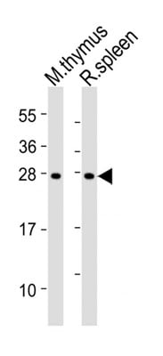
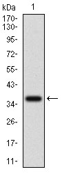
 WB (Western Blot)
(Western blot analysis of IRAK1 over-expressed 293 cell line, cotransfected with IRAK1 Validated Chimera RNAi ((Lane 2) or non-transfected control (Lane 1). Blot probed with IRAK1 monoclonal antibody. GAPDH (36.1kD) used as specificity and loading control.)
WB (Western Blot)
(Western blot analysis of IRAK1 over-expressed 293 cell line, cotransfected with IRAK1 Validated Chimera RNAi ((Lane 2) or non-transfected control (Lane 1). Blot probed with IRAK1 monoclonal antibody. GAPDH (36.1kD) used as specificity and loading control.)
IRAK1, Monoclonal Antibody (Cat# AAA24839)
Full Name
IRAK1 (Interleukin-1 Receptor-associated Kinase 1, IRAK-1, IRAK) (Biotin)
Gene Names
IRAK1; IRAK; pelle
Reactivity
Human
Applications
Immunofluorescence, Immunohistochemistry, Immunoprecipitation, Western Blot
Purity
Purified by Protein A Affinity Chromatography.
Pricing


 WB (Western Blot)
(Western blot analysis of IRAK1 over-expressed 293 cell line, cotransfected with IRAK1 Validated Chimera RNAi ((Lane 2) or non-transfected control (Lane 1). Blot probed with IRAK1 monoclonal antibody. GAPDH (36.1kD) used as specificity and loading control.)
WB (Western Blot)
(Western blot analysis of IRAK1 over-expressed 293 cell line, cotransfected with IRAK1 Validated Chimera RNAi ((Lane 2) or non-transfected control (Lane 1). Blot probed with IRAK1 monoclonal antibody. GAPDH (36.1kD) used as specificity and loading control.)
IRAK1, Monoclonal Antibody (Cat# AAA25136)
Full Name
IRAK1 (Interleukin-1 Receptor-associated Kinase 1, IRAK-1, IRAK) (FITC)
Gene Names
IRAK1; IRAK; pelle
Reactivity
Human
Applications
Immunofluorescence, Immunohistochemistry, Immunoprecipitation, Western Blot
Purity
Purified by Protein A Affinity Chromatography.
Pricing


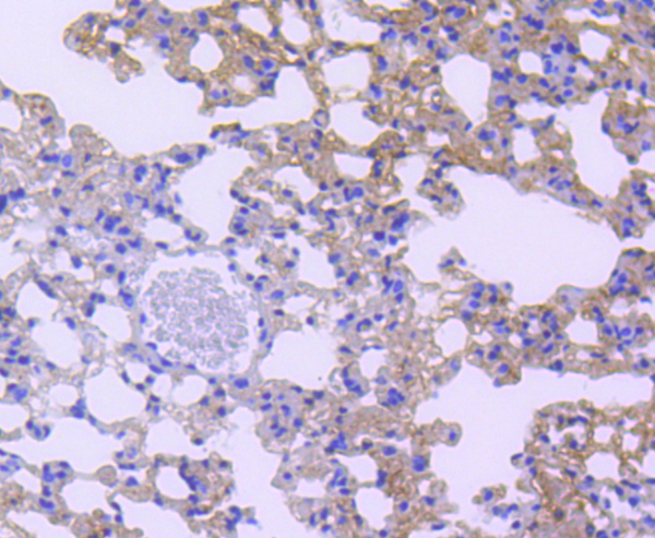
 IHC (Immunohistchemistry)
(Immunohistochemical analysis of paraffin-embedded lung cancer tissues using IL3RA mouse mAb with DAB staining.)
IHC (Immunohistchemistry)
(Immunohistochemical analysis of paraffin-embedded lung cancer tissues using IL3RA mouse mAb with DAB staining.)
IL3RA, Monoclonal Antibody (Cat# AAA14136)
Full Name
Anti-IL3RA Mouse mAb
Gene Names
IL3RA; IL3R; CD123; IL3RX; IL3RY; IL3RAY; hIL-3Ra
Reactivity
Human
Applications
Western Blot, Immunohistochemistry, Immunocytochemistry, Flow Cytometry
Pricing


 FCM (Flow Cytometry)
(Flow cytometric analysis of Jurkat cells with IRAK2 antibody at 1/50 dilution (purple) compared with an unlabelled control (cells without incubation with primary antibody; yellow). Alexa Fluor 488-conjugated goat anti-rabbit IgG was used as the secondary antibody.)
FCM (Flow Cytometry)
(Flow cytometric analysis of Jurkat cells with IRAK2 antibody at 1/50 dilution (purple) compared with an unlabelled control (cells without incubation with primary antibody; yellow). Alexa Fluor 488-conjugated goat anti-rabbit IgG was used as the secondary antibody.)
IRAK2, Monoclonal Antibody (Cat# AAA30491)
Full Name
IRAK2 Antibody
Gene Names
IRAK2; IRAK-2
Reactivity
Human
Applications
Western Blot, Immunocytochemistry, Immunofluorescence, Immunohistochemistry, Flow Cytometry, Immunoprecipitation
Purity
ProA affinity purified
Pricing


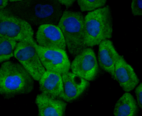
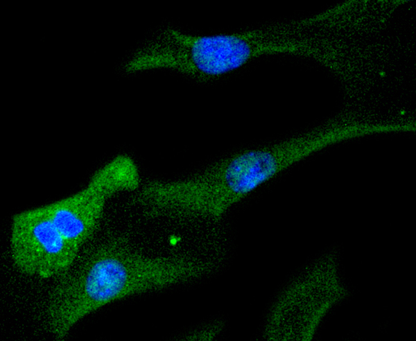
 ICC (Immunocytochemistry)
(ICC staining CD130 (green) in LOVO cells. The nuclear counter stain is DAPI (blue). Cells were fixed in paraformaldehyde, permeabilised with 0.25% Triton X100/PBS.)
ICC (Immunocytochemistry)
(ICC staining CD130 (green) in LOVO cells. The nuclear counter stain is DAPI (blue). Cells were fixed in paraformaldehyde, permeabilised with 0.25% Triton X100/PBS.)
CD130, Monoclonal Antibody (Cat# AAA29893)
Full Name
CD130 Antibody
Gene Names
IL6ST; CD130; GP130; CDW130; IL-6RB
Reactivity
Human
Applications
Western Blot, Immunohistochemistry, Immunocytochemistry
Purity
ProG affinity purified
Pricing


 Application Data
(Published customer image Infiltration of GFP+ BM-cells in infarct and peri-infarct regions. (A-B) Dot plots of viable macrophages/granulocytes (CD11b+CD45high, top right quadrants) and microglia (CD11b+CD45dim, bottom right quadrants) in cortex from BM-chimeric unmanipulated mice and mice exposed to pMCAO. (C) Bar graph showing mean numbers of CD11b+CD45dim microglia and CD11b+CD45high macrophages/granulocytes in BM-chimeric mice 24 hours after pMCAO, subdivided based on expression of GFP (n = 5). Approximately 92% of of the CD45high population were GFP+. (D) Estimation and comparison of mean numbers of CD11b+CD45dim microglia in non-chimeric (n = 10) versus BM-chimeric mice (n = 5) 24 hours after of pMCAO shows significantly fewer CD11b+CD45dim microglial cells in irradiated mice. (E) Overview, showing distribution of infiltrating GFP+ BM-derived cells into infarct (IF) and peri-infarct (P-IF) regions 24 hours after pMCAO. (E-G) By 24 hours, GFP+ single cells (F) and vessel-associated aggregates of GFP+ cells (arrows in G) were observed in infarct and peri-infarct regions. Some of the vessel-associated cells were round, leukocyte-like cells (arrows) while others were elongated cells lining the vasculature (arrow heads in G and in insert). (H) Bar graph showing mean numbers of single GFP+ cells and vessel-associated aggregates of GFP+ cells in ipsi- and contralateral cortex 24 hours after surgery (n = 10). (I-P) Immunohistochemical staining of CD45.1 (I, K), CD45.2 (J, L), IgG2a (M, O) and CD45 (N, P) in ischemic tissue in BM-chimeric (I, J, M, N) and non-chimeric mice (K, L, O, P) 24 hours after pMCAO. N.D, none detected. Scale bars: 200 um (A), 10 um (B, C). 50 um (I-P) *P < 0.05, **P < 0.01, and ***P < 0.001.From: Clausen BH, Lambertsen KL, Babcock AA, Holm TH, Dagnaes-Hansen F, Finsen B. Interleukin-1beta and tumor necrosis factor-alpha are expressed by different subsets of microglia and macrophages after ischemic stroke in mice. J Neuroinflammation. 2008 Oct 23;5:46.)
Application Data
(Published customer image Infiltration of GFP+ BM-cells in infarct and peri-infarct regions. (A-B) Dot plots of viable macrophages/granulocytes (CD11b+CD45high, top right quadrants) and microglia (CD11b+CD45dim, bottom right quadrants) in cortex from BM-chimeric unmanipulated mice and mice exposed to pMCAO. (C) Bar graph showing mean numbers of CD11b+CD45dim microglia and CD11b+CD45high macrophages/granulocytes in BM-chimeric mice 24 hours after pMCAO, subdivided based on expression of GFP (n = 5). Approximately 92% of of the CD45high population were GFP+. (D) Estimation and comparison of mean numbers of CD11b+CD45dim microglia in non-chimeric (n = 10) versus BM-chimeric mice (n = 5) 24 hours after of pMCAO shows significantly fewer CD11b+CD45dim microglial cells in irradiated mice. (E) Overview, showing distribution of infiltrating GFP+ BM-derived cells into infarct (IF) and peri-infarct (P-IF) regions 24 hours after pMCAO. (E-G) By 24 hours, GFP+ single cells (F) and vessel-associated aggregates of GFP+ cells (arrows in G) were observed in infarct and peri-infarct regions. Some of the vessel-associated cells were round, leukocyte-like cells (arrows) while others were elongated cells lining the vasculature (arrow heads in G and in insert). (H) Bar graph showing mean numbers of single GFP+ cells and vessel-associated aggregates of GFP+ cells in ipsi- and contralateral cortex 24 hours after surgery (n = 10). (I-P) Immunohistochemical staining of CD45.1 (I, K), CD45.2 (J, L), IgG2a (M, O) and CD45 (N, P) in ischemic tissue in BM-chimeric (I, J, M, N) and non-chimeric mice (K, L, O, P) 24 hours after pMCAO. N.D, none detected. Scale bars: 200 um (A), 10 um (B, C). 50 um (I-P) *P < 0.05, **P < 0.01, and ***P < 0.001.From: Clausen BH, Lambertsen KL, Babcock AA, Holm TH, Dagnaes-Hansen F, Finsen B. Interleukin-1beta and tumor necrosis factor-alpha are expressed by different subsets of microglia and macrophages after ischemic stroke in mice. J Neuroinflammation. 2008 Oct 23;5:46.)
CD11b, Monoclonal Antibody (Cat# AAA12185)
Full Name
RAT ANTI MOUSE CD11b
Gene Names
Itgam; CR3; CR3A; MAC1; Cd11b; Ly-40; Mac-1; Mac-1a; CD11b/CD18; F730045J24Rik
Applications
Immunohistochemistry, Flow Cytometry, Immunofluorescence, Immunoprecipitation
Pricing


 Application Data
(Published customer image Infiltration of GFP+ BM-cells in infarct and peri-infarct regions. (A-B) Dot plots of viable macrophages/granulocytes (CD11b+CD45high, top right quadrants) and microglia (CD11b+CD45dim, bottom right quadrants) in cortex from BM-chimeric unmanipulated mice and mice exposed to pMCAO. (C) Bar graph showing mean numbers of CD11b+CD45dim microglia and CD11b+CD45high macrophages/granulocytes in BM-chimeric mice 24 hours after pMCAO, subdivided based on expression of GFP (n = 5). Approximately 92% of of the CD45high population were GFP+. (D) Estimation and comparison of mean numbers of CD11b+CD45dim microglia in non-chimeric (n = 10) versus BM-chimeric mice (n = 5) 24 hours after of pMCAO shows significantly fewer CD11b+CD45dim microglial cells in irradiated mice. (E) Overview, showing distribution of infiltrating GFP+ BM-derived cells into infarct (IF) and peri-infarct (P-IF) regions 24 hours after pMCAO. (E-G) By 24 hours, GFP+ single cells (F) and vessel-associated aggregates of GFP+ cells (arrows in G) were observed in infarct and peri-infarct regions. Some of the vessel-associated cells were round, leukocyte-like cells (arrows) while others were elongated cells lining the vasculature (arrow heads in G and in insert). (H) Bar graph showing mean numbers of single GFP+ cells and vessel-associated aggregates of GFP+ cells in ipsi- and contralateral cortex 24 hours after surgery (n = 10). (I-P) Immunohistochemical staining of CD45.1 (I, K), CD45.2 (J, L), IgG2a (M, O) and CD45 (N, P) in ischemic tissue in BM-chimeric (I, J, M, N) and non-chimeric mice (K, L, O, P) 24 hours after pMCAO. N.D, none detected. Scale bars: 200 um (A), 10 um (B, C). 50 um (I-P) *P < 0.05, **P < 0.01, and ***P < 0.001.From: Clausen BH, Lambertsen KL, Babcock AA, Holm TH, Dagnaes-Hansen F, Finsen B. Interleukin-1beta and tumor necrosis factor-alpha are expressed by different subsets of microglia and macrophages after ischemic stroke in mice. J Neuroinflammation. 2008 Oct 23;5:46.)
Application Data
(Published customer image Infiltration of GFP+ BM-cells in infarct and peri-infarct regions. (A-B) Dot plots of viable macrophages/granulocytes (CD11b+CD45high, top right quadrants) and microglia (CD11b+CD45dim, bottom right quadrants) in cortex from BM-chimeric unmanipulated mice and mice exposed to pMCAO. (C) Bar graph showing mean numbers of CD11b+CD45dim microglia and CD11b+CD45high macrophages/granulocytes in BM-chimeric mice 24 hours after pMCAO, subdivided based on expression of GFP (n = 5). Approximately 92% of of the CD45high population were GFP+. (D) Estimation and comparison of mean numbers of CD11b+CD45dim microglia in non-chimeric (n = 10) versus BM-chimeric mice (n = 5) 24 hours after of pMCAO shows significantly fewer CD11b+CD45dim microglial cells in irradiated mice. (E) Overview, showing distribution of infiltrating GFP+ BM-derived cells into infarct (IF) and peri-infarct (P-IF) regions 24 hours after pMCAO. (E-G) By 24 hours, GFP+ single cells (F) and vessel-associated aggregates of GFP+ cells (arrows in G) were observed in infarct and peri-infarct regions. Some of the vessel-associated cells were round, leukocyte-like cells (arrows) while others were elongated cells lining the vasculature (arrow heads in G and in insert). (H) Bar graph showing mean numbers of single GFP+ cells and vessel-associated aggregates of GFP+ cells in ipsi- and contralateral cortex 24 hours after surgery (n = 10). (I-P) Immunohistochemical staining of CD45.1 (I, K), CD45.2 (J, L), IgG2a (M, O) and CD45 (N, P) in ischemic tissue in BM-chimeric (I, J, M, N) and non-chimeric mice (K, L, O, P) 24 hours after pMCAO. N.D, none detected. Scale bars: 200 um (A), 10 um (B, C). 50 um (I-P) *P < 0.05, **P < 0.01, and ***P < 0.001.From: Clausen BH, Lambertsen KL, Babcock AA, Holm TH, Dagnaes-Hansen F, Finsen B. Interleukin-1beta and tumor necrosis factor-alpha are expressed by different subsets of microglia and macrophages after ischemic stroke in mice. J Neuroinflammation. 2008 Oct 23;5:46.)
CD11b, Monoclonal Antibody (Cat# AAA12183)
Full Name
RAT ANTI MOUSE CD11b:FITC
Gene Names
Itgam; CR3; CR3A; MAC1; Cd11b; Ly-40; Mac-1; Mac-1a; CD11b/CD18; F730045J24Rik
Applications
Flow Cytometry
Pricing
 Standard Curve (Sample)
IL-18, ELISA Kit (Cat# AAA13491)
Full Name
IL-18 Rat ELISA Kit
Reactivity
Rat
Applications
ELISA
Pricing
 Standard Curve (Sample)
Interleukin 6 (IL-6), ELISA Kit (Cat# AAA12730)
Full Name
Goat Interleukin 6 (IL-6) ELISA Kit
Gene Names
IL6; HGF; HSF; BSF2; IL-6; IFNB2
Reactivity
Goat
Pricing
 Standard Curve (Sample)
interleukin 1 family, member 6 (epsilon), ELISA Kit (Cat# AAA18243)
Full Name
Human Interleukin-36 alpha, IL1F6 ELISA Kit
Gene Names
IL36A; FIL1; FIL1E; IL1F6; IL-1F6; IL1(EPSILON); FIL1(EPSILON)
Reactivity
Human
Pricing


 ICC (Immunocytochemistry)
(ICC staining STAT6 in NIH/3T3 cells (green). The nuclear counter stain is DAPI (blue). Cells were fixed in paraformaldehyde, permeabilised with 0.25% Triton X100/PBS.)
ICC (Immunocytochemistry)
(ICC staining STAT6 in NIH/3T3 cells (green). The nuclear counter stain is DAPI (blue). Cells were fixed in paraformaldehyde, permeabilised with 0.25% Triton X100/PBS.)
STAT6, Monoclonal Antibody (Cat# AAA30030)
Full Name
STAT6 Antibody
Gene Names
STAT6; STAT6B; STAT6C; D12S1644; IL-4-STAT
Reactivity
Human, Mouse
Applications
Western Blot, Immunocytochemistry, Immunohistochemistry, Immunoprecipitation
Purity
ProA affinity purified
Pricing
 Standard Curve (Sample)
interleukin 10, ELISA Kit (Cat# AAA15218)
Full Name
Chicken Interleukin 10, IL-10 ELISA Kit
Gene Names
IL10; IL-10
Reactivity
Chicken
Pricing


 Application Data
(Published customer image: Increased accumulation of repair-associated macrophages surrounding collaterals in ischemic hind limbs is PAR2-dependent. (A) Stainings of CD206-positive macrophages (green) and SMA-positive vessels (red) in non-ischemic (control) and ischemic (ligated) hind limbs of WT, PAR1-/- and PAR2-/- mice are shown. Nuclei were visualized with DAPI (blue). Arrows indicate single macrophages in the non-ischemic adductor. Quantification of the average number of repair-associated macrophages per vessel is indicated on the right. (B) Correlation between the number of CD206-positive macrophages in the ischemic tissues and the expression of CD11b and (C) CD115 on monocytes. ** p)
Application Data
(Published customer image: Increased accumulation of repair-associated macrophages surrounding collaterals in ischemic hind limbs is PAR2-dependent. (A) Stainings of CD206-positive macrophages (green) and SMA-positive vessels (red) in non-ischemic (control) and ischemic (ligated) hind limbs of WT, PAR1-/- and PAR2-/- mice are shown. Nuclei were visualized with DAPI (blue). Arrows indicate single macrophages in the non-ischemic adductor. Quantification of the average number of repair-associated macrophages per vessel is indicated on the right. (B) Correlation between the number of CD206-positive macrophages in the ischemic tissues and the expression of CD11b and (C) CD115 on monocytes. ** p)
CD206, Monoclonal Antibody (Cat# AAA12118)
Full Name
RAT ANTI MOUSE CD206:Biotin
Gene Names
Mrc1; MR; CD206; AW259686
Applications
Flow Cytometry
Pricing
 Standard Curve (Sample)
IL-1beta, ELISA Kit (Cat# AAA13508)
Full Name
IL-1beta Rat ELISA Kit
Gene Names
Il1b; Il-1b; IL-1beta
Reactivity
Rat
Applications
ELISA
Pricing


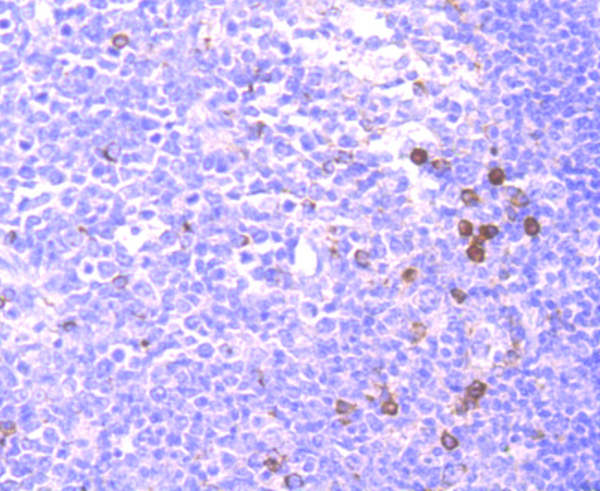
 Application Data
(Immunoperoxidase staining of rat lymph node cryosection with Mouse anti Rat CD25 followed by horseradish peroxidase conjugated Goat anti Mouse IgG1 as a detection reagent. High power)
Application Data
(Immunoperoxidase staining of rat lymph node cryosection with Mouse anti Rat CD25 followed by horseradish peroxidase conjugated Goat anti Mouse IgG1 as a detection reagent. High power)
CD25, Monoclonal Antibody (Cat# AAA11965)
Full Name
MOUSE ANTI RAT CD25
Gene Names
Il2ra; IL2RAC
Applications
Immunohistochemistry, Flow Cytometry, Immunoprecipitation, Immunohistochemistry
Pricing
 WB (Western Blot)
(Western Blot analysis using AAA14675 (1:1000)Lane 1: Human Interleukin 23, p40 fusion proteinLane 2: Human Interleukin 23, p19 fusion protein. Lane 3: Recombinant human single chain Interleukin 23 (DYKDDDDK-tagged))
WB (Western Blot)
(Western Blot analysis using AAA14675 (1:1000)Lane 1: Human Interleukin 23, p40 fusion proteinLane 2: Human Interleukin 23, p19 fusion protein. Lane 3: Recombinant human single chain Interleukin 23 (DYKDDDDK-tagged))
Interleukin 23, p40, Monoclonal Antibody (Cat# AAA14675)
Full Name
Interleukin 23, p40 (IL-23)
Gene Names
IL23A; P19; SGRF; IL-23; IL-23A; IL23P19
Reactivity
Human
Applications
Western Blot
Purity
Purified
Purified
Purified
Pricing


 WB (Western Blot)
(IL20 monoclonal antibody, Western Blot analysis of IL20 expression in K-562.)
WB (Western Blot)
(IL20 monoclonal antibody, Western Blot analysis of IL20 expression in K-562.)
Interleukin 20, Monoclonal Antibody (Cat# AAA25428)
Full Name
Interleukin 20 (Interleukin-20, IL-20, IL20, Cytokine Zcyto10, IL10D, MGC96907, UNQ852/PRO1801, ZCYTO10) (HRP)
Gene Names
IL20; IL-20; IL10D; ZCYTO10
Reactivity
Human
Applications
Immunoprecipitation, Western Blot
Purity
Purified by Protein A Affinity Chromatography.
Pricing
 Standard Curve (Sample)
Interleukin 12, ELISA Kit (Cat# AAA17599)
Full Name
Human Interleukin 12 ELISA Kit
Gene Names
IL12A; P35; CLMF; NFSK; NKSF1; IL-12A
Reactivity
Human
Pricing


 Application Data
(Immunoperoxidase staining of rat lymph node cryosection with Mouse anti Rat CD25 followed by horseradish peroxidase conjugated Goat anti Mouse IgG1 as a detection reagent. High power)
Application Data
(Immunoperoxidase staining of rat lymph node cryosection with Mouse anti Rat CD25 followed by horseradish peroxidase conjugated Goat anti Mouse IgG1 as a detection reagent. High power)
CD25, Monoclonal Antibody (Cat# AAA11966)
Full Name
MOUSE ANTI RAT CD25
Gene Names
Il2ra; IL2RAC
Applications
Immunohistochemistry, Flow Cytometry, Immunoprecipitation, Immunohistochemistry
Pricing


 WB (Western Blot)
(IL20 monoclonal antibody, Western Blot analysis of IL20 expression in K-562.)
WB (Western Blot)
(IL20 monoclonal antibody, Western Blot analysis of IL20 expression in K-562.)
Interleukin 20, Monoclonal Antibody (Cat# AAA25724)
Full Name
Interleukin 20 (Interleukin-20, IL-20, IL20, Cytokine Zcyto10, IL10D, MGC96907, UNQ852/PRO1801, ZCYTO10) (PE)
Gene Names
IL20; IL-20; IL10D; ZCYTO10
Reactivity
Human
Applications
Immunoprecipitation, Western Blot
Purity
Purified by Protein A Affinity Chromatography.
Pricing