Filters
Clonality
Type
Reactivity
Gene Name
Isotype
Host
Application
Clone
115 results for " Tumor B" - showing 1-50


 WB (Western Blot)
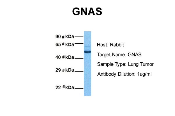
(WB Suggested Anti-GNAS Antibody Titration: 1 ug/mlPositive Control: MCF-7 whole cell lysatesGNAS is strongly supported by BioGPS gene expression data to be expressed in Human MCF7 cells)
WB (Western Blot)
(WB Suggested Anti-GNAS Antibody Titration: 1 ug/mlPositive Control: MCF-7 whole cell lysatesGNAS is strongly supported by BioGPS gene expression data to be expressed in Human MCF7 cells)
GNAS, Polyclonal Antibody (Cat# AAA23503)
Full Name
GNAS antibody - N-terminal region
Gene Names
GNAS; AHO; GSA; GSP; POH; GPSA; NESP; SCG6; SgVI; GNAS1; PITA3; C20orf45
Reactivity
Cow, Dog, Guinea Pig, Horse, Human, Mouse, Rabbit, Rat
Applications
IHC, WB
Purity
Affinity Purified
Pricing


 Application Data
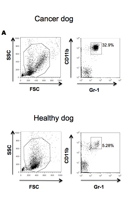
(Published customer image:RPE conjugated Mouse anti Canine CD4 antibody, clone YKIC302.9used for the assessment of CD4 levels on canine cells by flow cytometry.Image caption:Immunophenotypic profile of tumor infiltrating lymphocyte in canine mammary carcinomas. Analysis of tumor infiltrating T-cells, B-lymphocytes and T-cell subsets from MC-BMT or MC (A), further subcategorized according to the absence (-) or presence (+) of lymph node metastasis (-) (B). Lymphocyte populations and subsets were identified by flow cytometric immunostaining as described in Material and Methods. Data were expressed as percentage of positive cells within gated lymphocytes and CD4+/CD8+ T-cell ratio. Significant differences at p < 0.05 are highlighted by asterisk.)
Application Data
(Published customer image:RPE conjugated Mouse anti Canine CD4 antibody, clone YKIC302.9used for the assessment of CD4 levels on canine cells by flow cytometry.Image caption:Immunophenotypic profile of tumor infiltrating lymphocyte in canine mammary carcinomas. Analysis of tumor infiltrating T-cells, B-lymphocytes and T-cell subsets from MC-BMT or MC (A), further subcategorized according to the absence (-) or presence (+) of lymph node metastasis (-) (B). Lymphocyte populations and subsets were identified by flow cytometric immunostaining as described in Material and Methods. Data were expressed as percentage of positive cells within gated lymphocytes and CD4+/CD8+ T-cell ratio. Significant differences at p < 0.05 are highlighted by asterisk.)
CD4, Monoclonal Antibody (Cat# AAA12278)
Full Name
RAT ANTI DOG CD4: APC
Reactivity
Dog
Applications
Flow Cytometry
Pricing
 SDS-PAGE
Aldose reductase, Active Protein (Cat# AAA11762)
Full Name
Aldo-keta reductase family 1, member B1
Gene Names
AKR1B1; AR; ADR; ALR2; ALDR1
Applications
SDS-PAGE
Purity
> 95% by SDS-PAGE
Pricing


 Application Data
(Figure 10 KD Validation in HeLa cells (Horinaka et al., 2005)HeLa cells were transfected with DR4siRNA or LacZ control siRNA. At 24 h after transfection, the cells were treated with or without 20 ?M luteolin for 24 h. Western blot analysis was carried out with anti-DR4 antibodies (1139). DR4 expression was markedly reduced after DR4 knockdown.)
Application Data
(Figure 10 KD Validation in HeLa cells (Horinaka et al., 2005)HeLa cells were transfected with DR4siRNA or LacZ control siRNA. At 24 h after transfection, the cells were treated with or without 20 ?M luteolin for 24 h. Western blot analysis was carried out with anti-DR4 antibodies (1139). DR4 expression was markedly reduced after DR4 knockdown.)
DR4, Polyclonal Antibody (Cat# AAA10952)
Full Name
DR4 Antibody
Gene Names
TNFRSF10A; DR4; APO2; CD261; TRAILR1; TRAILR-1
Reactivity
Human
Applications
Western Blot, Immunocytochemistry
Purity
DR4 Antibody is Antibody is affinity chromatography purified via peptide column.
Pricing


 Application Data
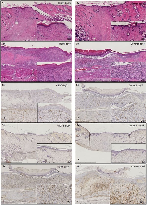
(Published customer image: Histological staining of control and HBOT wounds at post-wounding days 7 and 29. A -D) H&E staining. E -H) CD34 immunohistochemistry. I+J) CD68 immunohistochemistry.From: uk B, Tong M, Fijneman EMG, van Neck JW (2014) Hyperbaric Oxygen Therapy to Treat Diabetes Impaired Wound Healing in Rats. PLoS ONE 9(10): e108533.)
Application Data
(Published customer image: Histological staining of control and HBOT wounds at post-wounding days 7 and 29. A -D) H&E staining. E -H) CD34 immunohistochemistry. I+J) CD68 immunohistochemistry.From: uk B, Tong M, Fijneman EMG, van Neck JW (2014) Hyperbaric Oxygen Therapy to Treat Diabetes Impaired Wound Healing in Rats. PLoS ONE 9(10): e108533.)
CD68, Monoclonal Antibody (Cat# AAA12150)
Full Name
MOUSE ANTI RAT CD68:RPE
Applications
Flow Cytometry
Pricing


 Application Data
(Staining of canine peripheral blood lymphocytes with Rat anti Canine CD8:RPE)
Application Data
(Staining of canine peripheral blood lymphocytes with Rat anti Canine CD8:RPE)
CD8, Monoclonal Antibody (Cat# AAA12066)
Full Name
RAT ANTI DOG CD8:FITC
Applications
Flow Cytometry
Pricing


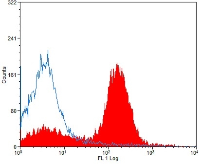
 FCM (Flow Cytometry)
(Flow cytometric analysis of THP-1 cells with CD40L antibody at 1/50 dilution (red) compared with an unlabelled control (cells without incubation with primary antibody; black). Alexa Fluor 488-conjugated goat anti rabbit IgG was used as the secondary antibody.)
FCM (Flow Cytometry)
(Flow cytometric analysis of THP-1 cells with CD40L antibody at 1/50 dilution (red) compared with an unlabelled control (cells without incubation with primary antibody; black). Alexa Fluor 488-conjugated goat anti rabbit IgG was used as the secondary antibody.)
CD40L, Monoclonal Antibody (Cat# AAA30328)
Full Name
CD40L Antibody
Gene Names
CD40LG; IGM; IMD3; TRAP; gp39; CD154; CD40L; HIGM1; T-BAM; TNFSF5; hCD40L
Reactivity
Human, Mouse
Applications
Western Blot, Immunocytochemistry, Immunofluorescence, Immunohistochemistry, Flow Cytometry
Purity
ProA affinity purified
Pricing


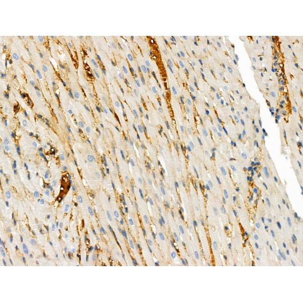
 IF (Immunofluorescence)
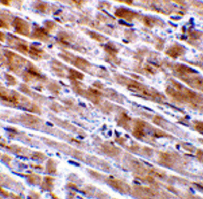
(Immunofluorescence of CTRP3 in Mouse Heart tissue with CTRP3 antibody at 10 μg/mL.)
IF (Immunofluorescence)
(Immunofluorescence of CTRP3 in Mouse Heart tissue with CTRP3 antibody at 10 μg/mL.)
CTRP3, Polyclonal Antibody (Cat# AAA10961)
Full Name
CTRP3 Antibody
Gene Names
C1QTNF3; CORS; CORCS; CTRP3; CORS26; C1ATNF3; CORS-26
Reactivity
Human, Mouse, Rat
Applications
Western Blot, Immunohistochemistry, Immunofluorescence
Purity
CTRP3 Antibody is affinity chromatography purified via peptide column.
Pricing


 Application Data
(Staining of canine peripheral blood lymphocytes with Rat anti Canine CD8:RPE)
Application Data
(Staining of canine peripheral blood lymphocytes with Rat anti Canine CD8:RPE)
CD8, Monoclonal Antibody (Cat# AAA12067)
Full Name
RAT ANTI DOG CD8:RPE
Applications
Flow Cytometry
Pricing


 Application Data
(Published customer image: Effects of EDTA versus media storage of patient blood samples on flow cytometric results. Peripheral blood cells were (a) stained immediate following collection with CD11b and CADO48A or maintained in EDTA collection tubes for (b) 24 or (c) 48 hours or in media for (d) 24 or (e) 48 hours prior to staining with CD11b and CADO48A. Both EDTA and media samples were kept refrigerated. These findings show a decrease over time of population distinction in EDTA with minimal changes when cells are kept in media up to 48 hours. All cells were gated on P1.From: Sherger et al. BMC Veterinary Research 2012 8:209)
Application Data
(Published customer image: Effects of EDTA versus media storage of patient blood samples on flow cytometric results. Peripheral blood cells were (a) stained immediate following collection with CD11b and CADO48A or maintained in EDTA collection tubes for (b) 24 or (c) 48 hours or in media for (d) 24 or (e) 48 hours prior to staining with CD11b and CADO48A. Both EDTA and media samples were kept refrigerated. These findings show a decrease over time of population distinction in EDTA with minimal changes when cells are kept in media up to 48 hours. All cells were gated on P1.From: Sherger et al. BMC Veterinary Research 2012 8:209)
CD11b, Monoclonal Antibody (Cat# AAA12092)
Full Name
MOUSE ANTI DOG CD11b
Gene Names
ITGAM; CD11B
Applications
Immunohistochemistry, Flow Cytometry, Immunoprecipitation
Pricing


 Application Data
(Published customer image: Histological staining of control and HBOT wounds at post-wounding days 7 and 29. A -D) H&E staining. E -H) CD34 immunohistochemistry. I+J) CD68 immunohistochemistry.From: uk B, Tong M, Fijneman EMG, van Neck JW (2014) Hyperbaric Oxygen Therapy to Treat Diabetes Impaired Wound Healing in Rats. PLoS ONE 9(10): e108533.)
Application Data
(Published customer image: Histological staining of control and HBOT wounds at post-wounding days 7 and 29. A -D) H&E staining. E -H) CD34 immunohistochemistry. I+J) CD68 immunohistochemistry.From: uk B, Tong M, Fijneman EMG, van Neck JW (2014) Hyperbaric Oxygen Therapy to Treat Diabetes Impaired Wound Healing in Rats. PLoS ONE 9(10): e108533.)
CD68, Monoclonal Antibody (Cat# AAA12149)
Full Name
MOUSE ANTI RAT CD68
Applications
Immunohistochemistry, Flow Cytometry, Immunofluorescence, Immunoprecipitation, Immunohistochemistry
Pricing


 Application Data
(Staining of mouse splenocytes with Rat anti Mouse CD40: RPE)
Application Data
(Staining of mouse splenocytes with Rat anti Mouse CD40: RPE)
CD40, Monoclonal Antibody (Cat# AAA12023)
Full Name
RAT ANTI MOUSE CD40:RPE
Gene Names
Cd40; IGM; p50; Bp50; GP39; IMD3; TRAP; HIGM1; T-BAM; Tnfrsf5; AI326936
Applications
Flow Cytometry
Pricing


 Application Data
(Detection limit for recombinant GST tagged NME1 is approximately 0.1ng/ml as a capture antibody.)
Application Data
(Detection limit for recombinant GST tagged NME1 is approximately 0.1ng/ml as a capture antibody.)
NME1, Monoclonal Antibody (Cat# AAA26300)
Full Name
NME1 (Non-Metastatic Cells 1, Protein (NM23A) Expressed in, AWD, GAAD, NB, NBS, NDPK-A, NDPKA, NM23, NM23-H1) (FITC)
Gene Names
NME1; NB; AWD; NBS; GAAD; NDKA; NM23; NDPKA; NDPK-A; NM23-H1
Applications
IF, IHC, IP, WB
Purity
Purified
Pricing


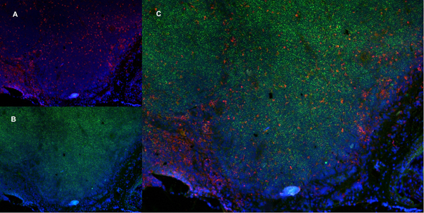
 Application Data
(Published Customer Image:Mouse CD31 antibody, clone ER-MP12 used for the demonstration of vasculature in mouse brain by immunofluorescence.Image caption:Inhibition of 2-AG hydrolysis reduces LPS-induced BBB permeability. a, b Fibrinogen levels in b plasma and the a ratio of brain to plasma fibrinogen were assessed by ELISA. n?=?5/7 mice per group. c, d Fluorescent immunostaining in the striatum for fibrinogen (red) and vascular marker (CD31; green) demonstrated leakage of fibrinogen into the brain with vehicle treatment, whereas vascular integrity was preserved when (e, f) MAGL was inhibited. g Extravascular fibrinogen was semi-quantitated in fluorescently labeled sections of the striatum. Bar graphs were plotted with mean?+/-SEM and data analyzed using one-way analysis of variance (ANOVA) with Tukey post-hoc comparisons. n = 5/7 mice per group. Significance is shown as *p?)
Application Data
(Published Customer Image:Mouse CD31 antibody, clone ER-MP12 used for the demonstration of vasculature in mouse brain by immunofluorescence.Image caption:Inhibition of 2-AG hydrolysis reduces LPS-induced BBB permeability. a, b Fibrinogen levels in b plasma and the a ratio of brain to plasma fibrinogen were assessed by ELISA. n?=?5/7 mice per group. c, d Fluorescent immunostaining in the striatum for fibrinogen (red) and vascular marker (CD31; green) demonstrated leakage of fibrinogen into the brain with vehicle treatment, whereas vascular integrity was preserved when (e, f) MAGL was inhibited. g Extravascular fibrinogen was semi-quantitated in fluorescently labeled sections of the striatum. Bar graphs were plotted with mean?+/-SEM and data analyzed using one-way analysis of variance (ANOVA) with Tukey post-hoc comparisons. n = 5/7 mice per group. Significance is shown as *p?)
CD31, Monoclonal Antibody (Cat# AAA12258)
Full Name
Rat Anti Mouse CD31: FITC
Gene Names
Pecam1; Cd31; Pecam; C85791; PECAM-1
Reactivity
Mouse
Applications
Flow Cytometry
Purity
Purified IgG prepared by affinity chromatography on Protein G from tissue culture supernatant
Pricing


 WB (Western Blot)
(Western blot analysis of Human Lysates showing detection of Hsp90 protein using Mouse Anti-Hsp90 Monoclonal Antibody, Clone H9010 . Primary Antibody: Mouse Anti-Hsp90 Monoclonal Antibody at 1:1000. Comparison of clone H9010 behavior with Hsp90 human beta (1) and Hsp90 human alpha (2). Courtesy of: David Toft, Mayo Clinic.)
WB (Western Blot)
(Western blot analysis of Human Lysates showing detection of Hsp90 protein using Mouse Anti-Hsp90 Monoclonal Antibody, Clone H9010 . Primary Antibody: Mouse Anti-Hsp90 Monoclonal Antibody at 1:1000. Comparison of clone H9010 behavior with Hsp90 human beta (1) and Hsp90 human alpha (2). Courtesy of: David Toft, Mayo Clinic.)
HSP90, Monoclonal Antibody (Cat# AAA27660)
Full Name
HSP90 Antibody
Gene Names
HSP90AB1; HSP84; HSPC2; HSPCB; D6S182; HSP90B
Reactivity
Human, Mouse, Rat, Rabbit, Chicken, Dog, Fish, Shark, Hamster
Applications
Western Blot, Immunohistochemistry, Immunocytochemistry, Immunofluorescence, Immunoprecipitation, Antibody Microarray
Purity
Protein G Purified
Pricing


 Application Data
(Published customer image Infiltration of GFP+ BM-cells in infarct and peri-infarct regions. (A-B) Dot plots of viable macrophages/granulocytes (CD11b+CD45high, top right quadrants) and microglia (CD11b+CD45dim, bottom right quadrants) in cortex from BM-chimeric unmanipulated mice and mice exposed to pMCAO. (C) Bar graph showing mean numbers of CD11b+CD45dim microglia and CD11b+CD45high macrophages/granulocytes in BM-chimeric mice 24 hours after pMCAO, subdivided based on expression of GFP (n = 5). Approximately 92% of of the CD45high population were GFP+. (D) Estimation and comparison of mean numbers of CD11b+CD45dim microglia in non-chimeric (n = 10) versus BM-chimeric mice (n = 5) 24 hours after of pMCAO shows significantly fewer CD11b+CD45dim microglial cells in irradiated mice. (E) Overview, showing distribution of infiltrating GFP+ BM-derived cells into infarct (IF) and peri-infarct (P-IF) regions 24 hours after pMCAO. (E-G) By 24 hours, GFP+ single cells (F) and vessel-associated aggregates of GFP+ cells (arrows in G) were observed in infarct and peri-infarct regions. Some of the vessel-associated cells were round, leukocyte-like cells (arrows) while others were elongated cells lining the vasculature (arrow heads in G and in insert). (H) Bar graph showing mean numbers of single GFP+ cells and vessel-associated aggregates of GFP+ cells in ipsi- and contralateral cortex 24 hours after surgery (n = 10). (I-P) Immunohistochemical staining of CD45.1 (I, K), CD45.2 (J, L), IgG2a (M, O) and CD45 (N, P) in ischemic tissue in BM-chimeric (I, J, M, N) and non-chimeric mice (K, L, O, P) 24 hours after pMCAO. N.D, none detected. Scale bars: 200 um (A), 10 um (B, C). 50 um (I-P) *P < 0.05, **P < 0.01, and ***P < 0.001.From: Clausen BH, Lambertsen KL, Babcock AA, Holm TH, Dagnaes-Hansen F, Finsen B. Interleukin-1beta and tumor necrosis factor-alpha are expressed by different subsets of microglia and macrophages after ischemic stroke in mice. J Neuroinflammation. 2008 Oct 23;5:46.)
Application Data
(Published customer image Infiltration of GFP+ BM-cells in infarct and peri-infarct regions. (A-B) Dot plots of viable macrophages/granulocytes (CD11b+CD45high, top right quadrants) and microglia (CD11b+CD45dim, bottom right quadrants) in cortex from BM-chimeric unmanipulated mice and mice exposed to pMCAO. (C) Bar graph showing mean numbers of CD11b+CD45dim microglia and CD11b+CD45high macrophages/granulocytes in BM-chimeric mice 24 hours after pMCAO, subdivided based on expression of GFP (n = 5). Approximately 92% of of the CD45high population were GFP+. (D) Estimation and comparison of mean numbers of CD11b+CD45dim microglia in non-chimeric (n = 10) versus BM-chimeric mice (n = 5) 24 hours after of pMCAO shows significantly fewer CD11b+CD45dim microglial cells in irradiated mice. (E) Overview, showing distribution of infiltrating GFP+ BM-derived cells into infarct (IF) and peri-infarct (P-IF) regions 24 hours after pMCAO. (E-G) By 24 hours, GFP+ single cells (F) and vessel-associated aggregates of GFP+ cells (arrows in G) were observed in infarct and peri-infarct regions. Some of the vessel-associated cells were round, leukocyte-like cells (arrows) while others were elongated cells lining the vasculature (arrow heads in G and in insert). (H) Bar graph showing mean numbers of single GFP+ cells and vessel-associated aggregates of GFP+ cells in ipsi- and contralateral cortex 24 hours after surgery (n = 10). (I-P) Immunohistochemical staining of CD45.1 (I, K), CD45.2 (J, L), IgG2a (M, O) and CD45 (N, P) in ischemic tissue in BM-chimeric (I, J, M, N) and non-chimeric mice (K, L, O, P) 24 hours after pMCAO. N.D, none detected. Scale bars: 200 um (A), 10 um (B, C). 50 um (I-P) *P < 0.05, **P < 0.01, and ***P < 0.001.From: Clausen BH, Lambertsen KL, Babcock AA, Holm TH, Dagnaes-Hansen F, Finsen B. Interleukin-1beta and tumor necrosis factor-alpha are expressed by different subsets of microglia and macrophages after ischemic stroke in mice. J Neuroinflammation. 2008 Oct 23;5:46.)
CD11b, Monoclonal Antibody (Cat# AAA12182)
Full Name
RAT ANTI MOUSE CD11b:FITC
Gene Names
Itgam; CR3; CR3A; MAC1; Cd11b; Ly-40; Mac-1; Mac-1a; CD11b/CD18; F730045J24Rik
Applications
Flow Cytometry
Pricing


 Application Data
(Published customer image Infiltration of GFP+ BM-cells in infarct and peri-infarct regions. (A-B) Dot plots of viable macrophages/granulocytes (CD11b+CD45high, top right quadrants) and microglia (CD11b+CD45dim, bottom right quadrants) in cortex from BM-chimeric unmanipulated mice and mice exposed to pMCAO. (C) Bar graph showing mean numbers of CD11b+CD45dim microglia and CD11b+CD45high macrophages/granulocytes in BM-chimeric mice 24 hours after pMCAO, subdivided based on expression of GFP (n = 5). Approximately 92% of of the CD45high population were GFP+. (D) Estimation and comparison of mean numbers of CD11b+CD45dim microglia in non-chimeric (n = 10) versus BM-chimeric mice (n = 5) 24 hours after of pMCAO shows significantly fewer CD11b+CD45dim microglial cells in irradiated mice. (E) Overview, showing distribution of infiltrating GFP+ BM-derived cells into infarct (IF) and peri-infarct (P-IF) regions 24 hours after pMCAO. (E-G) By 24 hours, GFP+ single cells (F) and vessel-associated aggregates of GFP+ cells (arrows in G) were observed in infarct and peri-infarct regions. Some of the vessel-associated cells were round, leukocyte-like cells (arrows) while others were elongated cells lining the vasculature (arrow heads in G and in insert). (H) Bar graph showing mean numbers of single GFP+ cells and vessel-associated aggregates of GFP+ cells in ipsi- and contralateral cortex 24 hours after surgery (n = 10). (I-P) Immunohistochemical staining of CD45.1 (I, K), CD45.2 (J, L), IgG2a (M, O) and CD45 (N, P) in ischemic tissue in BM-chimeric (I, J, M, N) and non-chimeric mice (K, L, O, P) 24 hours after pMCAO. N.D, none detected. Scale bars: 200 um (A), 10 um (B, C). 50 um (I-P) *P < 0.05, **P < 0.01, and ***P < 0.001.From: Clausen BH, Lambertsen KL, Babcock AA, Holm TH, Dagnaes-Hansen F, Finsen B. Interleukin-1beta and tumor necrosis factor-alpha are expressed by different subsets of microglia and macrophages after ischemic stroke in mice. J Neuroinflammation. 2008 Oct 23;5:46.)
Application Data
(Published customer image Infiltration of GFP+ BM-cells in infarct and peri-infarct regions. (A-B) Dot plots of viable macrophages/granulocytes (CD11b+CD45high, top right quadrants) and microglia (CD11b+CD45dim, bottom right quadrants) in cortex from BM-chimeric unmanipulated mice and mice exposed to pMCAO. (C) Bar graph showing mean numbers of CD11b+CD45dim microglia and CD11b+CD45high macrophages/granulocytes in BM-chimeric mice 24 hours after pMCAO, subdivided based on expression of GFP (n = 5). Approximately 92% of of the CD45high population were GFP+. (D) Estimation and comparison of mean numbers of CD11b+CD45dim microglia in non-chimeric (n = 10) versus BM-chimeric mice (n = 5) 24 hours after of pMCAO shows significantly fewer CD11b+CD45dim microglial cells in irradiated mice. (E) Overview, showing distribution of infiltrating GFP+ BM-derived cells into infarct (IF) and peri-infarct (P-IF) regions 24 hours after pMCAO. (E-G) By 24 hours, GFP+ single cells (F) and vessel-associated aggregates of GFP+ cells (arrows in G) were observed in infarct and peri-infarct regions. Some of the vessel-associated cells were round, leukocyte-like cells (arrows) while others were elongated cells lining the vasculature (arrow heads in G and in insert). (H) Bar graph showing mean numbers of single GFP+ cells and vessel-associated aggregates of GFP+ cells in ipsi- and contralateral cortex 24 hours after surgery (n = 10). (I-P) Immunohistochemical staining of CD45.1 (I, K), CD45.2 (J, L), IgG2a (M, O) and CD45 (N, P) in ischemic tissue in BM-chimeric (I, J, M, N) and non-chimeric mice (K, L, O, P) 24 hours after pMCAO. N.D, none detected. Scale bars: 200 um (A), 10 um (B, C). 50 um (I-P) *P < 0.05, **P < 0.01, and ***P < 0.001.From: Clausen BH, Lambertsen KL, Babcock AA, Holm TH, Dagnaes-Hansen F, Finsen B. Interleukin-1beta and tumor necrosis factor-alpha are expressed by different subsets of microglia and macrophages after ischemic stroke in mice. J Neuroinflammation. 2008 Oct 23;5:46.)
CD11b, Monoclonal Antibody (Cat# AAA12184)
Full Name
RAT ANTI MOUSE CD11b
Gene Names
Itgam; CR3; CR3A; MAC1; Cd11b; Ly-40; Mac-1; Mac-1a; CD11b/CD18; F730045J24Rik
Applications
Immunohistochemistry, Flow Cytometry, Immunofluorescence, Immunoprecipitation
Pricing


 WB (Western Blot)
(Figure 9 KD Validation in HeLa Cells (Horinaka et al., 2005)HeLa cells were transfected with DR4siRNA or LacZ control siRNA. At 24 h after transfection, the cells were treated with or without 20 μM luteolin for 24 h. Western blot analysis was carried out with anti-DR4 antibodies. DR4 expression was markedly reduced after DR4 knockdown.)
WB (Western Blot)
(Figure 9 KD Validation in HeLa Cells (Horinaka et al., 2005)HeLa cells were transfected with DR4siRNA or LacZ control siRNA. At 24 h after transfection, the cells were treated with or without 20 μM luteolin for 24 h. Western blot analysis was carried out with anti-DR4 antibodies. DR4 expression was markedly reduced after DR4 knockdown.)
DR4, Polyclonal Antibody (Cat# AAA10950)
Full Name
DR4 Antibody
Gene Names
TNFRSF10A; DR4; APO2; CD261; TRAILR1; TRAILR-1
Reactivity
Human
Applications
Western Blot
Purity
DR4 Antibody is affinity chromatography purified via peptide column.
Pricing


 Application Data
(At 25 degree C. Samples were then incubated with primary Ab(At 37 degree C. An AlexaFluor594 conjugated goat anti-rabbit IgG(H+L) Ab(Red) and an AlexaFluor488 conjugated goat anti-mouse IgG(H+L) Ab(Green) were used as the secondary antibody.The nuclear counter stain is DAPI (blue).)
Application Data
(At 25 degree C. Samples were then incubated with primary Ab(At 37 degree C. An AlexaFluor594 conjugated goat anti-rabbit IgG(H+L) Ab(Red) and an AlexaFluor488 conjugated goat anti-mouse IgG(H+L) Ab(Green) were used as the secondary antibody.The nuclear counter stain is DAPI (blue).)
SIRT2, Polyclonal Antibody (Cat# AAA31286)
Full Name
Phospho-SIRT2 (Ser368) Antibody
Gene Names
SIRT2; SIR2; SIR2L; SIR2L2
Reactivity
Human, Mouse, Rat
Applications
Immunohistochemistry, Immunofluorescence, Immunocytochemistry, Peptide ELISA
Purity
The antibody is from purified rabbit serum by affinity purification via sequential chromatography on phospho-peptide and non-phospho-peptide affinity columns.
Pricing


 Application Data
(Staining of canine peripheral blood lymphocytes with Rat anti Canine CD4: Alexa Fluor 488)
Application Data
(Staining of canine peripheral blood lymphocytes with Rat anti Canine CD4: Alexa Fluor 488)
CD4, Monoclonal Antibody (Cat# AAA12064)
Full Name
RAT ANTI DOG CD4:FITC
Applications
Flow Cytometry
Pricing


 Application Data
(Published customer image: Histological staining of control and HBOT wounds at post-wounding days 7 and 29. A -D) H&E staining. E -H) CD34 immunohistochemistry. I+J) CD68 immunohistochemistry.From: uk B, Tong M, Fijneman EMG, van Neck JW (2014) Hyperbaric Oxygen Therapy to Treat Diabetes Impaired Wound Healing in Rats. PLoS ONE 9(10): e108533.)
Application Data
(Published customer image: Histological staining of control and HBOT wounds at post-wounding days 7 and 29. A -D) H&E staining. E -H) CD34 immunohistochemistry. I+J) CD68 immunohistochemistry.From: uk B, Tong M, Fijneman EMG, van Neck JW (2014) Hyperbaric Oxygen Therapy to Treat Diabetes Impaired Wound Healing in Rats. PLoS ONE 9(10): e108533.)
CD68, Monoclonal Antibody (Cat# AAA12147)
Full Name
MOUSE ANTI RAT CD68:Biotin
Applications
Flow Cytometry
Pricing


 Application Data
(Staining of mouse splenocytes with Rat anti Mouse CD40: RPE)
Application Data
(Staining of mouse splenocytes with Rat anti Mouse CD40: RPE)
CD40, Monoclonal Antibody (Cat# AAA11919)
Full Name
RAT ANTI MOUSE CD40
Gene Names
Cd40; IGM; p50; Bp50; GP39; IMD3; TRAP; HIGM1; T-BAM; Tnfrsf5; AI326936
Applications
Immunohistochemistry, Flow Cytometry
Pricing


 IHC (Immunohistochemistry)
(At 1/100 staining Mouse lung tissue by IHC-P. The sample was formaldehyde fixed and a heat mediated antigen retrieval step in citrate buffer was performed. The sample was then blocked and incubated with the primary antibody at 4 degree C overnight. An HRP conjugated anti-Rabbit antibody was used as the secondary antibody.)
IHC (Immunohistochemistry)
(At 1/100 staining Mouse lung tissue by IHC-P. The sample was formaldehyde fixed and a heat mediated antigen retrieval step in citrate buffer was performed. The sample was then blocked and incubated with the primary antibody at 4 degree C overnight. An HRP conjugated anti-Rabbit antibody was used as the secondary antibody.)
Histone H2A, Polyclonal Antibody (Cat# AAA31347)
Full Name
Acetyl-Histone H2A (Lys5) Antibody
Gene Names
HIST1H2AB; H2A/m; H2AFM
Reactivity
Human, Mouse, Rat
Applications
Western Blot, Immunohistochemistry, Peptide ELISA
Purity
The antiserum was purified by peptide affinity chromatography using SulfoLink Coupling Resin
Pricing


 WB (Western Blot)
(WB Suggested Anti-HNF4A antibody Titration: 1 ug/mLSample Type: Human Liver)
WB (Western Blot)
(WB Suggested Anti-HNF4A antibody Titration: 1 ug/mLSample Type: Human Liver)
HNF4A, Polyclonal Antibody (Cat# AAA23401)
Full Name
HNF4A Antibody - N-terminal region
Gene Names
HNF4A; TCF; HNF4; MODY; FRTS4; MODY1; NR2A1; TCF14; HNF4a7; HNF4a8; HNF4a9; NR2A21; HNF4alpha
Reactivity
Cow, Dog, Guinea Pig, Horse, Human, Mouse, Rabbit, Rat, Sheep, Zebrafish
Applications
Western Blot
Purity
Affinity Purified
Pricing


 Application Data
(Published customer image Infiltration of GFP+ BM-cells in infarct and peri-infarct regions. (A-B) Dot plots of viable macrophages/granulocytes (CD11b+CD45high, top right quadrants) and microglia (CD11b+CD45dim, bottom right quadrants) in cortex from BM-chimeric unmanipulated mice and mice exposed to pMCAO. (C) Bar graph showing mean numbers of CD11b+CD45dim microglia and CD11b+CD45high macrophages/granulocytes in BM-chimeric mice 24 hours after pMCAO, subdivided based on expression of GFP (n = 5). Approximately 92% of of the CD45high population were GFP+. (D) Estimation and comparison of mean numbers of CD11b+CD45dim microglia in non-chimeric (n = 10) versus BM-chimeric mice (n = 5) 24 hours after of pMCAO shows significantly fewer CD11b+CD45dim microglial cells in irradiated mice. (E) Overview, showing distribution of infiltrating GFP+ BM-derived cells into infarct (IF) and peri-infarct (P-IF) regions 24 hours after pMCAO. (E-G) By 24 hours, GFP+ single cells (F) and vessel-associated aggregates of GFP+ cells (arrows in G) were observed in infarct and peri-infarct regions. Some of the vessel-associated cells were round, leukocyte-like cells (arrows) while others were elongated cells lining the vasculature (arrow heads in G and in insert). (H) Bar graph showing mean numbers of single GFP+ cells and vessel-associated aggregates of GFP+ cells in ipsi- and contralateral cortex 24 hours after surgery (n = 10). (I-P) Immunohistochemical staining of CD45.1 (I, K), CD45.2 (J, L), IgG2a (M, O) and CD45 (N, P) in ischemic tissue in BM-chimeric (I, J, M, N) and non-chimeric mice (K, L, O, P) 24 hours after pMCAO. N.D, none detected. Scale bars: 200 um (A), 10 um (B, C). 50 um (I-P) *P < 0.05, **P < 0.01, and ***P < 0.001.From: Clausen BH, Lambertsen KL, Babcock AA, Holm TH, Dagnaes-Hansen F, Finsen B. Interleukin-1beta and tumor necrosis factor-alpha are expressed by different subsets of microglia and macrophages after ischemic stroke in mice. J Neuroinflammation. 2008 Oct 23;5:46.)
Application Data
(Published customer image Infiltration of GFP+ BM-cells in infarct and peri-infarct regions. (A-B) Dot plots of viable macrophages/granulocytes (CD11b+CD45high, top right quadrants) and microglia (CD11b+CD45dim, bottom right quadrants) in cortex from BM-chimeric unmanipulated mice and mice exposed to pMCAO. (C) Bar graph showing mean numbers of CD11b+CD45dim microglia and CD11b+CD45high macrophages/granulocytes in BM-chimeric mice 24 hours after pMCAO, subdivided based on expression of GFP (n = 5). Approximately 92% of of the CD45high population were GFP+. (D) Estimation and comparison of mean numbers of CD11b+CD45dim microglia in non-chimeric (n = 10) versus BM-chimeric mice (n = 5) 24 hours after of pMCAO shows significantly fewer CD11b+CD45dim microglial cells in irradiated mice. (E) Overview, showing distribution of infiltrating GFP+ BM-derived cells into infarct (IF) and peri-infarct (P-IF) regions 24 hours after pMCAO. (E-G) By 24 hours, GFP+ single cells (F) and vessel-associated aggregates of GFP+ cells (arrows in G) were observed in infarct and peri-infarct regions. Some of the vessel-associated cells were round, leukocyte-like cells (arrows) while others were elongated cells lining the vasculature (arrow heads in G and in insert). (H) Bar graph showing mean numbers of single GFP+ cells and vessel-associated aggregates of GFP+ cells in ipsi- and contralateral cortex 24 hours after surgery (n = 10). (I-P) Immunohistochemical staining of CD45.1 (I, K), CD45.2 (J, L), IgG2a (M, O) and CD45 (N, P) in ischemic tissue in BM-chimeric (I, J, M, N) and non-chimeric mice (K, L, O, P) 24 hours after pMCAO. N.D, none detected. Scale bars: 200 um (A), 10 um (B, C). 50 um (I-P) *P < 0.05, **P < 0.01, and ***P < 0.001.From: Clausen BH, Lambertsen KL, Babcock AA, Holm TH, Dagnaes-Hansen F, Finsen B. Interleukin-1beta and tumor necrosis factor-alpha are expressed by different subsets of microglia and macrophages after ischemic stroke in mice. J Neuroinflammation. 2008 Oct 23;5:46.)
CD11b, Monoclonal Antibody (Cat# AAA12185)
Full Name
RAT ANTI MOUSE CD11b
Gene Names
Itgam; CR3; CR3A; MAC1; Cd11b; Ly-40; Mac-1; Mac-1a; CD11b/CD18; F730045J24Rik
Applications
Immunohistochemistry, Flow Cytometry, Immunofluorescence, Immunoprecipitation
Pricing


 Application Data
(Published customer image Infiltration of GFP+ BM-cells in infarct and peri-infarct regions. (A-B) Dot plots of viable macrophages/granulocytes (CD11b+CD45high, top right quadrants) and microglia (CD11b+CD45dim, bottom right quadrants) in cortex from BM-chimeric unmanipulated mice and mice exposed to pMCAO. (C) Bar graph showing mean numbers of CD11b+CD45dim microglia and CD11b+CD45high macrophages/granulocytes in BM-chimeric mice 24 hours after pMCAO, subdivided based on expression of GFP (n = 5). Approximately 92% of of the CD45high population were GFP+. (D) Estimation and comparison of mean numbers of CD11b+CD45dim microglia in non-chimeric (n = 10) versus BM-chimeric mice (n = 5) 24 hours after of pMCAO shows significantly fewer CD11b+CD45dim microglial cells in irradiated mice. (E) Overview, showing distribution of infiltrating GFP+ BM-derived cells into infarct (IF) and peri-infarct (P-IF) regions 24 hours after pMCAO. (E-G) By 24 hours, GFP+ single cells (F) and vessel-associated aggregates of GFP+ cells (arrows in G) were observed in infarct and peri-infarct regions. Some of the vessel-associated cells were round, leukocyte-like cells (arrows) while others were elongated cells lining the vasculature (arrow heads in G and in insert). (H) Bar graph showing mean numbers of single GFP+ cells and vessel-associated aggregates of GFP+ cells in ipsi- and contralateral cortex 24 hours after surgery (n = 10). (I-P) Immunohistochemical staining of CD45.1 (I, K), CD45.2 (J, L), IgG2a (M, O) and CD45 (N, P) in ischemic tissue in BM-chimeric (I, J, M, N) and non-chimeric mice (K, L, O, P) 24 hours after pMCAO. N.D, none detected. Scale bars: 200 um (A), 10 um (B, C). 50 um (I-P) *P < 0.05, **P < 0.01, and ***P < 0.001.From: Clausen BH, Lambertsen KL, Babcock AA, Holm TH, Dagnaes-Hansen F, Finsen B. Interleukin-1beta and tumor necrosis factor-alpha are expressed by different subsets of microglia and macrophages after ischemic stroke in mice. J Neuroinflammation. 2008 Oct 23;5:46.)
Application Data
(Published customer image Infiltration of GFP+ BM-cells in infarct and peri-infarct regions. (A-B) Dot plots of viable macrophages/granulocytes (CD11b+CD45high, top right quadrants) and microglia (CD11b+CD45dim, bottom right quadrants) in cortex from BM-chimeric unmanipulated mice and mice exposed to pMCAO. (C) Bar graph showing mean numbers of CD11b+CD45dim microglia and CD11b+CD45high macrophages/granulocytes in BM-chimeric mice 24 hours after pMCAO, subdivided based on expression of GFP (n = 5). Approximately 92% of of the CD45high population were GFP+. (D) Estimation and comparison of mean numbers of CD11b+CD45dim microglia in non-chimeric (n = 10) versus BM-chimeric mice (n = 5) 24 hours after of pMCAO shows significantly fewer CD11b+CD45dim microglial cells in irradiated mice. (E) Overview, showing distribution of infiltrating GFP+ BM-derived cells into infarct (IF) and peri-infarct (P-IF) regions 24 hours after pMCAO. (E-G) By 24 hours, GFP+ single cells (F) and vessel-associated aggregates of GFP+ cells (arrows in G) were observed in infarct and peri-infarct regions. Some of the vessel-associated cells were round, leukocyte-like cells (arrows) while others were elongated cells lining the vasculature (arrow heads in G and in insert). (H) Bar graph showing mean numbers of single GFP+ cells and vessel-associated aggregates of GFP+ cells in ipsi- and contralateral cortex 24 hours after surgery (n = 10). (I-P) Immunohistochemical staining of CD45.1 (I, K), CD45.2 (J, L), IgG2a (M, O) and CD45 (N, P) in ischemic tissue in BM-chimeric (I, J, M, N) and non-chimeric mice (K, L, O, P) 24 hours after pMCAO. N.D, none detected. Scale bars: 200 um (A), 10 um (B, C). 50 um (I-P) *P < 0.05, **P < 0.01, and ***P < 0.001.From: Clausen BH, Lambertsen KL, Babcock AA, Holm TH, Dagnaes-Hansen F, Finsen B. Interleukin-1beta and tumor necrosis factor-alpha are expressed by different subsets of microglia and macrophages after ischemic stroke in mice. J Neuroinflammation. 2008 Oct 23;5:46.)
CD11b, Monoclonal Antibody (Cat# AAA12183)
Full Name
RAT ANTI MOUSE CD11b:FITC
Gene Names
Itgam; CR3; CR3A; MAC1; Cd11b; Ly-40; Mac-1; Mac-1a; CD11b/CD18; F730045J24Rik
Applications
Flow Cytometry
Pricing


 Application Data
(Immunoperoxidase staining of a human tonsil cryosection with Mouse anti Human CD163 antibody, clone EDHu-1 followed by the Histar detection system . Low power)
Application Data
(Immunoperoxidase staining of a human tonsil cryosection with Mouse anti Human CD163 antibody, clone EDHu-1 followed by the Histar detection system . Low power)
CD163, Monoclonal Antibody (Cat# AAA12100)
Full Name
MOUSE ANTI HUMAN CD163
Gene Names
CD163; M130; MM130
Applications
Immunohistochemistry, Flow Cytometry, Immunofluorescence, Immunoassay, Immunohistochemistry, Western Blot
Pricing

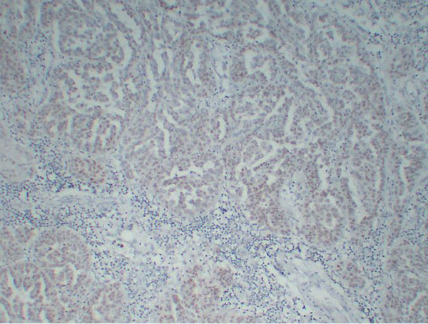
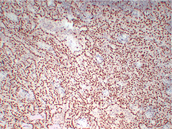


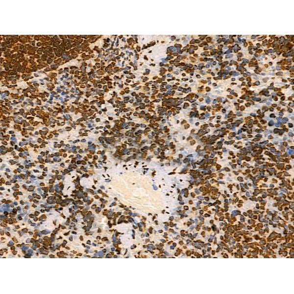
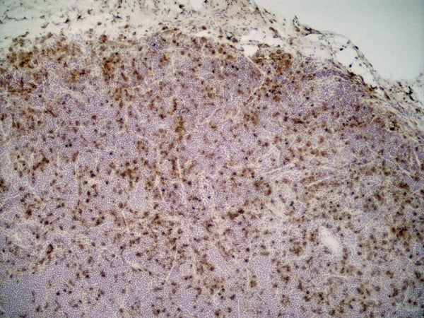
 IHC (Immunohistchemistry)
(Immunohistochemistry analysis of Formalin-fixed, paraffin-embedded Human Wilms' tumor using WT1, Wilms' Tumor 1 antibody.)
IHC (Immunohistchemistry)
(Immunohistochemistry analysis of Formalin-fixed, paraffin-embedded Human Wilms' tumor using WT1, Wilms' Tumor 1 antibody.)
Wilms' Tumor 1 (WT1), Monoclonal Antibody (Cat# AAA30741)
Full Name
Anti-Wilms' Tumor 1 (WT1) Antibody [ABT-WT1]
Gene Names
WT1; GUD; AWT1; WAGR; WT33; NPHS4; WIT-2
Reactivity
Human
Applications
Immunohistochemistry
Purity
Immunogen Affinity Purified
Pricing


 WB (Western Blot)
(WB Suggested Anti-PTX3 Antibody Titration: 0.2-1 ug/mlELISA Titer: 1:312500Positive Control: MCF7 cell lysate)
WB (Western Blot)
(WB Suggested Anti-PTX3 Antibody Titration: 0.2-1 ug/mlELISA Titer: 1:312500Positive Control: MCF7 cell lysate)
PTX3, Polyclonal Antibody (Cat# AAA23569)
Full Name
PTX3 antibody - N-terminal region
Gene Names
PTX3; TSG-14; TNFAIP5
Reactivity
Cow, Guinea Pig, Human, Mouse, Rabbit, Rat
Applications
Immunohistochemistry, Western Blot
Purity
Affinity Purified
Pricing


 ICC (Immunocytochemistry)
(ICC staining p53DINP1 in HUVEC cells (green). The nuclear counter stain is DAPI (blue). Cells were fixed in paraformaldehyde, permeabilised with 0.25% Triton X100/PBS.)
ICC (Immunocytochemistry)
(ICC staining p53DINP1 in HUVEC cells (green). The nuclear counter stain is DAPI (blue). Cells were fixed in paraformaldehyde, permeabilised with 0.25% Triton X100/PBS.)
p53DINP1, Monoclonal Antibody (Cat# AAA30494)
Full Name
p53 DINP1 Antibody
Gene Names
TP53INP1; SIP; Teap; p53DINP1; TP53DINP1; TP53INP1A; TP53INP1B
Reactivity
Human, Mouse, Rat
Applications
Western Blot, Immunocytochemistry, Immunofluorescence, Immunohistochemistry
Purity
ProA affinity purified
Pricing
 Standard Curve (Sample)
LY6/PLAUR domain containing 1, ELISA Kit (Cat# AAA18292)
Full Name
Human Ly6/PLAUR domain-containing protein 1, LYPD1 ELISA Kit
Gene Names
LYPD1; PHTS; LYPDC1
Reactivity
Human
Pricing
 WB (Western Blot)
(Host: RabbitTarget Name: FAN1Sample Tissue: Human Lung Tumor lysatesAntibody Dilution: 1ug/ml)
WB (Western Blot)
(Host: RabbitTarget Name: FAN1Sample Tissue: Human Lung Tumor lysatesAntibody Dilution: 1ug/ml)
FAN1, Polyclonal Antibody (Cat# AAA23600)
Full Name
FAN1 Antibody - N-terminal region
Gene Names
FAN1; KMIN; hFAN1; MTMR15; KIAA1018
Reactivity
Human
Applications
Western Blot
Purity
Affinity purified
Pricing


 Application Data
(Detection limit for recombinant GST tagged NME1 is approximately 0.1ng/ml as a capture antibody.)
Application Data
(Detection limit for recombinant GST tagged NME1 is approximately 0.1ng/ml as a capture antibody.)
NME1, Monoclonal Antibody (Cat# AAA26433)
Full Name
NME1 (Non-Metastatic Cells 1, Protein (NM23A) Expressed in, AWD, GAAD, NB, NBS, NDPK-A, NDPKA, NM23, NM23-H1) (HRP)
Gene Names
NME1; NB; AWD; NBS; GAAD; NDKA; NM23; NDPKA; NDPK-A; NM23-H1
Applications
Immunofluorescence, Immunohistochemistry, Immunoprecipitation, Western Blot
Purity
Purified
Pricing


 Application Data
(Detection limit for recombinant GST tagged NME1 is approximately 0.1ng/ml as a capture antibody.)
Application Data
(Detection limit for recombinant GST tagged NME1 is approximately 0.1ng/ml as a capture antibody.)
NME1, Monoclonal Antibody (Cat# AAA26167)
Full Name
NME1 (Non-Metastatic Cells 1, Protein (NM23A) Expressed in, AWD, GAAD, NB, NBS, NDPK-A, NDPKA, NM23, NM23-H1) (Biotin)
Gene Names
NME1; NB; AWD; NBS; GAAD; NDKA; NM23; NDPKA; NDPK-A; NM23-H1
Applications
Immunofluorescence, Immunohistochemistry, Immunoprecipitation, Western Blot
Purity
Purified
Pricing


 ELISA
ELISA
p63alpha, Monoclonal Antibody (Cat# AAA17943)
Full Name
p63alpha Mouse Monoclonal Antibody
Gene Names
TP63; AIS; KET; LMS; NBP; RHS; p40; p51; p63; EEC3; OFC8; p73H; p73L; SHFM4; TP53L; TP73L; p53CP; TP53CP; B(p51A); B(p51B)
Reactivity
Human, Mouse, Rat, Monkey
Applications
Western Blot, Immunohistochemistry
Pricing


 Application Data
(Published customer image: Histological staining of control and HBOT wounds at post-wounding days 7 and 29. A -D) H&E staining. E -H) CD34 immunohistochemistry. I+J) CD68 immunohistochemistry.From: uk B, Tong M, Fijneman EMG, van Neck JW (2014) Hyperbaric Oxygen Therapy to Treat Diabetes Impaired Wound Healing in Rats. PLoS ONE 9(10): e108533.)
Application Data
(Published customer image: Histological staining of control and HBOT wounds at post-wounding days 7 and 29. A -D) H&E staining. E -H) CD34 immunohistochemistry. I+J) CD68 immunohistochemistry.From: uk B, Tong M, Fijneman EMG, van Neck JW (2014) Hyperbaric Oxygen Therapy to Treat Diabetes Impaired Wound Healing in Rats. PLoS ONE 9(10): e108533.)
CD68, Monoclonal Antibody (Cat# AAA12151)
Full Name
MOUSE ANTI RAT CD68
Applications
Immunohistochemistry, Flow Cytometry, Immunofluorescence, Immunoprecipitation, Immunohistochemistry, Radioimmunoassay, Western Blot
Pricing


 Application Data
(Published customer image Infiltration of GFP+ BM-cells in infarct and peri-infarct regions. (A-B) Dot plots of viable macrophages/granulocytes (CD11b+CD45high, top right quadrants) and microglia (CD11b+CD45dim, bottom right quadrants) in cortex from BM-chimeric unmanipulated mice and mice exposed to pMCAO. (C) Bar graph showing mean numbers of CD11b+CD45dim microglia and CD11b+CD45high macrophages/granulocytes in BM-chimeric mice 24 hours after pMCAO, subdivided based on expression of GFP (n = 5). Approximately 92% of of the CD45high population were GFP+. (D) Estimation and comparison of mean numbers of CD11b+CD45dim microglia in non-chimeric (n = 10) versus BM-chimeric mice (n = 5) 24 hours after of pMCAO shows significantly fewer CD11b+CD45dim microglial cells in irradiated mice. (E) Overview, showing distribution of infiltrating GFP+ BM-derived cells into infarct (IF) and peri-infarct (P-IF) regions 24 hours after pMCAO. (E-G) By 24 hours, GFP+ single cells (F) and vessel-associated aggregates of GFP+ cells (arrows in G) were observed in infarct and peri-infarct regions. Some of the vessel-associated cells were round, leukocyte-like cells (arrows) while others were elongated cells lining the vasculature (arrow heads in G and in insert). (H) Bar graph showing mean numbers of single GFP+ cells and vessel-associated aggregates of GFP+ cells in ipsi- and contralateral cortex 24 hours after surgery (n = 10). (I-P) Immunohistochemical staining of CD45.1 (I, K), CD45.2 (J, L), IgG2a (M, O) and CD45 (N, P) in ischemic tissue in BM-chimeric (I, J, M, N) and non-chimeric mice (K, L, O, P) 24 hours after pMCAO. N.D, none detected. Scale bars: 200 um (A), 10 um (B, C). 50 um (I-P) *P < 0.05, **P < 0.01, and ***P < 0.001.From: Clausen BH, Lambertsen KL, Babcock AA, Holm TH, Dagnaes-Hansen F, Finsen B. Interleukin-1beta and tumor necrosis factor-alpha are expressed by different subsets of microglia and macrophages after ischemic stroke in mice. J Neuroinflammation. 2008 Oct 23;5:46.)
Application Data
(Published customer image Infiltration of GFP+ BM-cells in infarct and peri-infarct regions. (A-B) Dot plots of viable macrophages/granulocytes (CD11b+CD45high, top right quadrants) and microglia (CD11b+CD45dim, bottom right quadrants) in cortex from BM-chimeric unmanipulated mice and mice exposed to pMCAO. (C) Bar graph showing mean numbers of CD11b+CD45dim microglia and CD11b+CD45high macrophages/granulocytes in BM-chimeric mice 24 hours after pMCAO, subdivided based on expression of GFP (n = 5). Approximately 92% of of the CD45high population were GFP+. (D) Estimation and comparison of mean numbers of CD11b+CD45dim microglia in non-chimeric (n = 10) versus BM-chimeric mice (n = 5) 24 hours after of pMCAO shows significantly fewer CD11b+CD45dim microglial cells in irradiated mice. (E) Overview, showing distribution of infiltrating GFP+ BM-derived cells into infarct (IF) and peri-infarct (P-IF) regions 24 hours after pMCAO. (E-G) By 24 hours, GFP+ single cells (F) and vessel-associated aggregates of GFP+ cells (arrows in G) were observed in infarct and peri-infarct regions. Some of the vessel-associated cells were round, leukocyte-like cells (arrows) while others were elongated cells lining the vasculature (arrow heads in G and in insert). (H) Bar graph showing mean numbers of single GFP+ cells and vessel-associated aggregates of GFP+ cells in ipsi- and contralateral cortex 24 hours after surgery (n = 10). (I-P) Immunohistochemical staining of CD45.1 (I, K), CD45.2 (J, L), IgG2a (M, O) and CD45 (N, P) in ischemic tissue in BM-chimeric (I, J, M, N) and non-chimeric mice (K, L, O, P) 24 hours after pMCAO. N.D, none detected. Scale bars: 200 um (A), 10 um (B, C). 50 um (I-P) *P < 0.05, **P < 0.01, and ***P < 0.001.From: Clausen BH, Lambertsen KL, Babcock AA, Holm TH, Dagnaes-Hansen F, Finsen B. Interleukin-1beta and tumor necrosis factor-alpha are expressed by different subsets of microglia and macrophages after ischemic stroke in mice. J Neuroinflammation. 2008 Oct 23;5:46.)
CD11b, Monoclonal Antibody (Cat# AAA12186)
Full Name
RAT ANTI MOUSE CD11b:RPE
Gene Names
Itgam; CR3; CR3A; MAC1; Cd11b; Ly-40; Mac-1; Mac-1a; CD11b/CD18; F730045J24Rik
Applications
Flow Cytometry
Pricing
 SDS-PAGE
Cellular tumor antigen p53 (Tp53), Recombinant Protein (Cat# AAA18501)
Full Name
Recombinant Mouse Cellular tumor antigen p53 (Tp53)
Gene Names
Trp53; bbl; bfy; bhy; p44; p53; Tp53
Purity
Greater or equal to 85% purity as determined by SDS-PAGE.
Pricing


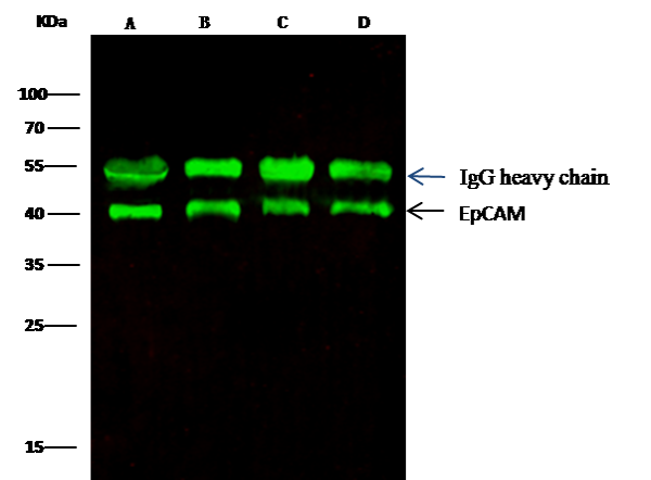 IP (Immunoprecipitation)
(EpCAM was immunoprecipitated using:Lane A:0.5 mg MCF-7 Whole Cell LysateLane B:0.5 mg A431 Whole Cell LysateLane C:0.5 mg HepG2 Whole Cell LysateLane D:0.5 mg Caco-2 Whole Cell Lysate0.5 uL anti-EpCAM rabbit monoclonal antibody and 15 ul of 50 % Protein G agarose.Primary antibody:Anti-EpCAM rabbit monoclonal antibody,at 1:5000 dilution Secondary antibody:Dylight 800-labeled antibody to rabbit IgG (H+L), at 1:5000 dilution Developed using the odssey technique.Performed under reducing conditions.Predicted band size: 40 kDaObserved band size: 40 kDa)
IP (Immunoprecipitation)
(EpCAM was immunoprecipitated using:Lane A:0.5 mg MCF-7 Whole Cell LysateLane B:0.5 mg A431 Whole Cell LysateLane C:0.5 mg HepG2 Whole Cell LysateLane D:0.5 mg Caco-2 Whole Cell Lysate0.5 uL anti-EpCAM rabbit monoclonal antibody and 15 ul of 50 % Protein G agarose.Primary antibody:Anti-EpCAM rabbit monoclonal antibody,at 1:5000 dilution Secondary antibody:Dylight 800-labeled antibody to rabbit IgG (H+L), at 1:5000 dilution Developed using the odssey technique.Performed under reducing conditions.Predicted band size: 40 kDaObserved band size: 40 kDa)
EpCAM, Monoclonal Antibody (Cat# AAA27736)
Full Name
Recombinant Anti-EpCAM Antibody, Rabbit Monoclonal
Gene Names
EPCAM; ESA; KSA; M4S1; MK-1; DIAR5; EGP-2; EGP40; KS1/4; MIC18; TROP1; EGP314; HNPCC8; TACSTD1
Reactivity
Human
Applications
Western Blot, Immunohistochemistry, Flow Cytometry, Immunocytochemistry, Immunofluorescence, Immunoprecipitation
Purity
Protein A
Pricing
 Standard Curve (Sample)
non-metastatic cells 1, protein (NM23A) expressed in, ELISA Kit (Cat# AAA18006)
Full Name
Human Nucleoside diphosphate kinase A, NME1 ELISA Kit
Gene Names
NME1; NB; AWD; NBS; GAAD; NDKA; NM23; NDPKA; NDPK-A; NM23-H1
Reactivity
Human
Pricing


 FCM (Flow Cytometry)
(Dual staining of pig peripheral blood lymphocytes with Mouse anti Pig CD335 detected with Goat anti Mouse IgG (H/L):FITC (STAR117F), and Mouse anti Pig wCD8a:RPE)
FCM (Flow Cytometry)
(Dual staining of pig peripheral blood lymphocytes with Mouse anti Pig CD335 detected with Goat anti Mouse IgG (H/L):FITC (STAR117F), and Mouse anti Pig wCD8a:RPE)
CD335, Monoclonal Antibody (Cat# AAA12253)
Full Name
MOUSE ANTI PIG CD335
Reactivity
Pig
Applications
Flow Cytometry, Immunofluorescence
Pricing


 Application Data
(Published customer image Infiltration of GFP+ BM-cells in infarct and peri-infarct regions. (A-B) Dot plots of viable macrophages/granulocytes (CD11b+CD45high, top right quadrants) and microglia (CD11b+CD45dim, bottom right quadrants) in cortex from BM-chimeric unmanipulated mice and mice exposed to pMCAO. (C) Bar graph showing mean numbers of CD11b+CD45dim microglia and CD11b+CD45high macrophages/granulocytes in BM-chimeric mice 24 hours after pMCAO, subdivided based on expression of GFP (n = 5). Approximately 92% of of the CD45high population were GFP+. (D) Estimation and comparison of mean numbers of CD11b+CD45dim microglia in non-chimeric (n = 10) versus BM-chimeric mice (n = 5) 24 hours after of pMCAO shows significantly fewer CD11b+CD45dim microglial cells in irradiated mice. (E) Overview, showing distribution of infiltrating GFP+ BM-derived cells into infarct (IF) and peri-infarct (P-IF) regions 24 hours after pMCAO. (E-G) By 24 hours, GFP+ single cells (F) and vessel-associated aggregates of GFP+ cells (arrows in G) were observed in infarct and peri-infarct regions. Some of the vessel-associated cells were round, leukocyte-like cells (arrows) while others were elongated cells lining the vasculature (arrow heads in G and in insert). (H) Bar graph showing mean numbers of single GFP+ cells and vessel-associated aggregates of GFP+ cells in ipsi- and contralateral cortex 24 hours after surgery (n = 10). (I-P) Immunohistochemical staining of CD45.1 (I, K), CD45.2 (J, L), IgG2a (M, O) and CD45 (N, P) in ischemic tissue in BM-chimeric (I, J, M, N) and non-chimeric mice (K, L, O, P) 24 hours after pMCAO. N.D, none detected. Scale bars: 200 um (A), 10 um (B, C). 50 um (I-P) *P < 0.05, **P < 0.01, and ***P < 0.001.From: Clausen BH, Lambertsen KL, Babcock AA, Holm TH, Dagnaes-Hansen F, Finsen B. Interleukin-1beta and tumor necrosis factor-alpha are expressed by different subsets of microglia and macrophages after ischemic stroke in mice. J Neuroinflammation. 2008 Oct 23;5:46.)
Application Data
(Published customer image Infiltration of GFP+ BM-cells in infarct and peri-infarct regions. (A-B) Dot plots of viable macrophages/granulocytes (CD11b+CD45high, top right quadrants) and microglia (CD11b+CD45dim, bottom right quadrants) in cortex from BM-chimeric unmanipulated mice and mice exposed to pMCAO. (C) Bar graph showing mean numbers of CD11b+CD45dim microglia and CD11b+CD45high macrophages/granulocytes in BM-chimeric mice 24 hours after pMCAO, subdivided based on expression of GFP (n = 5). Approximately 92% of of the CD45high population were GFP+. (D) Estimation and comparison of mean numbers of CD11b+CD45dim microglia in non-chimeric (n = 10) versus BM-chimeric mice (n = 5) 24 hours after of pMCAO shows significantly fewer CD11b+CD45dim microglial cells in irradiated mice. (E) Overview, showing distribution of infiltrating GFP+ BM-derived cells into infarct (IF) and peri-infarct (P-IF) regions 24 hours after pMCAO. (E-G) By 24 hours, GFP+ single cells (F) and vessel-associated aggregates of GFP+ cells (arrows in G) were observed in infarct and peri-infarct regions. Some of the vessel-associated cells were round, leukocyte-like cells (arrows) while others were elongated cells lining the vasculature (arrow heads in G and in insert). (H) Bar graph showing mean numbers of single GFP+ cells and vessel-associated aggregates of GFP+ cells in ipsi- and contralateral cortex 24 hours after surgery (n = 10). (I-P) Immunohistochemical staining of CD45.1 (I, K), CD45.2 (J, L), IgG2a (M, O) and CD45 (N, P) in ischemic tissue in BM-chimeric (I, J, M, N) and non-chimeric mice (K, L, O, P) 24 hours after pMCAO. N.D, none detected. Scale bars: 200 um (A), 10 um (B, C). 50 um (I-P) *P < 0.05, **P < 0.01, and ***P < 0.001.From: Clausen BH, Lambertsen KL, Babcock AA, Holm TH, Dagnaes-Hansen F, Finsen B. Interleukin-1beta and tumor necrosis factor-alpha are expressed by different subsets of microglia and macrophages after ischemic stroke in mice. J Neuroinflammation. 2008 Oct 23;5:46.)
CD11b, Monoclonal Antibody (Cat# AAA12231)
Full Name
RAT ANTI MOUSE CD11b:Low Endotoxin
Gene Names
Itgam; CR3; CR3A; MAC1; Cd11b; Ly-40; Mac-1; Mac-1a; CD11b/CD18; F730045J24Rik
Applications
Immunohistochemistry, Flow Cytometry, Functional Assay, Immunofluorescence, Immunoprecipitation
Pricing


 FCM (Flow Cytometry)
(Flow cytometric analysis of PC-3M cells with BAP31 antibody at 1/100 dilution (purple) compared with an unlabelled control (cells without incubation with primary antibody; yellow). Alexa Fluor 488-conjugated goat anti-rabbit IgG was used as the secondary antibody.)
FCM (Flow Cytometry)
(Flow cytometric analysis of PC-3M cells with BAP31 antibody at 1/100 dilution (purple) compared with an unlabelled control (cells without incubation with primary antibody; yellow). Alexa Fluor 488-conjugated goat anti-rabbit IgG was used as the secondary antibody.)
BAP31, Monoclonal Antibody (Cat# AAA30501)
Full Name
BAP31 Antibody
Gene Names
BCAP31; CDM; BAP31; 6C6-AG; DXS1357E
Reactivity
Human, Mouse
Applications
Western Blot, Immunocytochemistry, Immunofluorescence, Immunohistochemistry, Flow Cytometry
Purity
ProA affinity purified
Pricing


 Application Data
(Published customer image: Histological staining of control and HBOT wounds at post-wounding days 7 and 29. A -D) H&E staining. E -H) CD34 immunohistochemistry. I+J) CD68 immunohistochemistry.From: uk B, Tong M, Fijneman EMG, van Neck JW (2014) Hyperbaric Oxygen Therapy to Treat Diabetes Impaired Wound Healing in Rats. PLoS ONE 9(10): e108533.)
Application Data
(Published customer image: Histological staining of control and HBOT wounds at post-wounding days 7 and 29. A -D) H&E staining. E -H) CD34 immunohistochemistry. I+J) CD68 immunohistochemistry.From: uk B, Tong M, Fijneman EMG, van Neck JW (2014) Hyperbaric Oxygen Therapy to Treat Diabetes Impaired Wound Healing in Rats. PLoS ONE 9(10): e108533.)
CD68, Monoclonal Antibody (Cat# AAA12148)
Full Name
MOUSE ANTI RAT CD68:FITC
Applications
Flow Cytometry
Pricing


 Application Data
(Published customer image Infiltration of GFP+ BM-cells in infarct and peri-infarct regions. (A-B) Dot plots of viable macrophages/granulocytes (CD11b+CD45high, top right quadrants) and microglia (CD11b+CD45dim, bottom right quadrants) in cortex from BM-chimeric unmanipulated mice and mice exposed to pMCAO. (C) Bar graph showing mean numbers of CD11b+CD45dim microglia and CD11b+CD45high macrophages/granulocytes in BM-chimeric mice 24 hours after pMCAO, subdivided based on expression of GFP (n = 5). Approximately 92% of of the CD45high population were GFP+. (D) Estimation and comparison of mean numbers of CD11b+CD45dim microglia in non-chimeric (n = 10) versus BM-chimeric mice (n = 5) 24 hours after of pMCAO shows significantly fewer CD11b+CD45dim microglial cells in irradiated mice. (E) Overview, showing distribution of infiltrating GFP+ BM-derived cells into infarct (IF) and peri-infarct (P-IF) regions 24 hours after pMCAO. (E-G) By 24 hours, GFP+ single cells (F) and vessel-associated aggregates of GFP+ cells (arrows in G) were observed in infarct and peri-infarct regions. Some of the vessel-associated cells were round, leukocyte-like cells (arrows) while others were elongated cells lining the vasculature (arrow heads in G and in insert). (H) Bar graph showing mean numbers of single GFP+ cells and vessel-associated aggregates of GFP+ cells in ipsi- and contralateral cortex 24 hours after surgery (n = 10). (I-P) Immunohistochemical staining of CD45.1 (I, K), CD45.2 (J, L), IgG2a (M, O) and CD45 (N, P) in ischemic tissue in BM-chimeric (I, J, M, N) and non-chimeric mice (K, L, O, P) 24 hours after pMCAO. N.D, none detected. Scale bars: 200 um (A), 10 um (B, C). 50 um (I-P) *P < 0.05, **P < 0.01, and ***P < 0.001.From: Clausen BH, Lambertsen KL, Babcock AA, Holm TH, Dagnaes-Hansen F, Finsen B. Interleukin-1beta and tumor necrosis factor-alpha are expressed by different subsets of microglia and macrophages after ischemic stroke in mice. J Neuroinflammation. 2008 Oct 23;5:46.)
Application Data
(Published customer image Infiltration of GFP+ BM-cells in infarct and peri-infarct regions. (A-B) Dot plots of viable macrophages/granulocytes (CD11b+CD45high, top right quadrants) and microglia (CD11b+CD45dim, bottom right quadrants) in cortex from BM-chimeric unmanipulated mice and mice exposed to pMCAO. (C) Bar graph showing mean numbers of CD11b+CD45dim microglia and CD11b+CD45high macrophages/granulocytes in BM-chimeric mice 24 hours after pMCAO, subdivided based on expression of GFP (n = 5). Approximately 92% of of the CD45high population were GFP+. (D) Estimation and comparison of mean numbers of CD11b+CD45dim microglia in non-chimeric (n = 10) versus BM-chimeric mice (n = 5) 24 hours after of pMCAO shows significantly fewer CD11b+CD45dim microglial cells in irradiated mice. (E) Overview, showing distribution of infiltrating GFP+ BM-derived cells into infarct (IF) and peri-infarct (P-IF) regions 24 hours after pMCAO. (E-G) By 24 hours, GFP+ single cells (F) and vessel-associated aggregates of GFP+ cells (arrows in G) were observed in infarct and peri-infarct regions. Some of the vessel-associated cells were round, leukocyte-like cells (arrows) while others were elongated cells lining the vasculature (arrow heads in G and in insert). (H) Bar graph showing mean numbers of single GFP+ cells and vessel-associated aggregates of GFP+ cells in ipsi- and contralateral cortex 24 hours after surgery (n = 10). (I-P) Immunohistochemical staining of CD45.1 (I, K), CD45.2 (J, L), IgG2a (M, O) and CD45 (N, P) in ischemic tissue in BM-chimeric (I, J, M, N) and non-chimeric mice (K, L, O, P) 24 hours after pMCAO. N.D, none detected. Scale bars: 200 um (A), 10 um (B, C). 50 um (I-P) *P < 0.05, **P < 0.01, and ***P < 0.001.From: Clausen BH, Lambertsen KL, Babcock AA, Holm TH, Dagnaes-Hansen F, Finsen B. Interleukin-1beta and tumor necrosis factor-alpha are expressed by different subsets of microglia and macrophages after ischemic stroke in mice. J Neuroinflammation. 2008 Oct 23;5:46.)
CD11b, Monoclonal Antibody (Cat# AAA12181)
Full Name
RAT ANTI MOUSE CD11b
Gene Names
Itgam; CR3; CR3A; MAC1; Cd11b; Ly-40; Mac-1; Mac-1a; CD11b/CD18; F730045J24Rik
Reactivity
Human
Applications
Immunohistochemistry, Flow Cytometry, Immunofluorescence, Immunoprecipitation
Pricing


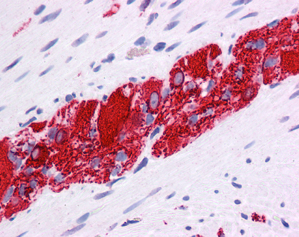
 IHC (Immunohistchemistry)
(Immunohistochemistry of TM4SF1 in mouse lung tissue with TM4SF1 antibody at 2 μg/mL.)
IHC (Immunohistchemistry)
(Immunohistochemistry of TM4SF1 in mouse lung tissue with TM4SF1 antibody at 2 μg/mL.)
TM4SF1, Polyclonal Antibody (Cat# AAA10947)
Full Name
TM4SF1 Antibody
Gene Names
TM4SF1; L6; H-L6; M3S1; TAAL6
Reactivity
Human, Mouse, Rat
Applications
Immunofluorescence, Immunohistochemistry, Western Blot
Purity
TM4SF1 Antibody is affinity chromatography purified via peptide column.
Pricing

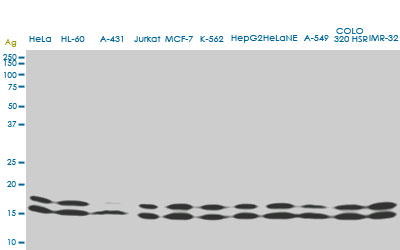
 WB (Western Blot)
((0.5ug/ml) staining of A549 (A), HepG2 (B) and K562 (C) lysates (35ug protein in RIPA buffer). Primary incubation was 1 hour. Detected by chemiluminescence.)
WB (Western Blot)
((0.5ug/ml) staining of A549 (A), HepG2 (B) and K562 (C) lysates (35ug protein in RIPA buffer). Primary incubation was 1 hour. Detected by chemiluminescence.)
TNFRSF1A/TNFR1, Polyclonal Antibody (Cat# AAA13672)
Full Name
Goat anti-TNFRSF1A/TNFR1 Antibody
Gene Names
TNFRSF1A; FPF; MS5; p55; p60; TBP1; TNF-R; TNFAR; TNFR1; p55-R; CD120a; TNFR55; TNFR60; TNF-R-I; TNF-R55; TNFR1-d2
Reactivity
Tested: Human; Expected from sequence similarity: Human, Rat, Pig
Applications
Peptide ELISA, Western Blot
Purity
Purified from goat serum by ammonium sulphate precipitation followed by antigen affinity chromatography using the immunizing peptide.
Pricing


 WB (Western Blot)
(WB Suggested Anti-NR2E1 Antibody Titration: 1 ug/mlPositive Control: Fetal Muscle cell lysate)
WB (Western Blot)
(WB Suggested Anti-NR2E1 Antibody Titration: 1 ug/mlPositive Control: Fetal Muscle cell lysate)
NR2E1, Polyclonal Antibody (Cat# AAA23403)
Full Name
NR2E1 antibody - N-terminal region
Gene Names
NR2E1; TLL; TLX; XTLL
Reactivity
Cow, Dog, Guinea Pig, Horse, Human, Mouse, Rabbit, Rat, Zebrafish
Applications
Immunohistochemistry, Western Blot
Purity
Affinity Purified
Pricing


 WB (Western Blot)
(Western BlotSample: Recombinant GAL9, Human;Antibody: Rabbit Anti-Human GAL9 Ab)
WB (Western Blot)
(Western BlotSample: Recombinant GAL9, Human;Antibody: Rabbit Anti-Human GAL9 Ab)
Galectin 9 (GAL9), Active Protein (Cat# AAA21103)
Full Name
Active Galectin 9 (GAL9)
Gene Names
LGALS9; HUAT; LGALS9A
Reactivity
Homo sapiens (Human)
Applications
Cell culture; Activity Assays.
Purity
>90%
Pricing
 Standard Curve (Sample)
heat shock protein 90kDa alpha (cytosolic) , class B member 1, ELISA Kit (Cat# AAA18180)
Full Name
Mouse Heat shock protein HSP 90-beta, HSP90AB1 ELISA Kit
Gene Names
Hsp90ab1; 90kDa; Hsp84; Hsp90; Hspcb; C81438; Hsp84-1; AL022974
Reactivity
Mouse
Pricing