Filters
Clonality
Type
Reactivity
Gene Name
Isotype
Host
Application
Clone
219 results for "Rat Anti Mouse IgG2a" - showing 1-50


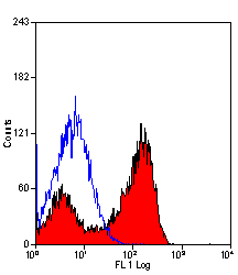
 Application Data
(Staining of mouse spleen cells with Rat anti Mouse CD3 Epsilon (T3): APC)
Application Data
(Staining of mouse spleen cells with Rat anti Mouse CD3 Epsilon (T3): APC)
CD3, Monoclonal Antibody (Cat# AAA11880)
Full Name
RAT ANTI MOUSE CD3:FITC
Gene Names
Cd3e; CD3; T3e; AI504783; CD3epsilon
Applications
Flow Cytometry
Pricing


 Application Data
(Published customer image:RPE conjugated Mouse anti Canine CD4 antibody, clone YKIC302.9used for the assessment of CD4 levels on canine cells by flow cytometry.Image caption:Immunophenotypic profile of tumor infiltrating lymphocyte in canine mammary carcinomas. Analysis of tumor infiltrating T-cells, B-lymphocytes and T-cell subsets from MC-BMT or MC (A), further subcategorized according to the absence (-) or presence (+) of lymph node metastasis (-) (B). Lymphocyte populations and subsets were identified by flow cytometric immunostaining as described in Material and Methods. Data were expressed as percentage of positive cells within gated lymphocytes and CD4+/CD8+ T-cell ratio. Significant differences at p < 0.05 are highlighted by asterisk.)
Application Data
(Published customer image:RPE conjugated Mouse anti Canine CD4 antibody, clone YKIC302.9used for the assessment of CD4 levels on canine cells by flow cytometry.Image caption:Immunophenotypic profile of tumor infiltrating lymphocyte in canine mammary carcinomas. Analysis of tumor infiltrating T-cells, B-lymphocytes and T-cell subsets from MC-BMT or MC (A), further subcategorized according to the absence (-) or presence (+) of lymph node metastasis (-) (B). Lymphocyte populations and subsets were identified by flow cytometric immunostaining as described in Material and Methods. Data were expressed as percentage of positive cells within gated lymphocytes and CD4+/CD8+ T-cell ratio. Significant differences at p < 0.05 are highlighted by asterisk.)
CD4, Monoclonal Antibody (Cat# AAA12278)
Full Name
RAT ANTI DOG CD4: APC
Reactivity
Dog
Applications
Flow Cytometry
Pricing


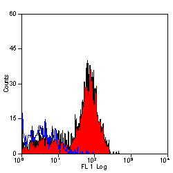
 Application Data
(Staining of mouse peripheral blood lymphocytes with Rat anti Mouse CD200R: Low Endotoxin)
Application Data
(Staining of mouse peripheral blood lymphocytes with Rat anti Mouse CD200R: Low Endotoxin)
CD200R, Monoclonal Antibody (Cat# AAA12039)
Full Name
RAT ANTI MOUSE CD200R:RPE
Gene Names
Cd200r1; OX2R; Mox2r; CD200R
Applications
Flow Cytometry
Pricing


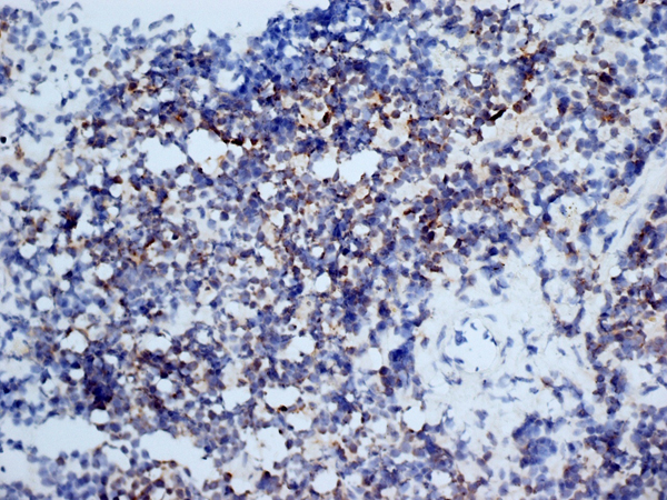
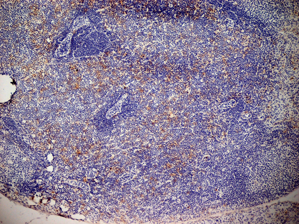
 Application Data
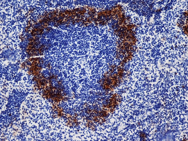
(Immunoperoxidase staining of mouse lymph node cryosection stained with Rat antii Mouse CD8alpha antibody, clone KT15 followed by horseradish peroxidase conjugatedGoat anti Rat IgG s a detection reagent. High power)
Application Data
(Immunoperoxidase staining of mouse lymph node cryosection stained with Rat antii Mouse CD8alpha antibody, clone KT15 followed by horseradish peroxidase conjugatedGoat anti Rat IgG s a detection reagent. High power)
CD8 ALPHA, Monoclonal Antibody (Cat# AAA12180)
Full Name
RAT ANTI MOUSE CD8 ALPHA
Gene Names
Cd8a; Ly-2; Ly-B; Ly-35; Lyt-2; BB154331
Applications
Immunohistochemistry, Flow Cytometry, Immunofluorescence
Pricing


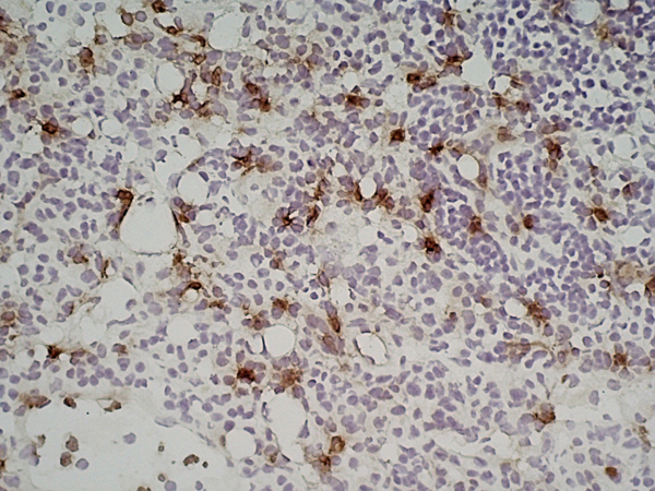
 Application Data
(Immunoperoxidase staining of mouse lymph node cryosection eith Rat anti Mouse antibody clone R3-63 followed by horseradish peroxidase Goat anti Rat IgG antibody . Medium power)
Application Data
(Immunoperoxidase staining of mouse lymph node cryosection eith Rat anti Mouse antibody clone R3-63 followed by horseradish peroxidase Goat anti Rat IgG antibody . Medium power)
CD13, Monoclonal Antibody (Cat# AAA12236)
Full Name
RAT ANTI MOUSE CD13:Low Endotoxin
Gene Names
Anpep; Apn; AP-M; AP-N; Cd13; P150
Applications
Immunohistochemistry, Flow Cytometry, Functional Assay, Immunohistochemistry
Pricing


 Application Data
(Immunoperoxidase staining of mouse lymph node cryosection stained with Rat antii Mouse CD8alpha antibody, clone KT15 followed by horseradish peroxidase conjugatedGoat anti Rat IgG s a detection reagent. High power)
Application Data
(Immunoperoxidase staining of mouse lymph node cryosection stained with Rat antii Mouse CD8alpha antibody, clone KT15 followed by horseradish peroxidase conjugatedGoat anti Rat IgG s a detection reagent. High power)
CD8 ALPHA, Monoclonal Antibody (Cat# AAA11842)
Full Name
RAT ANTI MOUSE CD8 ALPHA:APC
Gene Names
Cd8a; Ly-2; Ly-B; Ly-35; Lyt-2; BB154331
Applications
Flow Cytometry
Pricing


 Application Data
(Staining of Rat peripheral blood lymphocytes with Mouse anti Rat CD49d:RPE (MCA2872PE))
Application Data
(Staining of Rat peripheral blood lymphocytes with Mouse anti Rat CD49d:RPE (MCA2872PE))
CD49d, Monoclonal Antibody (Cat# AAA26784)
Full Name
CD49d (Antigen CD49d, CD49d Antigen, CDw49d, Alpha 4 Subunit of VLA-4 Receptor, Integrin alpha IV, Integrin alpha 4, IA4, ITGA4, LPAM23, MGC90518, Very Late Activation Protein 4 Receptor Alpha 4 Subunit, VLA4, VLA-4) (MaxLight 405)
Reactivity
Rat
Applications
Flow Cytometry, Immunoprecipitation
Purity
Purified by Protein G Affinity Chromatography
Pricing


 Application Data
(Published customer image: Increased accumulation of repair-associated macrophages surrounding collaterals in ischemic hind limbs is PAR2-dependent. (A) Stainings of CD206-positive macrophages (green) and SMA-positive vessels (red) in non-ischemic (control) and ischemic (ligated) hind limbs of WT, PAR1-/- and PAR2-/- mice are shown. Nuclei were visualized with DAPI (blue). Arrows indicate single macrophages in the non-ischemic adductor. Quantification of the average number of repair-associated macrophages per vessel is indicated on the right. (B) Correlation between the number of CD206-positive macrophages in the ischemic tissues and the expression of CD11b and (C) CD115 on monocytes. ** p)
Application Data
(Published customer image: Increased accumulation of repair-associated macrophages surrounding collaterals in ischemic hind limbs is PAR2-dependent. (A) Stainings of CD206-positive macrophages (green) and SMA-positive vessels (red) in non-ischemic (control) and ischemic (ligated) hind limbs of WT, PAR1-/- and PAR2-/- mice are shown. Nuclei were visualized with DAPI (blue). Arrows indicate single macrophages in the non-ischemic adductor. Quantification of the average number of repair-associated macrophages per vessel is indicated on the right. (B) Correlation between the number of CD206-positive macrophages in the ischemic tissues and the expression of CD11b and (C) CD115 on monocytes. ** p)
CD206, Monoclonal Antibody (Cat# AAA12117)
Full Name
RAT ANTI MOUSE CD206:Biotin
Gene Names
Mrc1; MR; CD206; AW259686
Applications
Flow Cytometry
Pricing


 Application Data
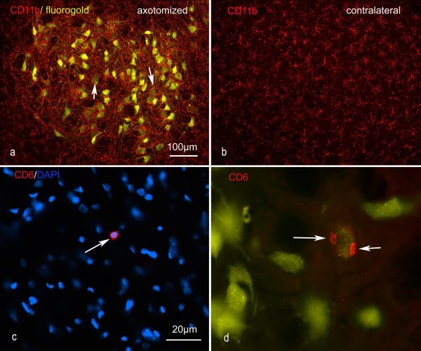
(Published customer image: Representative images of the inflammatory changes in the facial nucleus during axonal regeneration, one week following facial nerve transaction. a, b: CD11b immunoreactivity for microglia is increased in the axotomized facial nucleus, and microglia enwrap the facial motor neurons, e.g. at arrows. The regenerating neurons were retrogradely labelled with fluorogold. c, d: CD6- positive T-cells accumulated in the injured motor nucleus (arrows). They had little cytoplasm but dense nuclei (c) and were sometimes clustered around neurons retrogradely labelled with fluorogold (d). The scale bar in (a) also applies to (b) and that in (c) also applies to (d).From: Shokouhi et al. BMC Neuroscience 2010 11:13.)
Application Data
(Published customer image: Representative images of the inflammatory changes in the facial nucleus during axonal regeneration, one week following facial nerve transaction. a, b: CD11b immunoreactivity for microglia is increased in the axotomized facial nucleus, and microglia enwrap the facial motor neurons, e.g. at arrows. The regenerating neurons were retrogradely labelled with fluorogold. c, d: CD6- positive T-cells accumulated in the injured motor nucleus (arrows). They had little cytoplasm but dense nuclei (c) and were sometimes clustered around neurons retrogradely labelled with fluorogold (d). The scale bar in (a) also applies to (b) and that in (c) also applies to (d).From: Shokouhi et al. BMC Neuroscience 2010 11:13.)
CD11b, Monoclonal Antibody (Cat# AAA11853)
Full Name
MOUSE ANTI RAT CD11b:Biotin
Gene Names
ITGAM; CD11B
Applications
Immunohistochemistry, Flow Cytometry
Pricing


 Application Data
(Immunoperoxidase staining of mouse lymph node cryosection stained with Rat antii Mouse CD8alpha antibody, clone KT15 followed by horseradish peroxidase conjugatedGoat anti Rat IgG s a detection reagent. High power)
Application Data
(Immunoperoxidase staining of mouse lymph node cryosection stained with Rat antii Mouse CD8alpha antibody, clone KT15 followed by horseradish peroxidase conjugatedGoat anti Rat IgG s a detection reagent. High power)
CD8 ALPHA, Monoclonal Antibody (Cat# AAA11886)
Full Name
RAT ANTI MOUSE CD8 ALPHA:FITC
Gene Names
Cd8a; Ly-2; Ly-B; Ly-35; Lyt-2; BB154331
Applications
Flow Cytometry
Pricing


 WB (Western Blot)
(Western Blot analysis of TUBB2A expression in transfected 293T cell line by TUBB2A monoclonal antibody. Lane 1: TUBB2A transfected lysate (49.9kD). Lane 2: Non-transfected lysate.)
WB (Western Blot)
(Western Blot analysis of TUBB2A expression in transfected 293T cell line by TUBB2A monoclonal antibody. Lane 1: TUBB2A transfected lysate (49.9kD). Lane 2: Non-transfected lysate.)
Tubulin beta-2A Chain, Monoclonal Antibody (Cat# AAA24087)
Full Name
Tubulin beta-2A Chain (Tubulin beta Class IIa, TUBB2A, TUBB2, TUBB, dJ40E16.7)
Gene Names
TUBB2A; TUBB; TUBB2; dJ40E16.7
Reactivity
Human, Mouse, Rat
Applications
Western Blot, Immunohistochemistry
Purity
Affinity Purified
Purified by Protein A affinity chromatography.
Purified by Protein A affinity chromatography.
Pricing


 Application Data
(Immunoperoxidase staining of human tonsil cryosection using Mouse anti Human CD3 antibody, clone UCHT1 followed by horseradish peroxidase Goat anti Mouse IgG2a antibody as a detection reagent. Medium power)
Application Data
(Immunoperoxidase staining of human tonsil cryosection using Mouse anti Human CD3 antibody, clone UCHT1 followed by horseradish peroxidase Goat anti Mouse IgG2a antibody as a detection reagent. Medium power)
CD3, Monoclonal Antibody (Cat# AAA11877)
Full Name
MOUSE ANTI HUMAN CD3:FITC
Gene Names
CD3G; T3G; IMD17; CD3-GAMMA
Applications
Flow Cytometry
Pricing


 Application Data
(Frozen mouse spleen stained with Rat anti Mouse CD169)
Application Data
(Frozen mouse spleen stained with Rat anti Mouse CD169)
CD169, Monoclonal Antibody (Cat# AAA12204)
Full Name
RAT ANTI MOUSE CD169
Gene Names
Siglec1; Sn; Cd169; Siglec-1
Applications
Immunohistochemistry, Flow Cytometry, Immunofluorescence
Pricing


 Application Data
(Published customer image: Increased accumulation of repair-associated macrophages surrounding collaterals in ischemic hind limbs is PAR2-dependent. (A) Stainings of CD206-positive macrophages (green) and SMA-positive vessels (red) in non-ischemic (control) and ischemic (ligated) hind limbs of WT, PAR1-/- and PAR2-/- mice are shown. Nuclei were visualized with DAPI (blue). Arrows indicate single macrophages in the non-ischemic adductor. Quantification of the average number of repair-associated macrophages per vessel is indicated on the right. (B) Correlation between the number of CD206-positive macrophages in the ischemic tissues and the expression of CD11b and (C) CD115 on monocytes. ** p)
Application Data
(Published customer image: Increased accumulation of repair-associated macrophages surrounding collaterals in ischemic hind limbs is PAR2-dependent. (A) Stainings of CD206-positive macrophages (green) and SMA-positive vessels (red) in non-ischemic (control) and ischemic (ligated) hind limbs of WT, PAR1-/- and PAR2-/- mice are shown. Nuclei were visualized with DAPI (blue). Arrows indicate single macrophages in the non-ischemic adductor. Quantification of the average number of repair-associated macrophages per vessel is indicated on the right. (B) Correlation between the number of CD206-positive macrophages in the ischemic tissues and the expression of CD11b and (C) CD115 on monocytes. ** p)
CD206, Monoclonal Antibody (Cat# AAA12122)
Full Name
RAT ANTI MOUSE CD206:FITC
Gene Names
Mrc1; MR; CD206; AW259686
Applications
Flow Cytometry
Pricing


 Application Data
(Staining of mouse peripheral blood granulocytes with Rat anti Mouse CD13)
Application Data
(Staining of mouse peripheral blood granulocytes with Rat anti Mouse CD13)
CD13, Monoclonal Antibody (Cat# AAA12144)
Full Name
RAT ANTI MOUSE CD13
Gene Names
Anpep; Apn; AP-M; AP-N; Cd13; P150
Applications
Immunohistochemistry, Flow Cytometry
Purity
Affinity chromatography on Protein G from tissue culture supernatant
Pricing


 Application Data
(Frozen mouse spleen stained with Rat anti Mouse CD169)
Application Data
(Frozen mouse spleen stained with Rat anti Mouse CD169)
CD169, Monoclonal Antibody (Cat# AAA12206)
Full Name
RAT ANTI MOUSE CD169:FITC
Gene Names
Siglec1; Sn; Cd169; Siglec-1
Applications
Flow Cytometry
Pricing


 Application Data
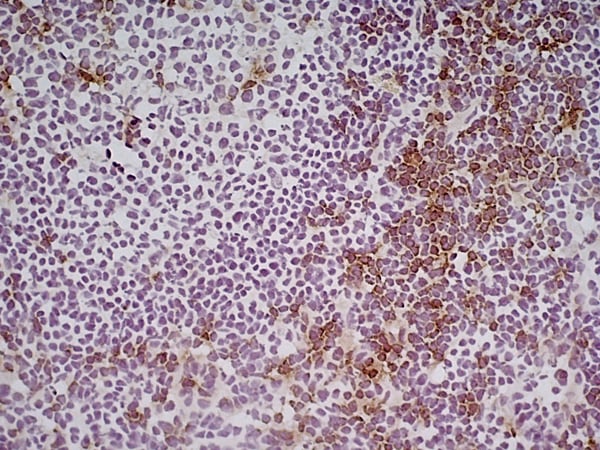
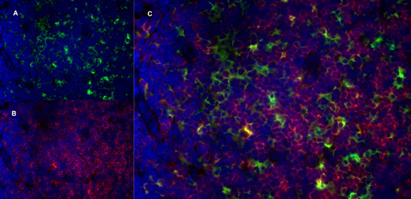
(Immunofluorescence staining of a mouse lymph node cryosection with Rat anti MouseCD19 antibody, clone 6D5 , green in A and Rat anti Mlouse CD8 antibody, clone YTS105.18 , red in B. Merged image in C with nuclei counterstained blue using DAPI. Low power)
Application Data
(Immunofluorescence staining of a mouse lymph node cryosection with Rat anti MouseCD19 antibody, clone 6D5 , green in A and Rat anti Mlouse CD8 antibody, clone YTS105.18 , red in B. Merged image in C with nuclei counterstained blue using DAPI. Low power)
CD19, Monoclonal Antibody (Cat# AAA12028)
Full Name
RAT ANTI MOUSE CD19:RPE
Gene Names
Cd19; AW495831
Applications
Flow Cytometry
Pricing


 Application Data
(Immunoperoxidase staining of mouse lymph node cryosection eith Rat anti Mouse antibody clone R3-63 followed by horseradish peroxidase Goat anti Rat IgG antibody . Medium power)
Application Data
(Immunoperoxidase staining of mouse lymph node cryosection eith Rat anti Mouse antibody clone R3-63 followed by horseradish peroxidase Goat anti Rat IgG antibody . Medium power)
CD13, Monoclonal Antibody (Cat# AAA11953)
Full Name
RAT ANTI MOUSE CD13
Gene Names
Anpep; Apn; AP-M; AP-N; Cd13; P150
Applications
Immunohistochemistry, Flow Cytometry, Immunofluorescence, Immunohistochemistry
Pricing


 Application Data
(Published customer image: Increased accumulation of repair-associated macrophages surrounding collaterals in ischemic hind limbs is PAR2-dependent. (A) Stainings of CD206-positive macrophages (green) and SMA-positive vessels (red) in non-ischemic (control) and ischemic (ligated) hind limbs of WT, PAR1-/- and PAR2-/- mice are shown. Nuclei were visualized with DAPI (blue). Arrows indicate single macrophages in the non-ischemic adductor. Quantification of the average number of repair-associated macrophages per vessel is indicated on the right. (B) Correlation between the number of CD206-positive macrophages in the ischemic tissues and the expression of CD11b and (C) CD115 on monocytes. ** p)
Application Data
(Published customer image: Increased accumulation of repair-associated macrophages surrounding collaterals in ischemic hind limbs is PAR2-dependent. (A) Stainings of CD206-positive macrophages (green) and SMA-positive vessels (red) in non-ischemic (control) and ischemic (ligated) hind limbs of WT, PAR1-/- and PAR2-/- mice are shown. Nuclei were visualized with DAPI (blue). Arrows indicate single macrophages in the non-ischemic adductor. Quantification of the average number of repair-associated macrophages per vessel is indicated on the right. (B) Correlation between the number of CD206-positive macrophages in the ischemic tissues and the expression of CD11b and (C) CD115 on monocytes. ** p)
CD206, Monoclonal Antibody (Cat# AAA12125)
Full Name
RAT ANTI MOUSE CD206:RPE
Gene Names
Mrc1; MR; CD206; AW259686
Applications
Flow Cytometry
Pricing


 Application Data
(Staining of mouse splenocytes with Rat anti Mouse CD40: RPE)
Application Data
(Staining of mouse splenocytes with Rat anti Mouse CD40: RPE)
CD40, Monoclonal Antibody (Cat# AAA12023)
Full Name
RAT ANTI MOUSE CD40:RPE
Gene Names
Cd40; IGM; p50; Bp50; GP39; IMD3; TRAP; HIGM1; T-BAM; Tnfrsf5; AI326936
Applications
Flow Cytometry
Pricing


 Application Data
(Immunoperoxidase staining of mouse lymph node cryosection stained with Rat antii Mouse CD8alpha antibody, clone KT15 followed by horseradish peroxidase conjugatedGoat anti Rat IgG s a detection reagent. High power)
Application Data
(Immunoperoxidase staining of mouse lymph node cryosection stained with Rat antii Mouse CD8alpha antibody, clone KT15 followed by horseradish peroxidase conjugatedGoat anti Rat IgG s a detection reagent. High power)
CD8 ALPHA, Monoclonal Antibody (Cat# AAA12000)
Full Name
RAT ANTI MOUSE CD8 ALPHA
Gene Names
Cd8a; Ly-2; Ly-B; Ly-35; Lyt-2; BB154331
Applications
Immunohistochemistry, Flow Cytometry, Immunofluorescence
Pricing


 Application Data
(Published customer image: Increased accumulation of repair-associated macrophages surrounding collaterals in ischemic hind limbs is PAR2-dependent. (A) Stainings of CD206-positive macrophages (green) and SMA-positive vessels (red) in non-ischemic (control) and ischemic (ligated) hind limbs of WT, PAR1-/- and PAR2-/- mice are shown. Nuclei were visualized with DAPI (blue). Arrows indicate single macrophages in the non-ischemic adductor. Quantification of the average number of repair-associated macrophages per vessel is indicated on the right. (B) Correlation between the number of CD206-positive macrophages in the ischemic tissues and the expression of CD11b and (C) CD115 on monocytes. ** p)
Application Data
(Published customer image: Increased accumulation of repair-associated macrophages surrounding collaterals in ischemic hind limbs is PAR2-dependent. (A) Stainings of CD206-positive macrophages (green) and SMA-positive vessels (red) in non-ischemic (control) and ischemic (ligated) hind limbs of WT, PAR1-/- and PAR2-/- mice are shown. Nuclei were visualized with DAPI (blue). Arrows indicate single macrophages in the non-ischemic adductor. Quantification of the average number of repair-associated macrophages per vessel is indicated on the right. (B) Correlation between the number of CD206-positive macrophages in the ischemic tissues and the expression of CD11b and (C) CD115 on monocytes. ** p)
CD206, Monoclonal Antibody (Cat# AAA12120)
Full Name
RAT ANTI MOUSE CD206:FITC
Gene Names
Mrc1; MR; CD206; AW259686
Applications
Flow Cytometry
Pricing


 Application Data
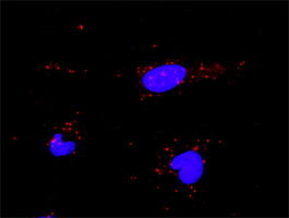
(Proximity Ligation Analysis of protein-protein interactions between TP53 and PML HeLa cells were stained with anti-TP53 rabbit purified polyclonal 1:1200 and anti-PML mouse monoclonal antibody 1:50. Each red dot represents the detection of protein-protein interaction complex, and nuclei were counterstained with DAPI (blue).)
Application Data
(Proximity Ligation Analysis of protein-protein interactions between TP53 and PML HeLa cells were stained with anti-TP53 rabbit purified polyclonal 1:1200 and anti-PML mouse monoclonal antibody 1:50. Each red dot represents the detection of protein-protein interaction complex, and nuclei were counterstained with DAPI (blue).)
PML, Monoclonal Antibody (Cat# AAA25211)
Full Name
PML (Probable Transcription Factor PML, RING Finger Protein 71, Tripartite Motif-containing Protein 19, PML, MYL, RNF71, TRIM19) (FITC)
Gene Names
PML; MYL; RNF71; PP8675; TRIM19
Reactivity
Human, Rat
Applications
Immunofluorescence, Western Blot
Purity
Purified by Protein A Affinity Chromatography.
Pricing


 Application Data
(Published Customer Image:Mouse CD31 antibody, clone ER-MP12 used for the demonstration of vasculature in mouse brain by immunofluorescence.Image caption:Inhibition of 2-AG hydrolysis reduces LPS-induced BBB permeability. a, b Fibrinogen levels in b plasma and the a ratio of brain to plasma fibrinogen were assessed by ELISA. n?=?5/7 mice per group. c, d Fluorescent immunostaining in the striatum for fibrinogen (red) and vascular marker (CD31; green) demonstrated leakage of fibrinogen into the brain with vehicle treatment, whereas vascular integrity was preserved when (e, f) MAGL was inhibited. g Extravascular fibrinogen was semi-quantitated in fluorescently labeled sections of the striatum. Bar graphs were plotted with mean?+/-SEM and data analyzed using one-way analysis of variance (ANOVA) with Tukey post-hoc comparisons. n = 5/7 mice per group. Significance is shown as *p?)
Application Data
(Published Customer Image:Mouse CD31 antibody, clone ER-MP12 used for the demonstration of vasculature in mouse brain by immunofluorescence.Image caption:Inhibition of 2-AG hydrolysis reduces LPS-induced BBB permeability. a, b Fibrinogen levels in b plasma and the a ratio of brain to plasma fibrinogen were assessed by ELISA. n?=?5/7 mice per group. c, d Fluorescent immunostaining in the striatum for fibrinogen (red) and vascular marker (CD31; green) demonstrated leakage of fibrinogen into the brain with vehicle treatment, whereas vascular integrity was preserved when (e, f) MAGL was inhibited. g Extravascular fibrinogen was semi-quantitated in fluorescently labeled sections of the striatum. Bar graphs were plotted with mean?+/-SEM and data analyzed using one-way analysis of variance (ANOVA) with Tukey post-hoc comparisons. n = 5/7 mice per group. Significance is shown as *p?)
CD31, Monoclonal Antibody (Cat# AAA12258)
Full Name
Rat Anti Mouse CD31: FITC
Gene Names
Pecam1; Cd31; Pecam; C85791; PECAM-1
Reactivity
Mouse
Applications
Flow Cytometry
Purity
Purified IgG prepared by affinity chromatography on Protein G from tissue culture supernatant
Pricing


 Application Data
(Proximity Ligation Analysis of protein-protein interactions between PRKCZ and AKT3. HeLa cells were stained with anti-PRKCZ rabbit purified polyclonal 1:1200 and anti-AKT3 mouse monoclonal antibody 1:50. Each red dot represents the detection of protein-protein interaction complex, and nuclei were counterstained with DAPI (blue).)
Application Data
(Proximity Ligation Analysis of protein-protein interactions between PRKCZ and AKT3. HeLa cells were stained with anti-PRKCZ rabbit purified polyclonal 1:1200 and anti-AKT3 mouse monoclonal antibody 1:50. Each red dot represents the detection of protein-protein interaction complex, and nuclei were counterstained with DAPI (blue).)
AKT3, Monoclonal Antibody (Cat# AAA24429)
Full Name
AKT3 (RAC-gamma Serine/Threonine-protein Kinase, Protein Kinase Akt-3, Protein Kinase B gamma, PKB gamma, RAC-PK-gamma, STK-2, PKBG) APC
Gene Names
AKT3; MPPH; PKBG; MPPH2; PRKBG; STK-2; PKB-GAMMA; RAC-gamma; RAC-PK-gamma
Reactivity
Human, Mouse, Rat
Applications
Western Blot
Purity
Purified by Protein A Affinity Chromatography.
Pricing


 Application Data
(Western Blot analysis of CD68 expression on J774 cells using Rat anti Mouse CD68 with Goat anti Rat IgG:HRP as a detection antibody)
Application Data
(Western Blot analysis of CD68 expression on J774 cells using Rat anti Mouse CD68 with Goat anti Rat IgG:HRP as a detection antibody)
CD68, Monoclonal Antibody (Cat# AAA12245)
Full Name
RAT ANTI MOUSE CD68:Endotoxin Low
Gene Names
Cd68; Lamp4; gp110; Scard1
Applications
Immunohistochemistry, Flow Cytometry, Immunofluorescence, Immunoprecipitation, Immunohistochemistry, Western Blot
Pricing


 SDS-PAGE
(SDS Page analysis of purified S100B Mouse Monoclonal Antibody (S100B/1012).)
SDS-PAGE
(SDS Page analysis of purified S100B Mouse Monoclonal Antibody (S100B/1012).)
S100B, Monoclonal Antibody (Cat# AAA23893)
Full Name
S100B (Astrocyte and Melanoma Marker)
Gene Names
S100B; NEF; S100; S100-B; S100beta
Reactivity
Human, Mouse, Rat, Cow. Others not known.
Applications
Flow Cytometry, Immunofluorescence, Western Blot, Immunohistochemistry
Pricing


 Application Data
(Frozen mouse spleen stained with Rat anti Mouse CD169)
Application Data
(Frozen mouse spleen stained with Rat anti Mouse CD169)
CD169, Monoclonal Antibody (Cat# AAA12223)
Full Name
RAT ANTI MOUSE CD169:Low Endotoxin
Gene Names
Siglec1; Sn; Cd169; Siglec-1
Applications
Immunohistochemistry, Flow Cytometry, Immunofluorescence
Pricing


 WB (Western Blot)
(Western blot analysis of Human Lysates showing detection of Hsp90 protein using Mouse Anti-Hsp90 Monoclonal Antibody, Clone H9010 . Primary Antibody: Mouse Anti-Hsp90 Monoclonal Antibody at 1:1000. Comparison of clone H9010 behavior with Hsp90 human beta (1) and Hsp90 human alpha (2). Courtesy of: David Toft, Mayo Clinic.)
WB (Western Blot)
(Western blot analysis of Human Lysates showing detection of Hsp90 protein using Mouse Anti-Hsp90 Monoclonal Antibody, Clone H9010 . Primary Antibody: Mouse Anti-Hsp90 Monoclonal Antibody at 1:1000. Comparison of clone H9010 behavior with Hsp90 human beta (1) and Hsp90 human alpha (2). Courtesy of: David Toft, Mayo Clinic.)
HSP90, Monoclonal Antibody (Cat# AAA27660)
Full Name
HSP90 Antibody
Gene Names
HSP90AB1; HSP84; HSPC2; HSPCB; D6S182; HSP90B
Reactivity
Human, Mouse, Rat, Rabbit, Chicken, Dog, Fish, Shark, Hamster
Applications
Western Blot, Immunohistochemistry, Immunocytochemistry, Immunofluorescence, Immunoprecipitation, Antibody Microarray
Purity
Protein G Purified
Pricing


 Application Data
(Staining of Rat peripheral blood lymphocytes with Mouse anti Rat CD49d:RPE (MCA2872PE))
Application Data
(Staining of Rat peripheral blood lymphocytes with Mouse anti Rat CD49d:RPE (MCA2872PE))
CD49d, Monoclonal Antibody (Cat# AAA26771)
Full Name
CD49d (Antigen CD49d, CD49d Antigen, CDw49d, Alpha 4 Subunit of VLA-4 Receptor, Integrin alpha IV, Integrin alpha 4, IA4, ITGA4, LPAM23, MGC90518, Very Late Activation Protein 4 Receptor Alpha 4 Subunit, VLA4, VLA-4) (PE)
Reactivity
Rat
Applications
Flow Cytometry, Immunoprecipitation
Purity
Purified by Protein G Affinity Chromatography
Pricing


 Application Data
(Proximity Ligation Analysis of protein-protein interactions between TP53 and PML HeLa cells were stained with anti-TP53 rabbit purified polyclonal 1:1200 and anti-PML mouse monoclonal antibody 1:50. Each red dot represents the detection of protein-protein interaction complex, and nuclei were counterstained with DAPI (blue).)
Application Data
(Proximity Ligation Analysis of protein-protein interactions between TP53 and PML HeLa cells were stained with anti-TP53 rabbit purified polyclonal 1:1200 and anti-PML mouse monoclonal antibody 1:50. Each red dot represents the detection of protein-protein interaction complex, and nuclei were counterstained with DAPI (blue).)
PML, Monoclonal Antibody (Cat# AAA24619)
Full Name
PML (Probable Transcription Factor PML, RING Finger Protein 71, Tripartite Motif-containing Protein 19, PML, MYL, RNF71, TRIM19) APC
Gene Names
PML; MYL; RNF71; PP8675; TRIM19
Reactivity
Human, Rat
Applications
Immunofluorescence, Western Blot
Purity
Purified by Protein A Affinity Chromatography.
Pricing


 Application Data
(Published customer image Infiltration of GFP+ BM-cells in infarct and peri-infarct regions. (A-B) Dot plots of viable macrophages/granulocytes (CD11b+CD45high, top right quadrants) and microglia (CD11b+CD45dim, bottom right quadrants) in cortex from BM-chimeric unmanipulated mice and mice exposed to pMCAO. (C) Bar graph showing mean numbers of CD11b+CD45dim microglia and CD11b+CD45high macrophages/granulocytes in BM-chimeric mice 24 hours after pMCAO, subdivided based on expression of GFP (n = 5). Approximately 92% of of the CD45high population were GFP+. (D) Estimation and comparison of mean numbers of CD11b+CD45dim microglia in non-chimeric (n = 10) versus BM-chimeric mice (n = 5) 24 hours after of pMCAO shows significantly fewer CD11b+CD45dim microglial cells in irradiated mice. (E) Overview, showing distribution of infiltrating GFP+ BM-derived cells into infarct (IF) and peri-infarct (P-IF) regions 24 hours after pMCAO. (E-G) By 24 hours, GFP+ single cells (F) and vessel-associated aggregates of GFP+ cells (arrows in G) were observed in infarct and peri-infarct regions. Some of the vessel-associated cells were round, leukocyte-like cells (arrows) while others were elongated cells lining the vasculature (arrow heads in G and in insert). (H) Bar graph showing mean numbers of single GFP+ cells and vessel-associated aggregates of GFP+ cells in ipsi- and contralateral cortex 24 hours after surgery (n = 10). (I-P) Immunohistochemical staining of CD45.1 (I, K), CD45.2 (J, L), IgG2a (M, O) and CD45 (N, P) in ischemic tissue in BM-chimeric (I, J, M, N) and non-chimeric mice (K, L, O, P) 24 hours after pMCAO. N.D, none detected. Scale bars: 200 um (A), 10 um (B, C). 50 um (I-P) *P < 0.05, **P < 0.01, and ***P < 0.001.From: Clausen BH, Lambertsen KL, Babcock AA, Holm TH, Dagnaes-Hansen F, Finsen B. Interleukin-1beta and tumor necrosis factor-alpha are expressed by different subsets of microglia and macrophages after ischemic stroke in mice. J Neuroinflammation. 2008 Oct 23;5:46.)
Application Data
(Published customer image Infiltration of GFP+ BM-cells in infarct and peri-infarct regions. (A-B) Dot plots of viable macrophages/granulocytes (CD11b+CD45high, top right quadrants) and microglia (CD11b+CD45dim, bottom right quadrants) in cortex from BM-chimeric unmanipulated mice and mice exposed to pMCAO. (C) Bar graph showing mean numbers of CD11b+CD45dim microglia and CD11b+CD45high macrophages/granulocytes in BM-chimeric mice 24 hours after pMCAO, subdivided based on expression of GFP (n = 5). Approximately 92% of of the CD45high population were GFP+. (D) Estimation and comparison of mean numbers of CD11b+CD45dim microglia in non-chimeric (n = 10) versus BM-chimeric mice (n = 5) 24 hours after of pMCAO shows significantly fewer CD11b+CD45dim microglial cells in irradiated mice. (E) Overview, showing distribution of infiltrating GFP+ BM-derived cells into infarct (IF) and peri-infarct (P-IF) regions 24 hours after pMCAO. (E-G) By 24 hours, GFP+ single cells (F) and vessel-associated aggregates of GFP+ cells (arrows in G) were observed in infarct and peri-infarct regions. Some of the vessel-associated cells were round, leukocyte-like cells (arrows) while others were elongated cells lining the vasculature (arrow heads in G and in insert). (H) Bar graph showing mean numbers of single GFP+ cells and vessel-associated aggregates of GFP+ cells in ipsi- and contralateral cortex 24 hours after surgery (n = 10). (I-P) Immunohistochemical staining of CD45.1 (I, K), CD45.2 (J, L), IgG2a (M, O) and CD45 (N, P) in ischemic tissue in BM-chimeric (I, J, M, N) and non-chimeric mice (K, L, O, P) 24 hours after pMCAO. N.D, none detected. Scale bars: 200 um (A), 10 um (B, C). 50 um (I-P) *P < 0.05, **P < 0.01, and ***P < 0.001.From: Clausen BH, Lambertsen KL, Babcock AA, Holm TH, Dagnaes-Hansen F, Finsen B. Interleukin-1beta and tumor necrosis factor-alpha are expressed by different subsets of microglia and macrophages after ischemic stroke in mice. J Neuroinflammation. 2008 Oct 23;5:46.)
CD11b, Monoclonal Antibody (Cat# AAA12182)
Full Name
RAT ANTI MOUSE CD11b:FITC
Gene Names
Itgam; CR3; CR3A; MAC1; Cd11b; Ly-40; Mac-1; Mac-1a; CD11b/CD18; F730045J24Rik
Applications
Flow Cytometry
Pricing


 Application Data
(Immunoperoxidase staining of mouse lymph node cryosection stained with Rat antii Mouse CD8alpha antibody, clone KT15 followed by horseradish peroxidase conjugatedGoat anti Rat IgG s a detection reagent. High power)
Application Data
(Immunoperoxidase staining of mouse lymph node cryosection stained with Rat antii Mouse CD8alpha antibody, clone KT15 followed by horseradish peroxidase conjugatedGoat anti Rat IgG s a detection reagent. High power)
CD8 ALPHA, Monoclonal Antibody (Cat# AAA12055)
Full Name
RAT ANTI MOUSE CD8 ALPHA:RPE
Gene Names
Cd8a; Ly-2; Ly-B; Ly-35; Lyt-2; BB154331
Applications
Flow Cytometry
Pricing


 Application Data
(Published customer image: Representative images of the inflammatory changes in the facial nucleus during axonal regeneration, one week following facial nerve transaction. a, b: CD11b immunoreactivity for microglia is increased in the axotomized facial nucleus, and microglia enwrap the facial motor neurons, e.g. at arrows. The regenerating neurons were retrogradely labelled with fluorogold. c, d: CD6- positive T-cells accumulated in the injured motor nucleus (arrows). They had little cytoplasm but dense nuclei (c) and were sometimes clustered around neurons retrogradely labelled with fluorogold (d). The scale bar in (a) also applies to (b) and that in (c) also applies to (d).From: Shokouhi et al. BMC Neuroscience 2010 11:13.)
Application Data
(Published customer image: Representative images of the inflammatory changes in the facial nucleus during axonal regeneration, one week following facial nerve transaction. a, b: CD11b immunoreactivity for microglia is increased in the axotomized facial nucleus, and microglia enwrap the facial motor neurons, e.g. at arrows. The regenerating neurons were retrogradely labelled with fluorogold. c, d: CD6- positive T-cells accumulated in the injured motor nucleus (arrows). They had little cytoplasm but dense nuclei (c) and were sometimes clustered around neurons retrogradely labelled with fluorogold (d). The scale bar in (a) also applies to (b) and that in (c) also applies to (d).From: Shokouhi et al. BMC Neuroscience 2010 11:13.)
CD11b, Monoclonal Antibody (Cat# AAA12146)
Full Name
MOUSE ANTI RAT CD11b:FITC
Gene Names
ITGAM; CD11B
Applications
Flow Cytometry
Pricing


 WB (Western Blot)
(UBQLN2 monoclonal antibody. Western Blot analysis of UBQLN2 expression in NIH/3T3.)
WB (Western Blot)
(UBQLN2 monoclonal antibody. Western Blot analysis of UBQLN2 expression in NIH/3T3.)
Ubiquilin-2, Monoclonal Antibody (Cat# AAA24086)
Full Name
Ubiquilin-2 (UBQLN2, Chap1, DSK2 Homolog, HRIHFB2157, N4BP4, Protein Linking IAP with Cytoskeleton 2, PLIC2, PLIC-2, hPLIC-2, Ubiquitin-like Product Chap1/Dsk2)
Gene Names
UBQLN2; DSK2; ALS15; CHAP1; N4BP4; PLIC2; HRIHFB2157
Reactivity
Human, Mouse, Rat
Applications
Western Blot, Immunofluorescence
Purity
Affinity Purified
Purified by Protein A affinity chromatography.
Purified by Protein A affinity chromatography.
Pricing


 Application Data
(Staining of mouse spleen cells with Rat anti Mouse CD3 Epsilon (T3): APC)
Application Data
(Staining of mouse spleen cells with Rat anti Mouse CD3 Epsilon (T3): APC)
CD3, Monoclonal Antibody (Cat# AAA12175)
Full Name
RAT ANTI MOUSE CD3
Gene Names
Cd3e; CD3; T3e; AI504783; CD3epsilon
Applications
Immunohistochemistry, Flow Cytometry
Pricing


 Application Data
(Immunoperoxidase staining of mouse lymph node cryosection stained with Rat antii Mouse CD8alpha antibody, clone KT15 followed by horseradish peroxidase conjugatedGoat anti Rat IgG s a detection reagent. High power)
Application Data
(Immunoperoxidase staining of mouse lymph node cryosection stained with Rat antii Mouse CD8alpha antibody, clone KT15 followed by horseradish peroxidase conjugatedGoat anti Rat IgG s a detection reagent. High power)
CD8 ALPHA, Monoclonal Antibody (Cat# AAA11885)
Full Name
RAT ANTI MOUSE CD8 ALPHA:FITC
Gene Names
Cd8a; Ly-2; Ly-B; Ly-35; Lyt-2; BB154331
Applications
Flow Cytometry
Pricing


 Application Data
(Immunoperoxidase staining of mouse lymph node cryosection eith Rat anti Mouse antibody clone R3-63 followed by horseradish peroxidase Goat anti Rat IgG antibody . Medium power)
Application Data
(Immunoperoxidase staining of mouse lymph node cryosection eith Rat anti Mouse antibody clone R3-63 followed by horseradish peroxidase Goat anti Rat IgG antibody . Medium power)
CD13, Monoclonal Antibody (Cat# AAA12115)
Full Name
RAT ANTI MOUSE CD13:RPE
Gene Names
Anpep; Apn; AP-M; AP-N; Cd13; P150
Applications
Flow Cytometry
Pricing


 Application Data
(Published customer image Infiltration of GFP+ BM-cells in infarct and peri-infarct regions. (A-B) Dot plots of viable macrophages/granulocytes (CD11b+CD45high, top right quadrants) and microglia (CD11b+CD45dim, bottom right quadrants) in cortex from BM-chimeric unmanipulated mice and mice exposed to pMCAO. (C) Bar graph showing mean numbers of CD11b+CD45dim microglia and CD11b+CD45high macrophages/granulocytes in BM-chimeric mice 24 hours after pMCAO, subdivided based on expression of GFP (n = 5). Approximately 92% of of the CD45high population were GFP+. (D) Estimation and comparison of mean numbers of CD11b+CD45dim microglia in non-chimeric (n = 10) versus BM-chimeric mice (n = 5) 24 hours after of pMCAO shows significantly fewer CD11b+CD45dim microglial cells in irradiated mice. (E) Overview, showing distribution of infiltrating GFP+ BM-derived cells into infarct (IF) and peri-infarct (P-IF) regions 24 hours after pMCAO. (E-G) By 24 hours, GFP+ single cells (F) and vessel-associated aggregates of GFP+ cells (arrows in G) were observed in infarct and peri-infarct regions. Some of the vessel-associated cells were round, leukocyte-like cells (arrows) while others were elongated cells lining the vasculature (arrow heads in G and in insert). (H) Bar graph showing mean numbers of single GFP+ cells and vessel-associated aggregates of GFP+ cells in ipsi- and contralateral cortex 24 hours after surgery (n = 10). (I-P) Immunohistochemical staining of CD45.1 (I, K), CD45.2 (J, L), IgG2a (M, O) and CD45 (N, P) in ischemic tissue in BM-chimeric (I, J, M, N) and non-chimeric mice (K, L, O, P) 24 hours after pMCAO. N.D, none detected. Scale bars: 200 um (A), 10 um (B, C). 50 um (I-P) *P < 0.05, **P < 0.01, and ***P < 0.001.From: Clausen BH, Lambertsen KL, Babcock AA, Holm TH, Dagnaes-Hansen F, Finsen B. Interleukin-1beta and tumor necrosis factor-alpha are expressed by different subsets of microglia and macrophages after ischemic stroke in mice. J Neuroinflammation. 2008 Oct 23;5:46.)
Application Data
(Published customer image Infiltration of GFP+ BM-cells in infarct and peri-infarct regions. (A-B) Dot plots of viable macrophages/granulocytes (CD11b+CD45high, top right quadrants) and microglia (CD11b+CD45dim, bottom right quadrants) in cortex from BM-chimeric unmanipulated mice and mice exposed to pMCAO. (C) Bar graph showing mean numbers of CD11b+CD45dim microglia and CD11b+CD45high macrophages/granulocytes in BM-chimeric mice 24 hours after pMCAO, subdivided based on expression of GFP (n = 5). Approximately 92% of of the CD45high population were GFP+. (D) Estimation and comparison of mean numbers of CD11b+CD45dim microglia in non-chimeric (n = 10) versus BM-chimeric mice (n = 5) 24 hours after of pMCAO shows significantly fewer CD11b+CD45dim microglial cells in irradiated mice. (E) Overview, showing distribution of infiltrating GFP+ BM-derived cells into infarct (IF) and peri-infarct (P-IF) regions 24 hours after pMCAO. (E-G) By 24 hours, GFP+ single cells (F) and vessel-associated aggregates of GFP+ cells (arrows in G) were observed in infarct and peri-infarct regions. Some of the vessel-associated cells were round, leukocyte-like cells (arrows) while others were elongated cells lining the vasculature (arrow heads in G and in insert). (H) Bar graph showing mean numbers of single GFP+ cells and vessel-associated aggregates of GFP+ cells in ipsi- and contralateral cortex 24 hours after surgery (n = 10). (I-P) Immunohistochemical staining of CD45.1 (I, K), CD45.2 (J, L), IgG2a (M, O) and CD45 (N, P) in ischemic tissue in BM-chimeric (I, J, M, N) and non-chimeric mice (K, L, O, P) 24 hours after pMCAO. N.D, none detected. Scale bars: 200 um (A), 10 um (B, C). 50 um (I-P) *P < 0.05, **P < 0.01, and ***P < 0.001.From: Clausen BH, Lambertsen KL, Babcock AA, Holm TH, Dagnaes-Hansen F, Finsen B. Interleukin-1beta and tumor necrosis factor-alpha are expressed by different subsets of microglia and macrophages after ischemic stroke in mice. J Neuroinflammation. 2008 Oct 23;5:46.)
CD11b, Monoclonal Antibody (Cat# AAA12184)
Full Name
RAT ANTI MOUSE CD11b
Gene Names
Itgam; CR3; CR3A; MAC1; Cd11b; Ly-40; Mac-1; Mac-1a; CD11b/CD18; F730045J24Rik
Applications
Immunohistochemistry, Flow Cytometry, Immunofluorescence, Immunoprecipitation
Pricing


 Application Data
(Published customer image: Representative images of the inflammatory changes in the facial nucleus during axonal regeneration, one week following facial nerve transaction. a, b: CD11b immunoreactivity for microglia is increased in the axotomized facial nucleus, and microglia enwrap the facial motor neurons, e.g. at arrows. The regenerating neurons were retrogradely labelled with fluorogold. c, d: CD6- positive T-cells accumulated in the injured motor nucleus (arrows). They had little cytoplasm but dense nuclei (c) and were sometimes clustered around neurons retrogradely labelled with fluorogold (d). The scale bar in (a) also applies to (b) and that in (c) also applies to (d).From: Shokouhi et al. BMC Neuroscience 2010 11:13.)
Application Data
(Published customer image: Representative images of the inflammatory changes in the facial nucleus during axonal regeneration, one week following facial nerve transaction. a, b: CD11b immunoreactivity for microglia is increased in the axotomized facial nucleus, and microglia enwrap the facial motor neurons, e.g. at arrows. The regenerating neurons were retrogradely labelled with fluorogold. c, d: CD6- positive T-cells accumulated in the injured motor nucleus (arrows). They had little cytoplasm but dense nuclei (c) and were sometimes clustered around neurons retrogradely labelled with fluorogold (d). The scale bar in (a) also applies to (b) and that in (c) also applies to (d).From: Shokouhi et al. BMC Neuroscience 2010 11:13.)
CD11b, Monoclonal Antibody (Cat# AAA12045)
Full Name
MOUSE ANTI RAT CD11b:RPE
Gene Names
ITGAM; CD11B
Applications
Flow Cytometry
Pricing


 Application Data
(Published customer image: Representative images of the inflammatory changes in the facial nucleus during axonal regeneration, one week following facial nerve transaction. a, b: CD11b immunoreactivity for microglia is increased in the axotomized facial nucleus, and microglia enwrap the facial motor neurons, e.g. at arrows. The regenerating neurons were retrogradely labelled with fluorogold. c, d: CD6- positive T-cells accumulated in the injured motor nucleus (arrows). They had little cytoplasm but dense nuclei (c) and were sometimes clustered around neurons retrogradely labelled with fluorogold (d). The scale bar in (a) also applies to (b) and that in (c) also applies to (d).From: Shokouhi et al. BMC Neuroscience 2010 11:13.)
Application Data
(Published customer image: Representative images of the inflammatory changes in the facial nucleus during axonal regeneration, one week following facial nerve transaction. a, b: CD11b immunoreactivity for microglia is increased in the axotomized facial nucleus, and microglia enwrap the facial motor neurons, e.g. at arrows. The regenerating neurons were retrogradely labelled with fluorogold. c, d: CD6- positive T-cells accumulated in the injured motor nucleus (arrows). They had little cytoplasm but dense nuclei (c) and were sometimes clustered around neurons retrogradely labelled with fluorogold (d). The scale bar in (a) also applies to (b) and that in (c) also applies to (d).From: Shokouhi et al. BMC Neuroscience 2010 11:13.)
CD11b, Monoclonal Antibody (Cat# AAA11969)
Full Name
MOUSE ANTI RAT CD11b
Gene Names
ITGAM; CD11B
Applications
Immunohistochemistry, Flow Cytometry, Immunofluorescence, Immunoprecipitation
Pricing


 Application Data
(Staining of mouse peripheral blood platelets with Rat anti Mouse CD36:FITC)
Application Data
(Staining of mouse peripheral blood platelets with Rat anti Mouse CD36:FITC)
CD36, Monoclonal Antibody (Cat# AAA12213)
Full Name
RAT ANTI MOUSE CD36
Gene Names
Cd36; FAT; GPIV; Scarb3
Applications
Flow Cytometry, Immunofluorescence, Immunoprecipitation, Western Blot
Pricing


 Application Data
(Proximity Ligation Analysis of protein-protein interactions between PRKCZ and AKT3. HeLa cells were stained with anti-PRKCZ rabbit purified polyclonal 1:1200 and anti-AKT3 mouse monoclonal antibody 1:50. Each red dot represents the detection of protein-protein interaction complex, and nuclei were counterstained with DAPI (blue).)
Application Data
(Proximity Ligation Analysis of protein-protein interactions between PRKCZ and AKT3. HeLa cells were stained with anti-PRKCZ rabbit purified polyclonal 1:1200 and anti-AKT3 mouse monoclonal antibody 1:50. Each red dot represents the detection of protein-protein interaction complex, and nuclei were counterstained with DAPI (blue).)
AKT3, Monoclonal Antibody (Cat# AAA25610)
Full Name
AKT3 (RAC-gamma Serine/Threonine-protein Kinase, Protein Kinase Akt-3, Protein Kinase B gamma, PKB gamma, RAC-PK-gamma, STK-2, PKBG) (PE)
Gene Names
AKT3; MPPH; PKBG; MPPH2; PRKBG; STK-2; PKB-GAMMA; RAC-gamma; RAC-PK-gamma
Reactivity
Human, Mouse, Rat
Applications
Western Blot
Purity
Purified by Protein A Affinity Chromatography.
Pricing


 Application Data
(Immunoperoxidase staining of mouse lymph node cryosection stained with Rat antii Mouse CD8alpha antibody, clone KT15 followed by horseradish peroxidase conjugatedGoat anti Rat IgG s a detection reagent. High power)
Application Data
(Immunoperoxidase staining of mouse lymph node cryosection stained with Rat antii Mouse CD8alpha antibody, clone KT15 followed by horseradish peroxidase conjugatedGoat anti Rat IgG s a detection reagent. High power)
CD8 ALPHA, Monoclonal Antibody (Cat# AAA12218)
Full Name
RAT ANTI MOUSE CD8 ALPHA:APC
Gene Names
Cd8a; Ly-2; Ly-B; Ly-35; Lyt-2; BB154331
Applications
Flow Cytometry
Pricing


 Application Data
(Staining of Rat peripheral blood lymphocytes with Mouse anti Rat CD49d:RPE (MCA2872PE))
Application Data
(Staining of Rat peripheral blood lymphocytes with Mouse anti Rat CD49d:RPE (MCA2872PE))
CD49d, Monoclonal Antibody (Cat# AAA26721)
Full Name
CD49d (Antigen CD49d, CD49d Antigen, CDw49d, Alpha 4 Subunit of VLA-4 Receptor, Integrin alpha IV, Integrin alpha 4, IA4, ITGA4, LPAM23, MGC90518, Very Late Activation Protein 4 Receptor Alpha 4 Subunit, VLA4, VLA-4) (AP)
Reactivity
Rat
Applications
Immunoprecipitation
Purity
Purified by Protein G Affinity Chromatography
Pricing


 Application Data
(Staining of mouse peripheral blood platelets with Rat anti Mouse CD36:FITC)
Application Data
(Staining of mouse peripheral blood platelets with Rat anti Mouse CD36:FITC)
CD36, Monoclonal Antibody (Cat# AAA12214)
Full Name
RAT ANTI MOUSE CD36:Low Endotoxin
Gene Names
Cd36; FAT; GPIV; Scarb3
Applications
Flow Cytometry, Functional Assay, Immunofluorescence, Immunoprecipitation, Western Blot
Pricing


 Application Data
(Published customer image: Increased accumulation of repair-associated macrophages surrounding collaterals in ischemic hind limbs is PAR2-dependent. (A) Stainings of CD206-positive macrophages (green) and SMA-positive vessels (red) in non-ischemic (control) and ischemic (ligated) hind limbs of WT, PAR1-/- and PAR2-/- mice are shown. Nuclei were visualized with DAPI (blue). Arrows indicate single macrophages in the non-ischemic adductor. Quantification of the average number of repair-associated macrophages per vessel is indicated on the right. (B) Correlation between the number of CD206-positive macrophages in the ischemic tissues and the expression of CD11b and (C) CD115 on monocytes. ** p)
Application Data
(Published customer image: Increased accumulation of repair-associated macrophages surrounding collaterals in ischemic hind limbs is PAR2-dependent. (A) Stainings of CD206-positive macrophages (green) and SMA-positive vessels (red) in non-ischemic (control) and ischemic (ligated) hind limbs of WT, PAR1-/- and PAR2-/- mice are shown. Nuclei were visualized with DAPI (blue). Arrows indicate single macrophages in the non-ischemic adductor. Quantification of the average number of repair-associated macrophages per vessel is indicated on the right. (B) Correlation between the number of CD206-positive macrophages in the ischemic tissues and the expression of CD11b and (C) CD115 on monocytes. ** p)
CD206, Monoclonal Antibody (Cat# AAA12123)
Full Name
RAT ANTI MOUSE CD206
Gene Names
Mrc1; MR; CD206; AW259686
Applications
Immunohistochemistry, Flow Cytometry, Immunofluorescence, Immunoprecipitation
Pricing


 Application Data
(Frozen mouse spleen stained with Rat anti Mouse CD169)
Application Data
(Frozen mouse spleen stained with Rat anti Mouse CD169)
CD169, Monoclonal Antibody (Cat# AAA12205)
Full Name
RAT ANTI MOUSE CD169:FITC
Gene Names
Siglec1; Sn; Cd169; Siglec-1
Applications
Flow Cytometry
Pricing


 Application Data
(Staining of mouse splenocytes with Rat anti Mouse CD40: RPE)
Application Data
(Staining of mouse splenocytes with Rat anti Mouse CD40: RPE)
CD40, Monoclonal Antibody (Cat# AAA11919)
Full Name
RAT ANTI MOUSE CD40
Gene Names
Cd40; IGM; p50; Bp50; GP39; IMD3; TRAP; HIGM1; T-BAM; Tnfrsf5; AI326936
Applications
Immunohistochemistry, Flow Cytometry
Pricing


 Application Data
(Staining of mouse peripheral blood granulocytes with Rat anti Mouse CD13)
Application Data
(Staining of mouse peripheral blood granulocytes with Rat anti Mouse CD13)
CD13, Monoclonal Antibody (Cat# AAA12145)
Full Name
RAT ANTI MOUSE CD13
Gene Names
Anpep; Apn; AP-M; AP-N; Cd13; P150
Applications
Immunohistochemistry, Flow Cytometry
Pricing