Filters
Clonality
Type
Reactivity
Gene Name
Isotype
Host
Application
Clone
14 results for "Molecular Weight Marker" - showing 1-14


 IHC (Immunohistchemistry)
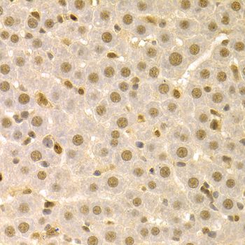
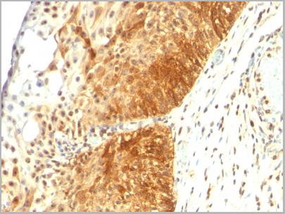
(Immunohistochemistry of paraffin-embedded mouse liver using SFN Antibody at dilution of 1:100 (40x lens).)
IHC (Immunohistchemistry)
(Immunohistochemistry of paraffin-embedded mouse liver using SFN Antibody at dilution of 1:100 (40x lens).)
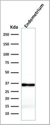
SFN, Polyclonal Antibody (Cat# AAA10757)
Full Name
SFN Polyclonal Antibody
Gene Names
SFN; YWHAS
Reactivity
Human, Mouse, Rat
Applications
Western Blot, Immunohistochemistry, Immunofluorescence
Purity
Affinity Purification
Pricing
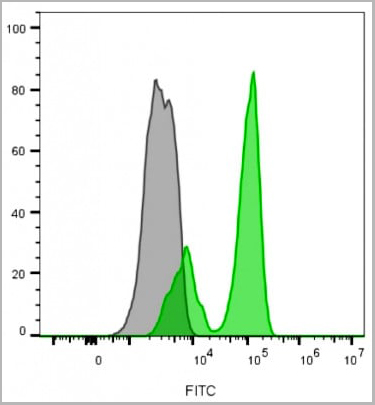
 FCM (Flow Cytometry)
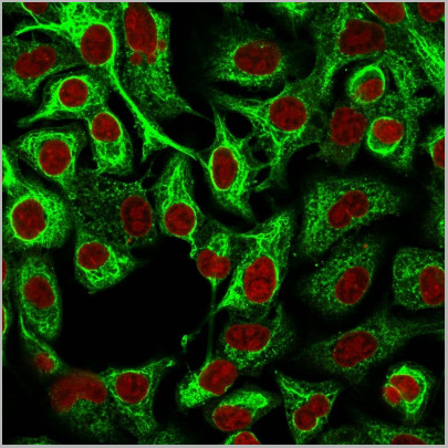
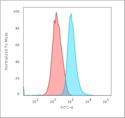
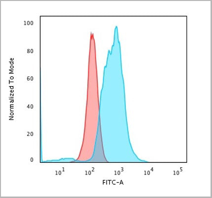
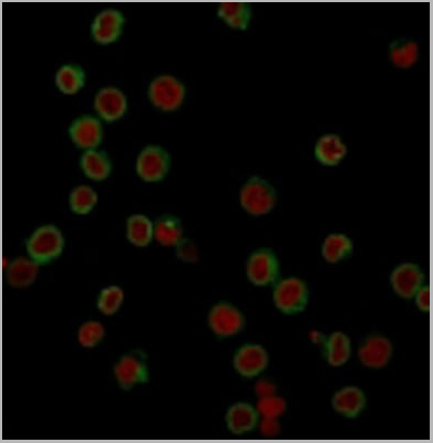
(Flow Cytometric Analysis of trypsinized MeOH-fixed HeLa cells using Cytokeratin 14 Mouse Monoclonal Antibody (LL002) followed by Goat anti-Mouse IgG-CF488 (Blue); Isotype Control (Red).)
FCM (Flow Cytometry)
(Flow Cytometric Analysis of trypsinized MeOH-fixed HeLa cells using Cytokeratin 14 Mouse Monoclonal Antibody (LL002) followed by Goat anti-Mouse IgG-CF488 (Blue); Isotype Control (Red).)
Cytokeratin 14 (KRT14), Monoclonal Antibody (Cat# AAA13813)
Full Name
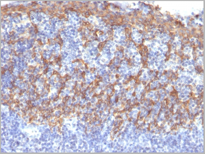
Cytokeratin 14 (KRT14) (Squamous Cell Marker) Mouse Monoclonal Antibody
Gene Names
KRT14; K14; NFJ; CK14; EBS3; EBS4
Reactivity
Human
Applications
Flow Cytometry, Immunofluorescence, Immunohistochemistry
Pricing
 Application Data
Prestained Multicolor Protein Molecular Weight Marker (1.7-40 kDa), Accessory Reagent (Cat# AAA13522)
Full Name
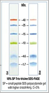
Prestained Multicolor Protein Molecular Weight Marker (1.7-40 kDa)
Applications
Western Blot
Pricing
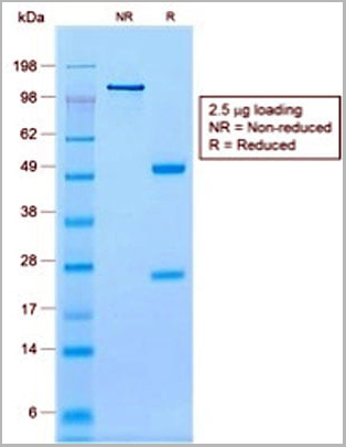
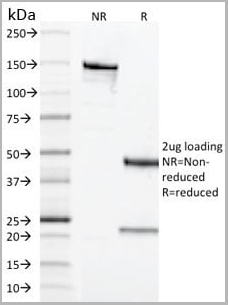
 SDS-PAGE
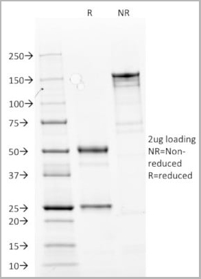
(The antibody wsas purified by protein G affinity achromatography from cell culture supernatants and verified by SDS-Page (Fig.2).Fig.2: SDS-PAGE analysis of purified YD-4E9 monoclonal antibody.Lane 1: molecular weight markerLane 2: 2ug of purified YD-4E9 antibody. Proteins were separated by SDSPAGE and stained with RAPID StainTM Reagent.)
SDS-PAGE
(The antibody wsas purified by protein G affinity achromatography from cell culture supernatants and verified by SDS-Page (Fig.2).Fig.2: SDS-PAGE analysis of purified YD-4E9 monoclonal antibody.Lane 1: molecular weight markerLane 2: 2ug of purified YD-4E9 antibody. Proteins were separated by SDSPAGE and stained with RAPID StainTM Reagent.)
CLAUDIN9, Monoclonal Antibody (Cat# AAA14098)
Full Name
anti-human Claudin 9 monoclonal antibody
Reactivity
Human
Applications
Flow Cytometry, Competitive ELISA
Purity
Protein G
Pricing
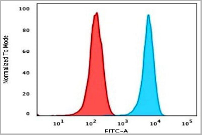
 FCM (Flow Cytometry)
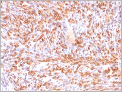
(Flow Cytometric Analysis of PFA-fixed K562 cells using Calponin-1 Recombinant Mouse Monoclonal Antibody (rCNN1/832) followed by Goat anti-Mouse IgG-CF488 (Blue); Isotype Control (Red).)
FCM (Flow Cytometry)
(Flow Cytometric Analysis of PFA-fixed K562 cells using Calponin-1 Recombinant Mouse Monoclonal Antibody (rCNN1/832) followed by Goat anti-Mouse IgG-CF488 (Blue); Isotype Control (Red).)
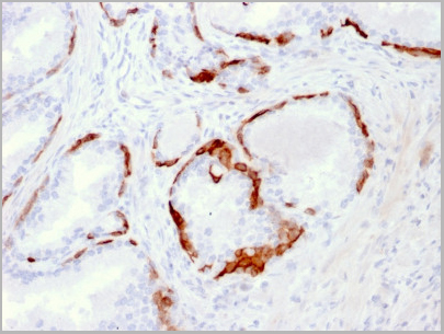
Calponin-1, Monoclonal Antibody (Cat# AAA13848)
Full Name
Calponin-1 (Smooth Muscle Marker)
Gene Names
CNN1; SMCC; Sm-Calp; HEL-S-14
Reactivity
Human, Rat
Applications
Flow Cytometry, Immunofluorescence, Immunohistochemistry
Pricing
 Application Data
(Fig.2: CGE analysis of purified EQ-8D11-C1 monoclonal antibody. Lane 1: molecular weight marker, Lane 2: 2ug of purified EQ-8D11-C1 antibody. Proteins were separated by CGE (capillary gel electrophoresis, Agilent 2100 Bioanalyzer). Internal control bands (240 kDa / 7 kDa / 4,5 kDa).The antibody was purified by protein G affinity chromatography from cell culture supernatants and verified by CGE (Fig.2).)
Application Data
(Fig.2: CGE analysis of purified EQ-8D11-C1 monoclonal antibody. Lane 1: molecular weight marker, Lane 2: 2ug of purified EQ-8D11-C1 antibody. Proteins were separated by CGE (capillary gel electrophoresis, Agilent 2100 Bioanalyzer). Internal control bands (240 kDa / 7 kDa / 4,5 kDa).The antibody was purified by protein G affinity chromatography from cell culture supernatants and verified by CGE (Fig.2).)
Ovine CD34, Monoclonal Antibody (Cat# AAA14097)
Full Name
anti-ovine CD34 monoclonal antibody
Applications
Flow Cytometry, Competitive ELISA
Purity
Protein G purified
Pricing


 Application Data
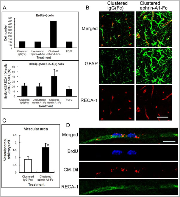
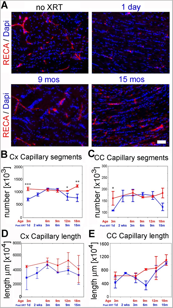
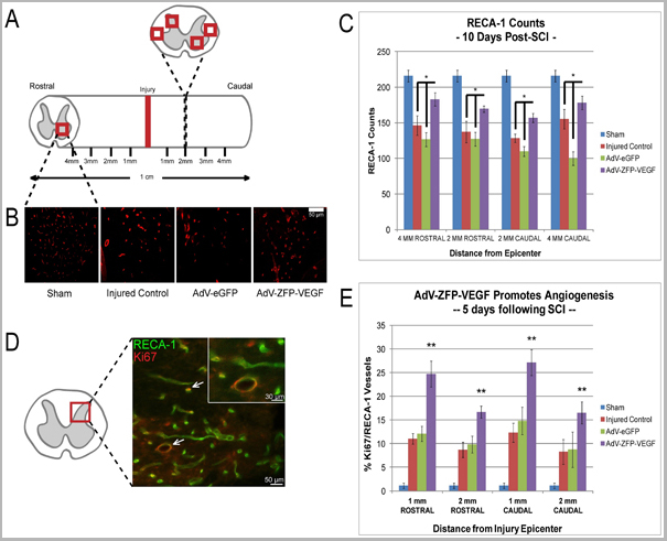
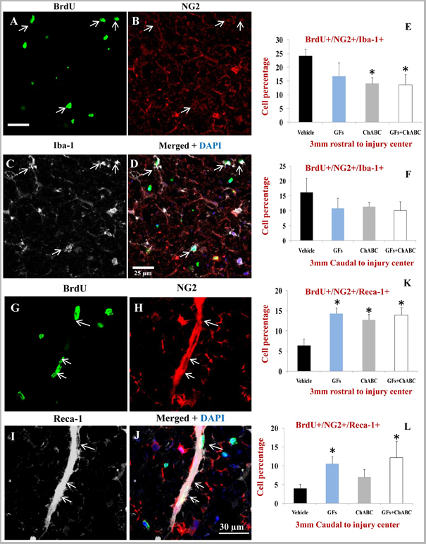
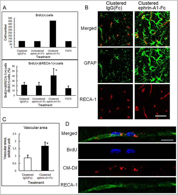
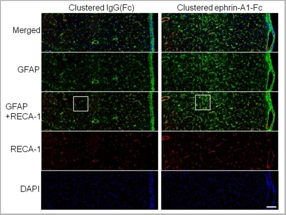
(Published customer image: Effect of clustered ephrin-A1-Fc on vascular formation in the rat striatum. Clustered ephrin-A1-Fc was injected into the lesioned side of the lateral ventricle in the unilaterally lesioned rats. Brains taken 6 weeks after injection were sectioned coronally and stained for GFAP (green) and RECA-1 (red) and with DAPI (nuclei; blue). The rectangular insets are shown in Fig. 8B. Scale bar: 100 um.From: Jing X, Miwa H, Sawada T, Nakanishi I, Kondo T, et al. (2012) Ephrin-A1-Mediated Dopaminergic Neurogenesis and Angiogenesis in a Rat Model of Parkinson's Disease. PLoS ONE 7(2): e32019.)
Application Data
(Published customer image: Effect of clustered ephrin-A1-Fc on vascular formation in the rat striatum. Clustered ephrin-A1-Fc was injected into the lesioned side of the lateral ventricle in the unilaterally lesioned rats. Brains taken 6 weeks after injection were sectioned coronally and stained for GFAP (green) and RECA-1 (red) and with DAPI (nuclei; blue). The rectangular insets are shown in Fig. 8B. Scale bar: 100 um.From: Jing X, Miwa H, Sawada T, Nakanishi I, Kondo T, et al. (2012) Ephrin-A1-Mediated Dopaminergic Neurogenesis and Angiogenesis in a Rat Model of Parkinson's Disease. PLoS ONE 7(2): e32019.)
RECA-1, Monoclonal Antibody (Cat# AAA12018)
Full Name
MOUSE ANTI RAT RECA-1
Applications
Immunohistochemistry, Immunofluorescence
Pricing


 Application Data
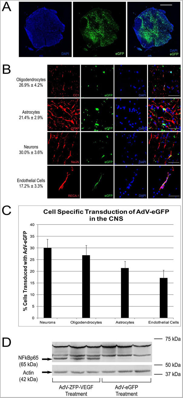
(Published customer image: Mouse anti V5 tag antibody, clone SV5-Pk1 used for the detection of V5 tagged WEEV_nsP3 protein by western blotting and immunofluorescenceImage caption: WEEV nsP3 interaction with host IKKbeta. A) U87MGs were transfected in a 6-well plate with 5 ug of pUC19 and WEEV_nsP3_HA for 24 hours. Cell lysates were resolved using SDS-PAGE and subsequently immunoblotted with V5 antibody and beta-actin served as a loading control. B) U87MGs were transfected with WEEV_nsP3_V5; cells were fixed after 24 hours and stained with antibodies against the endogenous IKKbeta and the V5 tag. Cells were incubated with appropriate secondary Alexa Fluor antibodies and the nuclei stained with DAPI. Co-localization of IKKbeta with WEEV_nsP3_V5 (yellow) was observed as shown by the arrows. B) Panels E -H serve as an example of transfected cells in a given field of view that show co-localization of IKKbeta and WEEV_nsP3_V5 24 hours post transfection. Panels I-L represent magnified images of other cells showing co-localization of IKKbeta and WEEV_nsP3_V5. Panel M is a magnified image of panel L. The co-localization was confirmed by Z-stack analysis. Co-localization was calculated to be approximately in 61% of cells (163 cells were counted of which 44% demonstrated expression of nsP3. Of those cells that expressed nsP3, 61% showed co-localization of both proteins). Images were taken using Nikon Eclipse TE2000-U at 60x magnification and are representative of 2 independent experiments.From: Amaya M, Voss K, Sampey G, Senina S, de la Fuente C, et al. (2014) The Role of IKKbeta in Venezuelan Equine Encephalitis Virus Infection. PLoS ONE 9(2): e86745.)
Application Data
(Published customer image: Mouse anti V5 tag antibody, clone SV5-Pk1 used for the detection of V5 tagged WEEV_nsP3 protein by western blotting and immunofluorescenceImage caption: WEEV nsP3 interaction with host IKKbeta. A) U87MGs were transfected in a 6-well plate with 5 ug of pUC19 and WEEV_nsP3_HA for 24 hours. Cell lysates were resolved using SDS-PAGE and subsequently immunoblotted with V5 antibody and beta-actin served as a loading control. B) U87MGs were transfected with WEEV_nsP3_V5; cells were fixed after 24 hours and stained with antibodies against the endogenous IKKbeta and the V5 tag. Cells were incubated with appropriate secondary Alexa Fluor antibodies and the nuclei stained with DAPI. Co-localization of IKKbeta with WEEV_nsP3_V5 (yellow) was observed as shown by the arrows. B) Panels E -H serve as an example of transfected cells in a given field of view that show co-localization of IKKbeta and WEEV_nsP3_V5 24 hours post transfection. Panels I-L represent magnified images of other cells showing co-localization of IKKbeta and WEEV_nsP3_V5. Panel M is a magnified image of panel L. The co-localization was confirmed by Z-stack analysis. Co-localization was calculated to be approximately in 61% of cells (163 cells were counted of which 44% demonstrated expression of nsP3. Of those cells that expressed nsP3, 61% showed co-localization of both proteins). Images were taken using Nikon Eclipse TE2000-U at 60x magnification and are representative of 2 independent experiments.From: Amaya M, Voss K, Sampey G, Senina S, de la Fuente C, et al. (2014) The Role of IKKbeta in Venezuelan Equine Encephalitis Virus Infection. PLoS ONE 9(2): e86745.)
V5-TAG, Monoclonal Antibody (Cat# AAA12081)
Full Name
MOUSE ANTI V5-TAG:HRP
Applications
Western Blot
Pricing


 Application Data
(Published customer image: Effect of clustered ephrin-A1-Fc on vascular formation in the rat striatum. Clustered ephrin-A1-Fc was injected into the lesioned side of the lateral ventricle in the unilaterally lesioned rats. Brains taken 6 weeks after injection were sectioned coronally and stained for GFAP (green) and RECA-1 (red) and with DAPI (nuclei; blue). The rectangular insets are shown in Fig. 8B. Scale bar: 100 um.From: Jing X, Miwa H, Sawada T, Nakanishi I, Kondo T, et al. (2012) Ephrin-A1-Mediated Dopaminergic Neurogenesis and Angiogenesis in a Rat Model of Parkinson's Disease. PLoS ONE 7(2): e32019.)
Application Data
(Published customer image: Effect of clustered ephrin-A1-Fc on vascular formation in the rat striatum. Clustered ephrin-A1-Fc was injected into the lesioned side of the lateral ventricle in the unilaterally lesioned rats. Brains taken 6 weeks after injection were sectioned coronally and stained for GFAP (green) and RECA-1 (red) and with DAPI (nuclei; blue). The rectangular insets are shown in Fig. 8B. Scale bar: 100 um.From: Jing X, Miwa H, Sawada T, Nakanishi I, Kondo T, et al. (2012) Ephrin-A1-Mediated Dopaminergic Neurogenesis and Angiogenesis in a Rat Model of Parkinson's Disease. PLoS ONE 7(2): e32019.)
RECA-1, Monoclonal Antibody (Cat# AAA12019)
Full Name
MOUSE ANTI RAT RECA-1
Applications
Immunohistochemistry, Immunofluorescence
Pricing


 Application Data
(Published customer image: Mouse anti V5 tag antibody, clone SV5-Pk1 used for the detection of V5 tagged WEEV_nsP3 protein by western blotting and immunofluorescenceImage caption: WEEV nsP3 interaction with host IKKbeta. A) U87MGs were transfected in a 6-well plate with 5 ug of pUC19 and WEEV_nsP3_HA for 24 hours. Cell lysates were resolved using SDS-PAGE and subsequently immunoblotted with V5 antibody and beta-actin served as a loading control. B) U87MGs were transfected with WEEV_nsP3_V5; cells were fixed after 24 hours and stained with antibodies against the endogenous IKKbeta and the V5 tag. Cells were incubated with appropriate secondary Alexa Fluor antibodies and the nuclei stained with DAPI. Co-localization of IKKbeta with WEEV_nsP3_V5 (yellow) was observed as shown by the arrows. B) Panels E -H serve as an example of transfected cells in a given field of view that show co-localization of IKKbeta and WEEV_nsP3_V5 24 hours post transfection. Panels I-L represent magnified images of other cells showing co-localization of IKKbeta and WEEV_nsP3_V5. Panel M is a magnified image of panel L. The co-localization was confirmed by Z-stack analysis. Co-localization was calculated to be approximately in 61% of cells (163 cells were counted of which 44% demonstrated expression of nsP3. Of those cells that expressed nsP3, 61% showed co-localization of both proteins). Images were taken using Nikon Eclipse TE2000-U at 60x magnification and are representative of 2 independent experiments.From: Amaya M, Voss K, Sampey G, Senina S, de la Fuente C, et al. (2014) The Role of IKKbeta in Venezuelan Equine Encephalitis Virus Infection. PLoS ONE 9(2): e86745.)
Application Data
(Published customer image: Mouse anti V5 tag antibody, clone SV5-Pk1 used for the detection of V5 tagged WEEV_nsP3 protein by western blotting and immunofluorescenceImage caption: WEEV nsP3 interaction with host IKKbeta. A) U87MGs were transfected in a 6-well plate with 5 ug of pUC19 and WEEV_nsP3_HA for 24 hours. Cell lysates were resolved using SDS-PAGE and subsequently immunoblotted with V5 antibody and beta-actin served as a loading control. B) U87MGs were transfected with WEEV_nsP3_V5; cells were fixed after 24 hours and stained with antibodies against the endogenous IKKbeta and the V5 tag. Cells were incubated with appropriate secondary Alexa Fluor antibodies and the nuclei stained with DAPI. Co-localization of IKKbeta with WEEV_nsP3_V5 (yellow) was observed as shown by the arrows. B) Panels E -H serve as an example of transfected cells in a given field of view that show co-localization of IKKbeta and WEEV_nsP3_V5 24 hours post transfection. Panels I-L represent magnified images of other cells showing co-localization of IKKbeta and WEEV_nsP3_V5. Panel M is a magnified image of panel L. The co-localization was confirmed by Z-stack analysis. Co-localization was calculated to be approximately in 61% of cells (163 cells were counted of which 44% demonstrated expression of nsP3. Of those cells that expressed nsP3, 61% showed co-localization of both proteins). Images were taken using Nikon Eclipse TE2000-U at 60x magnification and are representative of 2 independent experiments.From: Amaya M, Voss K, Sampey G, Senina S, de la Fuente C, et al. (2014) The Role of IKKbeta in Venezuelan Equine Encephalitis Virus Infection. PLoS ONE 9(2): e86745.)
V5-TAG, Monoclonal Antibody (Cat# AAA11864)
Full Name
MOUSE ANTI V5-TAG:FITC
Applications
Immunofluorescence
Pricing


 Application Data
(Published customer image: Mouse anti V5 tag antibody, clone SV5-Pk1 used for the detection of V5 tagged WEEV_nsP3 protein by western blotting and immunofluorescenceImage caption: WEEV nsP3 interaction with host IKKbeta. A) U87MGs were transfected in a 6-well plate with 5 ug of pUC19 and WEEV_nsP3_HA for 24 hours. Cell lysates were resolved using SDS-PAGE and subsequently immunoblotted with V5 antibody and beta-actin served as a loading control. B) U87MGs were transfected with WEEV_nsP3_V5; cells were fixed after 24 hours and stained with antibodies against the endogenous IKKbeta and the V5 tag. Cells were incubated with appropriate secondary Alexa Fluor antibodies and the nuclei stained with DAPI. Co-localization of IKKbeta with WEEV_nsP3_V5 (yellow) was observed as shown by the arrows. B) Panels E -H serve as an example of transfected cells in a given field of view that show co-localization of IKKbeta and WEEV_nsP3_V5 24 hours post transfection. Panels I-L represent magnified images of other cells showing co-localization of IKKbeta and WEEV_nsP3_V5. Panel M is a magnified image of panel L. The co-localization was confirmed by Z-stack analysis. Co-localization was calculated to be approximately in 61% of cells (163 cells were counted of which 44% demonstrated expression of nsP3. Of those cells that expressed nsP3, 61% showed co-localization of both proteins). Images were taken using Nikon Eclipse TE2000-U at 60x magnification and are representative of 2 independent experiments.From: Amaya M, Voss K, Sampey G, Senina S, de la Fuente C, et al. (2014) The Role of IKKbeta in Venezuelan Equine Encephalitis Virus Infection. PLoS ONE 9(2): e86745.)
Application Data
(Published customer image: Mouse anti V5 tag antibody, clone SV5-Pk1 used for the detection of V5 tagged WEEV_nsP3 protein by western blotting and immunofluorescenceImage caption: WEEV nsP3 interaction with host IKKbeta. A) U87MGs were transfected in a 6-well plate with 5 ug of pUC19 and WEEV_nsP3_HA for 24 hours. Cell lysates were resolved using SDS-PAGE and subsequently immunoblotted with V5 antibody and beta-actin served as a loading control. B) U87MGs were transfected with WEEV_nsP3_V5; cells were fixed after 24 hours and stained with antibodies against the endogenous IKKbeta and the V5 tag. Cells were incubated with appropriate secondary Alexa Fluor antibodies and the nuclei stained with DAPI. Co-localization of IKKbeta with WEEV_nsP3_V5 (yellow) was observed as shown by the arrows. B) Panels E -H serve as an example of transfected cells in a given field of view that show co-localization of IKKbeta and WEEV_nsP3_V5 24 hours post transfection. Panels I-L represent magnified images of other cells showing co-localization of IKKbeta and WEEV_nsP3_V5. Panel M is a magnified image of panel L. The co-localization was confirmed by Z-stack analysis. Co-localization was calculated to be approximately in 61% of cells (163 cells were counted of which 44% demonstrated expression of nsP3. Of those cells that expressed nsP3, 61% showed co-localization of both proteins). Images were taken using Nikon Eclipse TE2000-U at 60x magnification and are representative of 2 independent experiments.From: Amaya M, Voss K, Sampey G, Senina S, de la Fuente C, et al. (2014) The Role of IKKbeta in Venezuelan Equine Encephalitis Virus Infection. PLoS ONE 9(2): e86745.)
V5-TAG, Monoclonal Antibody (Cat# AAA11930)
Full Name
MOUSE ANTI V5-TAG
Applications
Immunohistochemistry, Flow Cytometry, Immunofluorescence, Immunoprecipitation, Western Blot, Radioimmunoassay
Pricing
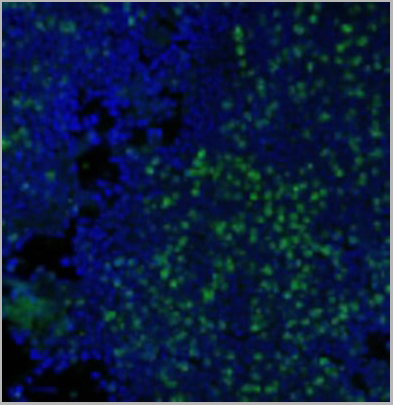 IF (Immunofluorescence)
(Immunofluorescence Analysis of methanol-fixed human tonsil cryosection stained with CF488A CD3e clone RIV9 (green), mounted in EverBrite’ Mounting Medium with DAPI (nuclei, blue).)
IF (Immunofluorescence)
(Immunofluorescence Analysis of methanol-fixed human tonsil cryosection stained with CF488A CD3e clone RIV9 (green), mounted in EverBrite’ Mounting Medium with DAPI (nuclei, blue).)
CD3, Monoclonal Antibody (Cat# AAA13839)
Full Name
CD3 (T-Cell Marker) Mouse Monoclonal Antibody
Gene Names
CD3E; T3E; TCRE; IMD18
Reactivity
Human, Mouse, Rat
Applications
Functional Assay, Flow Cytometry, Immunofluorescence
Pricing


 Application Data
(Published customer image: Mouse anti V5 tag antibody, clone SV5-Pk1 used for the detection of V5 tagged WEEV_nsP3 protein by western blotting and immunofluorescenceImage caption: WEEV nsP3 interaction with host IKKbeta. A) U87MGs were transfected in a 6-well plate with 5 ug of pUC19 and WEEV_nsP3_HA for 24 hours. Cell lysates were resolved using SDS-PAGE and subsequently immunoblotted with V5 antibody and beta-actin served as a loading control. B) U87MGs were transfected with WEEV_nsP3_V5; cells were fixed after 24 hours and stained with antibodies against the endogenous IKKbeta and the V5 tag. Cells were incubated with appropriate secondary Alexa Fluor antibodies and the nuclei stained with DAPI. Co-localization of IKKbeta with WEEV_nsP3_V5 (yellow) was observed as shown by the arrows. B) Panels E -H serve as an example of transfected cells in a given field of view that show co-localization of IKKbeta and WEEV_nsP3_V5 24 hours post transfection. Panels I-L represent magnified images of other cells showing co-localization of IKKbeta and WEEV_nsP3_V5. Panel M is a magnified image of panel L. The co-localization was confirmed by Z-stack analysis. Co-localization was calculated to be approximately in 61% of cells (163 cells were counted of which 44% demonstrated expression of nsP3. Of those cells that expressed nsP3, 61% showed co-localization of both proteins). Images were taken using Nikon Eclipse TE2000-U at 60x magnification and are representative of 2 independent experiments.From: Amaya M, Voss K, Sampey G, Senina S, de la Fuente C, et al. (2014) The Role of IKKbeta in Venezuelan Equine Encephalitis Virus Infection. PLoS ONE 9(2): e86745.)
Application Data
(Published customer image: Mouse anti V5 tag antibody, clone SV5-Pk1 used for the detection of V5 tagged WEEV_nsP3 protein by western blotting and immunofluorescenceImage caption: WEEV nsP3 interaction with host IKKbeta. A) U87MGs were transfected in a 6-well plate with 5 ug of pUC19 and WEEV_nsP3_HA for 24 hours. Cell lysates were resolved using SDS-PAGE and subsequently immunoblotted with V5 antibody and beta-actin served as a loading control. B) U87MGs were transfected with WEEV_nsP3_V5; cells were fixed after 24 hours and stained with antibodies against the endogenous IKKbeta and the V5 tag. Cells were incubated with appropriate secondary Alexa Fluor antibodies and the nuclei stained with DAPI. Co-localization of IKKbeta with WEEV_nsP3_V5 (yellow) was observed as shown by the arrows. B) Panels E -H serve as an example of transfected cells in a given field of view that show co-localization of IKKbeta and WEEV_nsP3_V5 24 hours post transfection. Panels I-L represent magnified images of other cells showing co-localization of IKKbeta and WEEV_nsP3_V5. Panel M is a magnified image of panel L. The co-localization was confirmed by Z-stack analysis. Co-localization was calculated to be approximately in 61% of cells (163 cells were counted of which 44% demonstrated expression of nsP3. Of those cells that expressed nsP3, 61% showed co-localization of both proteins). Images were taken using Nikon Eclipse TE2000-U at 60x magnification and are representative of 2 independent experiments.From: Amaya M, Voss K, Sampey G, Senina S, de la Fuente C, et al. (2014) The Role of IKKbeta in Venezuelan Equine Encephalitis Virus Infection. PLoS ONE 9(2): e86745.)
V5-TAG, Monoclonal Antibody (Cat# AAA12211)
Full Name
MOUSE ANTI V5-TAG
Applications
Immunohistochemistry, Flow Cytometry, Immunofluorescence, Immunoprecipitation, Western Blot, Radioimmunoassay
Pricing


 Application Data
(Published customer image: Mouse anti V5 tag antibody, clone SV5-Pk1 used for the detection of V5 tagged WEEV_nsP3 protein by western blotting and immunofluorescenceImage caption: WEEV nsP3 interaction with host IKKbeta. A) U87MGs were transfected in a 6-well plate with 5 ug of pUC19 and WEEV_nsP3_HA for 24 hours. Cell lysates were resolved using SDS-PAGE and subsequently immunoblotted with V5 antibody and beta-actin served as a loading control. B) U87MGs were transfected with WEEV_nsP3_V5; cells were fixed after 24 hours and stained with antibodies against the endogenous IKKbeta and the V5 tag. Cells were incubated with appropriate secondary Alexa Fluor antibodies and the nuclei stained with DAPI. Co-localization of IKKbeta with WEEV_nsP3_V5 (yellow) was observed as shown by the arrows. B) Panels E -H serve as an example of transfected cells in a given field of view that show co-localization of IKKbeta and WEEV_nsP3_V5 24 hours post transfection. Panels I-L represent magnified images of other cells showing co-localization of IKKbeta and WEEV_nsP3_V5. Panel M is a magnified image of panel L. The co-localization was confirmed by Z-stack analysis. Co-localization was calculated to be approximately in 61% of cells (163 cells were counted of which 44% demonstrated expression of nsP3. Of those cells that expressed nsP3, 61% showed co-localization of both proteins). Images were taken using Nikon Eclipse TE2000-U at 60x magnification and are representative of 2 independent experiments.From: Amaya M, Voss K, Sampey G, Senina S, de la Fuente C, et al. (2014) The Role of IKKbeta in Venezuelan Equine Encephalitis Virus Infection. PLoS ONE 9(2): e86745.)
Application Data
(Published customer image: Mouse anti V5 tag antibody, clone SV5-Pk1 used for the detection of V5 tagged WEEV_nsP3 protein by western blotting and immunofluorescenceImage caption: WEEV nsP3 interaction with host IKKbeta. A) U87MGs were transfected in a 6-well plate with 5 ug of pUC19 and WEEV_nsP3_HA for 24 hours. Cell lysates were resolved using SDS-PAGE and subsequently immunoblotted with V5 antibody and beta-actin served as a loading control. B) U87MGs were transfected with WEEV_nsP3_V5; cells were fixed after 24 hours and stained with antibodies against the endogenous IKKbeta and the V5 tag. Cells were incubated with appropriate secondary Alexa Fluor antibodies and the nuclei stained with DAPI. Co-localization of IKKbeta with WEEV_nsP3_V5 (yellow) was observed as shown by the arrows. B) Panels E -H serve as an example of transfected cells in a given field of view that show co-localization of IKKbeta and WEEV_nsP3_V5 24 hours post transfection. Panels I-L represent magnified images of other cells showing co-localization of IKKbeta and WEEV_nsP3_V5. Panel M is a magnified image of panel L. The co-localization was confirmed by Z-stack analysis. Co-localization was calculated to be approximately in 61% of cells (163 cells were counted of which 44% demonstrated expression of nsP3. Of those cells that expressed nsP3, 61% showed co-localization of both proteins). Images were taken using Nikon Eclipse TE2000-U at 60x magnification and are representative of 2 independent experiments.From: Amaya M, Voss K, Sampey G, Senina S, de la Fuente C, et al. (2014) The Role of IKKbeta in Venezuelan Equine Encephalitis Virus Infection. PLoS ONE 9(2): e86745.)
V5-TAG, Monoclonal Antibody (Cat# AAA11850)
Full Name
MOUSE ANTI V5-TAG:Biotin
Applications
Immunohistochemistry, Western Blot
Pricing