Filters
Clonality
Type
Reactivity
Gene Name
Isotype
Host
Application
Clone
64 results for " s Disease" - showing 1-50


 ELISA
(Figure 6 ELISA Validation of Delta Variant Spike S1 Protein with SARS-CoV-2 Spike S1 Antibodies Coating Antigen: SARS-CoV-2 spike S1 proteins, including WT and Delta variant (B.1.671.2), 5 μg/mL, incubated at 4 ˚C overnight.Detection Antibodies: SARS-CoV-2 Spike S1 antibody, dilution: 0.3-1000 ng/mL, incubate at RT for 1 hr.Secondary Antibodies: Goat anti-rabbit HRP at 1:20,000, incubate at RT for 1 hr.Antibody 9083 does not distinguish between WT and Delta.)
ELISA
(Figure 6 ELISA Validation of Delta Variant Spike S1 Protein with SARS-CoV-2 Spike S1 Antibodies Coating Antigen: SARS-CoV-2 spike S1 proteins, including WT and Delta variant (B.1.671.2), 5 μg/mL, incubated at 4 ˚C overnight.Detection Antibodies: SARS-CoV-2 Spike S1 antibody, dilution: 0.3-1000 ng/mL, incubate at RT for 1 hr.Secondary Antibodies: Goat anti-rabbit HRP at 1:20,000, incubate at RT for 1 hr.Antibody 9083 does not distinguish between WT and Delta.)
COVID 19 Delta Variant Spike S1 (His-Avi Tag) Coronavirus, Recombinant Protein (Cat# AAA11040)
Full Name
SARS-CoV-2 (COVID-19) Delta Variant Spike S1 (His-Avi Tag) Recombinant Protein
Applications
Western Blot
Purity
> 90% as determined by Bis-Tris PAGE
Pricing


 WB (Western Blot)
(Figure 6 WB Validation of Delta Variant Spike RBD Protein with SARS-CoV-2 Spike RBD Antibodies Loading: 20 ng of SARS-CoV-2 spike RBD proteins, including WT and Delta variant (B.1.671.2).Detection Antibodies: SARS-CoV-2 Spike RBD antibody, 1 μg/mL, incubate at RT for 1 hr.Secondary Antibodies: Goat anti-rabbit HRP at 1:20,000, incubate at RT for 1 hr.)
WB (Western Blot)
(Figure 6 WB Validation of Delta Variant Spike RBD Protein with SARS-CoV-2 Spike RBD Antibodies Loading: 20 ng of SARS-CoV-2 spike RBD proteins, including WT and Delta variant (B.1.671.2).Detection Antibodies: SARS-CoV-2 Spike RBD antibody, 1 μg/mL, incubate at RT for 1 hr.Secondary Antibodies: Goat anti-rabbit HRP at 1:20,000, incubate at RT for 1 hr.)
COVID 19 Delta Variant Spike RBD (His-Avi Tag) Coronavirus, Recombinant Protein (Cat# AAA11039)
Full Name
SARS-CoV-2 (COVID-19) Delta Variant Spike RBD (His-Avi Tag) Recombinant Protein
Applications
Western Blot
Purity
> 95% as determined by Bis-Tris PAGE
Pricing
HBsAg, Polyclonal Antibody (Cat# AAA14330)
Full Name
HBsAg antibody
Purity
HBsAg antibody was purified by affinity chromatography.
Pricing


 IP (Immunoprecipitation)
(IP/WB analysis of mouse brain, rat brain membrane lysate and mouse brain membrane lysate, were resolved by electrophoresis, transferred to PVDF membrane and probed using AAA14705 (1 :500). Proteins were visualized using a goat anti-rabbit secondary antibody conjugated to HRP and a chemiluminescence detection system. Arrow indicates protein AMYLOID OLIGOMER (-80kD).)
IP (Immunoprecipitation)
(IP/WB analysis of mouse brain, rat brain membrane lysate and mouse brain membrane lysate, were resolved by electrophoresis, transferred to PVDF membrane and probed using AAA14705 (1 :500). Proteins were visualized using a goat anti-rabbit secondary antibody conjugated to HRP and a chemiluminescence detection system. Arrow indicates protein AMYLOID OLIGOMER (-80kD).)
b-Amyloid, Oligomer A11, Polyclonal Antibody (Cat# AAA14705)
Full Name
b-Amyloid, Oligomer A11
Reactivity
Human, Mouse, Rat and Eukaryote
Applications
Western Blot, Immunoprecipitation, Immunohistochemistry, Immunofluorescence
Purity
Serum
Pricing


 IF (Immunofluorescence)
(Immunofluorescence analysis of HeLa cells using APP antibody at dilution of 1:100. Blue: DAPI for nuclear staining.)
IF (Immunofluorescence)
(Immunofluorescence analysis of HeLa cells using APP antibody at dilution of 1:100. Blue: DAPI for nuclear staining.)
APP, Polyclonal Antibody (Cat# AAA28326)
Full Name
APP Rabbit pAb
Gene Names
APP; AAA; AD1; PN2; ABPP; APPI; CVAP; ABETA; PN-II; CTFgamma
Reactivity
Human, Mouse, Rat
Applications
Western Blot, Immunohistochemistry, Immunofluorescence
Purity
Affinity purification
Pricing


 FCM (Flow Cytometry)
(Flow cytometric analysis of HepG2 cells with PARK7 antibody at 1/50 dilution (red) compared with an unlabelled control (cells without incubation with primary antibody; black). Alexa Fluor 488-conjugated goat anti rabbit IgG was used as the secondary antibody.)
FCM (Flow Cytometry)
(Flow cytometric analysis of HepG2 cells with PARK7 antibody at 1/50 dilution (red) compared with an unlabelled control (cells without incubation with primary antibody; black). Alexa Fluor 488-conjugated goat anti rabbit IgG was used as the secondary antibody.)
PARK7/DJ1, Monoclonal Antibody (Cat# AAA30163)
Full Name
PARK7/DJ1 Antibody
Gene Names
PARK7; DJ1; DJ-1
Reactivity
Human, Mouse, Rat
Applications
Western Blot, Immunocytochemistry, Immunofluorescence, Immunohistochemistry, Immunoprecipitation, Flow Cytometry
Purity
ProA affinity purified
Pricing

 SDS-PAGE
Amyloid Precursor Protein (APP), Recombinant Protein (Cat# AAA20280)
Full Name
Recombinant Amyloid Precursor Protein (APP)
Gene Names
App; Ag; Abpp; Adap; Cvap; Abeta; betaApp; E030013M08Rik
Reactivity
Mus musculus (Mouse)
Applications
Western Blot
Purity
> 95%
Pricing


 FCM (Flow Cytometry)
(Flow cytometric analysis of MCF-7 cells with MUC1 antibody at 1/50 dilution (red) compared with an unlabelled control (cells without incubation with primary antibody; black). Alexa Fluor 488-conjugated goat anti rabbit IgG was used as the secondary antibody.)
FCM (Flow Cytometry)
(Flow cytometric analysis of MCF-7 cells with MUC1 antibody at 1/50 dilution (red) compared with an unlabelled control (cells without incubation with primary antibody; black). Alexa Fluor 488-conjugated goat anti rabbit IgG was used as the secondary antibody.)
MUC1, Monoclonal Antibody (Cat# AAA30155)
Full Name
MUC1 Antibody
Gene Names
MUC1; EMA; PEM; PUM; KL-6; MAM6; PEMT; CD227; H23AG; MUC-1; CA 15-3; MUC-1/X; MUC1/ZD; MUC-1/SEC
Reactivity
Human
Applications
Western Blot, Immunocytochemistry, Immunofluorescence, Immunohistochemistry, Immunoprecipitation, Flow Cytometry
Purity
ProA affinity purified
Pricing


 Application Data
(Published customer image: Leukocyte infiltration in COX-2-M/-M and COX-2+/+ mice. MPO enzymatic activity (panel A) was statistically similar in COX-2-M/-M and COX-2+/+ livers at 6 h and 24 h post-IRI. Ly-6G+ neutrophil (panel B) and granulocyte (panel C) infiltration were also comparable in COX-2-M/-M and COX-2+/+ livers after IRI. Mac-1+ (panel D) and CD68 (panel E) infiltrating macrophages were significantly reduced in COX-2-M/-M livers at 24 h post-reperfusion, but were statistically indistinguishable in COX-2-M/-M and COX-2+/+ livers at 6 h after IRI. No statistical differences in MMP-9 expression (panel F) could be demonstrated in livers of COX-2-M/-M and COX-2+/+ mice post-IRI. Representative immunostaining (panel G) of infiltrating Ly-6G+ (a,b,e,f) and Mac-1+ (c,d,g,h) leukocytes in livers of COX-2+/+ (a,c,e,g) and COX-2-M/-M (b,d,f,h) mice at 6 h (a to d) and 24 h (e to h) post IRI; (n = 5 -6/group; * indicates p)
Application Data
(Published customer image: Leukocyte infiltration in COX-2-M/-M and COX-2+/+ mice. MPO enzymatic activity (panel A) was statistically similar in COX-2-M/-M and COX-2+/+ livers at 6 h and 24 h post-IRI. Ly-6G+ neutrophil (panel B) and granulocyte (panel C) infiltration were also comparable in COX-2-M/-M and COX-2+/+ livers after IRI. Mac-1+ (panel D) and CD68 (panel E) infiltrating macrophages were significantly reduced in COX-2-M/-M livers at 24 h post-reperfusion, but were statistically indistinguishable in COX-2-M/-M and COX-2+/+ livers at 6 h after IRI. No statistical differences in MMP-9 expression (panel F) could be demonstrated in livers of COX-2-M/-M and COX-2+/+ mice post-IRI. Representative immunostaining (panel G) of infiltrating Ly-6G+ (a,b,e,f) and Mac-1+ (c,d,g,h) leukocytes in livers of COX-2+/+ (a,c,e,g) and COX-2-M/-M (b,d,f,h) mice at 6 h (a to d) and 24 h (e to h) post IRI; (n = 5 -6/group; * indicates p)
CD68, Monoclonal Antibody (Cat# AAA12105)
Full Name
RAT ANTI MOUSE CD68:FITC
Gene Names
Cd68; Lamp4; gp110; Scard1
Applications
Flow Cytometry
Pricing


 IHC (Immunohistochemistry)
(At 1/100 staining Human prostate cancer and adjacent normal tissues by IHC-P. The sample was formaldehyde fixed and a heat mediated antigen retrieval step in citrate buffer was performed. The sample was then blocked and incubated with the primary antibody at 4 degree C overnight. An HRP conjugated anti-Rabbit antibody was used as the secondary antibody.)
IHC (Immunohistochemistry)
(At 1/100 staining Human prostate cancer and adjacent normal tissues by IHC-P. The sample was formaldehyde fixed and a heat mediated antigen retrieval step in citrate buffer was performed. The sample was then blocked and incubated with the primary antibody at 4 degree C overnight. An HRP conjugated anti-Rabbit antibody was used as the secondary antibody.)
S100A9, Polyclonal Antibody (Cat# AAA31466)
Full Name
Phospho-S100A9 (Thr113) Antibody
Reactivity
Human
Applications
Western Blot, Immunohistochemistry, Peptide ELISA
Purity
The antibody is from purified rabbit serum by affinity purification via sequential chromatography on phospho-peptide and non-phospho-peptide affinity columns.
Pricing


 Application Data
(Published customer image: Leukocyte infiltration in COX-2-M/-M and COX-2+/+ mice. MPO enzymatic activity (panel A) was statistically similar in COX-2-M/-M and COX-2+/+ livers at 6 h and 24 h post-IRI. Ly-6G+ neutrophil (panel B) and granulocyte (panel C) infiltration were also comparable in COX-2-M/-M and COX-2+/+ livers after IRI. Mac-1+ (panel D) and CD68 (panel E) infiltrating macrophages were significantly reduced in COX-2-M/-M livers at 24 h post-reperfusion, but were statistically indistinguishable in COX-2-M/-M and COX-2+/+ livers at 6 h after IRI. No statistical differences in MMP-9 expression (panel F) could be demonstrated in livers of COX-2-M/-M and COX-2+/+ mice post-IRI. Representative immunostaining (panel G) of infiltrating Ly-6G+ (a,b,e,f) and Mac-1+ (c,d,g,h) leukocytes in livers of COX-2+/+ (a,c,e,g) and COX-2-M/-M (b,d,f,h) mice at 6 h (a to d) and 24 h (e to h) post IRI; (n = 5 -6/group; * indicates p)
Application Data
(Published customer image: Leukocyte infiltration in COX-2-M/-M and COX-2+/+ mice. MPO enzymatic activity (panel A) was statistically similar in COX-2-M/-M and COX-2+/+ livers at 6 h and 24 h post-IRI. Ly-6G+ neutrophil (panel B) and granulocyte (panel C) infiltration were also comparable in COX-2-M/-M and COX-2+/+ livers after IRI. Mac-1+ (panel D) and CD68 (panel E) infiltrating macrophages were significantly reduced in COX-2-M/-M livers at 24 h post-reperfusion, but were statistically indistinguishable in COX-2-M/-M and COX-2+/+ livers at 6 h after IRI. No statistical differences in MMP-9 expression (panel F) could be demonstrated in livers of COX-2-M/-M and COX-2+/+ mice post-IRI. Representative immunostaining (panel G) of infiltrating Ly-6G+ (a,b,e,f) and Mac-1+ (c,d,g,h) leukocytes in livers of COX-2+/+ (a,c,e,g) and COX-2-M/-M (b,d,f,h) mice at 6 h (a to d) and 24 h (e to h) post IRI; (n = 5 -6/group; * indicates p)
CD68, Monoclonal Antibody (Cat# AAA12103)
Full Name
RAT ANTI MOUSE CD68:Biotin
Gene Names
Cd68; Lamp4; gp110; Scard1
Applications
Flow Cytometry
Pricing


 FCM (Flow Cytometry)
(Figure 6. Flow Cytometry analysis of PC-3 cells using anti-APP antibody (AAA11597).Overlay histogram showing PC-3 cells stained with AAA11597 (Blue line).The cells were blocked with 10% normal goat serum. And then incubated with rabbit anti-APP Antibody (AAA11597,1ug/1x10^6 cells) for 30 min at 20 degree C. DyLight®488 conjugated goat anti-rabbit IgG (5-10ug/1x10^6 cells) was used as secondary antibody for 30 minutes at 20 degree C. Isotype control antibody (Green line) was rabbit IgG (1ug/1x106) used under the same conditions. Unlabelled sample (Red line) was also used as a control.)
FCM (Flow Cytometry)
(Figure 6. Flow Cytometry analysis of PC-3 cells using anti-APP antibody (AAA11597).Overlay histogram showing PC-3 cells stained with AAA11597 (Blue line).The cells were blocked with 10% normal goat serum. And then incubated with rabbit anti-APP Antibody (AAA11597,1ug/1x10^6 cells) for 30 min at 20 degree C. DyLight®488 conjugated goat anti-rabbit IgG (5-10ug/1x10^6 cells) was used as secondary antibody for 30 minutes at 20 degree C. Isotype control antibody (Green line) was rabbit IgG (1ug/1x106) used under the same conditions. Unlabelled sample (Red line) was also used as a control.)
beta Amyloid, Polyclonal Antibody (Cat# AAA11597)
Full Name
Anti-beta Amyloid Antibody
Gene Names
APP; AAA; AD1; PN2; ABPP; APPI; CVAP; ABETA; PN-II; CTFgamma
Reactivity
Human, Mouse, Rat
Applications
Western Blot, Immunohistochemistry
Purity
Immunogen affinity purified.
Pricing


 Application Data
(Published customer image: Leukocyte infiltration in COX-2-M/-M and COX-2+/+ mice. MPO enzymatic activity (panel A) was statistically similar in COX-2-M/-M and COX-2+/+ livers at 6 h and 24 h post-IRI. Ly-6G+ neutrophil (panel B) and granulocyte (panel C) infiltration were also comparable in COX-2-M/-M and COX-2+/+ livers after IRI. Mac-1+ (panel D) and CD68 (panel E) infiltrating macrophages were significantly reduced in COX-2-M/-M livers at 24 h post-reperfusion, but were statistically indistinguishable in COX-2-M/-M and COX-2+/+ livers at 6 h after IRI. No statistical differences in MMP-9 expression (panel F) could be demonstrated in livers of COX-2-M/-M and COX-2+/+ mice post-IRI. Representative immunostaining (panel G) of infiltrating Ly-6G+ (a,b,e,f) and Mac-1+ (c,d,g,h) leukocytes in livers of COX-2+/+ (a,c,e,g) and COX-2-M/-M (b,d,f,h) mice at 6 h (a to d) and 24 h (e to h) post IRI; (n = 5 -6/group; * indicates p)
Application Data
(Published customer image: Leukocyte infiltration in COX-2-M/-M and COX-2+/+ mice. MPO enzymatic activity (panel A) was statistically similar in COX-2-M/-M and COX-2+/+ livers at 6 h and 24 h post-IRI. Ly-6G+ neutrophil (panel B) and granulocyte (panel C) infiltration were also comparable in COX-2-M/-M and COX-2+/+ livers after IRI. Mac-1+ (panel D) and CD68 (panel E) infiltrating macrophages were significantly reduced in COX-2-M/-M livers at 24 h post-reperfusion, but were statistically indistinguishable in COX-2-M/-M and COX-2+/+ livers at 6 h after IRI. No statistical differences in MMP-9 expression (panel F) could be demonstrated in livers of COX-2-M/-M and COX-2+/+ mice post-IRI. Representative immunostaining (panel G) of infiltrating Ly-6G+ (a,b,e,f) and Mac-1+ (c,d,g,h) leukocytes in livers of COX-2+/+ (a,c,e,g) and COX-2-M/-M (b,d,f,h) mice at 6 h (a to d) and 24 h (e to h) post IRI; (n = 5 -6/group; * indicates p)
CD68, Monoclonal Antibody (Cat# AAA12110)
Full Name
RAT ANTI MOUSE CD68
Gene Names
Cd68; Lamp4; gp110; Scard1
Applications
Immunohistochemistry, Flow Cytometry, Immunofluorescence, Immunoprecipitation, Immunohistochemistry, Western Blot
Pricing


 Application Data
(Published customer image: Leukocyte infiltration in COX-2-M/-M and COX-2+/+ mice. MPO enzymatic activity (panel A) was statistically similar in COX-2-M/-M and COX-2+/+ livers at 6 h and 24 h post-IRI. Ly-6G+ neutrophil (panel B) and granulocyte (panel C) infiltration were also comparable in COX-2-M/-M and COX-2+/+ livers after IRI. Mac-1+ (panel D) and CD68 (panel E) infiltrating macrophages were significantly reduced in COX-2-M/-M livers at 24 h post-reperfusion, but were statistically indistinguishable in COX-2-M/-M and COX-2+/+ livers at 6 h after IRI. No statistical differences in MMP-9 expression (panel F) could be demonstrated in livers of COX-2-M/-M and COX-2+/+ mice post-IRI. Representative immunostaining (panel G) of infiltrating Ly-6G+ (a,b,e,f) and Mac-1+ (c,d,g,h) leukocytes in livers of COX-2+/+ (a,c,e,g) and COX-2-M/-M (b,d,f,h) mice at 6 h (a to d) and 24 h (e to h) post IRI; (n = 5 -6/group; * indicates p)
Application Data
(Published customer image: Leukocyte infiltration in COX-2-M/-M and COX-2+/+ mice. MPO enzymatic activity (panel A) was statistically similar in COX-2-M/-M and COX-2+/+ livers at 6 h and 24 h post-IRI. Ly-6G+ neutrophil (panel B) and granulocyte (panel C) infiltration were also comparable in COX-2-M/-M and COX-2+/+ livers after IRI. Mac-1+ (panel D) and CD68 (panel E) infiltrating macrophages were significantly reduced in COX-2-M/-M livers at 24 h post-reperfusion, but were statistically indistinguishable in COX-2-M/-M and COX-2+/+ livers at 6 h after IRI. No statistical differences in MMP-9 expression (panel F) could be demonstrated in livers of COX-2-M/-M and COX-2+/+ mice post-IRI. Representative immunostaining (panel G) of infiltrating Ly-6G+ (a,b,e,f) and Mac-1+ (c,d,g,h) leukocytes in livers of COX-2+/+ (a,c,e,g) and COX-2-M/-M (b,d,f,h) mice at 6 h (a to d) and 24 h (e to h) post IRI; (n = 5 -6/group; * indicates p)
CD68, Monoclonal Antibody (Cat# AAA12104)
Full Name
RAT ANTI MOUSE CD68:Biotin
Gene Names
Cd68; Lamp4; gp110; Scard1
Applications
Flow Cytometry
Pricing


 IHC (Immunohistochemistry)
(At 1/100 staining Human colorectal cancer by IHC-P. The sample was formaldehyde fixed and a heat mediated antigen retrieval step in citrate buffer was performed. The sample was then blocked and incubated with the primary antibody at 4 degree C overnight. An HRP conjugated anti-Rabbit antibody was used as the secondary antibody.)
IHC (Immunohistochemistry)
(At 1/100 staining Human colorectal cancer by IHC-P. The sample was formaldehyde fixed and a heat mediated antigen retrieval step in citrate buffer was performed. The sample was then blocked and incubated with the primary antibody at 4 degree C overnight. An HRP conjugated anti-Rabbit antibody was used as the secondary antibody.)
CD227/MUC1, Polyclonal Antibody (Cat# AAA31426)
Full Name
Phospho-CD227/MUC1 (Ser1227) Antibody
Gene Names
MUC1; EMA; PEM; PUM; KL-6; MAM6; PEMT; CD227; H23AG; MUC-1; CA 15-3; MUC-1/X; MUC1/ZD; MUC-1/SEC
Reactivity
Human, Mouse, Rat
Applications
Western Blot, Immunohistochemistry, Peptide ELISA
Purity
The antibody is from purified rabbit serum by affinity purification via sequential chromatography on phospho-peptide and non-phospho-peptide affinity columns.
Pricing


 Application Data
(Published customer image: Leukocyte infiltration in COX-2-M/-M and COX-2+/+ mice. MPO enzymatic activity (panel A) was statistically similar in COX-2-M/-M and COX-2+/+ livers at 6 h and 24 h post-IRI. Ly-6G+ neutrophil (panel B) and granulocyte (panel C) infiltration were also comparable in COX-2-M/-M and COX-2+/+ livers after IRI. Mac-1+ (panel D) and CD68 (panel E) infiltrating macrophages were significantly reduced in COX-2-M/-M livers at 24 h post-reperfusion, but were statistically indistinguishable in COX-2-M/-M and COX-2+/+ livers at 6 h after IRI. No statistical differences in MMP-9 expression (panel F) could be demonstrated in livers of COX-2-M/-M and COX-2+/+ mice post-IRI. Representative immunostaining (panel G) of infiltrating Ly-6G+ (a,b,e,f) and Mac-1+ (c,d,g,h) leukocytes in livers of COX-2+/+ (a,c,e,g) and COX-2-M/-M (b,d,f,h) mice at 6 h (a to d) and 24 h (e to h) post IRI; (n = 5 -6/group; * indicates p)
Application Data
(Published customer image: Leukocyte infiltration in COX-2-M/-M and COX-2+/+ mice. MPO enzymatic activity (panel A) was statistically similar in COX-2-M/-M and COX-2+/+ livers at 6 h and 24 h post-IRI. Ly-6G+ neutrophil (panel B) and granulocyte (panel C) infiltration were also comparable in COX-2-M/-M and COX-2+/+ livers after IRI. Mac-1+ (panel D) and CD68 (panel E) infiltrating macrophages were significantly reduced in COX-2-M/-M livers at 24 h post-reperfusion, but were statistically indistinguishable in COX-2-M/-M and COX-2+/+ livers at 6 h after IRI. No statistical differences in MMP-9 expression (panel F) could be demonstrated in livers of COX-2-M/-M and COX-2+/+ mice post-IRI. Representative immunostaining (panel G) of infiltrating Ly-6G+ (a,b,e,f) and Mac-1+ (c,d,g,h) leukocytes in livers of COX-2+/+ (a,c,e,g) and COX-2-M/-M (b,d,f,h) mice at 6 h (a to d) and 24 h (e to h) post IRI; (n = 5 -6/group; * indicates p)
CD68, Monoclonal Antibody (Cat# AAA12107)
Full Name
RAT ANTI MOUSE CD68
Gene Names
Cd68; Lamp4; gp110; Scard1
Applications
Immunohistochemistry, Flow Cytometry, Immunofluorescence, Immunoprecipitation, Immunohistochemistry, Western Blot
Purity
Purified
Purified IgG - liquid
Purified IgG - liquid
Pricing


 Application Data
(Published customer image: Leukocyte infiltration in COX-2-M/-M and COX-2+/+ mice. MPO enzymatic activity (panel A) was statistically similar in COX-2-M/-M and COX-2+/+ livers at 6 h and 24 h post-IRI. Ly-6G+ neutrophil (panel B) and granulocyte (panel C) infiltration were also comparable in COX-2-M/-M and COX-2+/+ livers after IRI. Mac-1+ (panel D) and CD68 (panel E) infiltrating macrophages were significantly reduced in COX-2-M/-M livers at 24 h post-reperfusion, but were statistically indistinguishable in COX-2-M/-M and COX-2+/+ livers at 6 h after IRI. No statistical differences in MMP-9 expression (panel F) could be demonstrated in livers of COX-2-M/-M and COX-2+/+ mice post-IRI. Representative immunostaining (panel G) of infiltrating Ly-6G+ (a,b,e,f) and Mac-1+ (c,d,g,h) leukocytes in livers of COX-2+/+ (a,c,e,g) and COX-2-M/-M (b,d,f,h) mice at 6 h (a to d) and 24 h (e to h) post IRI; (n = 5 -6/group; * indicates p)
Application Data
(Published customer image: Leukocyte infiltration in COX-2-M/-M and COX-2+/+ mice. MPO enzymatic activity (panel A) was statistically similar in COX-2-M/-M and COX-2+/+ livers at 6 h and 24 h post-IRI. Ly-6G+ neutrophil (panel B) and granulocyte (panel C) infiltration were also comparable in COX-2-M/-M and COX-2+/+ livers after IRI. Mac-1+ (panel D) and CD68 (panel E) infiltrating macrophages were significantly reduced in COX-2-M/-M livers at 24 h post-reperfusion, but were statistically indistinguishable in COX-2-M/-M and COX-2+/+ livers at 6 h after IRI. No statistical differences in MMP-9 expression (panel F) could be demonstrated in livers of COX-2-M/-M and COX-2+/+ mice post-IRI. Representative immunostaining (panel G) of infiltrating Ly-6G+ (a,b,e,f) and Mac-1+ (c,d,g,h) leukocytes in livers of COX-2+/+ (a,c,e,g) and COX-2-M/-M (b,d,f,h) mice at 6 h (a to d) and 24 h (e to h) post IRI; (n = 5 -6/group; * indicates p)
CD68, Monoclonal Antibody (Cat# AAA12108)
Full Name
RAT ANTI MOUSE CD68:RPE
Gene Names
Cd68; Lamp4; gp110; Scard1
Applications
Flow Cytometry
Pricing


 IF (Immunofluorescence)
(Immunofluorescence Analysis of T98G cells labeling Pgp9.5 with Pgp9.5 / UchL1 Mouse Monoclonal Antibody (31A3) followed by Goat anti-Mouse IgG-CF488 (Green). The nuclear counterstain is Nucspot (Red))
IF (Immunofluorescence)
(Immunofluorescence Analysis of T98G cells labeling Pgp9.5 with Pgp9.5 / UchL1 Mouse Monoclonal Antibody (31A3) followed by Goat anti-Mouse IgG-CF488 (Green). The nuclear counterstain is Nucspot (Red))
PGP9.5 / UchL1, Monoclonal Antibody (Cat# AAA13809)
Full Name
PGP9.5 / UchL1 (pan-Neuronal Marker) Mouse Monoclonal Antibody
Gene Names
UCHL1; NDGOA; PARK5; PGP95; PGP9.5; Uch-L1; HEL-117; PGP 9.5; HEL-S-53
Reactivity
Human, Mouse, Rat, Cow, Pig, Dog
Applications
Western Blot, Immunohistochemistry
Pricing


 Application Data
(Published customer image: Leukocyte infiltration in COX-2-M/-M and COX-2+/+ mice. MPO enzymatic activity (panel A) was statistically similar in COX-2-M/-M and COX-2+/+ livers at 6 h and 24 h post-IRI. Ly-6G+ neutrophil (panel B) and granulocyte (panel C) infiltration were also comparable in COX-2-M/-M and COX-2+/+ livers after IRI. Mac-1+ (panel D) and CD68 (panel E) infiltrating macrophages were significantly reduced in COX-2-M/-M livers at 24 h post-reperfusion, but were statistically indistinguishable in COX-2-M/-M and COX-2+/+ livers at 6 h after IRI. No statistical differences in MMP-9 expression (panel F) could be demonstrated in livers of COX-2-M/-M and COX-2+/+ mice post-IRI. Representative immunostaining (panel G) of infiltrating Ly-6G+ (a,b,e,f) and Mac-1+ (c,d,g,h) leukocytes in livers of COX-2+/+ (a,c,e,g) and COX-2-M/-M (b,d,f,h) mice at 6 h (a to d) and 24 h (e to h) post IRI; (n = 5 -6/group; * indicates p)
Application Data
(Published customer image: Leukocyte infiltration in COX-2-M/-M and COX-2+/+ mice. MPO enzymatic activity (panel A) was statistically similar in COX-2-M/-M and COX-2+/+ livers at 6 h and 24 h post-IRI. Ly-6G+ neutrophil (panel B) and granulocyte (panel C) infiltration were also comparable in COX-2-M/-M and COX-2+/+ livers after IRI. Mac-1+ (panel D) and CD68 (panel E) infiltrating macrophages were significantly reduced in COX-2-M/-M livers at 24 h post-reperfusion, but were statistically indistinguishable in COX-2-M/-M and COX-2+/+ livers at 6 h after IRI. No statistical differences in MMP-9 expression (panel F) could be demonstrated in livers of COX-2-M/-M and COX-2+/+ mice post-IRI. Representative immunostaining (panel G) of infiltrating Ly-6G+ (a,b,e,f) and Mac-1+ (c,d,g,h) leukocytes in livers of COX-2+/+ (a,c,e,g) and COX-2-M/-M (b,d,f,h) mice at 6 h (a to d) and 24 h (e to h) post IRI; (n = 5 -6/group; * indicates p)
CD68, Monoclonal Antibody (Cat# AAA12102)
Full Name
RAT ANTI MOUSE CD68
Gene Names
Cd68; Lamp4; gp110; Scard1
Applications
Immunohistochemistry, Flow Cytometry, Immunofluorescence, Immunoprecipitation, Immunohistochemistry, Western Blot
Pricing
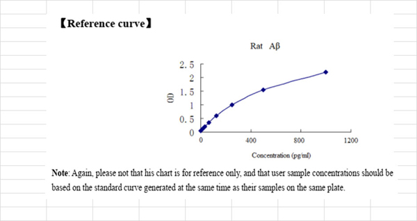
 Standard Curve (Sample)
amyloid beta (A4) precursor protein, ELISA Kit (Cat# AAA18393)
Full Name
Rat Amyloid beta A4 protein, APP ELISA Kit
Reactivity
Rat
Pricing


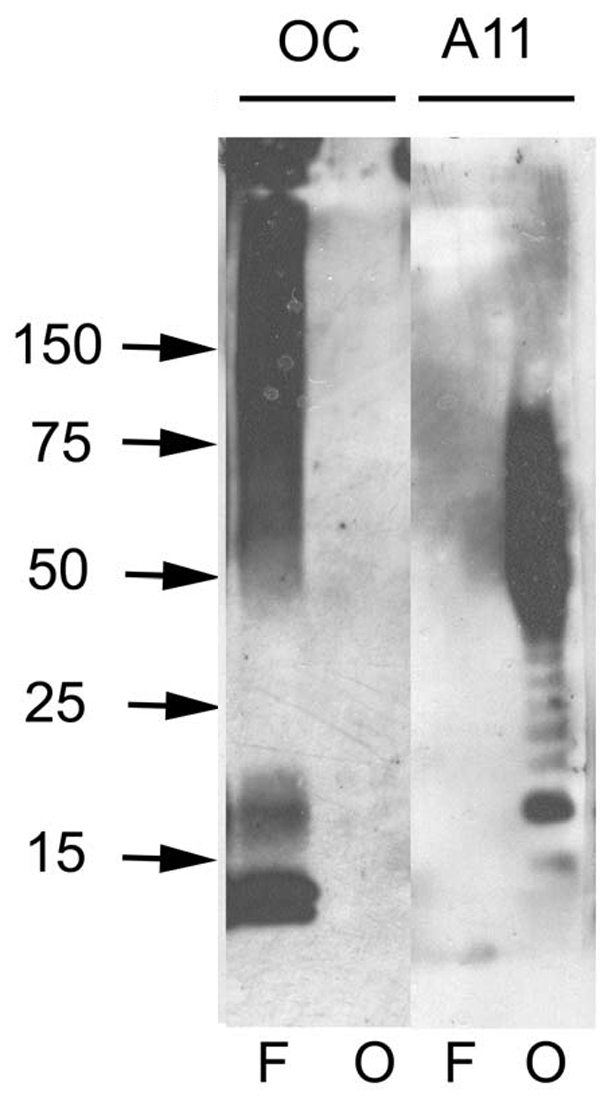
 DB (Dot Blot)
(Dot blot analysis using Rabbit Anti-Amyloid Fibrils (OC) Polyclonal Antibody (SPC-507). Tissue: Cell lysates. Species: Human. Primary Antibody: Rabbit Anti-Amyloid Fibrils (OC) Polyclonal Antibody (SPC-507) at 1:500, 1:5000. Beta Amyloid HEPES-NaCl aggregation, showing 1:500 (L) and 1:5000 (R) time lapse dot blot.)
DB (Dot Blot)
(Dot blot analysis using Rabbit Anti-Amyloid Fibrils (OC) Polyclonal Antibody (SPC-507). Tissue: Cell lysates. Species: Human. Primary Antibody: Rabbit Anti-Amyloid Fibrils (OC) Polyclonal Antibody (SPC-507) at 1:500, 1:5000. Beta Amyloid HEPES-NaCl aggregation, showing 1:500 (L) and 1:5000 (R) time lapse dot blot.)
Amyloid Fibrils (OC), Polyclonal Antibody (Cat# AAA17803)
Full Name
Amyloid Fibrils (OC) Antibody: ATTO 488
Reactivity
Human. Potentially mouse and rat based on species homology.
Applications
IP, ICC, IF, IHC, EIA, WB, DB
Purity
Protein A Purified
Pricing


 DB (Dot Blot)
(Dot blot analysis using Rabbit Anti-Amyloid Fibrils (OC) Polyclonal Antibody (SPC-507). Tissue: Cell lysates. Species: Human. Primary Antibody: Rabbit Anti-Amyloid Fibrils (OC) Polyclonal Antibody (SPC-507) at 1:500, 1:5000. Beta Amyloid HEPES-NaCl aggregation, showing 1:500 (L) and 1:5000 (R) time lapse dot blot.)
DB (Dot Blot)
(Dot blot analysis using Rabbit Anti-Amyloid Fibrils (OC) Polyclonal Antibody (SPC-507). Tissue: Cell lysates. Species: Human. Primary Antibody: Rabbit Anti-Amyloid Fibrils (OC) Polyclonal Antibody (SPC-507) at 1:500, 1:5000. Beta Amyloid HEPES-NaCl aggregation, showing 1:500 (L) and 1:5000 (R) time lapse dot blot.)
Amyloid Fibrils (OC), Polyclonal Antibody (Cat# AAA17800)
Full Name
Amyloid Fibrils (OC) Antibody: PerCP
Reactivity
Human. Potentially mouse and rat based on species homology.
Applications
IP, ICC, IHC, EIA, WB, DB
Pricing


 IF (Immunofluorescence)
(Immunofluorescence analysis of C6 cells using GAPDH Rabbit mAb at dilution of 1:100 (40x lens). Blue: DAPI for nuclear staining.)
IF (Immunofluorescence)
(Immunofluorescence analysis of C6 cells using GAPDH Rabbit mAb at dilution of 1:100 (40x lens). Blue: DAPI for nuclear staining.)
GAPDH, Monoclonal Antibody (Cat# AAA28392)
Full Name
GAPDH Rabbit mAb
Gene Names
GAPDH; G3PD; GAPD
Reactivity
Human, Mouse, Rat
Applications
WB, IHC, IF
Purity
Affinity purification
Pricing
 Sample Sheet
(Results for individual researchers may vary.)
Sample Sheet
(Results for individual researchers may vary.)
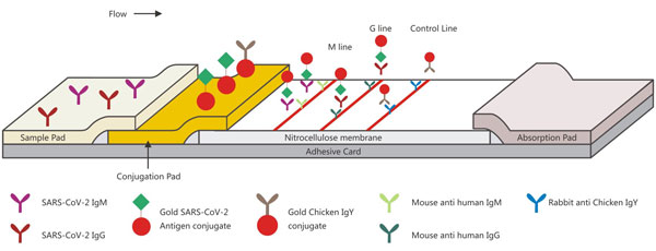
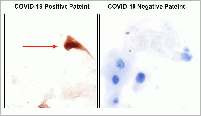
COVID 19 Nucleoprotein (N) / Spike Glycoprotein (S) Antibody Immunochromatography Coronavirus, Assay Kit (Cat# AAA27035)
Full Name
Human SARS-CoV-2 Nucleoprotein (N) / Spike Glycoprotein (S) Antibody Immunochromatography Assay Kit
Pricing
 Standard Curve (Sample)
Amyloid beta peptide 1-42 (Abeta1-42), ELISA Kit (Cat# AAA12964)
Full Name
Rat Amyloid beta peptide 1-42 (Abeta1-42) ELISA Kit
Gene Names
APP; AAA; AD1; PN2; ABPP; APPI; CVAP; ABETA; PN-II; CTFgamma
Reactivity
Rat
Pricing


 IF (Immunofluorescence)
(Immunofluorescence analysis of U-2 OS cells using GAPDH Rabbit pAb at dilution of 1:100 (40x lens). Blue: DAPI for nuclear staining.)
IF (Immunofluorescence)
(Immunofluorescence analysis of U-2 OS cells using GAPDH Rabbit pAb at dilution of 1:100 (40x lens). Blue: DAPI for nuclear staining.)
GAPDH, Polyclonal Antibody (Cat# AAA28397)
Full Name
GAPDH Rabbit pAb
Gene Names
GAPDH; G3PD; GAPD
Reactivity
Human, Mouse, Rat
Applications
WB, IHC, IF, IP
Purity
Affinity purification
Pricing
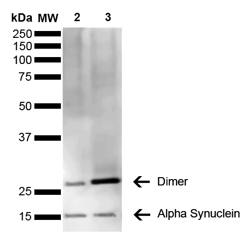

 WB (Western Blot)
(Western Blot analysis of Mouse, Rat Brain showing detection of 14 kDa Alpha Synuclein protein using Mouse Anti-Alpha Synuclein Monoclonal Antibody, Clone 3C11. Lane 1: Molecular Weight Ladder (MW). Lane 2: Mouse brain cell lysate. Lane 3: Rat brain cell lysate. Load: 15 ug. Block: 5% Skim Milk in 1X TBST. Primary Antibody: Mouse Anti-Alpha Synuclein Monoclonal Antibody at 1:1000 for 2 hours at RT. Secondary Antibody: Goat Anti-Mouse HRP:IgG at 1:3000 for 1 hour at RT. Color Development: ECL solution (Super Signal West Pico) for 5 min in RT. Predicted/Observed Size: 14 kDa. Other Band(s): ~30 kDa (dimer).)
WB (Western Blot)
(Western Blot analysis of Mouse, Rat Brain showing detection of 14 kDa Alpha Synuclein protein using Mouse Anti-Alpha Synuclein Monoclonal Antibody, Clone 3C11. Lane 1: Molecular Weight Ladder (MW). Lane 2: Mouse brain cell lysate. Lane 3: Rat brain cell lysate. Load: 15 ug. Block: 5% Skim Milk in 1X TBST. Primary Antibody: Mouse Anti-Alpha Synuclein Monoclonal Antibody at 1:1000 for 2 hours at RT. Secondary Antibody: Goat Anti-Mouse HRP:IgG at 1:3000 for 1 hour at RT. Color Development: ECL solution (Super Signal West Pico) for 5 min in RT. Predicted/Observed Size: 14 kDa. Other Band(s): ~30 kDa (dimer).)
Alpha Synuclein, Monoclonal Antibody (Cat# AAA17810)
Full Name
Alpha Synuclein Antibody, Clone 3C11
Gene Names
SNCA; PD1; NACP; PARK1; PARK4
Reactivity
Human; Mouse; Rat
Applications
WB, DB, ICC, IF, EIA
Purity
Protein G Purified
Pricing
 Standard Curve (Sample)
total amyloid beta peptide, ELISA Kit (Cat# AAA16628)
Full Name
Human total amyloid beta peptide ELISA Kit
Gene Names
APP; AAA; AD1; PN2; ABPP; APPI; CVAP; ABETA; PN-II; CTFgamma
Reactivity
Human
Pricing


 DB (Dot Blot)
(Dot blot analysis using Rabbit Anti-Amyloid Fibrils (OC) Polyclonal Antibody (SPC-507). Tissue: Cell lysates. Species: Human. Primary Antibody: Rabbit Anti-Amyloid Fibrils (OC) Polyclonal Antibody (SPC-507) at 1:500, 1:5000. Beta Amyloid HEPES-NaCl aggregation, showing 1:500 (L) and 1:5000 (R) time lapse dot blot.)
DB (Dot Blot)
(Dot blot analysis using Rabbit Anti-Amyloid Fibrils (OC) Polyclonal Antibody (SPC-507). Tissue: Cell lysates. Species: Human. Primary Antibody: Rabbit Anti-Amyloid Fibrils (OC) Polyclonal Antibody (SPC-507) at 1:500, 1:5000. Beta Amyloid HEPES-NaCl aggregation, showing 1:500 (L) and 1:5000 (R) time lapse dot blot.)
Amyloid Fibrils (OC), Polyclonal Antibody (Cat# AAA17802)
Full Name
Amyloid Fibrils (OC) Antibody: RPE
Reactivity
Human. Potentially mouse and rat based on species homology.
Applications
IP, ICC, IHC, EIA, WB, DB
Pricing


 IF (Immunofluorescence)
(Immunofluorescence analysis of U-2 OS cells using GAPDH Mouse mAb at dilution of 1:100 (40x lens). Blue: DAPI for nuclear staining.)
IF (Immunofluorescence)
(Immunofluorescence analysis of U-2 OS cells using GAPDH Mouse mAb at dilution of 1:100 (40x lens). Blue: DAPI for nuclear staining.)
GAPDH, Monoclonal Antibody (Cat# AAA28398)
Full Name
GAPDH Mouse mAb (High Dilution)
Gene Names
GAPDH; G3PD; GAPD
Reactivity
Human, Mouse, Rat
Applications
WB, IHC, IF
Purity
Affinity purification
Pricing


 Application Data
(Analysis of Protein Array containing more than 19, 000 full-length human proteins using Mouse Emerin Monoclonal Antibody (EMD/2167) Z- and S- Score: The Z-score represents the strength of a signal that a monoclonal antibody (MAb) (in combination with a fluorescently-tagged anti-IgG secondary antibody) produces when binding to a particular protein on the HuProtTM array. Z-scores are described in units of standard deviations (SD's) above the mean value of all signals generated on that array. If targets on HuProtTM are arranged in descending order of the Z-score, the S-score is the difference (also in units of SD's) between the Z-score. S-score therefore represents the relative target specificity of a MAb to its intended target. A MAb is considered to specific to its intended target, if the MAb has an S-score of at least 2.5. For example, if a MAb binds to protein X with a Z-score of 43 and to protein Y with a Z-score of 14, then the S-score for the binding of that MAb to protein X is equal to 29.)
Application Data
(Analysis of Protein Array containing more than 19, 000 full-length human proteins using Mouse Emerin Monoclonal Antibody (EMD/2167) Z- and S- Score: The Z-score represents the strength of a signal that a monoclonal antibody (MAb) (in combination with a fluorescently-tagged anti-IgG secondary antibody) produces when binding to a particular protein on the HuProtTM array. Z-scores are described in units of standard deviations (SD's) above the mean value of all signals generated on that array. If targets on HuProtTM are arranged in descending order of the Z-score, the S-score is the difference (also in units of SD's) between the Z-score. S-score therefore represents the relative target specificity of a MAb to its intended target. A MAb is considered to specific to its intended target, if the MAb has an S-score of at least 2.5. For example, if a MAb binds to protein X with a Z-score of 43 and to protein Y with a Z-score of 14, then the S-score for the binding of that MAb to protein X is equal to 29.)
Emerin, Monoclonal Antibody (Cat# AAA23898)
Full Name
Emerin (Papillary Thyroid Carcinoma and EDMD Marker)
Gene Names
EMD; STA; EDMD; LEMD5
Reactivity
Human. Others not known.
Applications
Immunohistochemistry
Pricing
 Standard Curve (Sample)
Amyloid beta Protein 42, ELISA Kit (Cat# AAA16338)
Full Name
Human Amyloid beta Protein 42 ELISA Kit
Gene Names
APP; AAA; AD1; PN2; ABPP; APPI; CVAP; ABETA; PN-II; CTFgamma
Reactivity
Human
Pricing


 DB (Dot Blot)
(Dot blot analysis using Rabbit Anti-Amyloid Fibrils (OC) Polyclonal Antibody (SPC-507). Tissue: Cell lysates. Species: Human. Primary Antibody: Rabbit Anti-Amyloid Fibrils (OC) Polyclonal Antibody (SPC-507) at 1:500, 1:5000. Beta Amyloid HEPES-NaCl aggregation, showing 1:500 (L) and 1:5000 (R) time lapse dot blot.)
DB (Dot Blot)
(Dot blot analysis using Rabbit Anti-Amyloid Fibrils (OC) Polyclonal Antibody (SPC-507). Tissue: Cell lysates. Species: Human. Primary Antibody: Rabbit Anti-Amyloid Fibrils (OC) Polyclonal Antibody (SPC-507) at 1:500, 1:5000. Beta Amyloid HEPES-NaCl aggregation, showing 1:500 (L) and 1:5000 (R) time lapse dot blot.)
Amyloid Fibrils (OC), Polyclonal Antibody (Cat# AAA17797)
Full Name
Amyloid Fibrils (OC) Antibody: FITC
Reactivity
Human. Potentially mouse and rat based on species homology.
Applications
IP, ICC, IHC, EIA, WB, DB
Pricing


 DB (Dot Blot)
(Dot blot analysis using Rabbit Anti-Amyloid Fibrils (OC) Polyclonal Antibody (SPC-507). Tissue: Cell lysates. Species: Human. Primary Antibody: Rabbit Anti-Amyloid Fibrils (OC) Polyclonal Antibody (SPC-507) at 1:500, 1:5000. Beta Amyloid HEPES-NaCl aggregation, showing 1:500 (L) and 1:5000 (R) time lapse dot blot.)
DB (Dot Blot)
(Dot blot analysis using Rabbit Anti-Amyloid Fibrils (OC) Polyclonal Antibody (SPC-507). Tissue: Cell lysates. Species: Human. Primary Antibody: Rabbit Anti-Amyloid Fibrils (OC) Polyclonal Antibody (SPC-507) at 1:500, 1:5000. Beta Amyloid HEPES-NaCl aggregation, showing 1:500 (L) and 1:5000 (R) time lapse dot blot.)
Amyloid Fibrils (OC), Polyclonal Antibody (Cat# AAA17801)
Full Name
Amyloid Fibrils (OC) Antibody: ATTO 594
Reactivity
Human. Potentially mouse and rat based on species homology.
Applications
IP, ICC, IHC, EIA, WB, DB
Pricing
 Standard Curve (Sample)
total amyloid beta peptide, ELISA Kit (Cat# AAA16420)
Full Name
Rat total amyloid beta peptide ELISA Kit
Gene Names
APP; AAA; AD1; PN2; ABPP; APPI; CVAP; ABETA; PN-II; CTFgamma
Reactivity
Rat
Pricing


 DB (Dot Blot)
(Dot blot analysis using Rabbit Anti-Amyloid Fibrils (OC) Polyclonal Antibody (SPC-507). Tissue: Cell lysates. Species: Human. Primary Antibody: Rabbit Anti-Amyloid Fibrils (OC) Polyclonal Antibody (SPC-507) at 1:500, 1:5000. Beta Amyloid HEPES-NaCl aggregation, showing 1:500 (L) and 1:5000 (R) time lapse dot blot.)
DB (Dot Blot)
(Dot blot analysis using Rabbit Anti-Amyloid Fibrils (OC) Polyclonal Antibody (SPC-507). Tissue: Cell lysates. Species: Human. Primary Antibody: Rabbit Anti-Amyloid Fibrils (OC) Polyclonal Antibody (SPC-507) at 1:500, 1:5000. Beta Amyloid HEPES-NaCl aggregation, showing 1:500 (L) and 1:5000 (R) time lapse dot blot.)
Amyloid Fibrils (OC), Polyclonal Antibody (Cat# AAA17798)
Full Name
Amyloid Fibrils (OC) Antibody: ATTO 390
Reactivity
Human. Potentially mouse and rat based on species homology.
Applications
IP, ICC, IHC, EIA, WB, DB
Pricing


 Application Data
(Analysis of Protein Array containing more than 19, 000 full-length human proteins using Mouse Emerin Monoclonal Antibody (EMD/2168) Z- and S- Score: The Z-score represents the strength of a signal that a monoclonal antibody (MAb) (in combination with a fluorescently-tagged anti-IgG secondary antibody) produces when binding to a particular protein on the HuProtTM array. Z-scores are described in units of standard deviations (SD's) above the mean value of all signals generated on that array. If targets on HuProtTM are arranged in descending order of the Z-score, the S-score is the difference (also in units of SD's) between the Z-score. S-score therefore represents the relative target specificity of a MAb to its intended target. A MAb is considered to specific to its intended target, if the MAb has an S-score of at least 2.5. For example, if a MAb binds to protein X with a Z-score of 43 and to protein Y with a Z-score of 14, then the S-score for the binding of that MAb to protein X is equal to 29.)
Application Data
(Analysis of Protein Array containing more than 19, 000 full-length human proteins using Mouse Emerin Monoclonal Antibody (EMD/2168) Z- and S- Score: The Z-score represents the strength of a signal that a monoclonal antibody (MAb) (in combination with a fluorescently-tagged anti-IgG secondary antibody) produces when binding to a particular protein on the HuProtTM array. Z-scores are described in units of standard deviations (SD's) above the mean value of all signals generated on that array. If targets on HuProtTM are arranged in descending order of the Z-score, the S-score is the difference (also in units of SD's) between the Z-score. S-score therefore represents the relative target specificity of a MAb to its intended target. A MAb is considered to specific to its intended target, if the MAb has an S-score of at least 2.5. For example, if a MAb binds to protein X with a Z-score of 43 and to protein Y with a Z-score of 14, then the S-score for the binding of that MAb to protein X is equal to 29.)
Emerin, Monoclonal Antibody (Cat# AAA23899)
Full Name
Emerin (Papillary Thyroid Carcinoma and EDMD Marker)
Gene Names
EMD; STA; EDMD; LEMD5
Reactivity
Human. Others not known.
Applications
Immunohistochemistry
Pricing
 Standard Curve (Sample)
Amyloid beta peptide 1-42 (Abeta1-42), ELISA Kit (Cat# AAA12846)
Full Name
Mouse Amyloid beta peptide 1-42 (Abeta1-42) ELISA Kit
Gene Names
APP; AAA; AD1; PN2; ABPP; APPI; CVAP; ABETA; PN-II; CTFgamma
Reactivity
Mouse
Pricing
 Standard Curve (Sample)
Amyloid beta peptide (Abeta), ELISA Kit (Cat# AAA13122)
Full Name
Rat Amyloid beta peptide (Abeta) ELISA Kit
Gene Names
APP; AAA; AD1; PN2; ABPP; APPI; CVAP; ABETA; PN-II; CTFgamma
Reactivity
Rat
Pricing
 Standard Curve (Sample)
amyloid beta peptide 1-42, ELISA Kit (Cat# AAA16315)
Full Name
Rat amyloid beta peptide 1-42 ELISA Kit
Gene Names
App; Ag; Abpp; Adap; Cvap; Abeta; betaApp; E030013M08Rik
Reactivity
Rat
Pricing


 WB (Western Blot)
(WB Suggested Anti-LDHB AntibodyTitration: 1 ug/mlPositive Control: 721_B Whole CellLDHB is strongly supported by BioGPS gene expression data to be expressed in Human 721_B cells)
WB (Western Blot)
(WB Suggested Anti-LDHB AntibodyTitration: 1 ug/mlPositive Control: 721_B Whole CellLDHB is strongly supported by BioGPS gene expression data to be expressed in Human 721_B cells)
LDHB, Polyclonal Antibody (Cat# AAA23530)
Full Name
LDHB Antibody - C-terminal region
Gene Names
LDHB; LDH-B; LDH-H; LDHBD; TRG-5; HEL-S-281
Reactivity
Cow, Dog, Guinea Pig, Horse, Human, Mouse, Rabbit, Rat, Zebrafish
Applications
IHC, WB
Purity
Affinity Purified
Pricing
 Standard Curve (Sample)
amyloid beta A4 protein, ELISA Kit (Cat# AAA27296)
Full Name
Monkey amyloid beta A4 protein ELISA Kit
Gene Names
APP; AAA; AD1; PN2; ABPP; APPI; CVAP; ABETA; PN-II; CTFgamma
Reactivity
Monkey
Pricing


 IHC (Immunohistchemistry)
(Figure 6. IHC analysis of AMD1 using anti-AMD1 antibody (AAA19177).AMD1 was detected in paraffin-embedded section of human mammary cancer tissue. Heat mediated antigen retrieval was performed in citrate buffer (pH6, epitope retrieval solution) for 20 mins. The tissue section was blocked with 10% goat serum. The tissue section was then incubated with 1ug/ml rabbit anti-AMD1 Antibody (AAA19177) overnight at 4 degree C. Biotinylated goat anti-rabbit IgG was used as secondary antibody and incubated for 30 minutes at 37 degree C. The tissue section was developed using Strepavidin-Biotin-Complex (SABC) with DAB as the chromogen.)
IHC (Immunohistchemistry)
(Figure 6. IHC analysis of AMD1 using anti-AMD1 antibody (AAA19177).AMD1 was detected in paraffin-embedded section of human mammary cancer tissue. Heat mediated antigen retrieval was performed in citrate buffer (pH6, epitope retrieval solution) for 20 mins. The tissue section was blocked with 10% goat serum. The tissue section was then incubated with 1ug/ml rabbit anti-AMD1 Antibody (AAA19177) overnight at 4 degree C. Biotinylated goat anti-rabbit IgG was used as secondary antibody and incubated for 30 minutes at 37 degree C. The tissue section was developed using Strepavidin-Biotin-Complex (SABC) with DAB as the chromogen.)
AMD1/Adometdc, Polyclonal Antibody (Cat# AAA19177)
Full Name
Anti-AMD1/Adometdc Antibody
Gene Names
AMD1; AMD; SAMDC; ADOMETDC
Reactivity
Human, Mouse, Rat
No cross reactivity with other proteins.
No cross reactivity with other proteins.
Applications
IHC, WB
Purity
Immunogen affinity purified
Pricing


 WB (Western Blot)
(WB Suggested Anti-PHB Antibody Titration: 0.2-1 ug/mlPositive Control: Jurkat cell lysatePHB is supported by BioGPS gene expression data to be expressed in Jurkat)
WB (Western Blot)
(WB Suggested Anti-PHB Antibody Titration: 0.2-1 ug/mlPositive Control: Jurkat cell lysatePHB is supported by BioGPS gene expression data to be expressed in Jurkat)
PHB, Polyclonal Antibody (Cat# AAA23420)
Full Name
PHB antibody - C-terminal region
Gene Names
PHB; PHB1; HEL-215; HEL-S-54e
Reactivity
Cow, Dog, Guinea Pig, Horse, Human, Mouse, Rabbit, Rat, Zebrafish
Applications
WB, IHC
Purity
Affinity Purified
Pricing
 Standard Curve (Sample)
amyloid beta peptide 1-42, ELISA Kit (Cat# AAA16487)
Full Name
Human amyloid beta peptide 1-42 ELISA Kit
Gene Names
App; Ag; Abpp; Adap; Cvap; Abeta; betaApp; E030013M08Rik
Reactivity
Human
Pricing


 DB (Dot Blot)
(Dot blot analysis using Rabbit Anti-Amyloid Fibrils (OC) Polyclonal Antibody (SPC-507). Tissue: Cell lysates. Species: Human. Primary Antibody: Rabbit Anti-Amyloid Fibrils (OC) Polyclonal Antibody (SPC-507) at 1:500, 1:5000. Beta Amyloid HEPES-NaCl aggregation, showing 1:500 (L) and 1:5000 (R) time lapse dot blot.)
DB (Dot Blot)
(Dot blot analysis using Rabbit Anti-Amyloid Fibrils (OC) Polyclonal Antibody (SPC-507). Tissue: Cell lysates. Species: Human. Primary Antibody: Rabbit Anti-Amyloid Fibrils (OC) Polyclonal Antibody (SPC-507) at 1:500, 1:5000. Beta Amyloid HEPES-NaCl aggregation, showing 1:500 (L) and 1:5000 (R) time lapse dot blot.)
Amyloid Fibrils (OC), Polyclonal Antibody (Cat# AAA17804)
Full Name
Amyloid Fibrils (OC) Antibody
Reactivity
Human. Potentially mouse and rat based on species homology.
Applications
IP, ICC, IHC, EIA, WB, DB
Pricing
Amyloid beta peptide, Polyclonal Antibody (Cat# AAA13671)
Full Name
Goat anti-Amyloid beta peptide (aa1-16) Antibody
Gene Names
APP; AAA; AD1; PN2; ABPP; APPI; CVAP; ABETA; PN-II; CTFgamma
Reactivity
Tested: Human
Expected from sequence similarity: Human, Dog, Pig, Cow
Expected from sequence similarity: Human, Dog, Pig, Cow
Applications
Peptide ELISA, Western Blot
Purity
Purified from goat serum by ammonium sulphate precipitation followed by antigen affinity chromatography using the immunizing peptide.
Pricing
HBsAg, Recombinant Protein (Cat# AAA14349)
Full Name
HBsAg protein (Subtype adw)
Purity
> 98% by SDS-PAGE
Pricing


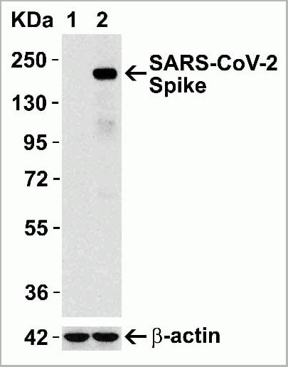 WB (Western Blot)
(Figure 12 Overexpression Validation in Spike Transfected 293 Cells Loading: 15 ug per lane of 293 cell lysate. Antibodies: SARS-CoV-2 (COVID-19) Spike, AAA10931 (1 ug/mL), 1h incubation at RT in 5% NFDM/TBST. Secondary: Goat anti rabbit IgG HRP conjugate at 1:10000 dilution. Lane 1: WT293 cells and Lane 2: SARS-CoV-2 Spike overexpressed 293 cells.)
WB (Western Blot)
(Figure 12 Overexpression Validation in Spike Transfected 293 Cells Loading: 15 ug per lane of 293 cell lysate. Antibodies: SARS-CoV-2 (COVID-19) Spike, AAA10931 (1 ug/mL), 1h incubation at RT in 5% NFDM/TBST. Secondary: Goat anti rabbit IgG HRP conjugate at 1:10000 dilution. Lane 1: WT293 cells and Lane 2: SARS-CoV-2 Spike overexpressed 293 cells.)
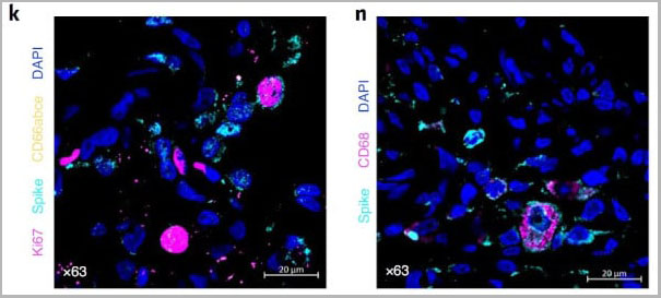
COVID 19 Spike Protein Coronavirus, Polyclonal Antibody (Cat# AAA10931)
Full Name
SARS-CoV-2 (COVID-19, 2019-nCoV) Spike Antibody
Reactivity
Virus
Applications
Immunofluorescence, Immunohistochemistry, Western Blot
Purity
SARS-CoV-2 (COVID-19, 2019-nCoV) Spike Antibody is affinity chromatography purified via peptide column.
Pricing