Filters
Clonality
Type
Reactivity
Gene Name
Isotype
Host
Application
Clone
227 results for " Other Antibodies" - showing 1-50
 WB (Western Blot)
(Western Blot: 1: 293 cell extract control2: 293 cell expressing NA (H5N1))
WB (Western Blot)
(Western Blot: 1: 293 cell extract control2: 293 cell expressing NA (H5N1))
NA (A/Vietnam/1203/2004)(H5N1, human), Polyclonal Antibody (Cat# AAA13721)
Full Name
Anti-NA (A/Vietnam/1203/2004)(H5N1, human)
Applications
Western Blot
Purity
Immunoaffinity chromatography
Pricing


 ELISA
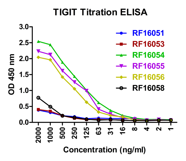
(2https://www.prosci-inc.com/tigit-antibody-7e5-16051.htmlSeptember 16, 2020Titration curve analysis of TIGIT mAbs to detectrecombinant TIGIT in ELISA with AAA11002 and antibodies atdecreasing concentrations.)
ELISA
(2https://www.prosci-inc.com/tigit-antibody-7e5-16051.htmlSeptember 16, 2020Titration curve analysis of TIGIT mAbs to detectrecombinant TIGIT in ELISA with AAA11002 and antibodies atdecreasing concentrations.)
TIGIT, Monoclonal Antibody (Cat# AAA11002)
Full Name
TIGIT Antibody [7E5]
Gene Names
TIGIT; VSIG9; VSTM3; WUCAM
Reactivity
Human
Applications
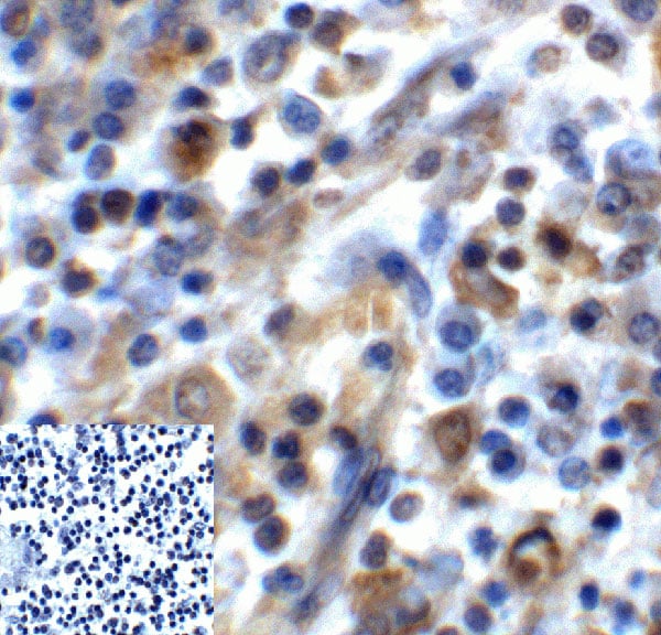
Immunohistochemistry, Immunocytochemistry, Immunofluorescence, Flow Cytometry
Purity
Protein A purified
Pricing
 Application Data
HA1 (A/chicken/HongKong/NT366/03)(H9N2), Polyclonal Antibody (Cat# AAA13717)
Full Name
Anti-HA1 (A/chicken/HongKong/NT366/03)(H9N2)
Applications
Western Blot
Purity
Immunoaffinity chromatography
Pricing
 Application Data
HA (A/California/06/09)(H1N1), Polyclonal Antibody (Cat# AAA13733)
Full Name
Anti-HA (A/California/06/09)(H1N1)
Applications
Western Blot
Purity
Immunoaffinity chromatography
Pricing


 IHC (Immunohistchemistry)
(Formalin-fixed, paraffin-embedded human Uterus stained with Vimentin Monoclonal Antibody (VM1170).)
IHC (Immunohistchemistry)
(Formalin-fixed, paraffin-embedded human Uterus stained with Vimentin Monoclonal Antibody (VM1170).)
Vimentin, Monoclonal Antibody (Cat# AAA13829)
Full Name
Vimentin (Mesenchymal Cell Marker) Mouse Monoclonal Antibody
Gene Names
VIM; HEL113; CTRCT30
Reactivity
Human.
Does not react with Mouse and Rat
Does not react with Mouse and Rat
Applications
Flow Cytometry, Immunofluorescence, Western Blot, Immunohistochemistry
Pricing
 Application Data
(Western Blot Data: purified HA1 (H7N9)(A/Shanghai/2/2013))
Application Data
(Western Blot Data: purified HA1 (H7N9)(A/Shanghai/2/2013))
H7 (H7N9)(A/Shanghai/2/2013), Polyclonal Antibody (Cat# AAA13735)
Full Name
Anti-H7 (H7N9)(A/Shanghai/2/2013)
Applications
Western Blot, Immunoprecipitation
Purity
Immunoaffinity chromatography
Pricing


 Application Data
(Published customer image: Increased accumulation of repair-associated macrophages surrounding collaterals in ischemic hind limbs is PAR2-dependent. (A) Stainings of CD206-positive macrophages (green) and SMA-positive vessels (red) in non-ischemic (control) and ischemic (ligated) hind limbs of WT, PAR1-/- and PAR2-/- mice are shown. Nuclei were visualized with DAPI (blue). Arrows indicate single macrophages in the non-ischemic adductor. Quantification of the average number of repair-associated macrophages per vessel is indicated on the right. (B) Correlation between the number of CD206-positive macrophages in the ischemic tissues and the expression of CD11b and (C) CD115 on monocytes. ** p)
Application Data
(Published customer image: Increased accumulation of repair-associated macrophages surrounding collaterals in ischemic hind limbs is PAR2-dependent. (A) Stainings of CD206-positive macrophages (green) and SMA-positive vessels (red) in non-ischemic (control) and ischemic (ligated) hind limbs of WT, PAR1-/- and PAR2-/- mice are shown. Nuclei were visualized with DAPI (blue). Arrows indicate single macrophages in the non-ischemic adductor. Quantification of the average number of repair-associated macrophages per vessel is indicated on the right. (B) Correlation between the number of CD206-positive macrophages in the ischemic tissues and the expression of CD11b and (C) CD115 on monocytes. ** p)
CD206, Monoclonal Antibody (Cat# AAA12117)
Full Name
RAT ANTI MOUSE CD206:Biotin
Gene Names
Mrc1; MR; CD206; AW259686
Applications
Flow Cytometry
Pricing


 IF (Immunofluorescence)
(Confocal immunofluorescent analysis of HeLa cells, untreated (left) or anisomycin-treated (right), using AAA14726 (green). Actin filaments have been labeled with Alexa Fluor® 555 phalloidin (red). Blue pseudocolor = DRAQ5™ (fluorescent DNA dye).)
IF (Immunofluorescence)
(Confocal immunofluorescent analysis of HeLa cells, untreated (left) or anisomycin-treated (right), using AAA14726 (green). Actin filaments have been labeled with Alexa Fluor® 555 phalloidin (red). Blue pseudocolor = DRAQ5™ (fluorescent DNA dye).)
Heat Shock Protein 27, Polyclonal Antibody (Cat# AAA14726)
Full Name
Heat Shock Protein 27, phosphorylated (Ser82) (HSP27)
Gene Names
Hsp27; 27K; 28; anon-WO0140519.69; CG4466; Dhsp27; DmelCG4466; DmHsp27; hsp 27; hsp-27; hsp26; hsp27; HSP27; hsp27/26; hsp28; Hsp28; HSP28; small hsp locus 67B
Reactivity
Human, Monkey, Mouse, Rat
Applications
Western Blot, Immunohistochemistry, Flow Cytometry, Immunofluorescence
Purity
Affinity Purified
Purified by protein A and peptide affinity chromatography.
Purified by protein A and peptide affinity chromatography.
Pricing


 IHC (Immunohistchemistry)
(Validation of PKR in Rat Lung Immunohistochemical analysis of paraffin-embedded rat lung tissue using anti-PKR antibody (AAA10917) at 2.5 ug/ml. Tissue was fixed with formaldehyde and blocked with 10% serum for 1h at RT; antigen retrieval was by heat mediation with a citrate buffer (pH6). Samples were incubated with primary antibody overnight at 4 degree C. A goat anti-rabbit IgG H&L (HRP) at 1/250 was used as secondary. Counter stained with Hematoxylin.)
IHC (Immunohistchemistry)
(Validation of PKR in Rat Lung Immunohistochemical analysis of paraffin-embedded rat lung tissue using anti-PKR antibody (AAA10917) at 2.5 ug/ml. Tissue was fixed with formaldehyde and blocked with 10% serum for 1h at RT; antigen retrieval was by heat mediation with a citrate buffer (pH6). Samples were incubated with primary antibody overnight at 4 degree C. A goat anti-rabbit IgG H&L (HRP) at 1/250 was used as secondary. Counter stained with Hematoxylin.)
PKR, Polyclonal Antibody (Cat# AAA10917)
Full Name
PKR Antibody
Gene Names
EIF2AK2; PKR; PRKR; EIF2AK1; PPP1R83
Reactivity
Human, Mouse, Rat
Applications
Western Blot, Immunohistochemistry, Immunofluorescence
Purity
PKR Antibody is affinity chromatography purified via peptide column.
Pricing
 Application Data
(C: 293 cell extract control; E: 293 cell expressing gp120 (Clade B))
Application Data
(C: 293 cell extract control; E: 293 cell expressing gp120 (Clade B))
gp120 (Clade B), Polyclonal Antibody (Cat# AAA13711)
Full Name
Anti-gp120 (Clade B)
Applications
Western Blot
Purity
Immunoaffinity chromatography.
Pricing
 Application Data
(Western Blot Data: C: 293 cell extract controlE: 293 cell expressing NS1, band at *)
Application Data
(Western Blot Data: C: 293 cell extract controlE: 293 cell expressing NS1, band at *)
NS1 (A/Vietnam/1203/2004)(H5N1), Polyclonal Antibody (Cat# AAA13723)
Full Name
Anti-NS1 (A/Vietnam/1203/2004)(H5N1)
Applications
Western Blot
Purity
Immunoaffinity chromatography
Pricing


 Application Data
(Published customer image: Increased accumulation of repair-associated macrophages surrounding collaterals in ischemic hind limbs is PAR2-dependent. (A) Stainings of CD206-positive macrophages (green) and SMA-positive vessels (red) in non-ischemic (control) and ischemic (ligated) hind limbs of WT, PAR1-/- and PAR2-/- mice are shown. Nuclei were visualized with DAPI (blue). Arrows indicate single macrophages in the non-ischemic adductor. Quantification of the average number of repair-associated macrophages per vessel is indicated on the right. (B) Correlation between the number of CD206-positive macrophages in the ischemic tissues and the expression of CD11b and (C) CD115 on monocytes. ** p)
Application Data
(Published customer image: Increased accumulation of repair-associated macrophages surrounding collaterals in ischemic hind limbs is PAR2-dependent. (A) Stainings of CD206-positive macrophages (green) and SMA-positive vessels (red) in non-ischemic (control) and ischemic (ligated) hind limbs of WT, PAR1-/- and PAR2-/- mice are shown. Nuclei were visualized with DAPI (blue). Arrows indicate single macrophages in the non-ischemic adductor. Quantification of the average number of repair-associated macrophages per vessel is indicated on the right. (B) Correlation between the number of CD206-positive macrophages in the ischemic tissues and the expression of CD11b and (C) CD115 on monocytes. ** p)
CD206, Monoclonal Antibody (Cat# AAA12122)
Full Name
RAT ANTI MOUSE CD206:FITC
Gene Names
Mrc1; MR; CD206; AW259686
Applications
Flow Cytometry
Pricing


 FCM (Flow Cytometry)
(Flow cytometric analysis of Pam212 cells, untreated or sulindac sulfone- treated (to induce apoptosis), using AAA14725 Results were similar to those obtained by analyzing DNA content.)
FCM (Flow Cytometry)
(Flow cytometric analysis of Pam212 cells, untreated or sulindac sulfone- treated (to induce apoptosis), using AAA14725 Results were similar to those obtained by analyzing DNA content.)
Caspase 3, Cleaved, Polyclonal Antibody (Cat# AAA14725)
Full Name
Caspase 3, Cleaved (Asp175) (CPP-32, Apoptain, Yama, SCA-1)
Reactivity
Human, Monkey, Mouse, Rat
Applications
Western Blot, Immunohistochemistry, Flow Cytometry, Immunofluorescence
Purity
Affinity Purified
Purified by Protein A and immunoaffinity chromatography.
Purified by Protein A and immunoaffinity chromatography.
Pricing
 WB (Western Blot)
(C:293 cell extract controlE:293 cell expressing HBcAg(HBV))
WB (Western Blot)
(C:293 cell extract controlE:293 cell expressing HBcAg(HBV))
HBcAg (HBV), Polyclonal Antibody (Cat# AAA13719)
Full Name
Anti-HBcAg (HBV), rabbit IgG
Applications
Western Blot
Purity
Immunoaffinity chromatography
Pricing


 Application Data
(Published customer image: Increased accumulation of repair-associated macrophages surrounding collaterals in ischemic hind limbs is PAR2-dependent. (A) Stainings of CD206-positive macrophages (green) and SMA-positive vessels (red) in non-ischemic (control) and ischemic (ligated) hind limbs of WT, PAR1-/- and PAR2-/- mice are shown. Nuclei were visualized with DAPI (blue). Arrows indicate single macrophages in the non-ischemic adductor. Quantification of the average number of repair-associated macrophages per vessel is indicated on the right. (B) Correlation between the number of CD206-positive macrophages in the ischemic tissues and the expression of CD11b and (C) CD115 on monocytes. ** p)
Application Data
(Published customer image: Increased accumulation of repair-associated macrophages surrounding collaterals in ischemic hind limbs is PAR2-dependent. (A) Stainings of CD206-positive macrophages (green) and SMA-positive vessels (red) in non-ischemic (control) and ischemic (ligated) hind limbs of WT, PAR1-/- and PAR2-/- mice are shown. Nuclei were visualized with DAPI (blue). Arrows indicate single macrophages in the non-ischemic adductor. Quantification of the average number of repair-associated macrophages per vessel is indicated on the right. (B) Correlation between the number of CD206-positive macrophages in the ischemic tissues and the expression of CD11b and (C) CD115 on monocytes. ** p)
CD206, Monoclonal Antibody (Cat# AAA12125)
Full Name
RAT ANTI MOUSE CD206:RPE
Gene Names
Mrc1; MR; CD206; AW259686
Applications
Flow Cytometry
Pricing


 ICC (Immunocytochemistry)
(Figure 10 Immunocytochemistry Validation of PD-L1Immunocytochemical analysis of 4% paraformaldehyde-fixed PD-L1 transfected 293 cells labeling PD-L1 with at 1 μg/ml, followed by Goat anti-mouse IgG secondary antibody at 1/250 dilution (red). Lower left: Use mouse IgG antibody at 1 μg/ml as control.)
ICC (Immunocytochemistry)
(Figure 10 Immunocytochemistry Validation of PD-L1Immunocytochemical analysis of 4% paraformaldehyde-fixed PD-L1 transfected 293 cells labeling PD-L1 with at 1 μg/ml, followed by Goat anti-mouse IgG secondary antibody at 1/250 dilution (red). Lower left: Use mouse IgG antibody at 1 μg/ml as control.)
PDL1, Monoclonal Antibody (Cat# AAA10992)
Full Name
PDL1 Antibody [6H10]
Gene Names
CD274; B7-H; B7H1; PDL1; PD-L1; PDCD1L1; PDCD1LG1
Reactivity
Human
Applications
Western Blot, Immunohistochemistry, Immunocytochemistry, Immunofluorescence
Purity
Protein A purified
Pricing


 Titration Curve
(Titration curve analysis of LAG-3 mAbs to detect recombinant LAG-3 in ELISA with AAA11016 antibodies at decreasing concentrations.)
Titration Curve
(Titration curve analysis of LAG-3 mAbs to detect recombinant LAG-3 in ELISA with AAA11016 antibodies at decreasing concentrations.)
LAG3, Monoclonal Antibody (Cat# AAA11016)
Full Name
LAG3 Antibody [2G8]
Gene Names
LAG3; CD223
Reactivity
Human
Applications
Western Blot, Immunohistochemistry, Immunocytochemistry, Immunofluorescence, Flow Cytometry
Purity
LAG-3 Antibody is supplied as protein A purified IgG1.
Pricing


 Application Data
(Published customer image: Increased accumulation of repair-associated macrophages surrounding collaterals in ischemic hind limbs is PAR2-dependent. (A) Stainings of CD206-positive macrophages (green) and SMA-positive vessels (red) in non-ischemic (control) and ischemic (ligated) hind limbs of WT, PAR1-/- and PAR2-/- mice are shown. Nuclei were visualized with DAPI (blue). Arrows indicate single macrophages in the non-ischemic adductor. Quantification of the average number of repair-associated macrophages per vessel is indicated on the right. (B) Correlation between the number of CD206-positive macrophages in the ischemic tissues and the expression of CD11b and (C) CD115 on monocytes. ** p)
Application Data
(Published customer image: Increased accumulation of repair-associated macrophages surrounding collaterals in ischemic hind limbs is PAR2-dependent. (A) Stainings of CD206-positive macrophages (green) and SMA-positive vessels (red) in non-ischemic (control) and ischemic (ligated) hind limbs of WT, PAR1-/- and PAR2-/- mice are shown. Nuclei were visualized with DAPI (blue). Arrows indicate single macrophages in the non-ischemic adductor. Quantification of the average number of repair-associated macrophages per vessel is indicated on the right. (B) Correlation between the number of CD206-positive macrophages in the ischemic tissues and the expression of CD11b and (C) CD115 on monocytes. ** p)
CD206, Monoclonal Antibody (Cat# AAA12120)
Full Name
RAT ANTI MOUSE CD206:FITC
Gene Names
Mrc1; MR; CD206; AW259686
Applications
Flow Cytometry
Pricing


 FCM (Flow Cytometry)
(HAS2 Antibody (Center) (AAA28714) flow cytometric analysis of K562 cells (bottom histogram) compared to a Rabbit IgG Isotype control (negative control-top histogram).FITC-conjugated goat-anti-rabbit secondary antibodies were used for the analysis.)
FCM (Flow Cytometry)
(HAS2 Antibody (Center) (AAA28714) flow cytometric analysis of K562 cells (bottom histogram) compared to a Rabbit IgG Isotype control (negative control-top histogram).FITC-conjugated goat-anti-rabbit secondary antibodies were used for the analysis.)
HAS2, Polyclonal Antibody (Cat# AAA28714)
Full Name
HAS2 Antibody (Center)
Reactivity
Human, mouse
Applications
Western Blot, Immunohistochemistry, Flow Cytometry
Purity
Peptide Affinity Purified Rabbit Polyclonal Antibody (Pab)
Pricing


 IF (Immunofluorescence)
(Figure 6 Immunofluorescence Validation of TMEM41B in Rat LungImmunofluorescent analysis of 4% paraformaldehyde-fixed rat lung labeling TMEM41B at 20ug/mL, followed by goat anti-rabbit IgG secondary antibody at 1/500 dilution (green) and DAPI staining (blue).)
IF (Immunofluorescence)
(Figure 6 Immunofluorescence Validation of TMEM41B in Rat LungImmunofluorescent analysis of 4% paraformaldehyde-fixed rat lung labeling TMEM41B at 20ug/mL, followed by goat anti-rabbit IgG secondary antibody at 1/500 dilution (green) and DAPI staining (blue).)
TMEM41B, Polyclonal Antibody (Cat# AAA11035)
Full Name
TMEM41B (CT) Antibody
Reactivity
Human, Mouse, Rat
Predicted species reactivity based on immunogen sequence: Bovine (100%); Chicken (100%); Chimpanzee (100%).
Predicted species reactivity based on immunogen sequence: Bovine (100%); Chicken (100%); Chimpanzee (100%).
Applications
Immunofluorescence, Western Blot
Purity
TMEM41B Antibody is affinity chromatography purified via peptide column.
Pricing
 Application Data
Ectodomain of glycoprotein, Polyclonal Antibody (Cat# AAA13732)
Full Name
Anti-Ectodomain of glycoprotein (Rabies virus), rabbit IgG
Applications
Western Blot
Purity
Immunoaffinity chromatography
Pricing

 WB (Western Blot)
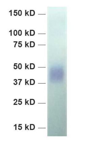
(Western Blot analysis of RNASE2 expression in transfected 293T cell line using AAA14729. Lane 1: RNASE2 transfected lysate (18.40kD). Lane 2: Non-transfected lysate.)
WB (Western Blot)
(Western Blot analysis of RNASE2 expression in transfected 293T cell line using AAA14729. Lane 1: RNASE2 transfected lysate (18.40kD). Lane 2: Non-transfected lysate.)
RNASE2, Polyclonal Antibody (Cat# AAA14729)
Full Name
RNASE2 (Non-secretory Ribonuclease, Eosinophil-derived Neurotoxin, RNase UpI-2, Ribonuclease 2, RNase 2, Ribonuclease US, EDN, RNS2)
Gene Names
Rnase2; R5; R14; R15; Ear4
Reactivity
Human
Applications
Western Blot
Purity
Purified
Purified.
Purified.
Pricing


 IHC (Immunohistochemistry)
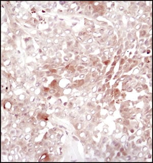
(Immunohistochemical analysis of paraffin-embedded Human brain section using Pink 1 (AAA28759). AAA28759 was diluted at 1:1000 dilution. A undiluted binotinylated goat polyvalent antibody was used as the secondary, followed by DAB staining)
IHC (Immunohistochemistry)
(Immunohistochemical analysis of paraffin-embedded Human brain section using Pink 1 (AAA28759). AAA28759 was diluted at 1:1000 dilution. A undiluted binotinylated goat polyvalent antibody was used as the secondary, followed by DAB staining)
S100B, Polyclonal Antibody (Cat# AAA28759)
Full Name
S100B Antibody
Gene Names
S100B; NEF; S100; S100-B; S100beta
Reactivity
Human, Mouse, Rat (Predicted: Rabbit)
Applications
Western Blot, Immunohistochemistry, Flow Cytometry, Immunohistochemistry
Purity
Peptide Affinity Purified Rabbit Polyclonal Antibody (Pab)
Pricing

 Application Data
H1 (A/California/06/2009)(H1N1), Monoclonal Antibody (Cat# AAA13689)
Full Name
Anti-H1 (A/California/06/2009)(H1N1) Monoclonal Antibody, clone IT-26D11
Applications
Immunoprecipitation, Immunofluorescence
Pricing


 ELISA
(A sandwich ELISA was performed using the anti-LAG3 mAbs as the capture antibodies for the LAG3 extracellular domain antigen with biotin-labeled Risk-Free anti-LAG3 mAbs as the detection antibodies.)
ELISA
(A sandwich ELISA was performed using the anti-LAG3 mAbs as the capture antibodies for the LAG3 extracellular domain antigen with biotin-labeled Risk-Free anti-LAG3 mAbs as the detection antibodies.)
LAG3, Monoclonal Antibody (Cat# AAA11020)
Full Name
LAG3 Antibody [5F11]
Gene Names
LAG3; CD223
Reactivity
Human
Applications
Western Blot, Immunohistochemistry, Immunocytochemistry, Immunofluorescence, Flow Cytometry
Purity
Protein A purified
Pricing


 IF (Immunofluorescence)
(Figure 7 Immunofluorescence Validation of TMEM41B in Rat LungImmunofluorescent analysis of 4% paraformaldehyde-fixed rat lung labeling TMEM41B at 20ug/mL, followed by goat anti-rabbit IgG secondary antibody at 1/500 dilution (green) and DAPI staining (blue).)
IF (Immunofluorescence)
(Figure 7 Immunofluorescence Validation of TMEM41B in Rat LungImmunofluorescent analysis of 4% paraformaldehyde-fixed rat lung labeling TMEM41B at 20ug/mL, followed by goat anti-rabbit IgG secondary antibody at 1/500 dilution (green) and DAPI staining (blue).)
TMEM41B, Polyclonal Antibody (Cat# AAA11036)
Full Name
TMEM41B (NT) Antibody
Reactivity
Human, Mouse, Rat
Predicted species reactivity based on immunogen sequence: Horse (100%); Chimpanzee (100%).
Predicted species reactivity based on immunogen sequence: Horse (100%); Chimpanzee (100%).
Applications
Immunofluorescence, Western Blot
Purity
TMEM41B Antibody is affinity chromatography purified via peptide column.
Pricing


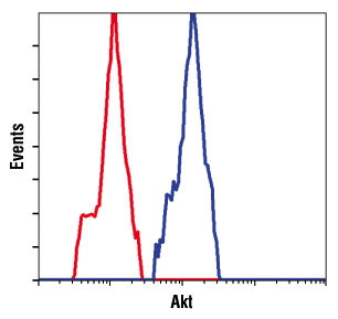 FCM (Flow Cytometry)
(Flow cytometric analysis of untreated Jurkat cells, using AAA14693 (blue) compared to a nonspecific negative control antibody (red).)
FCM (Flow Cytometry)
(Flow cytometric analysis of untreated Jurkat cells, using AAA14693 (blue) compared to a nonspecific negative control antibody (red).)
Akt, pan, Monoclonal Antibody (Cat# AAA14693)
Full Name
Akt, pan (Rac PKa, PKBa)
Gene Names
Akt1; akt; Akt; AKT; Akt/PKB; AKT/PKB; akt1; AKT1; Akt1|PKB; CG4006; D-Akt; dakt; dAkt; dAKT; Dakt; DAkt; dAKT/dPKB; dAkt/PKB; dakt1; dAkt1; dAKT1; Dakt1; DAkt1; DAKT1; DAKT1/PKB; DmelCG4006; dPKB; Dpkb; DPKB; DRAC-PK; DRAC-PK66; DRAC-PK85; l(3)04226; l(3)89Bq;
Reactivity
Human, Monkey, Mouse, Rat
Applications
Western Blot, Immunoprecipitation, Immunohistochemistry, Flow Cytometry, Immunofluorescence
Purity
Supernatant
Supernatant
Supernatant
Pricing


 Strength of Signal
(Analysis of Protein Array containing more than 19,000 full-length human proteins using Vimentin (VIM) Mouse Monoclonal Antibody (VM452)Z- and S- Score: The Z-score represents the strength of a signal that a monoclonal antibody (MAb) (in combination with a fluorescently-tagged anti-IgG secondary antibody) produces when binding to a particular protein on the HuProtTM array. Z-scores are described in units of standard deviations (SD's) above the mean value of all signals generated on that array. If targets on HuProtTM are arranged in descending order of the Z-score, the S-score is the difference (also in units of SD's) between the Z-score. S-score therefore represents the relative target specificity of a MAb to its intended target. A MAb is considered to specific to its intended target, if the MAb has an S-score of at least 2.5. For example, if a MAb binds to protein X with a Z-score of 43 and to protein Y with a Z-score of 14, then the S-score for the binding of that MAb to protein X is equal to 29.)
Strength of Signal
(Analysis of Protein Array containing more than 19,000 full-length human proteins using Vimentin (VIM) Mouse Monoclonal Antibody (VM452)Z- and S- Score: The Z-score represents the strength of a signal that a monoclonal antibody (MAb) (in combination with a fluorescently-tagged anti-IgG secondary antibody) produces when binding to a particular protein on the HuProtTM array. Z-scores are described in units of standard deviations (SD's) above the mean value of all signals generated on that array. If targets on HuProtTM are arranged in descending order of the Z-score, the S-score is the difference (also in units of SD's) between the Z-score. S-score therefore represents the relative target specificity of a MAb to its intended target. A MAb is considered to specific to its intended target, if the MAb has an S-score of at least 2.5. For example, if a MAb binds to protein X with a Z-score of 43 and to protein Y with a Z-score of 14, then the S-score for the binding of that MAb to protein X is equal to 29.)
Vimentin, Monoclonal Antibody (Cat# AAA13810)
Full Name
Vimentin (Mesenchymal Cell Marker) Mouse Monoclonal Antibody
Gene Names
VIM; HEL113; CTRCT30
Reactivity
Human, Cow, Dog, Cat, Pig, Goat, Chicken.
Does not react with Mouse and Rat
Does not react with Mouse and Rat
Applications
Flow Cytometry, Immunofluorescence, Western Blot, Immunohistochemistry
Pricing


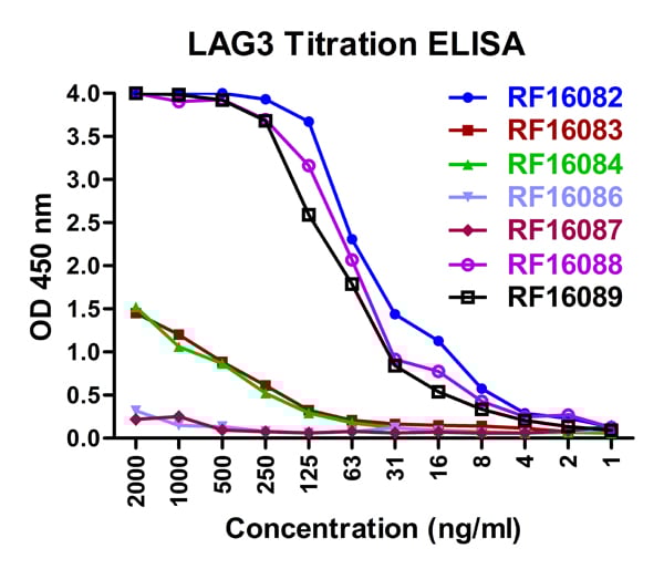
 ELISA
(Titration curve analysis of LAG-3 mAbs to detect recombinant LAG-3 in ELISA with AAA11018 and antibodies at decreasing concentrations.)
ELISA
(Titration curve analysis of LAG-3 mAbs to detect recombinant LAG-3 in ELISA with AAA11018 and antibodies at decreasing concentrations.)
LAG3, Monoclonal Antibody (Cat# AAA11018)
Full Name
LAG3 Antibody [6B12]
Gene Names
LAG3; CD223
Reactivity
Human
Applications
Immunohistochemistry, Immunocytochemistry, Immunofluorescence
Purity
LAG-3 Antibody is supplied as protein A purified IgG1.
Pricing


 FCM (Flow Cytometry)
(Figure-6: Epitope binding study by flow cytometric analysis. MCF-7 cells expressing HER2 antigen were treated with Herceptin and Samceptin (2 ug/10^6 Cells). Surface staining was done using FITC conjugated antibodies.)
FCM (Flow Cytometry)
(Figure-6: Epitope binding study by flow cytometric analysis. MCF-7 cells expressing HER2 antigen were treated with Herceptin and Samceptin (2 ug/10^6 Cells). Surface staining was done using FITC conjugated antibodies.)
ErbB2/HER2, Monoclonal Recombinant Antibody (Cat# AAA14887)
Full Name
Recombinant anti-human ErbB2/HER2 Antibody (Trastuzumab) FITC Conjugated
Gene Names
ERBB2; NEU; NGL; HER2; TKR1; CD340; HER-2; MLN 19; HER-2/neu
Reactivity
Human
Applications
Functional Assay, Flow Cytometry, Antibody-Dependent Cellular Cytotoxicity
Purity
Purity: >98.0% as determined by SEC-HPLC & SDS-PAGE
Pricing


 ELISA
(A sandwich ELISA was performed using the anti-LAG3 mAbs as the capture antibodies for the LAG3 extracellular domain antigen with biotin-labeled Risk-Free anti-LAG3 mAbs as the detection antibodies.)
ELISA
(A sandwich ELISA was performed using the anti-LAG3 mAbs as the capture antibodies for the LAG3 extracellular domain antigen with biotin-labeled Risk-Free anti-LAG3 mAbs as the detection antibodies.)
LAG3, Monoclonal Antibody (Cat# AAA11021)
Full Name
LAG3 Antibody [1G4]
Gene Names
LAG3; CD223
Reactivity
Human
Applications
Western Blot, Immunohistochemistry, Immunocytochemistry, Immunofluorescence, Flow Cytometry
Purity
Protein A purified
Pricing


 IF (Immunofluorescence)
(Figure 7 Immunofluorescence Validation of TMPRSS2 in Rat TestisImmunofluorescent analysis of 4% paraformaldehyde-fixed rat Testis labeling TMPRSS2 with 9569 at 20ug/mL, followed by goat anti-rabbit IgG secondary antibody at 1/500 dilution (green) and DAPI staining (blue).)
IF (Immunofluorescence)
(Figure 7 Immunofluorescence Validation of TMPRSS2 in Rat TestisImmunofluorescent analysis of 4% paraformaldehyde-fixed rat Testis labeling TMPRSS2 with 9569 at 20ug/mL, followed by goat anti-rabbit IgG secondary antibody at 1/500 dilution (green) and DAPI staining (blue).)
TMPRSS2, Polyclonal Antibody (Cat# AAA11037)
Full Name
TMPRSS2 (CT) Antibody
Gene Names
TMPRSS2; PP9284; PRSS10
Reactivity
Human, Mouse, Rat
Predicted species reactivity based on immunogen sequence: Monkey (100%); Gorilla (100%); Cat (92%).
Predicted species reactivity based on immunogen sequence: Monkey (100%); Gorilla (100%); Cat (92%).
Applications
Immunofluorescence, Western Blot
Purity
TMPRSS2 Antibody is affinity chromatography purified via peptide column.
Pricing


 IF (Immunofluorescence)
(Confocal immunofluorescent analysis of C2C12 cells, either LY294002-treated (left) or insulin-treated (right), using AAA14695 (green). Actin filaments have been labeled with DY554 phalloidin (red). Blue pseudocolor = DRAQ5™ (fluorescent DNA dye).)
IF (Immunofluorescence)
(Confocal immunofluorescent analysis of C2C12 cells, either LY294002-treated (left) or insulin-treated (right), using AAA14695 (green). Actin filaments have been labeled with DY554 phalloidin (red). Blue pseudocolor = DRAQ5™ (fluorescent DNA dye).)
Akt, pan, Monoclonal Antibody (Cat# AAA14695)
Full Name
Akt, pan (Rac PKa, PKBa)
Gene Names
Akt1; akt; Akt; AKT; Akt/PKB; AKT/PKB; akt1; AKT1; Akt1|PKB; CG4006; D-Akt; dakt; dAkt; dAKT; Dakt; DAkt; dAKT/dPKB; dAkt/PKB; dakt1; dAkt1; dAKT1; Dakt1; DAkt1; DAKT1; DAKT1/PKB; DmelCG4006; dPKB; Dpkb; DPKB; DRAC-PK; DRAC-PK66; DRAC-PK85; l(3)04226; l(3)89Bq;
Reactivity
Human, Monkey, Mouse, Rat
Applications
Western Blot, Immunoprecipitation, Immunohistochemistry, Flow Cytometry, Immunofluorescence
Purity
Ascites
Ascites
Ascites
Pricing


 FCM (Flow Cytometry)
(Figure-6: Epitope binding study by flow cytometric analysis. MCF-7 cells expressing HER2 antigen were treated with Either Herceptin or Samceptin (1 & 2 ug/10^6 Cells). Surface staining was done using FITC conjugated antibodies.)
FCM (Flow Cytometry)
(Figure-6: Epitope binding study by flow cytometric analysis. MCF-7 cells expressing HER2 antigen were treated with Either Herceptin or Samceptin (1 & 2 ug/10^6 Cells). Surface staining was done using FITC conjugated antibodies.)
ErbB2/HER2, Monoclonal Recombinant Antibody (Cat# AAA14881)
Full Name
Recombinant anti- human ErbB2/HER2 Antibody (Trastuzumab)
Gene Names
ERBB2; NEU; NGL; HER2; TKR1; CD340; HER-2; MLN 19; HER-2/neu
Reactivity
Human
Applications
Functional Assay, Flow Cytometry
Purity
>98.0% as determined by SEC-HPLC & SDS-PAGE.
Pricing
 Application Data
(C: 293 cell extract control; E: 293 cell expressing gp120 (Clade C))
Application Data
(C: 293 cell extract control; E: 293 cell expressing gp120 (Clade C))
gp120 (Clade C), Polyclonal Antibody (Cat# AAA13712)
Full Name
Anti-gp120 (Clade C)
Applications
Western Blot
Purity
Immunoaffinity chromatography.
Pricing


 IHC (Immunohistchemistry)
(Figure 9 Immunohistochemistry Validation of hRIP3 in Human Colon Tissue Immunohistochemical analysis of paraffin-embedded human colon tissue using anti-hRIP3 antibody (8963) at 2 μg/ml. Tissue was fixed with formaldehyde and blocked with 10% serum for 1 h at RT; antigen retrieval was by heat mediation with a citrate buffer (pH6). Samples were incubated with primary antibody overnight at 4˚C. A goat anti-rabbit IgG H&L (HRP) at 1/250 was used as secondary. Counter stained with Hematoxylin.)
IHC (Immunohistchemistry)
(Figure 9 Immunohistochemistry Validation of hRIP3 in Human Colon Tissue Immunohistochemical analysis of paraffin-embedded human colon tissue using anti-hRIP3 antibody (8963) at 2 μg/ml. Tissue was fixed with formaldehyde and blocked with 10% serum for 1 h at RT; antigen retrieval was by heat mediation with a citrate buffer (pH6). Samples were incubated with primary antibody overnight at 4˚C. A goat anti-rabbit IgG H&L (HRP) at 1/250 was used as secondary. Counter stained with Hematoxylin.)
hRIP3, Polyclonal Antibody (Cat# AAA11031)
Full Name
hRIP3 Antibody
Gene Names
RIPK3; RIP3
Reactivity
Human
Applications
Immunofluorescence, Immunohistochemistry, Western Blot
Purity
hRIP3 Antibody is Protein A purified.
Pricing


 Application Data
(Published customer image: Increased accumulation of repair-associated macrophages surrounding collaterals in ischemic hind limbs is PAR2-dependent. (A) Stainings of CD206-positive macrophages (green) and SMA-positive vessels (red) in non-ischemic (control) and ischemic (ligated) hind limbs of WT, PAR1-/- and PAR2-/- mice are shown. Nuclei were visualized with DAPI (blue). Arrows indicate single macrophages in the non-ischemic adductor. Quantification of the average number of repair-associated macrophages per vessel is indicated on the right. (B) Correlation between the number of CD206-positive macrophages in the ischemic tissues and the expression of CD11b and (C) CD115 on monocytes. ** p)
Application Data
(Published customer image: Increased accumulation of repair-associated macrophages surrounding collaterals in ischemic hind limbs is PAR2-dependent. (A) Stainings of CD206-positive macrophages (green) and SMA-positive vessels (red) in non-ischemic (control) and ischemic (ligated) hind limbs of WT, PAR1-/- and PAR2-/- mice are shown. Nuclei were visualized with DAPI (blue). Arrows indicate single macrophages in the non-ischemic adductor. Quantification of the average number of repair-associated macrophages per vessel is indicated on the right. (B) Correlation between the number of CD206-positive macrophages in the ischemic tissues and the expression of CD11b and (C) CD115 on monocytes. ** p)
CD206, Monoclonal Antibody (Cat# AAA12118)
Full Name
RAT ANTI MOUSE CD206:Biotin
Gene Names
Mrc1; MR; CD206; AW259686
Applications
Flow Cytometry
Pricing
 WB (Western Blot)
(C: 293 cell extract control; E: 293 cell expressing VP1 (HAV))
WB (Western Blot)
(C: 293 cell extract control; E: 293 cell expressing VP1 (HAV))
VP1, Polyclonal Antibody (Cat# AAA13727)
Full Name
Anti-VP1 (HAV) rabbit polyclonal antibody
Applications
Western Blot
Purity
Immunoaffinity chromatography
Pricing
 Standard Curve (Sample)
Antineutrophil cytoplasmic antibodies (ANCA), ELISA Kit (Cat# AAA12637)
Full Name
Human Antineutrophil cytoplasmic antibodies (ANCA) ELISA Kit
Reactivity
Human
Pricing
 Application Data
(C: 293 cell extract control; E: 293 cell expressing E1 (HCV))
Application Data
(C: 293 cell extract control; E: 293 cell expressing E1 (HCV))
E1 (HCV), Polyclonal Antibody (Cat# AAA13708)
Full Name
Anti-E1 (HCV), rabbit IgG
Applications
Western Blot
Purity
Immunoaffinity chromatography.
Pricing


 Application Data
(Published customer image: Increased accumulation of repair-associated macrophages surrounding collaterals in ischemic hind limbs is PAR2-dependent. (A) Stainings of CD206-positive macrophages (green) and SMA-positive vessels (red) in non-ischemic (control) and ischemic (ligated) hind limbs of WT, PAR1-/- and PAR2-/- mice are shown. Nuclei were visualized with DAPI (blue). Arrows indicate single macrophages in the non-ischemic adductor. Quantification of the average number of repair-associated macrophages per vessel is indicated on the right. (B) Correlation between the number of CD206-positive macrophages in the ischemic tissues and the expression of CD11b and (C) CD115 on monocytes. ** p)
Application Data
(Published customer image: Increased accumulation of repair-associated macrophages surrounding collaterals in ischemic hind limbs is PAR2-dependent. (A) Stainings of CD206-positive macrophages (green) and SMA-positive vessels (red) in non-ischemic (control) and ischemic (ligated) hind limbs of WT, PAR1-/- and PAR2-/- mice are shown. Nuclei were visualized with DAPI (blue). Arrows indicate single macrophages in the non-ischemic adductor. Quantification of the average number of repair-associated macrophages per vessel is indicated on the right. (B) Correlation between the number of CD206-positive macrophages in the ischemic tissues and the expression of CD11b and (C) CD115 on monocytes. ** p)
CD206, Monoclonal Antibody (Cat# AAA12124)
Full Name
RAT ANTI MOUSE CD206:RPE
Gene Names
Mrc1; MR; CD206; AW259686
Applications
Flow Cytometry
Pricing


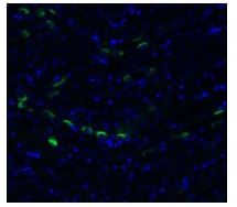
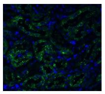
 Application Data
(C:FGFR2/isolectinB4 (C) and FGFR1/isolectinB4 (D) staining of apparent mesenchymal cells and the subpopulation of endothelial cells. Virtually all other dispersed apparent mesenchymal cells express FGFR1 and FGFR2 (merged image in E). F: FGFR2 (F) and FGFR1 (G) staining in clustered cells of epithelial origin (inferred by morphology here) demonstrating that epithelial cells express both FGFR1 and FGFR2 (merged image with DAPI staining in H).)
Application Data
(C:FGFR2/isolectinB4 (C) and FGFR1/isolectinB4 (D) staining of apparent mesenchymal cells and the subpopulation of endothelial cells. Virtually all other dispersed apparent mesenchymal cells express FGFR1 and FGFR2 (merged image in E). F: FGFR2 (F) and FGFR1 (G) staining in clustered cells of epithelial origin (inferred by morphology here) demonstrating that epithelial cells express both FGFR1 and FGFR2 (merged image with DAPI staining in H).)
FGFR2, Polyclonal Antibody (Cat# AAA26853)
Full Name
FGFR2, NT (FGFR2, BEK, KGFR, KSAM, Fibroblast growth factor receptor 2, K-sam, Keratinocyte growth factor receptor, CD332) (Biotin)
Gene Names
FGFR2; BEK; JWS; BBDS; CEK3; CFD1; ECT1; KGFR; TK14; TK25; BFR-1; CD332; K-SAM
Reactivity
Human, Monkey, Mouse, Rat
Applications
FC/FACS, EIA, IF, IHC, WB
Purity
Purified by Protein G Affinity Chromatography.
Pricing


 ELISA
(A sandwich ELISA was performed using the anti-LAG3 mAbs as the capture antibodies for the LAG3 extracellular domain antigen with biotin-labeled Risk-Free anti-LAG3 mAbs as the detection antibodies.)
ELISA
(A sandwich ELISA was performed using the anti-LAG3 mAbs as the capture antibodies for the LAG3 extracellular domain antigen with biotin-labeled Risk-Free anti-LAG3 mAbs as the detection antibodies.)
LAG3, Monoclonal Antibody (Cat# AAA11017)
Full Name
LAG3 Antibody [9F9]
Gene Names
LAG3; CD223
Reactivity
Human
Applications
Immunohistochemistry, Immunocytochemistry, Immunofluorescence, Flow Cytometry
Purity
Protein A purified
Pricing


 IF (Immunofluorescence)
(Figure 7 Immunofluorescence Validation of TMPRSS2 in Rat BrainImmunofluorescent analysis of 4% paraformaldehyde-fixed rat brain labeling TMPRSS2 at 20ug/mL, followed by goat anti-rabbit IgG secondary antibody at 1/500 dilution (green) and DAPI staining (blue).)
IF (Immunofluorescence)
(Figure 7 Immunofluorescence Validation of TMPRSS2 in Rat BrainImmunofluorescent analysis of 4% paraformaldehyde-fixed rat brain labeling TMPRSS2 at 20ug/mL, followed by goat anti-rabbit IgG secondary antibody at 1/500 dilution (green) and DAPI staining (blue).)
TMPRSS2, Polyclonal Antibody (Cat# AAA11038)
Full Name
TMPRSS2 (IN) Antibody
Gene Names
TMPRSS2; PP9284; PRSS10
Reactivity
Human, Mouse, Rat
Predicted species reactivity based on immunogen sequence: Horse (100%); Rabbit (100%); Monkey (100%); Sheep (100%); Gorilla (100%); Cat (100%).
Predicted species reactivity based on immunogen sequence: Horse (100%); Rabbit (100%); Monkey (100%); Sheep (100%); Gorilla (100%); Cat (100%).
Applications
Immunofluorescence, Western Blot
Purity
TMPRSS2 Antibody is affinity chromatography purified via peptide column.
Pricing


 Application Data
(C:FGFR2/isolectinB4 (C) and FGFR1/isolectinB4 (D) staining of apparent mesenchymal cells and the subpopulation of endothelial cells. Virtually all other dispersed apparent mesenchymal cells express FGFR1 and FGFR2 (merged image in E). F: FGFR2 (F) and FGFR1 (G) staining in clustered cells of epithelial origin (inferred by morphology here) demonstrating that epithelial cells express both FGFR1 and FGFR2 (merged image with DAPI staining in H).)
Application Data
(C:FGFR2/isolectinB4 (C) and FGFR1/isolectinB4 (D) staining of apparent mesenchymal cells and the subpopulation of endothelial cells. Virtually all other dispersed apparent mesenchymal cells express FGFR1 and FGFR2 (merged image in E). F: FGFR2 (F) and FGFR1 (G) staining in clustered cells of epithelial origin (inferred by morphology here) demonstrating that epithelial cells express both FGFR1 and FGFR2 (merged image with DAPI staining in H).)
FGFR2, Polyclonal Antibody (Cat# AAA26855)
Full Name
FGFR2, NT (FGFR2, BEK, KGFR, KSAM, Fibroblast growth factor receptor 2, K-sam, Keratinocyte growth factor receptor, CD332) (Azide free) (HRP)
Gene Names
FGFR2; BEK; JWS; BBDS; CEK3; CFD1; ECT1; KGFR; TK14; TK25; BFR-1; CD332; K-SAM
Reactivity
Human, Monkey, Mouse, Rat
Applications
IHC, EIA, WB
Purity
Purified by Protein G Affinity Chromatography.
Pricing


 Application Data
Quantitative, For Hepatitis B Virus Specific T Cells, Detection Kit (Cat# AAA27976)
Full Name
Quantitative Detection Kit, For Hepatitis B Virus Specific T Cells
Pricing
 Application Data
NP (Influenza B), Polyclonal Antibody (Cat# AAA13722)
Full Name
Anti-NP (Influenza B), rabbit IgG
Applications
Western Blot
Purity
Immunoaffinity chromatography
Pricing


 Application Data
(Published customer image: Mouse anti Human CD49d antibody, clone HP2/1 used for binding efficiency determinationImage caption:Binding efficiencies (BE) of different a4beta7 molecules composed of distinct a4 mutants to monoclonal antibodies against a4, beta7 or the a4beta7 heterodimer. Binding efficiency is determined by the ratio between the mean fluorescence of antibody binding to each a4 molecule and of the binding in a mock-transfected cell culture (see Materials and Methods for details). Dark gray bars represent binding to the human (wild type) a4 clone, whereas light gray bars are those of binding to the different a4 mutants (as shown in the x-axis). a4 mutants which included substitutions at codon 201 are boxed. A, binding of anti-a4 2b4 antibody. B, binding of anti-a4 HP2/1 antibody. C, BE of different anti-a4 and beta7 antibodies to the human a4 and the quintuple a4 mutant (5 aa mut). Bars represent the range of standard errors deduced from triplicate experiments. p-values of Student's t tests are shown above each comparison. NS, non-significant (> 0.05).From:Darc M, Hait SH, Soares EA, Cicala C, Seuanez HN, et al. (2011) Polymorphisms in the a4 Integrin of Neotropical Primates: Insights for Binding of Natural Ligands and HIV-1 gp120 to the Human a4beta7. PLoS ONE 6(9): e24461.)
Application Data
(Published customer image: Mouse anti Human CD49d antibody, clone HP2/1 used for binding efficiency determinationImage caption:Binding efficiencies (BE) of different a4beta7 molecules composed of distinct a4 mutants to monoclonal antibodies against a4, beta7 or the a4beta7 heterodimer. Binding efficiency is determined by the ratio between the mean fluorescence of antibody binding to each a4 molecule and of the binding in a mock-transfected cell culture (see Materials and Methods for details). Dark gray bars represent binding to the human (wild type) a4 clone, whereas light gray bars are those of binding to the different a4 mutants (as shown in the x-axis). a4 mutants which included substitutions at codon 201 are boxed. A, binding of anti-a4 2b4 antibody. B, binding of anti-a4 HP2/1 antibody. C, BE of different anti-a4 and beta7 antibodies to the human a4 and the quintuple a4 mutant (5 aa mut). Bars represent the range of standard errors deduced from triplicate experiments. p-values of Student's t tests are shown above each comparison. NS, non-significant (> 0.05).From:Darc M, Hait SH, Soares EA, Cicala C, Seuanez HN, et al. (2011) Polymorphisms in the a4 Integrin of Neotropical Primates: Insights for Binding of Natural Ligands and HIV-1 gp120 to the Human a4beta7. PLoS ONE 6(9): e24461.)
CD49d, Monoclonal Antibody (Cat# AAA12003)
Full Name
MOUSE ANTI HUMAN CD49d
Gene Names
ITGA4; IA4; CD49D
Applications
Immunohistochemistry, Flow Cytometry, Functional Assay, Immunoprecipitation
Pricing
 Application Data
HIV-1 p24, Monoclonal Antibody (Cat# AAA13685)
Full Name
Anti-HIV-1 p24
Reactivity
Reacts with P24(HIC-1) proteins
Applications
Western Blot
Purity
Ion exchange column
Pricing


 Application Data
(Published customer image: Increased accumulation of repair-associated macrophages surrounding collaterals in ischemic hind limbs is PAR2-dependent. (A) Stainings of CD206-positive macrophages (green) and SMA-positive vessels (red) in non-ischemic (control) and ischemic (ligated) hind limbs of WT, PAR1-/- and PAR2-/- mice are shown. Nuclei were visualized with DAPI (blue). Arrows indicate single macrophages in the non-ischemic adductor. Quantification of the average number of repair-associated macrophages per vessel is indicated on the right. (B) Correlation between the number of CD206-positive macrophages in the ischemic tissues and the expression of CD11b and (C) CD115 on monocytes. ** p)
Application Data
(Published customer image: Increased accumulation of repair-associated macrophages surrounding collaterals in ischemic hind limbs is PAR2-dependent. (A) Stainings of CD206-positive macrophages (green) and SMA-positive vessels (red) in non-ischemic (control) and ischemic (ligated) hind limbs of WT, PAR1-/- and PAR2-/- mice are shown. Nuclei were visualized with DAPI (blue). Arrows indicate single macrophages in the non-ischemic adductor. Quantification of the average number of repair-associated macrophages per vessel is indicated on the right. (B) Correlation between the number of CD206-positive macrophages in the ischemic tissues and the expression of CD11b and (C) CD115 on monocytes. ** p)
CD206, Monoclonal Antibody (Cat# AAA12119)
Full Name
RAT ANTI MOUSE CD206:FITC
Gene Names
Mrc1; MR; CD206; AW259686
Applications
Flow Cytometry
Pricing