Filters
Clonality
Type
Reactivity
Gene Name
Isotype
Host
Application
Clone
30 results for " MB Proteins" - showing 1-30


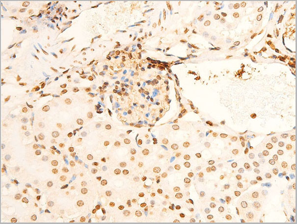
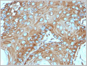
 IHC (Immunohistochemistry)
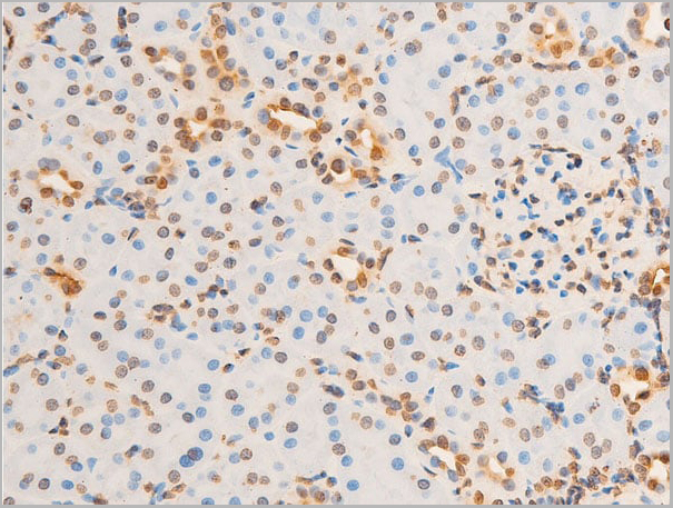
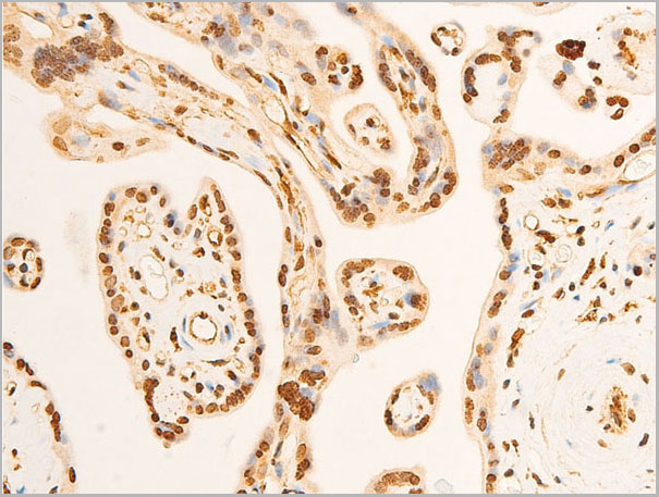
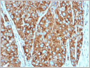
(At 1/100 staining Human esophageal cancer by IHC-P. The sample was formaldehyde fixed and a heat mediated antigen retrieval step in citrate buffer was performed. The sample was then blocked and incubated with the primary antibody at 4 degree C overnight. An HRP conjugated anti-Rabbit antibody was used as the secondary antibody.)
IHC (Immunohistochemistry)
(At 1/100 staining Human esophageal cancer by IHC-P. The sample was formaldehyde fixed and a heat mediated antigen retrieval step in citrate buffer was performed. The sample was then blocked and incubated with the primary antibody at 4 degree C overnight. An HRP conjugated anti-Rabbit antibody was used as the secondary antibody.)
p53, Polyclonal Antibody (Cat# AAA31354)
Full Name
Acetyl-p53 (Lys373) Antibody
Gene Names
TP53; P53; BCC7; LFS1; TRP53
Reactivity
Human, Mouse, Rat
Predicted Reactivity: Pig (91%), Sheep (80%), Rabbit (91%), Dog (91%)
Predicted Reactivity: Pig (91%), Sheep (80%), Rabbit (91%), Dog (91%)
Applications
Western Blot, Immunohistochemistry, Peptide ELISA
Purity
The antiserum was purified by peptide affinity chromatography using SulfoLink Coupling Resin
Pricing


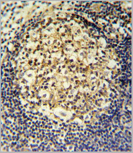
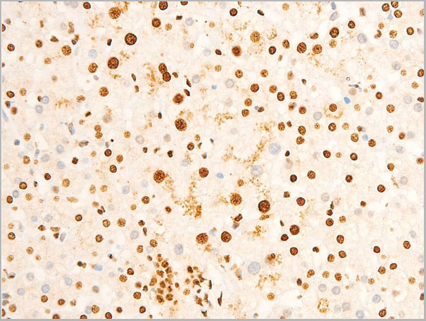
 IHC (Immunohistochemistry)
(At 1/100 staining Human esophageal cancer by IHC-P. The sample was formaldehyde fixed and a heat mediated antigen retrieval step in citrate buffer was performed. The sample was then blocked and incubated with the primary antibody at 4 degree C overnight. An HRP conjugated anti-Rabbit antibody was used as the secondary antibody.)
IHC (Immunohistochemistry)
(At 1/100 staining Human esophageal cancer by IHC-P. The sample was formaldehyde fixed and a heat mediated antigen retrieval step in citrate buffer was performed. The sample was then blocked and incubated with the primary antibody at 4 degree C overnight. An HRP conjugated anti-Rabbit antibody was used as the secondary antibody.)
p53, Polyclonal Antibody (Cat# AAA31377)
Full Name
p53 Antibody
Gene Names
TP53; P53; BCC7; LFS1; TRP53
Reactivity
Human, Mouse, Rat
Predicted Reactivity: Pig (91%), Sheep (80%), Rabbit (91%), Dog (91%)
Predicted Reactivity: Pig (91%), Sheep (80%), Rabbit (91%), Dog (91%)
Applications
Western Blot, Immunohistochemistry, Peptide ELISA
Purity
The antiserum was purified by peptide affinity chromatography using SulfoLink Coupling Resin
Pricing


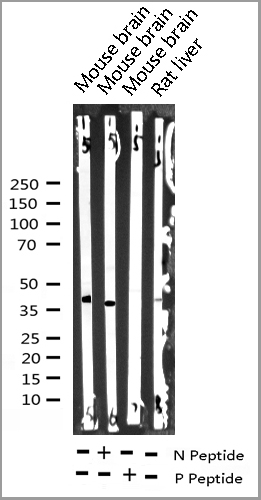
 WB (Western Blot)
(Anti-CD63 Antibody (C-term)at 1:2000 dilution + human plasma lysatesLysates/proteins at 20 ug per lane.SecondaryGoat Anti-Rabbit IgG, (H+L), Peroxidase conjugated at 1/10000 dilutionPredicted band size : 25 kDaBlocking/Dilution buffer: 5% NFDM/TBST.)
WB (Western Blot)
(Anti-CD63 Antibody (C-term)at 1:2000 dilution + human plasma lysatesLysates/proteins at 20 ug per lane.SecondaryGoat Anti-Rabbit IgG, (H+L), Peroxidase conjugated at 1/10000 dilutionPredicted band size : 25 kDaBlocking/Dilution buffer: 5% NFDM/TBST.)
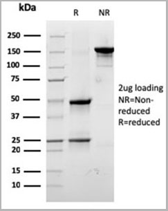
CD63, Polyclonal Antibody (Cat# AAA28725)
Full Name
CD63 Antibody (C-term)
Gene Names
CD63; MLA1; ME491; LAMP-3; OMA81H; TSPAN30
Reactivity
Human
Applications
Western Blot, Immunohistochemistry, Flow Cytometry
Purity
Peptide Affinity Purified Rabbit Polyclonal Antibody (Pab)
Pricing


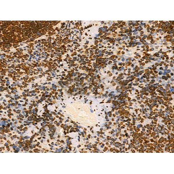
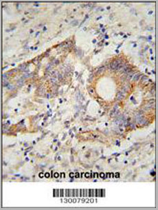
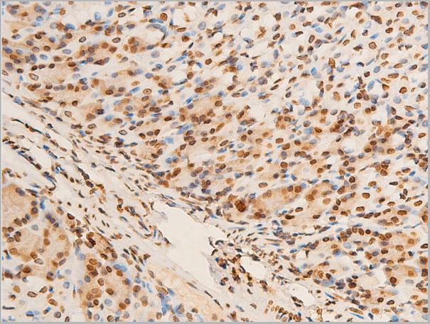
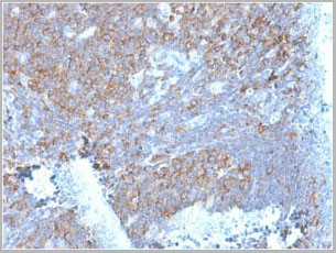
 IHC (Immunohistochemistry)
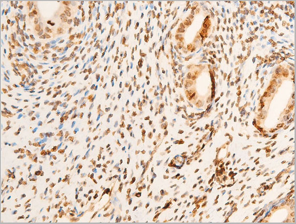
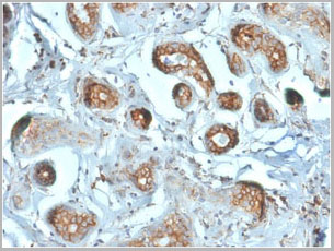
(At 1/100 staining Human pancreatic cancer by IHC-P. The sample was formaldehyde fixed and a heat mediated antigen retrieval step in citrate buffer was performed. The sample was then blocked and incubated with the primary antibody at 4 degree C overnight. An HRP conjugated anti-Rabbit antibody was used as the secondary antibody.)
IHC (Immunohistochemistry)
(At 1/100 staining Human pancreatic cancer by IHC-P. The sample was formaldehyde fixed and a heat mediated antigen retrieval step in citrate buffer was performed. The sample was then blocked and incubated with the primary antibody at 4 degree C overnight. An HRP conjugated anti-Rabbit antibody was used as the secondary antibody.)
p53, Polyclonal Antibody (Cat# AAA31355)
Full Name
Acetyl-p53 (Lys381) Antibody
Gene Names
TP53; P53; BCC7; LFS1; TRP53
Reactivity
Human, Mouse, Rat
Predicted Reactivity: Rabbit (90%), Dog (100%)
Predicted Reactivity: Rabbit (90%), Dog (100%)
Applications
Western Blot, Immunohistochemistry, Peptide ELISA
Purity
The antiserum was purified by peptide affinity chromatography using SulfoLink Coupling Resin
Pricing


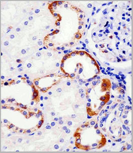
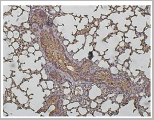
 IHC (Immunohistochemistry)
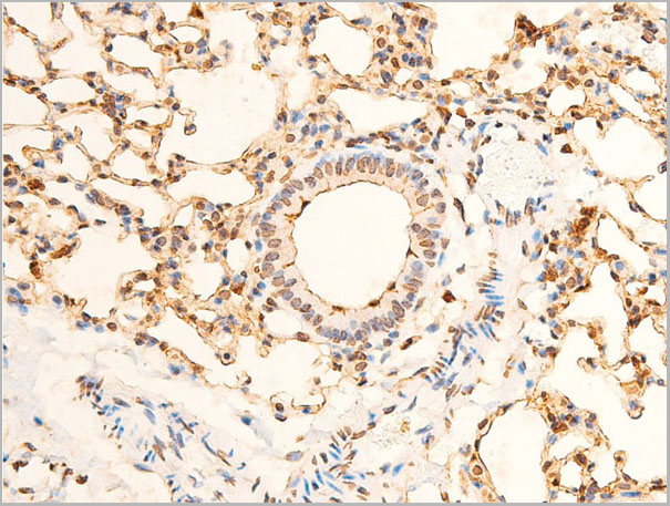
(At 1/100 staining Mouse lung tissue by IHC-P. The sample was formaldehyde fixed and a heat mediated antigen retrieval step in citrate buffer was performed. The sample was then blocked and incubated with the primary antibody at 4 degree C overnight. An HRP conjugated anti-Rabbit antibody was used as the secondary antibody.)
IHC (Immunohistochemistry)
(At 1/100 staining Mouse lung tissue by IHC-P. The sample was formaldehyde fixed and a heat mediated antigen retrieval step in citrate buffer was performed. The sample was then blocked and incubated with the primary antibody at 4 degree C overnight. An HRP conjugated anti-Rabbit antibody was used as the secondary antibody.)
Histone H2A, Polyclonal Antibody (Cat# AAA31347)
Full Name
Acetyl-Histone H2A (Lys5) Antibody
Gene Names
HIST1H2AB; H2A/m; H2AFM
Reactivity
Human, Mouse, Rat
Applications
Western Blot, Immunohistochemistry, Peptide ELISA
Purity
The antiserum was purified by peptide affinity chromatography using SulfoLink Coupling Resin
Pricing


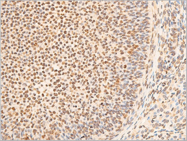
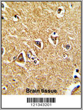
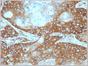
 IHC (Immunohistochemistry)
(Figure 8. IHC analysis of COMT using anti-COMT antibody (AAA11647).COMT was detected in frozen section of rat lung tissue. Heat mediated antigen retrieval was performed in citrate buffer (pH6, epitope retrieval solution) for 20 mins. The tissue section was blocked with 10% goat serum. The tissue section was then incubated with 1ug/ml rabbit anti-COMT Antibody (AAA11647) overnight at 4 degree C. Biotinylated goat anti-rabbit IgG was used as secondary antibody and incubated for 30 minutes at 37 degree C. The tissue section was developed using Strepavidin-Biotin-Complex (SABC) with DAB as the chromogen.)
IHC (Immunohistochemistry)
(Figure 8. IHC analysis of COMT using anti-COMT antibody (AAA11647).COMT was detected in frozen section of rat lung tissue. Heat mediated antigen retrieval was performed in citrate buffer (pH6, epitope retrieval solution) for 20 mins. The tissue section was blocked with 10% goat serum. The tissue section was then incubated with 1ug/ml rabbit anti-COMT Antibody (AAA11647) overnight at 4 degree C. Biotinylated goat anti-rabbit IgG was used as secondary antibody and incubated for 30 minutes at 37 degree C. The tissue section was developed using Strepavidin-Biotin-Complex (SABC) with DAB as the chromogen.)
COMT, Polyclonal Antibody (Cat# AAA11647)
Full Name
Anti-COMT Antibody
Gene Names
COMT; HEL-S-98n
Reactivity
Human, Mouse, Rat
Applications
Western Blot, Immunohistochemistry, Immunocytochemistry
Purity
Immunogen Affinity Purified
Pricing


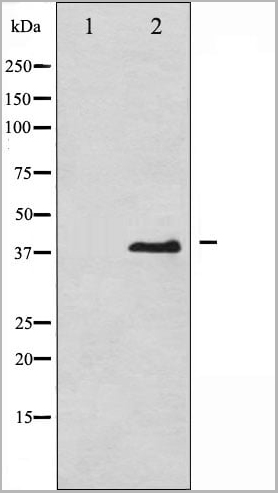
 WB (Western Blot)
(WB Suggested Anti-FOXM1 Antibody Titration: 1 ug/mlPositive Control: Jurkat cell lysate)
WB (Western Blot)
(WB Suggested Anti-FOXM1 Antibody Titration: 1 ug/mlPositive Control: Jurkat cell lysate)
FOXM1, Polyclonal Antibody (Cat# AAA23606)
Full Name
FOXM1 antibody - middle region
Gene Names
FOXM1; MPP2; HFH11; HNF-3; INS-1; MPP-2; PIG29; FKHL16; FOXM1A; FOXM1B; FOXM1C; HFH-11; TRIDENT; MPHOSPH2
Reactivity
Dog, Guinea Pig, Human, Mouse, Rabbit, Rat
Applications
Immunohistochemistry, Western Blot
Purity
Affinity Purified
Pricing


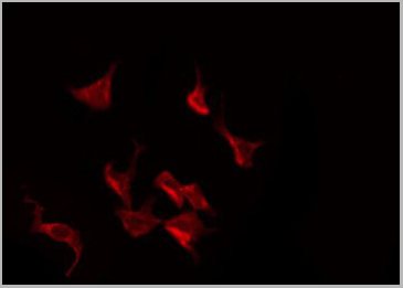
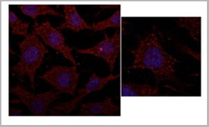
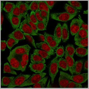
 IF (Immunofluorescence)
(Immunofluorescent analysis of 4% paraformaldehyde-fixed, 0.1% Triton X-100 permeabilized MCF-7 (human breast cancer cell line) cells labeling Pdx1 with at 1:25 dilution, followed by DyLight 488-conjugated IgG goat anti-rabbit secondary antibody at 1:200 dilution (green). Immunofluorescence image showing cytoplasm staining on MCF-7 cell line. Cytoplasmic actin is detected with DyLight 554 Phalloidin (PD18466410) at 1:100 dilution (red). The nuclear counter stain is DAPI (blue).)
IF (Immunofluorescence)
(Immunofluorescent analysis of 4% paraformaldehyde-fixed, 0.1% Triton X-100 permeabilized MCF-7 (human breast cancer cell line) cells labeling Pdx1 with at 1:25 dilution, followed by DyLight 488-conjugated IgG goat anti-rabbit secondary antibody at 1:200 dilution (green). Immunofluorescence image showing cytoplasm staining on MCF-7 cell line. Cytoplasmic actin is detected with DyLight 554 Phalloidin (PD18466410) at 1:100 dilution (red). The nuclear counter stain is DAPI (blue).)
OPN-a/b, Polyclonal Antibody (Cat# AAA26865)
Full Name
OPN-a/b, NT (SPP1, BNSP, OPN, Osteopontin, Bone sialoprotein 1, Nephropontin, Secreted phosphoprotein 1, Urinary stone protein, Uropontin) (PE)
Gene Names
SPP1; OPN; BNSP; BSPI; ETA-1
Reactivity
Human
Applications
WB, IHC, IF
Purity
Purified by Protein A and Peptide Affinity Chromatography.
Pricing


 IF (Immunofluorescence)
(Immunofluorescent analysis of 4% paraformaldehyde-fixed, 0.1% Triton X-100 permeabilized MCF-7 (human breast cancer cell line) cells labeling Pdx1 with at 1:25 dilution, followed by DyLight 488-conjugated IgG goat anti-rabbit secondary antibody at 1:200 dilution (green). Immunofluorescence image showing cytoplasm staining on MCF-7 cell line. Cytoplasmic actin is detected with DyLight 554 Phalloidin (PD18466410) at 1:100 dilution (red). The nuclear counter stain is DAPI (blue).)
IF (Immunofluorescence)
(Immunofluorescent analysis of 4% paraformaldehyde-fixed, 0.1% Triton X-100 permeabilized MCF-7 (human breast cancer cell line) cells labeling Pdx1 with at 1:25 dilution, followed by DyLight 488-conjugated IgG goat anti-rabbit secondary antibody at 1:200 dilution (green). Immunofluorescence image showing cytoplasm staining on MCF-7 cell line. Cytoplasmic actin is detected with DyLight 554 Phalloidin (PD18466410) at 1:100 dilution (red). The nuclear counter stain is DAPI (blue).)
OPN-a/b, Polyclonal Antibody (Cat# AAA26857)
Full Name
OPN-a/b, NT (SPP1, BNSP, OPN, Osteopontin, Bone sialoprotein 1, Nephropontin, Secreted phosphoprotein 1, Urinary stone protein, Uropontin) (APC)
Gene Names
SPP1; OPN; BNSP; BSPI; ETA-1
Reactivity
Human
Applications
WB, IHC, IF
Purity
Purified by Protein A and Peptide Affinity Chromatography.
Pricing


 IF (Immunofluorescence)
(Immunofluorescent analysis of 4% paraformaldehyde-fixed, 0.1% Triton X-100 permeabilized MCF-7 (human breast cancer cell line) cells labeling Pdx1 with at 1:25 dilution, followed by DyLight 488-conjugated IgG goat anti-rabbit secondary antibody at 1:200 dilution (green). Immunofluorescence image showing cytoplasm staining on MCF-7 cell line. Cytoplasmic actin is detected with DyLight 554 Phalloidin (PD18466410) at 1:100 dilution (red). The nuclear counter stain is DAPI (blue).)
IF (Immunofluorescence)
(Immunofluorescent analysis of 4% paraformaldehyde-fixed, 0.1% Triton X-100 permeabilized MCF-7 (human breast cancer cell line) cells labeling Pdx1 with at 1:25 dilution, followed by DyLight 488-conjugated IgG goat anti-rabbit secondary antibody at 1:200 dilution (green). Immunofluorescence image showing cytoplasm staining on MCF-7 cell line. Cytoplasmic actin is detected with DyLight 554 Phalloidin (PD18466410) at 1:100 dilution (red). The nuclear counter stain is DAPI (blue).)
OPN-a/b, Polyclonal Antibody (Cat# AAA26861)
Full Name
OPN-a/b, NT (SPP1, BNSP, OPN, Osteopontin, Bone sialoprotein 1, Nephropontin, Secreted phosphoprotein 1, Urinary stone protein, Uropontin) (MaxLight 490)
Gene Names
SPP1; OPN; BNSP; BSPI; ETA-1
Reactivity
Human
Applications
WB, IHC, IF
Purity
Purified by Protein A and Peptide Affinity Chromatography.
Pricing


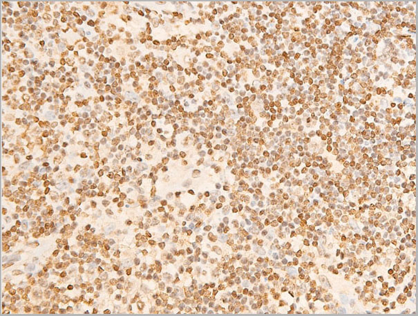
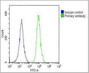
 FCM (Flow Cytometry)
(Figure 8. Flow Cytometry analysis of U-87 cells using anti-MVP antibody (AAA19137).Overlay histogram showing U-87 cells stained with AAA19137 (Blue line).The cells were blocked with 10% normal goat serum. And then incubated with rabbit anti-MVP Antibody (AAA19137,1ug/1x10^6 cells) for 30 min at 20 degree C. DyLight®488 conjugated goat anti-rabbit IgG (5-10ug/1x10^6 cells) was used as secondary antibody for 30 minutes at 20 degree C. Isotype control antibody (Green line) was rabbit IgG (1ug/1x106) used under the same conditions. Unlabelled sample (Red line) was also used as a control.)
FCM (Flow Cytometry)
(Figure 8. Flow Cytometry analysis of U-87 cells using anti-MVP antibody (AAA19137).Overlay histogram showing U-87 cells stained with AAA19137 (Blue line).The cells were blocked with 10% normal goat serum. And then incubated with rabbit anti-MVP Antibody (AAA19137,1ug/1x10^6 cells) for 30 min at 20 degree C. DyLight®488 conjugated goat anti-rabbit IgG (5-10ug/1x10^6 cells) was used as secondary antibody for 30 minutes at 20 degree C. Isotype control antibody (Green line) was rabbit IgG (1ug/1x106) used under the same conditions. Unlabelled sample (Red line) was also used as a control.)
MVP, Polyclonal Antibody (Cat# AAA19137)
Full Name
Anti-MVP Picoband antibody
Gene Names
MVP; LRP; VAULT1
Reactivity
Human, Mouse, Rat
Applications
EIA, FC/FACS, IHC, ICC, WB
Pricing


 IF (Immunofluorescence)
(Immunofluorescent analysis of 4% paraformaldehyde-fixed, 0.1% Triton X-100 permeabilized MCF-7 (human breast cancer cell line) cells labeling Pdx1 with at 1:25 dilution, followed by DyLight 488-conjugated IgG goat anti-rabbit secondary antibody at 1:200 dilution (green). Immunofluorescence image showing cytoplasm staining on MCF-7 cell line. Cytoplasmic actin is detected with DyLight 554 Phalloidin (PD18466410) at 1:100 dilution (red). The nuclear counter stain is DAPI (blue).)
IF (Immunofluorescence)
(Immunofluorescent analysis of 4% paraformaldehyde-fixed, 0.1% Triton X-100 permeabilized MCF-7 (human breast cancer cell line) cells labeling Pdx1 with at 1:25 dilution, followed by DyLight 488-conjugated IgG goat anti-rabbit secondary antibody at 1:200 dilution (green). Immunofluorescence image showing cytoplasm staining on MCF-7 cell line. Cytoplasmic actin is detected with DyLight 554 Phalloidin (PD18466410) at 1:100 dilution (red). The nuclear counter stain is DAPI (blue).)
OPN-a/b, Polyclonal Antibody (Cat# AAA26863)
Full Name
OPN-a/b, NT (SPP1, BNSP, OPN, Osteopontin, Bone sialoprotein 1, Nephropontin, Secreted phosphoprotein 1, Urinary stone protein, Uropontin) (MaxLight 650)
Gene Names
SPP1; OPN; BNSP; BSPI; ETA-1
Reactivity
Human
Applications
WB, IHC, IF
Purity
Purified by Protein A and Peptide Affinity Chromatography.
Pricing


 IF (Immunofluorescence)
(Immunofluorescent analysis of 4% paraformaldehyde-fixed, 0.1% Triton X-100 permeabilized MCF-7 (human breast cancer cell line) cells labeling Pdx1 with at 1:25 dilution, followed by DyLight 488-conjugated IgG goat anti-rabbit secondary antibody at 1:200 dilution (green). Immunofluorescence image showing cytoplasm staining on MCF-7 cell line. Cytoplasmic actin is detected with DyLight 554 Phalloidin (PD18466410) at 1:100 dilution (red). The nuclear counter stain is DAPI (blue).)
IF (Immunofluorescence)
(Immunofluorescent analysis of 4% paraformaldehyde-fixed, 0.1% Triton X-100 permeabilized MCF-7 (human breast cancer cell line) cells labeling Pdx1 with at 1:25 dilution, followed by DyLight 488-conjugated IgG goat anti-rabbit secondary antibody at 1:200 dilution (green). Immunofluorescence image showing cytoplasm staining on MCF-7 cell line. Cytoplasmic actin is detected with DyLight 554 Phalloidin (PD18466410) at 1:100 dilution (red). The nuclear counter stain is DAPI (blue).)
OPN-a/b, Polyclonal Antibody (Cat# AAA26864)
Full Name
OPN-a/b, NT (SPP1, BNSP, OPN, Osteopontin, Bone sialoprotein 1, Nephropontin, Secreted phosphoprotein 1, Urinary stone protein, Uropontin) (MaxLight 750)
Gene Names
SPP1; OPN; BNSP; BSPI; ETA-1
Reactivity
Human
Applications
WB, IHC, IF
Purity
Purified by Protein A and Peptide Affinity Chromatography.
Pricing


 IF (Immunofluorescence)
(Immunofluorescent analysis of 4% paraformaldehyde-fixed, 0.1% Triton X-100 permeabilized MCF-7 (human breast cancer cell line) cells labeling Pdx1 with at 1:25 dilution, followed by DyLight 488-conjugated IgG goat anti-rabbit secondary antibody at 1:200 dilution (green). Immunofluorescence image showing cytoplasm staining on MCF-7 cell line. Cytoplasmic actin is detected with DyLight 554 Phalloidin (PD18466410) at 1:100 dilution (red). The nuclear counter stain is DAPI (blue).)
IF (Immunofluorescence)
(Immunofluorescent analysis of 4% paraformaldehyde-fixed, 0.1% Triton X-100 permeabilized MCF-7 (human breast cancer cell line) cells labeling Pdx1 with at 1:25 dilution, followed by DyLight 488-conjugated IgG goat anti-rabbit secondary antibody at 1:200 dilution (green). Immunofluorescence image showing cytoplasm staining on MCF-7 cell line. Cytoplasmic actin is detected with DyLight 554 Phalloidin (PD18466410) at 1:100 dilution (red). The nuclear counter stain is DAPI (blue).)
OPN-a/b, Polyclonal Antibody (Cat# AAA26862)
Full Name
OPN-a/b, NT (SPP1, BNSP, OPN, Osteopontin, Bone sialoprotein 1, Nephropontin, Secreted phosphoprotein 1, Urinary stone protein, Uropontin) (MaxLight 550)
Gene Names
SPP1; OPN; BNSP; BSPI; ETA-1
Reactivity
Human
Applications
WB, IHC, IF
Purity
Purified by Protein A and Peptide Affinity Chromatography.
Pricing


 IF (Immunofluorescence)
(Immunofluorescent analysis of 4% paraformaldehyde-fixed, 0.1% Triton X-100 permeabilized MCF-7 (human breast cancer cell line) cells labeling Pdx1 with at 1:25 dilution, followed by DyLight 488-conjugated IgG goat anti-rabbit secondary antibody at 1:200 dilution (green). Immunofluorescence image showing cytoplasm staining on MCF-7 cell line. Cytoplasmic actin is detected with DyLight 554 Phalloidin (PD18466410) at 1:100 dilution (red). The nuclear counter stain is DAPI (blue).)
IF (Immunofluorescence)
(Immunofluorescent analysis of 4% paraformaldehyde-fixed, 0.1% Triton X-100 permeabilized MCF-7 (human breast cancer cell line) cells labeling Pdx1 with at 1:25 dilution, followed by DyLight 488-conjugated IgG goat anti-rabbit secondary antibody at 1:200 dilution (green). Immunofluorescence image showing cytoplasm staining on MCF-7 cell line. Cytoplasmic actin is detected with DyLight 554 Phalloidin (PD18466410) at 1:100 dilution (red). The nuclear counter stain is DAPI (blue).)
OPN-a/b, Polyclonal Antibody (Cat# AAA26859)
Full Name
OPN-a/b, NT (SPP1, BNSP, OPN, Osteopontin, Bone sialoprotein 1, Nephropontin, Secreted phosphoprotein 1, Urinary stone protein, Uropontin) (FITC)
Gene Names
SPP1; OPN; BNSP; BSPI; ETA-1
Reactivity
Human
Applications
WB, IHC, IF
Purity
Purified by Protein A and Peptide Affinity Chromatography.
Pricing


 IF (Immunofluorescence)
(Immunofluorescent analysis of 4% paraformaldehyde-fixed, 0.1% Triton X-100 permeabilized MCF-7 (human breast cancer cell line) cells labeling Pdx1 with at 1:25 dilution, followed by DyLight 488-conjugated IgG goat anti-rabbit secondary antibody at 1:200 dilution (green). Immunofluorescence image showing cytoplasm staining on MCF-7 cell line. Cytoplasmic actin is detected with DyLight 554 Phalloidin (PD18466410) at 1:100 dilution (red). The nuclear counter stain is DAPI (blue).)
IF (Immunofluorescence)
(Immunofluorescent analysis of 4% paraformaldehyde-fixed, 0.1% Triton X-100 permeabilized MCF-7 (human breast cancer cell line) cells labeling Pdx1 with at 1:25 dilution, followed by DyLight 488-conjugated IgG goat anti-rabbit secondary antibody at 1:200 dilution (green). Immunofluorescence image showing cytoplasm staining on MCF-7 cell line. Cytoplasmic actin is detected with DyLight 554 Phalloidin (PD18466410) at 1:100 dilution (red). The nuclear counter stain is DAPI (blue).)
OPN-a/b, Polyclonal Antibody (Cat# AAA26860)
Full Name
OPN-a/b, NT (SPP1, BNSP, OPN, Osteopontin, Bone sialoprotein 1, Nephropontin, Secreted phosphoprotein 1, Urinary stone protein, Uropontin) (MaxLight 405)
Gene Names
SPP1; OPN; BNSP; BSPI; ETA-1
Reactivity
Human
Applications
WB, IHC, IF
Purity
Purified by Protein A and Peptide Affinity Chromatography.
Pricing


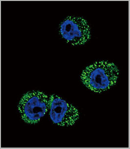
 IF (Immunofluorescence)
(Immunofluorescent analysis of 4% paraformaldehyde-fixed, 0.1% Triton X-100 permeabilized MCF-7 (human breast cancer cell line) cells labeling Pdx1 with AAA14796 at 1:25 dilution, followed by Dylight® 488-conjugated IgG goat anti-rabbit secondary antibody at 1:200 dilution (green). Immunofluorescence image showing cytoplasm staining on MCF-7 cell line. Cytoplasmic actin is detected with Dylight® 554 Phalloidin (PD18466410) at 1:100 dilution (red). The nuclear counter stain is DAPI (blue).)
IF (Immunofluorescence)
(Immunofluorescent analysis of 4% paraformaldehyde-fixed, 0.1% Triton X-100 permeabilized MCF-7 (human breast cancer cell line) cells labeling Pdx1 with AAA14796 at 1:25 dilution, followed by Dylight® 488-conjugated IgG goat anti-rabbit secondary antibody at 1:200 dilution (green). Immunofluorescence image showing cytoplasm staining on MCF-7 cell line. Cytoplasmic actin is detected with Dylight® 554 Phalloidin (PD18466410) at 1:100 dilution (red). The nuclear counter stain is DAPI (blue).)
OPN-a/b, Polyclonal Antibody (Cat# AAA14796)
Full Name
OPN-a/b, NT (SPP1, BNSP, OPN, Osteopontin, Bone sialoprotein 1, Nephropontin, Secreted phosphoprotein 1, Urinary stone protein, Uropontin)
Reactivity
Human
Applications
EL/EIA, WB, IHC, IF
Purity
Affinity Purified
Purified by Protein A affinity chromatography.
Purified by Protein A affinity chromatography.
Pricing


 IF (Immunofluorescence)
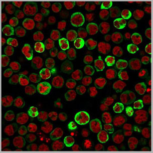
(Immunofluorescence analysis of U2OS cells using H2AFY antibody. Blue: DAPI for nuclear staining.)
IF (Immunofluorescence)
(Immunofluorescence analysis of U2OS cells using H2AFY antibody. Blue: DAPI for nuclear staining.)
H2AFY, Polyclonal Antibody (Cat# AAA28109)
Full Name
H2AFY Polyclonal Antibody
Gene Names
H2AFY; H2A.y; H2A/y; H2AFJ; mH2A1; H2AF12M; MACROH2A1.1; macroH2A1.2
Reactivity
Human, Mouse, Rat
Applications
WB, IHC, IF
Purity
Affinity Purification
Pricing


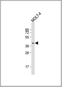
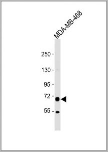
 WB (Western Blot)
(Western blot analysis of lysates from MDA-MB-468, SW620, T47D cell line, mouse spleen, mouse testis tissue (from left to right), using EZH2 Antibody. AAA28665 was diluted at 1:1000 at each lane. A goat anti-rabbit IgG H&L(HRP) at 1:10000 dilution was used as the secondary antibody. Lysates at 20ug per lane.)
WB (Western Blot)
(Western blot analysis of lysates from MDA-MB-468, SW620, T47D cell line, mouse spleen, mouse testis tissue (from left to right), using EZH2 Antibody. AAA28665 was diluted at 1:1000 at each lane. A goat anti-rabbit IgG H&L(HRP) at 1:10000 dilution was used as the secondary antibody. Lysates at 20ug per lane.)
EZH2, Polyclonal Antibody (Cat# AAA28665)
Full Name
EZH2 Antibody
Gene Names
EZH2; WVS; ENX1; EZH1; KMT6; WVS2; ENX-1; EZH2b; KMT6A
Reactivity
Human, mouse
Applications
WB, EIA, IHC, IF, FC/FACS
Purity
Peptide Affinity Purified Rabbit Polyclonal Antibody (Pab)
Pricing


 IF (Immunofluorescence)
(Immunofluorescent analysis of 4% paraformaldehyde-fixed, 0.1% Triton X-100 permeabilized MCF-7 (human breast cancer cell line) cells labeling Pdx1 with at 1:25 dilution, followed by DyLight 488-conjugated IgG goat anti-rabbit secondary antibody at 1:200 dilution (green). Immunofluorescence image showing cytoplasm staining on MCF-7 cell line. Cytoplasmic actin is detected with DyLight 554 Phalloidin (PD18466410) at 1:100 dilution (red). The nuclear counter stain is DAPI (blue).)
IF (Immunofluorescence)
(Immunofluorescent analysis of 4% paraformaldehyde-fixed, 0.1% Triton X-100 permeabilized MCF-7 (human breast cancer cell line) cells labeling Pdx1 with at 1:25 dilution, followed by DyLight 488-conjugated IgG goat anti-rabbit secondary antibody at 1:200 dilution (green). Immunofluorescence image showing cytoplasm staining on MCF-7 cell line. Cytoplasmic actin is detected with DyLight 554 Phalloidin (PD18466410) at 1:100 dilution (red). The nuclear counter stain is DAPI (blue).)
OPN-a/b, Polyclonal Antibody (Cat# AAA26858)
Full Name
OPN-a/b, NT (SPP1, BNSP, OPN, Osteopontin, Bone sialoprotein 1, Nephropontin, Secreted phosphoprotein 1, Urinary stone protein, Uropontin) (Biotin)
Gene Names
SPP1; OPN; BNSP; BSPI; ETA-1
Reactivity
Human
Applications
WB, IHC, IF, EIA
Purity
Purified by Protein A and Peptide Affinity Chromatography.
Pricing
Actin, beta Human, Active Protein (Cat# AAA14745)
Full Name
Actin, beta Human
Gene Names
ACTB; BRWS1; PS1TP5BP1
Applications
EL/EIA, WB
Purity
>99%. May contain traces of gamma-actin.
Pricing
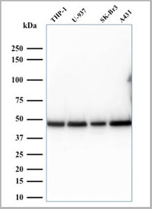
 WB (Western Blot)
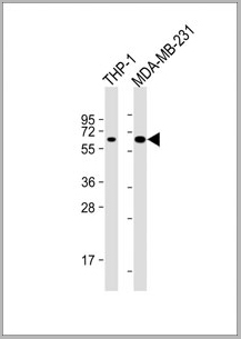
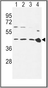
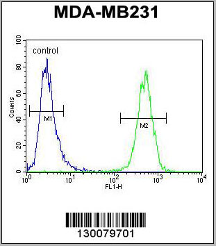
(All lanes : Anti-MB21D1 Antibody at 1:4000 dilutionLane 1: THP-1 whole cell lysateLane 2: MDA-MB-231 whole cell lysateLysates/proteins at 20 ug per lane. SecondaryGoat Anti-mouse IgG, (H+L), Peroxidase conjugated at 1/10000 dilution. Predicted band size : 59kDaBlocking/Dilution buffer: 5% NFDM/TBST.)
WB (Western Blot)
(All lanes : Anti-MB21D1 Antibody at 1:4000 dilutionLane 1: THP-1 whole cell lysateLane 2: MDA-MB-231 whole cell lysateLysates/proteins at 20 ug per lane. SecondaryGoat Anti-mouse IgG, (H+L), Peroxidase conjugated at 1/10000 dilution. Predicted band size : 59kDaBlocking/Dilution buffer: 5% NFDM/TBST.)
MB21D1, Monoclonal Antibody (Cat# AAA28800)
Full Name
MB21D1 Antibody
Gene Names
MB21D1; cGAS; h-cGAS; C6orf150
Reactivity
Human
Applications
Western Blot
Pricing
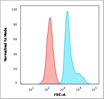
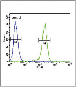 FCM (Flow Cytometry)
(CCR7 Antibody (N-term) (AAA28701) flow cytometric analysis of 293 cells (right histogram) compared to a negative controlcell (left histogram). FITC-conjugated goat-anti-rabbit secondary antibodies were used for the analysis.)
FCM (Flow Cytometry)
(CCR7 Antibody (N-term) (AAA28701) flow cytometric analysis of 293 cells (right histogram) compared to a negative controlcell (left histogram). FITC-conjugated goat-anti-rabbit secondary antibodies were used for the analysis.)
CCR7, Polyclonal Antibody (Cat# AAA28701)
Full Name
CCR7 Antibody (N-term)
Gene Names
CCR7; BLR2; EBI1; CCR-7; CD197; CDw197; CMKBR7; CC-CKR-7
Reactivity
Human, mouse
Applications
Western Blot, Immunohistochemistry, Immunofluorescence, Flow Cytometry
Purity
Peptide Affinity Purified Rabbit Polyclonal Antibody (Pab)
Pricing
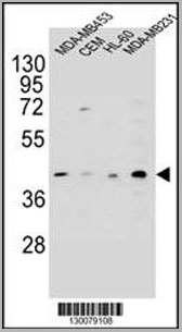 WB (Western Blot)
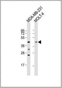
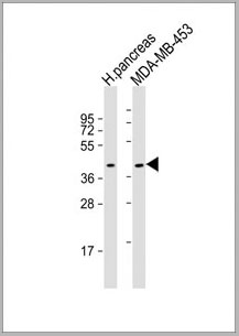
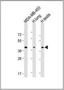
(HHLA2 Antibody (N-term) western blot analysis in MDA-MB453,CEM,HL-60,MDA-MB231 cell line lysates (35ug/lane).This demonstrates the HHLA2 antibody detected the HHLA2 protein (arrow).)
WB (Western Blot)
(HHLA2 Antibody (N-term) western blot analysis in MDA-MB453,CEM,HL-60,MDA-MB231 cell line lysates (35ug/lane).This demonstrates the HHLA2 antibody detected the HHLA2 protein (arrow).)
HHLA2, Polyclonal Antibody (Cat# AAA28722)
Full Name
HHLA2 Antibody (N-term)
Gene Names
HHLA2; B7H7; B7-H7
Reactivity
Human
Applications
Western Blot, Immunohistochemistry, Flow Cytometry
Purity
Peptide Affinity Purified Rabbit Polyclonal Antibody (Pab)
Pricing
 WB (Western Blot)
(Western blot analysis of eIF2 alpha phosphorylation expression in IFN-alpha treated K562 whole cell lysates, The lane on the left is treated with the antigen-specific peptide.)
WB (Western Blot)
(Western blot analysis of eIF2 alpha phosphorylation expression in IFN-alpha treated K562 whole cell lysates, The lane on the left is treated with the antigen-specific peptide.)
eIF2 alpha, Polyclonal Antibody (Cat# AAA30978)
Full Name
Phospho-eIF2 alpha (Ser51) Antibody
Gene Names
EIF2S1; EIF2; EIF-2; EIF2A; EIF-2A; EIF-2alpha
Reactivity
Human, Mouse, Rat
Applications
Western Blot, Immunohistochemistry, Immunofluorescence, Immunocytochemistry
Purity
From purified rabbit serum by affinity purification via sequential chromatography on phospho-and non-phospho-peptide affinity columns.
Pricing
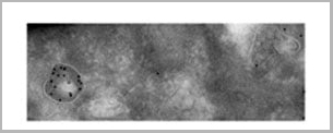
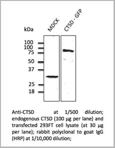
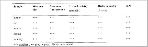
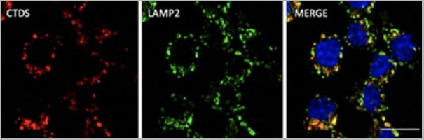 IF (Immunofluorescence)
(Immunofluorescence - anti-CTSD Ab at 1/100 dilution in RAW264.7 cells; cells were fixed with PFA and permeabilized with 0.05% saponin)
IF (Immunofluorescence)
(Immunofluorescence - anti-CTSD Ab at 1/100 dilution in RAW264.7 cells; cells were fixed with PFA and permeabilized with 0.05% saponin)
Cathepsin D, Polyclonal Antibody (Cat# AAA13859)
Full Name
Cathepsin D Polyclonal Antibody
Gene Names
CTSD; CPSD; CLN10; HEL-S-130P
Reactivity
Reacts against human, rat, mouse, canine and monkey proteins.
Applications
Western Blot, Immunofluorescence, Immunohistochemistry, Immunohistochemistry
Pricing
Ureaplasma parvum, Monoclonal Antibody (Cat# AAA13463)
Full Name
Ureaplasma parvum
Reactivity
These antibody do not cross react with Mycoplasma hominis and Mycoplasma genitallium.
Applications
Immunofluorescence
Purity
>90% pure mouse monoclonal antibody which has been purified from ascites fluid or culture medium by protein A chromatography or sequential differential precipitations.
Pricing
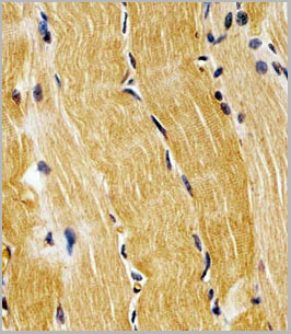
 IHC (Immunohistochemistry)
(Formalin-fixed and paraffin-embedded human brain tissue reacted with mouse Prodh Antibody (Center), which was peroxidase-conjugated to the secondary antibody, followed by DAB staining. This data demonstrates the use of this antibody for immunohistochemistry; clinical relevance has not been evaluated.)
IHC (Immunohistochemistry)
(Formalin-fixed and paraffin-embedded human brain tissue reacted with mouse Prodh Antibody (Center), which was peroxidase-conjugated to the secondary antibody, followed by DAB staining. This data demonstrates the use of this antibody for immunohistochemistry; clinical relevance has not been evaluated.)
PRODH, Polyclonal Antibody (Cat# AAA28717)
Full Name
PRODH Antibody (Center)
Gene Names
Prodh; Pro1; Pro-1; Ym24d07
Reactivity
Human, mouse
Applications
Flow Cytometry, Immunohistochemistry, Western Blot
Purity
Peptide Affinity Purified Rabbit Polyclonal Antibody (Pab)
Pricing
 Application Data
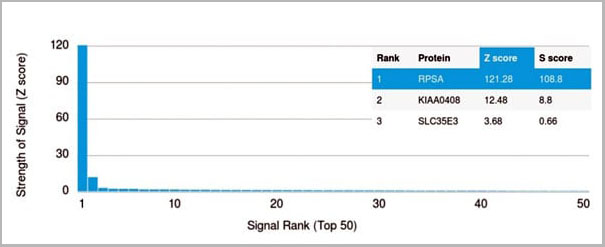
(Analysis of Protein Array containing more than 19,000 full-length human proteins using Laminin Receptor Monospecific Mouse Monoclonal Antibody (RPSA/2699) Z- and S- Score: The Z-score represents the strength of a signal that a monoclonal antibody (MAb) (in combination with a fluorescently-tagged anti-IgG secondary antibody) produces when binding to a particular protein on the HuProtTM array. Z-scores are described in units of standard deviations (SD's) above the mean value of all signals generated on that array. If targets on HuProtTM are arranged in descending order of the Z-score, the S-score is the difference (also in units of SD's) between the Z-score. S-score therefore represents the relative target specificity of a MAb to its intended target. A MAb is considered to specific to its intended target, if the MAb has an S-score of at least 2.5. For example, if a MAb binds to protein X with a Z-score of 43 and to protein Y with a Z-score of 14, then the S-score for the binding of that MAb to protein X is equal to 29.)
Application Data
(Analysis of Protein Array containing more than 19,000 full-length human proteins using Laminin Receptor Monospecific Mouse Monoclonal Antibody (RPSA/2699) Z- and S- Score: The Z-score represents the strength of a signal that a monoclonal antibody (MAb) (in combination with a fluorescently-tagged anti-IgG secondary antibody) produces when binding to a particular protein on the HuProtTM array. Z-scores are described in units of standard deviations (SD's) above the mean value of all signals generated on that array. If targets on HuProtTM are arranged in descending order of the Z-score, the S-score is the difference (also in units of SD's) between the Z-score. S-score therefore represents the relative target specificity of a MAb to its intended target. A MAb is considered to specific to its intended target, if the MAb has an S-score of at least 2.5. For example, if a MAb binds to protein X with a Z-score of 43 and to protein Y with a Z-score of 14, then the S-score for the binding of that MAb to protein X is equal to 29.)
Laminin Receptor/RPSA, Monoclonal Antibody (Cat# AAA23908)
Full Name
Laminin Receptor/RPSA (Marker of Metastatic Potential)
Gene Names
RPSA; SA; LBP; LRP; p40; 67LR; ICAS; lamR; 37LRP; LAMBR; LAMR1; LRP/LR; LBP/p40; NEM/1CHD4
Reactivity
Human. Others not known.
Applications
Flow Cytometry, Immunofluorescence, Western Blot, Immunohistochemistry
Pricing
S100 Protein, Protein (Cat# AAA14736)
Full Name
S100 Protein, Human
Gene Names
S100B; NEF; S100; S100-B; S100beta
Applications
Immunoassay, Antiserum Production, Iodination
Purity
>95%
Pricing