Filters
Clonality
Type
Reactivity
Gene Name
Isotype
Host
Application
Clone
54 results for " GFP" - showing 1-50
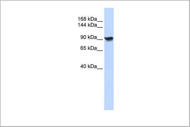
 WB (Western Blot)
(Positive WB detected in Recombinant proteinAll lanes: ompA antibody at 1:2000SecondaryGoat polyclonal to rabbit IgG at 1/50000 dilutionPredicted band size: 35.6 kDaObserved band size: 36 kDa)
WB (Western Blot)
(Positive WB detected in Recombinant proteinAll lanes: ompA antibody at 1:2000SecondaryGoat polyclonal to rabbit IgG at 1/50000 dilutionPredicted band size: 35.6 kDaObserved band size: 36 kDa)
ompA, Polyclonal Antibody (Cat# AAA27060)
Full Name
Rabbit anti-Escherichia coli O157:H7 ompA Polyclonal Antibody
Reactivity
Escherichia coli O157:H7
Applications
Western Blot
Purity
Protein G
Pricing


 Application Data
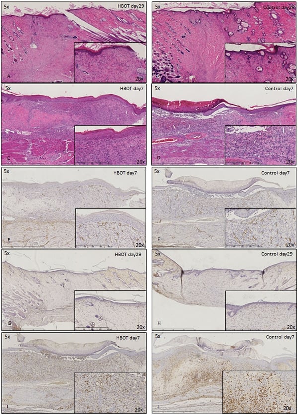
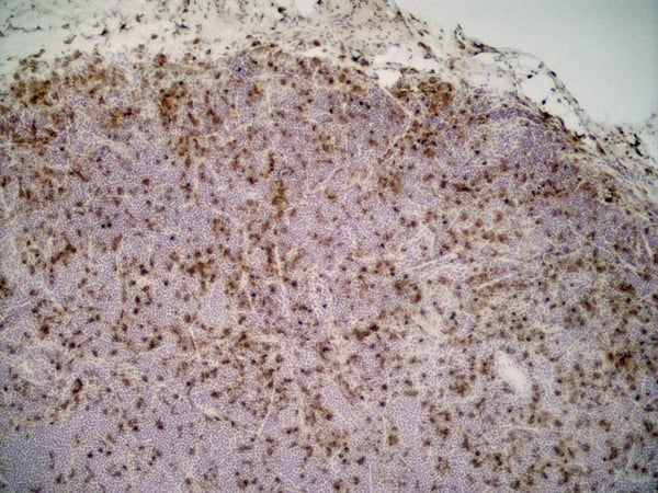
(Published customer image: Histological staining of control and HBOT wounds at post-wounding days 7 and 29. A -D) H&E staining. E -H) CD34 immunohistochemistry. I+J) CD68 immunohistochemistry.From: uk B, Tong M, Fijneman EMG, van Neck JW (2014) Hyperbaric Oxygen Therapy to Treat Diabetes Impaired Wound Healing in Rats. PLoS ONE 9(10): e108533.)
Application Data
(Published customer image: Histological staining of control and HBOT wounds at post-wounding days 7 and 29. A -D) H&E staining. E -H) CD34 immunohistochemistry. I+J) CD68 immunohistochemistry.From: uk B, Tong M, Fijneman EMG, van Neck JW (2014) Hyperbaric Oxygen Therapy to Treat Diabetes Impaired Wound Healing in Rats. PLoS ONE 9(10): e108533.)
CD68, Monoclonal Antibody (Cat# AAA12150)
Full Name
MOUSE ANTI RAT CD68:RPE
Applications
Flow Cytometry
Pricing


 WB (Western Blot)
(Anti-tdTomato Ab conjugated to DyLight 550 at 1/2,500 dilution using HEK293 transfected cell lysates at 50 ug per lane;)
WB (Western Blot)
(Anti-tdTomato Ab conjugated to DyLight 550 at 1/2,500 dilution using HEK293 transfected cell lysates at 50 ug per lane;)
tdTomato, Polyclonal Antibody (Cat# AAA13880)
Full Name
Anti-tdTomato, DyLight550
Reactivity
Reacts with Transfected cells proteins
Applications
Western Blot, Immunofluorescence, Immunohistochemistry
Purity
This antibody is epitope-affinity purified from goat antiserum.
Pricing


 Application Data
(Published Customer Image:Rat anti Mouse Gr-1 antibody, clone RB6-8C5 used for the identification of neutrophils by immunofluorescence.Image caption:S. typhimurium-Infected Macrophages Containing Phagocytosed Neutrophils and T Cells Confocal fluorescence microscopy of 50-mum-thick liver sections from 1-wk-infected Slc11a1 wild-type mice. (A-C) S.Typhimurium (O-antigen, arrows) are red, macrophages (F4-80 and MOMA-2) are blue, DNA (DAPI) is gray, phalloidin is green, and neutrophils (Gr-1/Ly-6G/RB6-8C5) are pink (arrowheads). (A) Collapsed image from a 40-mum Z-stack. Scale bar is 20 mum. (B and C) Sections from (A) that are 4 mum apart. The video from which (A-C) were derived (Video S2) is available online. (D-G) T cells within multinucleate macrophages.Macrophages (F4-80 and MOMA-2) are blue (D, G, and H), T cells (CD3zeta) are red (D, G, arrowheads), DAPI is gray (E, G), actin-bound phalloidin is green (F, G). (G) Is a composite of (D, E, and F). Scale bars are 16 mum. (H) An image from a different mouse stained and labeled as described for (D-G). Scale bar is 8 mum. A video showing a T cell inside of a macrophage is available online (Video S3).From: Nix RN, Altschuler SE, Henson PM, Detweiler CS (2007) Hemophagocytic Macrophages Harbor Salmonella enterica during Persistent Infection.PLoS Pathog 3(12): e193.)
Application Data
(Published Customer Image:Rat anti Mouse Gr-1 antibody, clone RB6-8C5 used for the identification of neutrophils by immunofluorescence.Image caption:S. typhimurium-Infected Macrophages Containing Phagocytosed Neutrophils and T Cells Confocal fluorescence microscopy of 50-mum-thick liver sections from 1-wk-infected Slc11a1 wild-type mice. (A-C) S.Typhimurium (O-antigen, arrows) are red, macrophages (F4-80 and MOMA-2) are blue, DNA (DAPI) is gray, phalloidin is green, and neutrophils (Gr-1/Ly-6G/RB6-8C5) are pink (arrowheads). (A) Collapsed image from a 40-mum Z-stack. Scale bar is 20 mum. (B and C) Sections from (A) that are 4 mum apart. The video from which (A-C) were derived (Video S2) is available online. (D-G) T cells within multinucleate macrophages.Macrophages (F4-80 and MOMA-2) are blue (D, G, and H), T cells (CD3zeta) are red (D, G, arrowheads), DAPI is gray (E, G), actin-bound phalloidin is green (F, G). (G) Is a composite of (D, E, and F). Scale bars are 16 mum. (H) An image from a different mouse stained and labeled as described for (D-G). Scale bar is 8 mum. A video showing a T cell inside of a macrophage is available online (Video S3).From: Nix RN, Altschuler SE, Henson PM, Detweiler CS (2007) Hemophagocytic Macrophages Harbor Salmonella enterica during Persistent Infection.PLoS Pathog 3(12): e193.)
Gr-1, Monoclonal Antibody (Cat# AAA12257)
Full Name
Rat Anti Mouse Gr-1: FITC
Reactivity
Mouse
Applications
Flow Cytometry
Purity
Purified IgG prepared by affinity chromatography on Protein G from tissue culture supernatant
Pricing
 WB (Western Blot)
(Positive WB detected in: recombinant proteinAll lanes: gH Antibody at 1:1000SecondaryGoat polyclonal to rabbit IgG at 1/50000 dilutionPredicted band size: 38 kDaObserved band size: 38 kDa)
WB (Western Blot)
(Positive WB detected in: recombinant proteinAll lanes: gH Antibody at 1:1000SecondaryGoat polyclonal to rabbit IgG at 1/50000 dilutionPredicted band size: 38 kDaObserved band size: 38 kDa)
gH, Polyclonal Antibody (Cat# AAA27061)
Full Name
Rabbit anti-Epstein-Barr virus (strain B95-8)(HHV-4)(Human herpesvirus 4) gH Polyclonal Antibody
Reactivity
Epstein-Barr virus
Applications
Western Blot
Purity
>95%, Protein G purified
Pricing


 WB (Western Blot)
(Western blot analysis of extracts of various cells.)
WB (Western Blot)
(Western blot analysis of extracts of various cells.)
POLK/DNA Polymerase Kappa, Polyclonal Antibody (Cat# AAA21279)
Full Name
Anti-POLK/DNA Polymerase Kappa Antibody
Gene Names
POLK; DINP; POLQ; DINB1
Reactivity
Human
Predicted: Mouse, Rat
Predicted: Mouse, Rat
Applications
Immunofluorescence, Immunohistochemistry, Immunohistochemistry, Western Blot
Purity
Affinity Purified
Pricing


 Application Data
(Published customer image: Histological staining of control and HBOT wounds at post-wounding days 7 and 29. A -D) H&E staining. E -H) CD34 immunohistochemistry. I+J) CD68 immunohistochemistry.From: uk B, Tong M, Fijneman EMG, van Neck JW (2014) Hyperbaric Oxygen Therapy to Treat Diabetes Impaired Wound Healing in Rats. PLoS ONE 9(10): e108533.)
Application Data
(Published customer image: Histological staining of control and HBOT wounds at post-wounding days 7 and 29. A -D) H&E staining. E -H) CD34 immunohistochemistry. I+J) CD68 immunohistochemistry.From: uk B, Tong M, Fijneman EMG, van Neck JW (2014) Hyperbaric Oxygen Therapy to Treat Diabetes Impaired Wound Healing in Rats. PLoS ONE 9(10): e108533.)
CD68, Monoclonal Antibody (Cat# AAA12149)
Full Name
MOUSE ANTI RAT CD68
Applications
Immunohistochemistry, Flow Cytometry, Immunofluorescence, Immunoprecipitation, Immunohistochemistry
Pricing
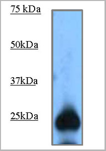
 WB (Western Blot)
(Positive WB detected in: recombinant proteinAll lanes: MELT Antibody at 1:1000Secondary: Goat polyclonal to rabbit IgG at 1/50000 dilutionPredicted band size: 17 kDaObserved band size: 17 kDa)
WB (Western Blot)
(Positive WB detected in: recombinant proteinAll lanes: MELT Antibody at 1:1000Secondary: Goat polyclonal to rabbit IgG at 1/50000 dilutionPredicted band size: 17 kDaObserved band size: 17 kDa)
MELT, Polyclonal Antibody (Cat# AAA27057)
Full Name
Rabbit anti-Apis mellifera (Honeybee) MELT Polyclonal Antibody
Gene Names
Melt; GB10355
Reactivity
Apis mellifera
Applications
Western Blot
Purity
>95%, Protein G purified
Pricing


 Application Data
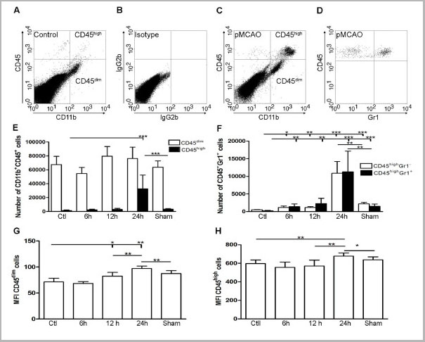
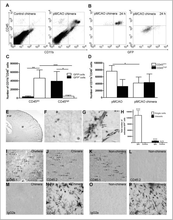
(Published customer image Infiltration of GFP+ BM-cells in infarct and peri-infarct regions. (A-B) Dot plots of viable macrophages/granulocytes (CD11b+CD45high, top right quadrants) and microglia (CD11b+CD45dim, bottom right quadrants) in cortex from BM-chimeric unmanipulated mice and mice exposed to pMCAO. (C) Bar graph showing mean numbers of CD11b+CD45dim microglia and CD11b+CD45high macrophages/granulocytes in BM-chimeric mice 24 hours after pMCAO, subdivided based on expression of GFP (n = 5). Approximately 92% of of the CD45high population were GFP+. (D) Estimation and comparison of mean numbers of CD11b+CD45dim microglia in non-chimeric (n = 10) versus BM-chimeric mice (n = 5) 24 hours after of pMCAO shows significantly fewer CD11b+CD45dim microglial cells in irradiated mice. (E) Overview, showing distribution of infiltrating GFP+ BM-derived cells into infarct (IF) and peri-infarct (P-IF) regions 24 hours after pMCAO. (E-G) By 24 hours, GFP+ single cells (F) and vessel-associated aggregates of GFP+ cells (arrows in G) were observed in infarct and peri-infarct regions. Some of the vessel-associated cells were round, leukocyte-like cells (arrows) while others were elongated cells lining the vasculature (arrow heads in G and in insert). (H) Bar graph showing mean numbers of single GFP+ cells and vessel-associated aggregates of GFP+ cells in ipsi- and contralateral cortex 24 hours after surgery (n = 10). (I-P) Immunohistochemical staining of CD45.1 (I, K), CD45.2 (J, L), IgG2a (M, O) and CD45 (N, P) in ischemic tissue in BM-chimeric (I, J, M, N) and non-chimeric mice (K, L, O, P) 24 hours after pMCAO. N.D, none detected. Scale bars: 200 um (A), 10 um (B, C). 50 um (I-P) *P < 0.05, **P < 0.01, and ***P < 0.001.From: Clausen BH, Lambertsen KL, Babcock AA, Holm TH, Dagnaes-Hansen F, Finsen B. Interleukin-1beta and tumor necrosis factor-alpha are expressed by different subsets of microglia and macrophages after ischemic stroke in mice. J Neuroinflammation. 2008 Oct 23;5:46.)
Application Data
(Published customer image Infiltration of GFP+ BM-cells in infarct and peri-infarct regions. (A-B) Dot plots of viable macrophages/granulocytes (CD11b+CD45high, top right quadrants) and microglia (CD11b+CD45dim, bottom right quadrants) in cortex from BM-chimeric unmanipulated mice and mice exposed to pMCAO. (C) Bar graph showing mean numbers of CD11b+CD45dim microglia and CD11b+CD45high macrophages/granulocytes in BM-chimeric mice 24 hours after pMCAO, subdivided based on expression of GFP (n = 5). Approximately 92% of of the CD45high population were GFP+. (D) Estimation and comparison of mean numbers of CD11b+CD45dim microglia in non-chimeric (n = 10) versus BM-chimeric mice (n = 5) 24 hours after of pMCAO shows significantly fewer CD11b+CD45dim microglial cells in irradiated mice. (E) Overview, showing distribution of infiltrating GFP+ BM-derived cells into infarct (IF) and peri-infarct (P-IF) regions 24 hours after pMCAO. (E-G) By 24 hours, GFP+ single cells (F) and vessel-associated aggregates of GFP+ cells (arrows in G) were observed in infarct and peri-infarct regions. Some of the vessel-associated cells were round, leukocyte-like cells (arrows) while others were elongated cells lining the vasculature (arrow heads in G and in insert). (H) Bar graph showing mean numbers of single GFP+ cells and vessel-associated aggregates of GFP+ cells in ipsi- and contralateral cortex 24 hours after surgery (n = 10). (I-P) Immunohistochemical staining of CD45.1 (I, K), CD45.2 (J, L), IgG2a (M, O) and CD45 (N, P) in ischemic tissue in BM-chimeric (I, J, M, N) and non-chimeric mice (K, L, O, P) 24 hours after pMCAO. N.D, none detected. Scale bars: 200 um (A), 10 um (B, C). 50 um (I-P) *P < 0.05, **P < 0.01, and ***P < 0.001.From: Clausen BH, Lambertsen KL, Babcock AA, Holm TH, Dagnaes-Hansen F, Finsen B. Interleukin-1beta and tumor necrosis factor-alpha are expressed by different subsets of microglia and macrophages after ischemic stroke in mice. J Neuroinflammation. 2008 Oct 23;5:46.)
CD11b, Monoclonal Antibody (Cat# AAA12182)
Full Name
RAT ANTI MOUSE CD11b:FITC
Gene Names
Itgam; CR3; CR3A; MAC1; Cd11b; Ly-40; Mac-1; Mac-1a; CD11b/CD18; F730045J24Rik
Applications
Flow Cytometry
Pricing


 Application Data
(Published customer image Infiltration of GFP+ BM-cells in infarct and peri-infarct regions. (A-B) Dot plots of viable macrophages/granulocytes (CD11b+CD45high, top right quadrants) and microglia (CD11b+CD45dim, bottom right quadrants) in cortex from BM-chimeric unmanipulated mice and mice exposed to pMCAO. (C) Bar graph showing mean numbers of CD11b+CD45dim microglia and CD11b+CD45high macrophages/granulocytes in BM-chimeric mice 24 hours after pMCAO, subdivided based on expression of GFP (n = 5). Approximately 92% of of the CD45high population were GFP+. (D) Estimation and comparison of mean numbers of CD11b+CD45dim microglia in non-chimeric (n = 10) versus BM-chimeric mice (n = 5) 24 hours after of pMCAO shows significantly fewer CD11b+CD45dim microglial cells in irradiated mice. (E) Overview, showing distribution of infiltrating GFP+ BM-derived cells into infarct (IF) and peri-infarct (P-IF) regions 24 hours after pMCAO. (E-G) By 24 hours, GFP+ single cells (F) and vessel-associated aggregates of GFP+ cells (arrows in G) were observed in infarct and peri-infarct regions. Some of the vessel-associated cells were round, leukocyte-like cells (arrows) while others were elongated cells lining the vasculature (arrow heads in G and in insert). (H) Bar graph showing mean numbers of single GFP+ cells and vessel-associated aggregates of GFP+ cells in ipsi- and contralateral cortex 24 hours after surgery (n = 10). (I-P) Immunohistochemical staining of CD45.1 (I, K), CD45.2 (J, L), IgG2a (M, O) and CD45 (N, P) in ischemic tissue in BM-chimeric (I, J, M, N) and non-chimeric mice (K, L, O, P) 24 hours after pMCAO. N.D, none detected. Scale bars: 200 um (A), 10 um (B, C). 50 um (I-P) *P < 0.05, **P < 0.01, and ***P < 0.001.From: Clausen BH, Lambertsen KL, Babcock AA, Holm TH, Dagnaes-Hansen F, Finsen B. Interleukin-1beta and tumor necrosis factor-alpha are expressed by different subsets of microglia and macrophages after ischemic stroke in mice. J Neuroinflammation. 2008 Oct 23;5:46.)
Application Data
(Published customer image Infiltration of GFP+ BM-cells in infarct and peri-infarct regions. (A-B) Dot plots of viable macrophages/granulocytes (CD11b+CD45high, top right quadrants) and microglia (CD11b+CD45dim, bottom right quadrants) in cortex from BM-chimeric unmanipulated mice and mice exposed to pMCAO. (C) Bar graph showing mean numbers of CD11b+CD45dim microglia and CD11b+CD45high macrophages/granulocytes in BM-chimeric mice 24 hours after pMCAO, subdivided based on expression of GFP (n = 5). Approximately 92% of of the CD45high population were GFP+. (D) Estimation and comparison of mean numbers of CD11b+CD45dim microglia in non-chimeric (n = 10) versus BM-chimeric mice (n = 5) 24 hours after of pMCAO shows significantly fewer CD11b+CD45dim microglial cells in irradiated mice. (E) Overview, showing distribution of infiltrating GFP+ BM-derived cells into infarct (IF) and peri-infarct (P-IF) regions 24 hours after pMCAO. (E-G) By 24 hours, GFP+ single cells (F) and vessel-associated aggregates of GFP+ cells (arrows in G) were observed in infarct and peri-infarct regions. Some of the vessel-associated cells were round, leukocyte-like cells (arrows) while others were elongated cells lining the vasculature (arrow heads in G and in insert). (H) Bar graph showing mean numbers of single GFP+ cells and vessel-associated aggregates of GFP+ cells in ipsi- and contralateral cortex 24 hours after surgery (n = 10). (I-P) Immunohistochemical staining of CD45.1 (I, K), CD45.2 (J, L), IgG2a (M, O) and CD45 (N, P) in ischemic tissue in BM-chimeric (I, J, M, N) and non-chimeric mice (K, L, O, P) 24 hours after pMCAO. N.D, none detected. Scale bars: 200 um (A), 10 um (B, C). 50 um (I-P) *P < 0.05, **P < 0.01, and ***P < 0.001.From: Clausen BH, Lambertsen KL, Babcock AA, Holm TH, Dagnaes-Hansen F, Finsen B. Interleukin-1beta and tumor necrosis factor-alpha are expressed by different subsets of microglia and macrophages after ischemic stroke in mice. J Neuroinflammation. 2008 Oct 23;5:46.)
CD11b, Monoclonal Antibody (Cat# AAA12184)
Full Name
RAT ANTI MOUSE CD11b
Gene Names
Itgam; CR3; CR3A; MAC1; Cd11b; Ly-40; Mac-1; Mac-1a; CD11b/CD18; F730045J24Rik
Applications
Immunohistochemistry, Flow Cytometry, Immunofluorescence, Immunoprecipitation
Pricing


 Application Data
(Published customer image: Histological staining of control and HBOT wounds at post-wounding days 7 and 29. A -D) H&E staining. E -H) CD34 immunohistochemistry. I+J) CD68 immunohistochemistry.From: uk B, Tong M, Fijneman EMG, van Neck JW (2014) Hyperbaric Oxygen Therapy to Treat Diabetes Impaired Wound Healing in Rats. PLoS ONE 9(10): e108533.)
Application Data
(Published customer image: Histological staining of control and HBOT wounds at post-wounding days 7 and 29. A -D) H&E staining. E -H) CD34 immunohistochemistry. I+J) CD68 immunohistochemistry.From: uk B, Tong M, Fijneman EMG, van Neck JW (2014) Hyperbaric Oxygen Therapy to Treat Diabetes Impaired Wound Healing in Rats. PLoS ONE 9(10): e108533.)
CD68, Monoclonal Antibody (Cat# AAA12147)
Full Name
MOUSE ANTI RAT CD68:Biotin
Applications
Flow Cytometry
Pricing
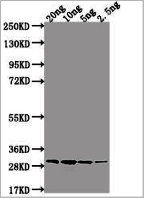
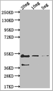
 WB (Western Blot)
(Western BlotPositive WB detected in Recombinant proteinAll lanes: omp38 antibody at 1:2000SecondaryGoat polyclonal to rabbit IgG at 1/50000 dilutionPredicted band size: 52.5 kDaObserved band size: 66 kDa.)
WB (Western Blot)
(Western BlotPositive WB detected in Recombinant proteinAll lanes: omp38 antibody at 1:2000SecondaryGoat polyclonal to rabbit IgG at 1/50000 dilutionPredicted band size: 52.5 kDaObserved band size: 66 kDa.)
omp38, Polyclonal Antibody (Cat# AAA27062)
Full Name
Rabbit anti-Acinetobacter baumannii (strain 17978/CIP 53.77/LMG 1025/NCDC KC755/5377) omp38 Polyclonal Antibody
Reactivity
Acinetobacter baumannii
Applications
Western Blot
Purity
Protein G
Pricing


 Application Data
(Published customer image Infiltration of GFP+ BM-cells in infarct and peri-infarct regions. (A-B) Dot plots of viable macrophages/granulocytes (CD11b+CD45high, top right quadrants) and microglia (CD11b+CD45dim, bottom right quadrants) in cortex from BM-chimeric unmanipulated mice and mice exposed to pMCAO. (C) Bar graph showing mean numbers of CD11b+CD45dim microglia and CD11b+CD45high macrophages/granulocytes in BM-chimeric mice 24 hours after pMCAO, subdivided based on expression of GFP (n = 5). Approximately 92% of of the CD45high population were GFP+. (D) Estimation and comparison of mean numbers of CD11b+CD45dim microglia in non-chimeric (n = 10) versus BM-chimeric mice (n = 5) 24 hours after of pMCAO shows significantly fewer CD11b+CD45dim microglial cells in irradiated mice. (E) Overview, showing distribution of infiltrating GFP+ BM-derived cells into infarct (IF) and peri-infarct (P-IF) regions 24 hours after pMCAO. (E-G) By 24 hours, GFP+ single cells (F) and vessel-associated aggregates of GFP+ cells (arrows in G) were observed in infarct and peri-infarct regions. Some of the vessel-associated cells were round, leukocyte-like cells (arrows) while others were elongated cells lining the vasculature (arrow heads in G and in insert). (H) Bar graph showing mean numbers of single GFP+ cells and vessel-associated aggregates of GFP+ cells in ipsi- and contralateral cortex 24 hours after surgery (n = 10). (I-P) Immunohistochemical staining of CD45.1 (I, K), CD45.2 (J, L), IgG2a (M, O) and CD45 (N, P) in ischemic tissue in BM-chimeric (I, J, M, N) and non-chimeric mice (K, L, O, P) 24 hours after pMCAO. N.D, none detected. Scale bars: 200 um (A), 10 um (B, C). 50 um (I-P) *P < 0.05, **P < 0.01, and ***P < 0.001.From: Clausen BH, Lambertsen KL, Babcock AA, Holm TH, Dagnaes-Hansen F, Finsen B. Interleukin-1beta and tumor necrosis factor-alpha are expressed by different subsets of microglia and macrophages after ischemic stroke in mice. J Neuroinflammation. 2008 Oct 23;5:46.)
Application Data
(Published customer image Infiltration of GFP+ BM-cells in infarct and peri-infarct regions. (A-B) Dot plots of viable macrophages/granulocytes (CD11b+CD45high, top right quadrants) and microglia (CD11b+CD45dim, bottom right quadrants) in cortex from BM-chimeric unmanipulated mice and mice exposed to pMCAO. (C) Bar graph showing mean numbers of CD11b+CD45dim microglia and CD11b+CD45high macrophages/granulocytes in BM-chimeric mice 24 hours after pMCAO, subdivided based on expression of GFP (n = 5). Approximately 92% of of the CD45high population were GFP+. (D) Estimation and comparison of mean numbers of CD11b+CD45dim microglia in non-chimeric (n = 10) versus BM-chimeric mice (n = 5) 24 hours after of pMCAO shows significantly fewer CD11b+CD45dim microglial cells in irradiated mice. (E) Overview, showing distribution of infiltrating GFP+ BM-derived cells into infarct (IF) and peri-infarct (P-IF) regions 24 hours after pMCAO. (E-G) By 24 hours, GFP+ single cells (F) and vessel-associated aggregates of GFP+ cells (arrows in G) were observed in infarct and peri-infarct regions. Some of the vessel-associated cells were round, leukocyte-like cells (arrows) while others were elongated cells lining the vasculature (arrow heads in G and in insert). (H) Bar graph showing mean numbers of single GFP+ cells and vessel-associated aggregates of GFP+ cells in ipsi- and contralateral cortex 24 hours after surgery (n = 10). (I-P) Immunohistochemical staining of CD45.1 (I, K), CD45.2 (J, L), IgG2a (M, O) and CD45 (N, P) in ischemic tissue in BM-chimeric (I, J, M, N) and non-chimeric mice (K, L, O, P) 24 hours after pMCAO. N.D, none detected. Scale bars: 200 um (A), 10 um (B, C). 50 um (I-P) *P < 0.05, **P < 0.01, and ***P < 0.001.From: Clausen BH, Lambertsen KL, Babcock AA, Holm TH, Dagnaes-Hansen F, Finsen B. Interleukin-1beta and tumor necrosis factor-alpha are expressed by different subsets of microglia and macrophages after ischemic stroke in mice. J Neuroinflammation. 2008 Oct 23;5:46.)
CD11b, Monoclonal Antibody (Cat# AAA12183)
Full Name
RAT ANTI MOUSE CD11b:FITC
Gene Names
Itgam; CR3; CR3A; MAC1; Cd11b; Ly-40; Mac-1; Mac-1a; CD11b/CD18; F730045J24Rik
Applications
Flow Cytometry
Pricing


 Application Data
(Published customer image Infiltration of GFP+ BM-cells in infarct and peri-infarct regions. (A-B) Dot plots of viable macrophages/granulocytes (CD11b+CD45high, top right quadrants) and microglia (CD11b+CD45dim, bottom right quadrants) in cortex from BM-chimeric unmanipulated mice and mice exposed to pMCAO. (C) Bar graph showing mean numbers of CD11b+CD45dim microglia and CD11b+CD45high macrophages/granulocytes in BM-chimeric mice 24 hours after pMCAO, subdivided based on expression of GFP (n = 5). Approximately 92% of of the CD45high population were GFP+. (D) Estimation and comparison of mean numbers of CD11b+CD45dim microglia in non-chimeric (n = 10) versus BM-chimeric mice (n = 5) 24 hours after of pMCAO shows significantly fewer CD11b+CD45dim microglial cells in irradiated mice. (E) Overview, showing distribution of infiltrating GFP+ BM-derived cells into infarct (IF) and peri-infarct (P-IF) regions 24 hours after pMCAO. (E-G) By 24 hours, GFP+ single cells (F) and vessel-associated aggregates of GFP+ cells (arrows in G) were observed in infarct and peri-infarct regions. Some of the vessel-associated cells were round, leukocyte-like cells (arrows) while others were elongated cells lining the vasculature (arrow heads in G and in insert). (H) Bar graph showing mean numbers of single GFP+ cells and vessel-associated aggregates of GFP+ cells in ipsi- and contralateral cortex 24 hours after surgery (n = 10). (I-P) Immunohistochemical staining of CD45.1 (I, K), CD45.2 (J, L), IgG2a (M, O) and CD45 (N, P) in ischemic tissue in BM-chimeric (I, J, M, N) and non-chimeric mice (K, L, O, P) 24 hours after pMCAO. N.D, none detected. Scale bars: 200 um (A), 10 um (B, C). 50 um (I-P) *P < 0.05, **P < 0.01, and ***P < 0.001.From: Clausen BH, Lambertsen KL, Babcock AA, Holm TH, Dagnaes-Hansen F, Finsen B. Interleukin-1beta and tumor necrosis factor-alpha are expressed by different subsets of microglia and macrophages after ischemic stroke in mice. J Neuroinflammation. 2008 Oct 23;5:46.)
Application Data
(Published customer image Infiltration of GFP+ BM-cells in infarct and peri-infarct regions. (A-B) Dot plots of viable macrophages/granulocytes (CD11b+CD45high, top right quadrants) and microglia (CD11b+CD45dim, bottom right quadrants) in cortex from BM-chimeric unmanipulated mice and mice exposed to pMCAO. (C) Bar graph showing mean numbers of CD11b+CD45dim microglia and CD11b+CD45high macrophages/granulocytes in BM-chimeric mice 24 hours after pMCAO, subdivided based on expression of GFP (n = 5). Approximately 92% of of the CD45high population were GFP+. (D) Estimation and comparison of mean numbers of CD11b+CD45dim microglia in non-chimeric (n = 10) versus BM-chimeric mice (n = 5) 24 hours after of pMCAO shows significantly fewer CD11b+CD45dim microglial cells in irradiated mice. (E) Overview, showing distribution of infiltrating GFP+ BM-derived cells into infarct (IF) and peri-infarct (P-IF) regions 24 hours after pMCAO. (E-G) By 24 hours, GFP+ single cells (F) and vessel-associated aggregates of GFP+ cells (arrows in G) were observed in infarct and peri-infarct regions. Some of the vessel-associated cells were round, leukocyte-like cells (arrows) while others were elongated cells lining the vasculature (arrow heads in G and in insert). (H) Bar graph showing mean numbers of single GFP+ cells and vessel-associated aggregates of GFP+ cells in ipsi- and contralateral cortex 24 hours after surgery (n = 10). (I-P) Immunohistochemical staining of CD45.1 (I, K), CD45.2 (J, L), IgG2a (M, O) and CD45 (N, P) in ischemic tissue in BM-chimeric (I, J, M, N) and non-chimeric mice (K, L, O, P) 24 hours after pMCAO. N.D, none detected. Scale bars: 200 um (A), 10 um (B, C). 50 um (I-P) *P < 0.05, **P < 0.01, and ***P < 0.001.From: Clausen BH, Lambertsen KL, Babcock AA, Holm TH, Dagnaes-Hansen F, Finsen B. Interleukin-1beta and tumor necrosis factor-alpha are expressed by different subsets of microglia and macrophages after ischemic stroke in mice. J Neuroinflammation. 2008 Oct 23;5:46.)
CD11b, Monoclonal Antibody (Cat# AAA12185)
Full Name
RAT ANTI MOUSE CD11b
Gene Names
Itgam; CR3; CR3A; MAC1; Cd11b; Ly-40; Mac-1; Mac-1a; CD11b/CD18; F730045J24Rik
Applications
Immunohistochemistry, Flow Cytometry, Immunofluorescence, Immunoprecipitation
Pricing


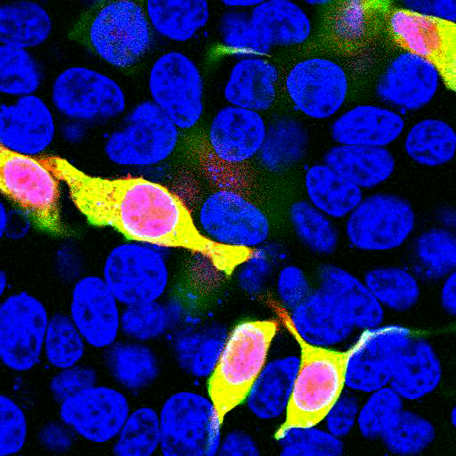
 IF (Immunofluorescence)
(Immunofluorescence analysis of GFP-RNF168 transgenic U2OS cells using PNKP antibody. Green:GFP-RNF168 fusion protein expression for DNA damage marker.Blue: DAPI for nuclear staining. RNF168(GFP) can be used to mark cells damaged by UV-A laser for they always gather around DNA damage region.)
IF (Immunofluorescence)
(Immunofluorescence analysis of GFP-RNF168 transgenic U2OS cells using PNKP antibody. Green:GFP-RNF168 fusion protein expression for DNA damage marker.Blue: DAPI for nuclear staining. RNF168(GFP) can be used to mark cells damaged by UV-A laser for they always gather around DNA damage region.)
PNKP, Polyclonal Antibody (Cat# AAA28091)
Full Name
PNKP Polyclonal Antibody
Gene Names
PNKP; PNK; MCSZ; EIEE10
Reactivity
Human, Mouse, Rat
Applications
Western Blot, Immunohistochemistry
Purity
Affinity Purification
Pricing


 IF (Immunofluorescence)
(Immunofluorescence analysis of A549 cells using MSH6 antibody.)
IF (Immunofluorescence)
(Immunofluorescence analysis of A549 cells using MSH6 antibody.)
MSH6, Polyclonal Antibody (Cat# AAA10686)
Full Name
MSH6 Polyclonal Antibody
Gene Names
MSH6; GTBP; HSAP; p160; GTMBP; HNPCC5
Reactivity
Human, Mouse, Monkey
Applications
Western Blot, Immunohistochemistry, Immunofluorescence
Purity
Affinity Purification
Pricing

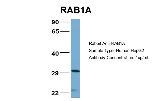


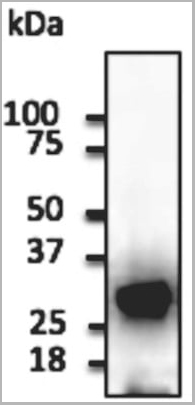
 WB (Western Blot)
(WB Suggested Anti-RAB1A Antibody Titration: 0.2-1 ug/mlELISA Titer: 1:312500Positive Control: Human Muscle)
WB (Western Blot)
(WB Suggested Anti-RAB1A Antibody Titration: 0.2-1 ug/mlELISA Titer: 1:312500Positive Control: Human Muscle)
RAB1A, Polyclonal Antibody (Cat# AAA23570)
Full Name
RAB1A antibody - middle region
Gene Names
RAB1A; RAB1; YPT1
Reactivity
Cow, Dog, Goat, Guinea Pig, Horse, Human, Mouse, Rabbit, Rat, Zebrafish
Applications
Immunohistochemistry, Western Blot
Purity
Affinity Purified
Pricing


 IP (Immunoprecipitation)
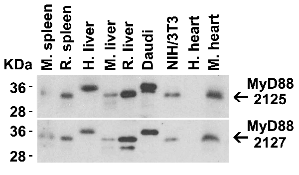
(Figure 13 Immunoprecipitation Validation in HEK293 cells (Kawai et al., 2004)HEK293 cells were transiently transfected with DYKDDDDK-IRF7. Ccell lysates were immunoprecipitated with control rabbit anti-mouse immunoglobulin serum (IgG) or anti-MyD88 (Ab1 and Ab2), followed by immunoblotting with anti-DYKDDDDK.)
IP (Immunoprecipitation)
(Figure 13 Immunoprecipitation Validation in HEK293 cells (Kawai et al., 2004)HEK293 cells were transiently transfected with DYKDDDDK-IRF7. Ccell lysates were immunoprecipitated with control rabbit anti-mouse immunoglobulin serum (IgG) or anti-MyD88 (Ab1 and Ab2), followed by immunoblotting with anti-DYKDDDDK.)
MYD88, Polyclonal Antibody (Cat# AAA10930)
Full Name
MYD88 Antibody
Gene Names
MYD88; MYD88D
Reactivity
Human, Mouse
Applications
Western Blot, Immunohistochemistry, Immunofluorescence
Purity
MYD88 Antibody is affinity chromatography purified via peptide column.
Pricing


 IP (Immunoprecipitation)
(Immunoprecipitating GFP in 293F whole cell lysate transfected with GFPLane 1: Mouse control IgG2b instead of AAA28064 in 293F whole cell lysate transfected with GFPLane 2: AAA28064 (4ug) + 293F whole cell lysate transfected with GFP (500ug)Lane 3: 293F whole cell lysate transfected with GFP (5ug)For western blotting, the blot was detected with AAA28064 at 1:2000, and a HRP-conjugated Protein G antibody was used as the secondary antibody at 1:50000)
IP (Immunoprecipitation)
(Immunoprecipitating GFP in 293F whole cell lysate transfected with GFPLane 1: Mouse control IgG2b instead of AAA28064 in 293F whole cell lysate transfected with GFPLane 2: AAA28064 (4ug) + 293F whole cell lysate transfected with GFP (500ug)Lane 3: 293F whole cell lysate transfected with GFP (5ug)For western blotting, the blot was detected with AAA28064 at 1:2000, and a HRP-conjugated Protein G antibody was used as the secondary antibody at 1:50000)
GFP, Monoclonal Antibody (Cat# AAA28064)
Full Name
GFP Monoclonal Antibody
Reactivity
All
Applications
Western Blot, Immunofluorescence, Flow Cytometry, Immunoprecipitation
Purity
>95%,Protein G purified
Pricing


 IF (Immunofluorescence)
(Immunofluorescence analysis of A549 cell using HMGB1 antibody. Blue: DAPI for nuclear staining.)
IF (Immunofluorescence)
(Immunofluorescence analysis of A549 cell using HMGB1 antibody. Blue: DAPI for nuclear staining.)
HMGB1, Polyclonal Antibody (Cat# AAA29657)
Full Name
HMGB1 antibody
Gene Names
HMGB1; HMG1; HMG3; HMG-1; SBP-1
Reactivity
Human, Mouse, Rat
Applications
Western Blot, Immunohistochemistry, Immunofluorescence
Purity
Antibodies were purified by affinity purification using immunogen.
Pricing


 IF (Immunofluorescence)
tdTomato, Polyclonal Antibody (Cat# AAA13876)
Full Name
anti-tdTomato
Reactivity
Reacts with Transfected cells proteins
Applications
Western Blot, Immunofluorescence, Immunohistochemistry, Immunohistochemistry
Purity
This antibody is epitope- affinity purified from goat antiserum.
Pricing


 Application Data
(Anti-mApple Ab at 1/2,500 dilution using HEK293 transfected cell lysates at 50 ug per lane; rabbit polyclonal to goat IgG (HRP) at 1/10,000 dilution;)
Application Data
(Anti-mApple Ab at 1/2,500 dilution using HEK293 transfected cell lysates at 50 ug per lane; rabbit polyclonal to goat IgG (HRP) at 1/10,000 dilution;)
mApple, Polyclonal Antibody (Cat# AAA13883)
Full Name
anti-mApple
Reactivity
Reacts with transfected cells proteins
Applications
Western Blot, Immunofluorescence, Immunohistochemistry, Immunohistochemistry, Immunoelectron Microscopy
Purity
This antibody is epitope-affinity purified from goat antiserum.
Pricing


 Application Data
(Published customer image: Histological staining of control and HBOT wounds at post-wounding days 7 and 29. A -D) H&E staining. E -H) CD34 immunohistochemistry. I+J) CD68 immunohistochemistry.From: uk B, Tong M, Fijneman EMG, van Neck JW (2014) Hyperbaric Oxygen Therapy to Treat Diabetes Impaired Wound Healing in Rats. PLoS ONE 9(10): e108533.)
Application Data
(Published customer image: Histological staining of control and HBOT wounds at post-wounding days 7 and 29. A -D) H&E staining. E -H) CD34 immunohistochemistry. I+J) CD68 immunohistochemistry.From: uk B, Tong M, Fijneman EMG, van Neck JW (2014) Hyperbaric Oxygen Therapy to Treat Diabetes Impaired Wound Healing in Rats. PLoS ONE 9(10): e108533.)
CD68, Monoclonal Antibody (Cat# AAA12151)
Full Name
MOUSE ANTI RAT CD68
Applications
Immunohistochemistry, Flow Cytometry, Immunofluorescence, Immunoprecipitation, Immunohistochemistry, Radioimmunoassay, Western Blot
Pricing


 Application Data
(Published customer image Infiltration of GFP+ BM-cells in infarct and peri-infarct regions. (A-B) Dot plots of viable macrophages/granulocytes (CD11b+CD45high, top right quadrants) and microglia (CD11b+CD45dim, bottom right quadrants) in cortex from BM-chimeric unmanipulated mice and mice exposed to pMCAO. (C) Bar graph showing mean numbers of CD11b+CD45dim microglia and CD11b+CD45high macrophages/granulocytes in BM-chimeric mice 24 hours after pMCAO, subdivided based on expression of GFP (n = 5). Approximately 92% of of the CD45high population were GFP+. (D) Estimation and comparison of mean numbers of CD11b+CD45dim microglia in non-chimeric (n = 10) versus BM-chimeric mice (n = 5) 24 hours after of pMCAO shows significantly fewer CD11b+CD45dim microglial cells in irradiated mice. (E) Overview, showing distribution of infiltrating GFP+ BM-derived cells into infarct (IF) and peri-infarct (P-IF) regions 24 hours after pMCAO. (E-G) By 24 hours, GFP+ single cells (F) and vessel-associated aggregates of GFP+ cells (arrows in G) were observed in infarct and peri-infarct regions. Some of the vessel-associated cells were round, leukocyte-like cells (arrows) while others were elongated cells lining the vasculature (arrow heads in G and in insert). (H) Bar graph showing mean numbers of single GFP+ cells and vessel-associated aggregates of GFP+ cells in ipsi- and contralateral cortex 24 hours after surgery (n = 10). (I-P) Immunohistochemical staining of CD45.1 (I, K), CD45.2 (J, L), IgG2a (M, O) and CD45 (N, P) in ischemic tissue in BM-chimeric (I, J, M, N) and non-chimeric mice (K, L, O, P) 24 hours after pMCAO. N.D, none detected. Scale bars: 200 um (A), 10 um (B, C). 50 um (I-P) *P < 0.05, **P < 0.01, and ***P < 0.001.From: Clausen BH, Lambertsen KL, Babcock AA, Holm TH, Dagnaes-Hansen F, Finsen B. Interleukin-1beta and tumor necrosis factor-alpha are expressed by different subsets of microglia and macrophages after ischemic stroke in mice. J Neuroinflammation. 2008 Oct 23;5:46.)
Application Data
(Published customer image Infiltration of GFP+ BM-cells in infarct and peri-infarct regions. (A-B) Dot plots of viable macrophages/granulocytes (CD11b+CD45high, top right quadrants) and microglia (CD11b+CD45dim, bottom right quadrants) in cortex from BM-chimeric unmanipulated mice and mice exposed to pMCAO. (C) Bar graph showing mean numbers of CD11b+CD45dim microglia and CD11b+CD45high macrophages/granulocytes in BM-chimeric mice 24 hours after pMCAO, subdivided based on expression of GFP (n = 5). Approximately 92% of of the CD45high population were GFP+. (D) Estimation and comparison of mean numbers of CD11b+CD45dim microglia in non-chimeric (n = 10) versus BM-chimeric mice (n = 5) 24 hours after of pMCAO shows significantly fewer CD11b+CD45dim microglial cells in irradiated mice. (E) Overview, showing distribution of infiltrating GFP+ BM-derived cells into infarct (IF) and peri-infarct (P-IF) regions 24 hours after pMCAO. (E-G) By 24 hours, GFP+ single cells (F) and vessel-associated aggregates of GFP+ cells (arrows in G) were observed in infarct and peri-infarct regions. Some of the vessel-associated cells were round, leukocyte-like cells (arrows) while others were elongated cells lining the vasculature (arrow heads in G and in insert). (H) Bar graph showing mean numbers of single GFP+ cells and vessel-associated aggregates of GFP+ cells in ipsi- and contralateral cortex 24 hours after surgery (n = 10). (I-P) Immunohistochemical staining of CD45.1 (I, K), CD45.2 (J, L), IgG2a (M, O) and CD45 (N, P) in ischemic tissue in BM-chimeric (I, J, M, N) and non-chimeric mice (K, L, O, P) 24 hours after pMCAO. N.D, none detected. Scale bars: 200 um (A), 10 um (B, C). 50 um (I-P) *P < 0.05, **P < 0.01, and ***P < 0.001.From: Clausen BH, Lambertsen KL, Babcock AA, Holm TH, Dagnaes-Hansen F, Finsen B. Interleukin-1beta and tumor necrosis factor-alpha are expressed by different subsets of microglia and macrophages after ischemic stroke in mice. J Neuroinflammation. 2008 Oct 23;5:46.)
CD11b, Monoclonal Antibody (Cat# AAA12186)
Full Name
RAT ANTI MOUSE CD11b:RPE
Gene Names
Itgam; CR3; CR3A; MAC1; Cd11b; Ly-40; Mac-1; Mac-1a; CD11b/CD18; F730045J24Rik
Applications
Flow Cytometry
Pricing


 Application Data
(Published customer image Infiltration of GFP+ BM-cells in infarct and peri-infarct regions. (A-B) Dot plots of viable macrophages/granulocytes (CD11b+CD45high, top right quadrants) and microglia (CD11b+CD45dim, bottom right quadrants) in cortex from BM-chimeric unmanipulated mice and mice exposed to pMCAO. (C) Bar graph showing mean numbers of CD11b+CD45dim microglia and CD11b+CD45high macrophages/granulocytes in BM-chimeric mice 24 hours after pMCAO, subdivided based on expression of GFP (n = 5). Approximately 92% of of the CD45high population were GFP+. (D) Estimation and comparison of mean numbers of CD11b+CD45dim microglia in non-chimeric (n = 10) versus BM-chimeric mice (n = 5) 24 hours after of pMCAO shows significantly fewer CD11b+CD45dim microglial cells in irradiated mice. (E) Overview, showing distribution of infiltrating GFP+ BM-derived cells into infarct (IF) and peri-infarct (P-IF) regions 24 hours after pMCAO. (E-G) By 24 hours, GFP+ single cells (F) and vessel-associated aggregates of GFP+ cells (arrows in G) were observed in infarct and peri-infarct regions. Some of the vessel-associated cells were round, leukocyte-like cells (arrows) while others were elongated cells lining the vasculature (arrow heads in G and in insert). (H) Bar graph showing mean numbers of single GFP+ cells and vessel-associated aggregates of GFP+ cells in ipsi- and contralateral cortex 24 hours after surgery (n = 10). (I-P) Immunohistochemical staining of CD45.1 (I, K), CD45.2 (J, L), IgG2a (M, O) and CD45 (N, P) in ischemic tissue in BM-chimeric (I, J, M, N) and non-chimeric mice (K, L, O, P) 24 hours after pMCAO. N.D, none detected. Scale bars: 200 um (A), 10 um (B, C). 50 um (I-P) *P < 0.05, **P < 0.01, and ***P < 0.001.From: Clausen BH, Lambertsen KL, Babcock AA, Holm TH, Dagnaes-Hansen F, Finsen B. Interleukin-1beta and tumor necrosis factor-alpha are expressed by different subsets of microglia and macrophages after ischemic stroke in mice. J Neuroinflammation. 2008 Oct 23;5:46.)
Application Data
(Published customer image Infiltration of GFP+ BM-cells in infarct and peri-infarct regions. (A-B) Dot plots of viable macrophages/granulocytes (CD11b+CD45high, top right quadrants) and microglia (CD11b+CD45dim, bottom right quadrants) in cortex from BM-chimeric unmanipulated mice and mice exposed to pMCAO. (C) Bar graph showing mean numbers of CD11b+CD45dim microglia and CD11b+CD45high macrophages/granulocytes in BM-chimeric mice 24 hours after pMCAO, subdivided based on expression of GFP (n = 5). Approximately 92% of of the CD45high population were GFP+. (D) Estimation and comparison of mean numbers of CD11b+CD45dim microglia in non-chimeric (n = 10) versus BM-chimeric mice (n = 5) 24 hours after of pMCAO shows significantly fewer CD11b+CD45dim microglial cells in irradiated mice. (E) Overview, showing distribution of infiltrating GFP+ BM-derived cells into infarct (IF) and peri-infarct (P-IF) regions 24 hours after pMCAO. (E-G) By 24 hours, GFP+ single cells (F) and vessel-associated aggregates of GFP+ cells (arrows in G) were observed in infarct and peri-infarct regions. Some of the vessel-associated cells were round, leukocyte-like cells (arrows) while others were elongated cells lining the vasculature (arrow heads in G and in insert). (H) Bar graph showing mean numbers of single GFP+ cells and vessel-associated aggregates of GFP+ cells in ipsi- and contralateral cortex 24 hours after surgery (n = 10). (I-P) Immunohistochemical staining of CD45.1 (I, K), CD45.2 (J, L), IgG2a (M, O) and CD45 (N, P) in ischemic tissue in BM-chimeric (I, J, M, N) and non-chimeric mice (K, L, O, P) 24 hours after pMCAO. N.D, none detected. Scale bars: 200 um (A), 10 um (B, C). 50 um (I-P) *P < 0.05, **P < 0.01, and ***P < 0.001.From: Clausen BH, Lambertsen KL, Babcock AA, Holm TH, Dagnaes-Hansen F, Finsen B. Interleukin-1beta and tumor necrosis factor-alpha are expressed by different subsets of microglia and macrophages after ischemic stroke in mice. J Neuroinflammation. 2008 Oct 23;5:46.)
CD11b, Monoclonal Antibody (Cat# AAA12231)
Full Name
RAT ANTI MOUSE CD11b:Low Endotoxin
Gene Names
Itgam; CR3; CR3A; MAC1; Cd11b; Ly-40; Mac-1; Mac-1a; CD11b/CD18; F730045J24Rik
Applications
Immunohistochemistry, Flow Cytometry, Functional Assay, Immunofluorescence, Immunoprecipitation
Pricing


 Application Data
(Published customer image Infiltration of GFP+ BM-cells in infarct and peri-infarct regions. (A-B) Dot plots of viable macrophages/granulocytes (CD11b+CD45high, top right quadrants) and microglia (CD11b+CD45dim, bottom right quadrants) in cortex from BM-chimeric unmanipulated mice and mice exposed to pMCAO. (C) Bar graph showing mean numbers of CD11b+CD45dim microglia and CD11b+CD45high macrophages/granulocytes in BM-chimeric mice 24 hours after pMCAO, subdivided based on expression of GFP (n = 5). Approximately 92% of of the CD45high population were GFP+. (D) Estimation and comparison of mean numbers of CD11b+CD45dim microglia in non-chimeric (n = 10) versus BM-chimeric mice (n = 5) 24 hours after of pMCAO shows significantly fewer CD11b+CD45dim microglial cells in irradiated mice. (E) Overview, showing distribution of infiltrating GFP+ BM-derived cells into infarct (IF) and peri-infarct (P-IF) regions 24 hours after pMCAO. (E-G) By 24 hours, GFP+ single cells (F) and vessel-associated aggregates of GFP+ cells (arrows in G) were observed in infarct and peri-infarct regions. Some of the vessel-associated cells were round, leukocyte-like cells (arrows) while others were elongated cells lining the vasculature (arrow heads in G and in insert). (H) Bar graph showing mean numbers of single GFP+ cells and vessel-associated aggregates of GFP+ cells in ipsi- and contralateral cortex 24 hours after surgery (n = 10). (I-P) Immunohistochemical staining of CD45.1 (I, K), CD45.2 (J, L), IgG2a (M, O) and CD45 (N, P) in ischemic tissue in BM-chimeric (I, J, M, N) and non-chimeric mice (K, L, O, P) 24 hours after pMCAO. N.D, none detected. Scale bars: 200 um (A), 10 um (B, C). 50 um (I-P) *P < 0.05, **P < 0.01, and ***P < 0.001.From: Clausen BH, Lambertsen KL, Babcock AA, Holm TH, Dagnaes-Hansen F, Finsen B. Interleukin-1beta and tumor necrosis factor-alpha are expressed by different subsets of microglia and macrophages after ischemic stroke in mice. J Neuroinflammation. 2008 Oct 23;5:46.)
Application Data
(Published customer image Infiltration of GFP+ BM-cells in infarct and peri-infarct regions. (A-B) Dot plots of viable macrophages/granulocytes (CD11b+CD45high, top right quadrants) and microglia (CD11b+CD45dim, bottom right quadrants) in cortex from BM-chimeric unmanipulated mice and mice exposed to pMCAO. (C) Bar graph showing mean numbers of CD11b+CD45dim microglia and CD11b+CD45high macrophages/granulocytes in BM-chimeric mice 24 hours after pMCAO, subdivided based on expression of GFP (n = 5). Approximately 92% of of the CD45high population were GFP+. (D) Estimation and comparison of mean numbers of CD11b+CD45dim microglia in non-chimeric (n = 10) versus BM-chimeric mice (n = 5) 24 hours after of pMCAO shows significantly fewer CD11b+CD45dim microglial cells in irradiated mice. (E) Overview, showing distribution of infiltrating GFP+ BM-derived cells into infarct (IF) and peri-infarct (P-IF) regions 24 hours after pMCAO. (E-G) By 24 hours, GFP+ single cells (F) and vessel-associated aggregates of GFP+ cells (arrows in G) were observed in infarct and peri-infarct regions. Some of the vessel-associated cells were round, leukocyte-like cells (arrows) while others were elongated cells lining the vasculature (arrow heads in G and in insert). (H) Bar graph showing mean numbers of single GFP+ cells and vessel-associated aggregates of GFP+ cells in ipsi- and contralateral cortex 24 hours after surgery (n = 10). (I-P) Immunohistochemical staining of CD45.1 (I, K), CD45.2 (J, L), IgG2a (M, O) and CD45 (N, P) in ischemic tissue in BM-chimeric (I, J, M, N) and non-chimeric mice (K, L, O, P) 24 hours after pMCAO. N.D, none detected. Scale bars: 200 um (A), 10 um (B, C). 50 um (I-P) *P < 0.05, **P < 0.01, and ***P < 0.001.From: Clausen BH, Lambertsen KL, Babcock AA, Holm TH, Dagnaes-Hansen F, Finsen B. Interleukin-1beta and tumor necrosis factor-alpha are expressed by different subsets of microglia and macrophages after ischemic stroke in mice. J Neuroinflammation. 2008 Oct 23;5:46.)
CD11b, Monoclonal Antibody (Cat# AAA12181)
Full Name
RAT ANTI MOUSE CD11b
Gene Names
Itgam; CR3; CR3A; MAC1; Cd11b; Ly-40; Mac-1; Mac-1a; CD11b/CD18; F730045J24Rik
Reactivity
Human
Applications
Immunohistochemistry, Flow Cytometry, Immunofluorescence, Immunoprecipitation
Pricing


 Application Data
(Published customer image: Histological staining of control and HBOT wounds at post-wounding days 7 and 29. A -D) H&E staining. E -H) CD34 immunohistochemistry. I+J) CD68 immunohistochemistry.From: uk B, Tong M, Fijneman EMG, van Neck JW (2014) Hyperbaric Oxygen Therapy to Treat Diabetes Impaired Wound Healing in Rats. PLoS ONE 9(10): e108533.)
Application Data
(Published customer image: Histological staining of control and HBOT wounds at post-wounding days 7 and 29. A -D) H&E staining. E -H) CD34 immunohistochemistry. I+J) CD68 immunohistochemistry.From: uk B, Tong M, Fijneman EMG, van Neck JW (2014) Hyperbaric Oxygen Therapy to Treat Diabetes Impaired Wound Healing in Rats. PLoS ONE 9(10): e108533.)
CD68, Monoclonal Antibody (Cat# AAA12148)
Full Name
MOUSE ANTI RAT CD68:FITC
Applications
Flow Cytometry
Pricing


 IF (Immunofluorescence)
(Immunofluorescence analysis of GFP-RNF168 transgenic U2OS cells using TP53BP1 antibody. Green:GFP-RNF168 fusion protein expression for DNA damage marker.Blue: DAPI for nuclear staining.RNF168(GFP) can be used to mark cells damaged by UV-A laser for they always gather around DNA damage region.)
IF (Immunofluorescence)
(Immunofluorescence analysis of GFP-RNF168 transgenic U2OS cells using TP53BP1 antibody. Green:GFP-RNF168 fusion protein expression for DNA damage marker.Blue: DAPI for nuclear staining.RNF168(GFP) can be used to mark cells damaged by UV-A laser for they always gather around DNA damage region.)
TP53BP1, Polyclonal Antibody (Cat# AAA28106)
Full Name
TP53BP1 Polyclonal Antibody
Gene Names
TP53BP1; p202; 53BP1
Reactivity
Human, Mouse, Rat
Applications
Western Blot, Immunohistochemistry, Immunofluorescence
Purity
Affinity Purification
Pricing


 IF (Immunofluorescence)
(Immunofluorescence - anti-mScarlet Ab using hCEC cells transduced with mScarlet-Rab5a; cells were fixed with methanol and anti-mScarlet at 1/250;)
IF (Immunofluorescence)
(Immunofluorescence - anti-mScarlet Ab using hCEC cells transduced with mScarlet-Rab5a; cells were fixed with methanol and anti-mScarlet at 1/250;)
mScarlet, Polyclonal Antibody (Cat# AAA13885)
Full Name
anti-mScarlet
Reactivity
Reacts with Transfected cells proteins
Applications
Western Blot, Immunofluorescence, Immunohistochemistry, Immunohistochemistry, Immunoelectron Microscopy
Purity
This antibody is epitope-affinity purified from goat antiserum.
Pricing

 IP (Immunoprecipitation)
(IP:Result of anti-GFP-tag Monoclonal antibodyLine 1:Control IgGLine 2:Precipitateing GFP-tagged fusion proteinLine 3:GFP Transfected HEK-293 cells lysateSecondaryGoat polyclonal to Mouse IgG at 1/5000 dilutionPredicted band size: 30kdObserved band size: 30kd)
IP (Immunoprecipitation)
(IP:Result of anti-GFP-tag Monoclonal antibodyLine 1:Control IgGLine 2:Precipitateing GFP-tagged fusion proteinLine 3:GFP Transfected HEK-293 cells lysateSecondaryGoat polyclonal to Mouse IgG at 1/5000 dilutionPredicted band size: 30kdObserved band size: 30kd)
GFP, Monoclonal Antibody (Cat# AAA18855)
Full Name
Mouse Anti-GFP monoclonal Antibody
Applications
EIA, WB, IP
Purity
>95%, Protein G Purified
Pricing


 Application Data
(Source:Arthrobacter luteus)
Application Data
(Source:Arthrobacter luteus)
Zymolyase 20T, Enzyme (Cat# AAA14844)
Full Name
Zymolyase 20T (Lyticase, Yeast Lytic Enzyme)
Pricing
 FCM (Flow Cytometry)
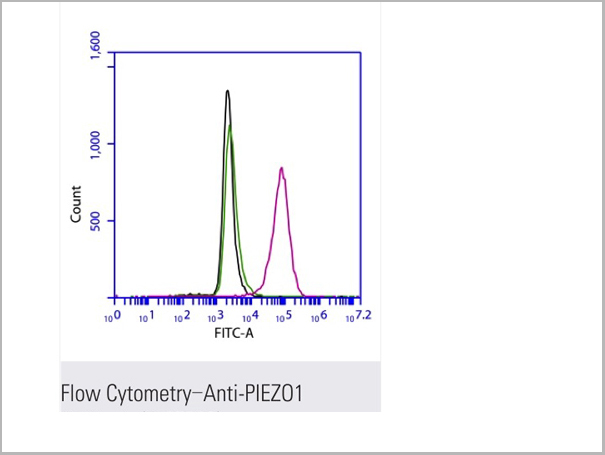
(Flow cytometric analysis of 2% paraformaldehyde-fixed THP1 (Human acute monocytic leukemia cell line) cells labeling PIEZO1 with AAA13800 at 1/200 dilution (red) compared with a mouse monoclonal IgG isotype control (black) and an unlabelled control (cells without incubation with primary antibody, green). Goat anti-mouse IgG (FITC) at 1/300 dilution was used as the secondary antibody.)
FCM (Flow Cytometry)
(Flow cytometric analysis of 2% paraformaldehyde-fixed THP1 (Human acute monocytic leukemia cell line) cells labeling PIEZO1 with AAA13800 at 1/200 dilution (red) compared with a mouse monoclonal IgG isotype control (black) and an unlabelled control (cells without incubation with primary antibody, green). Goat anti-mouse IgG (FITC) at 1/300 dilution was used as the secondary antibody.)
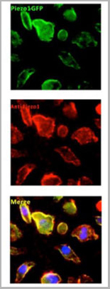
PIEZO1, Monoclonal Antibody (Cat# AAA13800)
Full Name
PIEZO1 Mouse mAb
Gene Names
PIEZO1; DHS; Mib; LMPH3; FAM38A
Reactivity
Homo Sapiens
Applications
Immunoprecipitation, Immunofluorescence, Flow Cytometry
Pricing
 Application Data
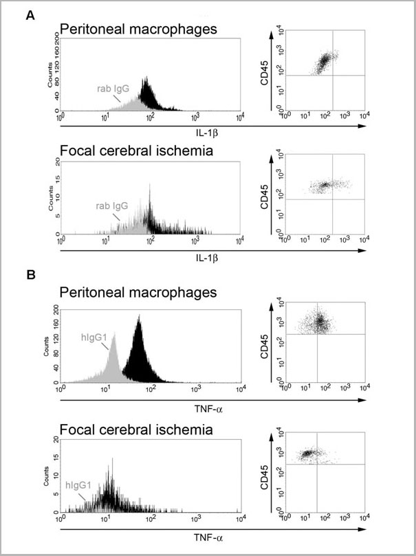
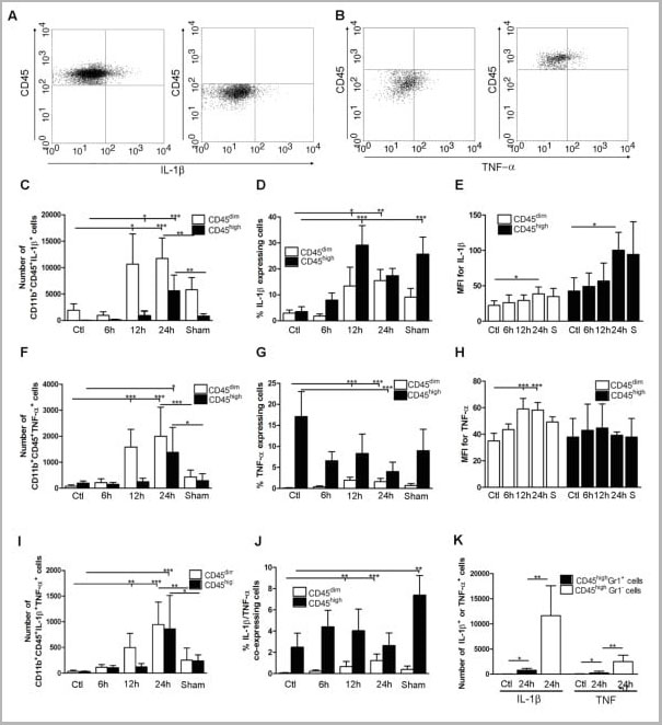
(Published customer image: Cytokine expression in segregated populations of cells following stroke. (A, B) Dot plots showing CD11b+CD45high macrophages/granulocytes (upper right quadrants) and CD11b+CD45dim microglia (bottom right quadrants) expressing IL-1beta (A) or TNF-a (B). (C-J) Bar graphs showing numbers and proportions of IL-1beta (C, D), TNF-a (F, G) and IL-1beta/TNF-a co-expressing (I, J) CD11b+CD45dim microglia and CD11b+CD45high macrophages/granulocytes in unmanipulated control mice (n = 10), in mice 6 (n = 7), 12 (n = 7), or 24 hours after pMCAO (n = 10), and in sham-operated mice 24 hours after pMCAO (n = 7). (E, H) Comparison of the MFI values for IL-1beta (E) and TNF-a (H) in viable CD11b+CD45dim microglia and CD11b+CD45high macrophages/granulocytes in unmanipulated mice, in mice 6, 12, or 24 hours after pMCAO, and in sham-operated mice 24 hours after pMCAO. Macrophages/granulocytes express significantly more IL-1beta than do microglial in unmanipulated mice, in mice 6, 12, or 24 hours after pMCAO, and in sham-operated mice 24 hours after pMCAO (E), whereas microglial cells express significantly higher levels of TNF-a than do macrophages/granulocytes at 12 h and 24 hours, and in sham-operated mice 24 hours after pMCAO (H). (K) CD11b+CD45highGr1- macrophages and not CD11b+CD45highGr1+ granulocytes are the main producers of IL-1beta and TNF-a 24 hours after pMCAO. *P < 0.05, **P < 0.01, and ***P < 0.001.From: http://www.jneuroinflammation.com/content/5/1/46.)
Application Data
(Published customer image: Cytokine expression in segregated populations of cells following stroke. (A, B) Dot plots showing CD11b+CD45high macrophages/granulocytes (upper right quadrants) and CD11b+CD45dim microglia (bottom right quadrants) expressing IL-1beta (A) or TNF-a (B). (C-J) Bar graphs showing numbers and proportions of IL-1beta (C, D), TNF-a (F, G) and IL-1beta/TNF-a co-expressing (I, J) CD11b+CD45dim microglia and CD11b+CD45high macrophages/granulocytes in unmanipulated control mice (n = 10), in mice 6 (n = 7), 12 (n = 7), or 24 hours after pMCAO (n = 10), and in sham-operated mice 24 hours after pMCAO (n = 7). (E, H) Comparison of the MFI values for IL-1beta (E) and TNF-a (H) in viable CD11b+CD45dim microglia and CD11b+CD45high macrophages/granulocytes in unmanipulated mice, in mice 6, 12, or 24 hours after pMCAO, and in sham-operated mice 24 hours after pMCAO. Macrophages/granulocytes express significantly more IL-1beta than do microglial in unmanipulated mice, in mice 6, 12, or 24 hours after pMCAO, and in sham-operated mice 24 hours after pMCAO (E), whereas microglial cells express significantly higher levels of TNF-a than do macrophages/granulocytes at 12 h and 24 hours, and in sham-operated mice 24 hours after pMCAO (H). (K) CD11b+CD45highGr1- macrophages and not CD11b+CD45highGr1+ granulocytes are the main producers of IL-1beta and TNF-a 24 hours after pMCAO. *P < 0.05, **P < 0.01, and ***P < 0.001.From: http://www.jneuroinflammation.com/content/5/1/46.)
CD45, Monoclonal Antibody (Cat# AAA11896)
Full Name
RAT ANTI MOUSE CD45
Gene Names
Ptprc; loc; B220; Cd45; L-CA; Ly-5; T200; CD45R; Lyt-4
Applications
Immunohistochemistry, Flow Cytometry, Immunofluorescence, Immunoprecipitation
Pricing
 WB (Western Blot)
(Western BlotPositive WB detected in Recombinant proteinAll lanes: ftnA antibody at 1:2000SecondaryGoat polyclonal to rabbit IgG at 1/50000 dilutionPredicted band size: 24.8 kDaObserved band size: 30 kDa)
WB (Western Blot)
(Western BlotPositive WB detected in Recombinant proteinAll lanes: ftnA antibody at 1:2000SecondaryGoat polyclonal to rabbit IgG at 1/50000 dilutionPredicted band size: 24.8 kDaObserved band size: 30 kDa)
ftnA, Polyclonal Antibody (Cat# AAA27053)
Full Name
Rabbit anti-Helicobacter pylori (strain 700392/26695)(Campylobacter pylori) ftnA Polyclonal antibody
Reactivity
Helicobacter pylori
Applications
Western Blot
Purity
Protein A/G
Pricing
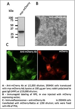
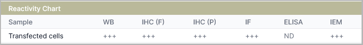
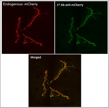 IF (Immunofluorescence)
(Immunofluorescence in Drosophila larvae NMJ muscle 6/7 expressing mCherry in neurons (DyGlutmCherry) using 1st Ab anti-mCherry at 1/1,000 and 2nd Ab anti-goat IgY conjugated to DyLight®488 at 1/500;)
IF (Immunofluorescence)
(Immunofluorescence in Drosophila larvae NMJ muscle 6/7 expressing mCherry in neurons (DyGlutmCherry) using 1st Ab anti-mCherry at 1/1,000 and 2nd Ab anti-goat IgY conjugated to DyLight®488 at 1/500;)
mCherry, Polyclonal Antibody (Cat# AAA13871)
Full Name
mCherry Polyclonal Antibody
Reactivity
Reacts with Transfected cells proteins
Applications
Western Blot, Immunofluorescence, Immunohistochemistry, Immunohistochemistry
Pricing
 Application Data
(Western blot analysis of recombinant EYFP protein with AAA27910 diluted at 1:10,000.)
Application Data
(Western blot analysis of recombinant EYFP protein with AAA27910 diluted at 1:10,000.)
EYFP, Monoclonal Antibody (Cat# AAA27910)
Full Name
EYFP Mouse Monoclonal Antibody (Mix-mA)
Applications
Western Blot, Immunohistochemistry, Immunoprecipitation
Pricing
 WB (Western Blot)
(Positive WB detected in Recombinant proteinAll lanes: gB antibody at 1:2000SecondaryGoat polyclonal to rabbit IgG at 1/50000 dilutionPredicted band size: 43.3 kDaObserved band size: 46 kDa)
WB (Western Blot)
(Positive WB detected in Recombinant proteinAll lanes: gB antibody at 1:2000SecondaryGoat polyclonal to rabbit IgG at 1/50000 dilutionPredicted band size: 43.3 kDaObserved band size: 46 kDa)
gB, Polyclonal Antibody (Cat# AAA27058)
Full Name
Rabbit anti-Epstein-Barr virus (strain B95-8)(HHV-4)(Human herpesvirus 4) gB Polyclonal Antibody
Reactivity
Epstein-Barr virus
Applications
Western Blot
Purity
Protein G
Pricing
 WB (Western Blot)
(Western Blot: The cell lysate of T293 with over-expressed recombinant protein GFP was resolved onto 10% SDS-PAGE, then transferred onto NC membrane. Followed by an immune-blotting with Rabbit anti- GFP (AAA14083) at 1:1000.)
WB (Western Blot)
(Western Blot: The cell lysate of T293 with over-expressed recombinant protein GFP was resolved onto 10% SDS-PAGE, then transferred onto NC membrane. Followed by an immune-blotting with Rabbit anti- GFP (AAA14083) at 1:1000.)
GFP, Antibody (Cat# AAA14083)
Full Name
Rabbit anti GFP Polyclonal antibody
Reactivity
Tag Antibody
Applications
Western Blot, Immunoprecipitation
Purity
The Rabbit IgG is purified by Affinity Chromatography.
Pricing
 WB (Western Blot)
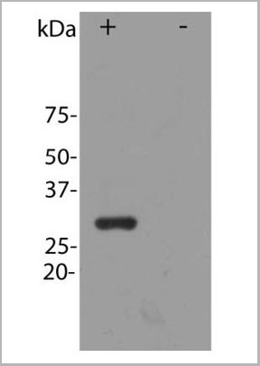
(Image: Blot of HEK293 cells transfected with pFin-EF1-mCherry vector, in the lane marked "+". HEK293 cells which were not transfected with this vector show no protein band in lane marked)
WB (Western Blot)
(Image: Blot of HEK293 cells transfected with pFin-EF1-mCherry vector, in the lane marked "+". HEK293 cells which were not transfected with this vector show no protein band in lane marked)
mCherry, Antibody (Cat# AAA14440)
Full Name
mCherry, Affinity Purified Antibody, Rabbit
Reactivity
All mammalian
Applications
Immunofluorescence, Western Blot
Pricing
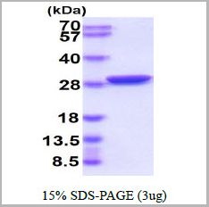 SDS-PAGE
(3ug by SDS-PAGE under reducing condition and visualized by coomassie blue stain.)
SDS-PAGE
(3ug by SDS-PAGE under reducing condition and visualized by coomassie blue stain.)
GFP, Recombinant Protein (Cat# AAA11761)
Full Name
GFP, 1-238aa, Aequorea victoria, E Coli
Applications
SDS-PAGE
Purity
> 95% by SDS-PAGE
Pricing
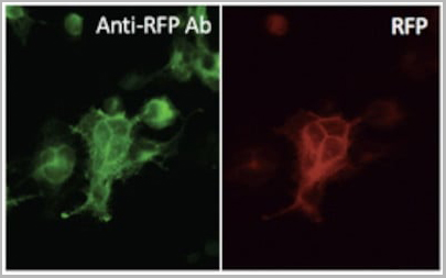
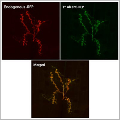
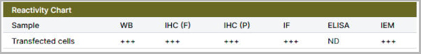
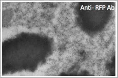 IHC (Immunohistochemistry)
(Immunogold labeling of epithelium cells, in vivo injected with RFP expressing vector;)
IHC (Immunohistochemistry)
(Immunogold labeling of epithelium cells, in vivo injected with RFP expressing vector;)
RFP, Polyclonal Antibody (Cat# AAA13877)
Full Name
RFP Polyclonal Antibody
Reactivity
Reacts with Transfected cells proteins.
Applications
Western Blot, Immunofluorescence, Immunohistochemistry
Purity
This antibody is epitope-affinity purified from goat antiserum.
Pricing
 WB (Western Blot)
(Western BlotPositive WB detected in: recombinant proteinAll lanes: psaA Antibody at 1:1000SecondaryGoat polyclonal to rabbit IgG at 1/50000 dilutionPredicted band size: 42 kDaObserved band size: 42 kDa)
WB (Western Blot)
(Western BlotPositive WB detected in: recombinant proteinAll lanes: psaA Antibody at 1:1000SecondaryGoat polyclonal to rabbit IgG at 1/50000 dilutionPredicted band size: 42 kDaObserved band size: 42 kDa)
psaA, Polyclonal Antibody (Cat# AAA27055)
Full Name
psaA Antibody
Reactivity
Streptococcus pneumoniae
Applications
Western Blot
Purity
Affinity-chromatography
Pricing
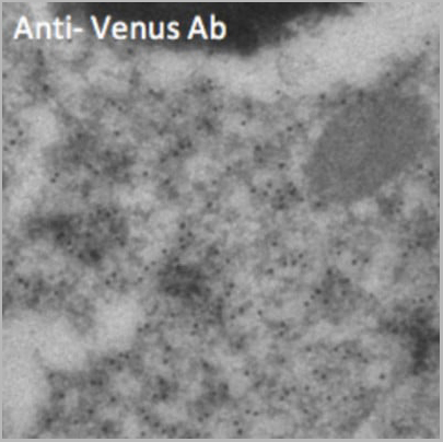
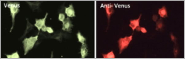

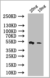
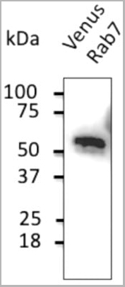 WB (Western Blot)
(Anti-Venus Ab at 1/2,000 dilution; 293HEK cells transduced with Venus-Rab7 Ad; lysates at 50 µg per lane; rabbit polyclonal to goat IgG (HRP) at 1/10,000 dilution;)
WB (Western Blot)
(Anti-Venus Ab at 1/2,000 dilution; 293HEK cells transduced with Venus-Rab7 Ad; lysates at 50 µg per lane; rabbit polyclonal to goat IgG (HRP) at 1/10,000 dilution;)
Venus, Polyclonal Antibody (Cat# AAA13879)
Full Name
Venus Polyclonal Antibody
Reactivity
Transfected cells proteins
Applications
Western Blot, Immunofluorescence, Immunohistochemistry, Immunohistochemistry
Pricing
 WB (Western Blot)
(Positive WB detected in Recombinant proteinAll lanes: BZLF1 antibody at 1:1000SecondaryGoat polyclonal to rabbit IgG at 1/50000 dilutionPredicted band size: 46 kDaObserved band size: 50 kDa)
WB (Western Blot)
(Positive WB detected in Recombinant proteinAll lanes: BZLF1 antibody at 1:1000SecondaryGoat polyclonal to rabbit IgG at 1/50000 dilutionPredicted band size: 46 kDaObserved band size: 50 kDa)
BZLF1, Polyclonal Antibody (Cat# AAA27059)
Full Name
Rabbit anti-Epstein-Barr virus (strain B95-8)(HHV-4)(Human herpesvirus 4) BZLF1 Polyclonal Antibody
Reactivity
Epstein-Barr virus
Applications
Western Blot
Purity
> 95%, Protein G purified
Pricing


 Application Data
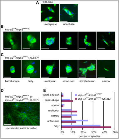
(Published customer image: Spindle abnormalities in embryos derived from imp-a2D14/imp-betaKetRE34 and imp-a2D14/imp-betac02473; NLSB-/+ females. (A -D) Wild-type and mutant embryos stained for a-tubulin (green) and DNA (blue). (A) Mitotic spindles in wild-type embryos at metaphase and anaphase. (B, C) Categories of spindle abnormalities found in embryos derived from (B) imp-a2D14/imp-betaKetRE34 and (C) imp-a2D14/imp-betac02743; NLSB-/+ females. (D) Formation of aster networks found in both genotypes. Scale bar: 10 um. (E) Frequency of spindle defects in embryos from both types of mutant females. Female genotypes are displayed at the upper right corner. At least 200 spindles were scored for both genotypes.From: Specific Cooperation Between Imp-a2 and Imp-beta/Ketel in Spindle Assembly During Drosophila Early Nuclear Divisions Erika Vir¡gh, M¡ty¡s Gorj¡n¡cz, Istv¡n T¶r¶k, Tolga Eichhorn, Sowjanya Kallakuri, Tam¡s Szlanka, Istv¡n Kiss, and Bernard M. Mechler G3 January 2012 2:1-14.)
Application Data
(Published customer image: Spindle abnormalities in embryos derived from imp-a2D14/imp-betaKetRE34 and imp-a2D14/imp-betac02473; NLSB-/+ females. (A -D) Wild-type and mutant embryos stained for a-tubulin (green) and DNA (blue). (A) Mitotic spindles in wild-type embryos at metaphase and anaphase. (B, C) Categories of spindle abnormalities found in embryos derived from (B) imp-a2D14/imp-betaKetRE34 and (C) imp-a2D14/imp-betac02743; NLSB-/+ females. (D) Formation of aster networks found in both genotypes. Scale bar: 10 um. (E) Frequency of spindle defects in embryos from both types of mutant females. Female genotypes are displayed at the upper right corner. At least 200 spindles were scored for both genotypes.From: Specific Cooperation Between Imp-a2 and Imp-beta/Ketel in Spindle Assembly During Drosophila Early Nuclear Divisions Erika Vir¡gh, M¡ty¡s Gorj¡n¡cz, Istv¡n T¶r¶k, Tolga Eichhorn, Sowjanya Kallakuri, Tam¡s Szlanka, Istv¡n Kiss, and Bernard M. Mechler G3 January 2012 2:1-14.)
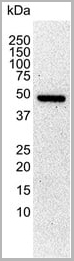
TUBULIN ALPHA, Monoclonal Antibody (Cat# AAA12232)
Full Name
RAT ANTI TUBULIN ALPHA:HRP
Applications
Immunohistochemistry, Western Blot
Pricing


 Application Data
(Published customer image: Spindle abnormalities in embryos derived from imp-a2D14/imp-betaKetRE34 and imp-a2D14/imp-betac02473; NLSB-/+ females. (A -D) Wild-type and mutant embryos stained for a-tubulin (green) and DNA (blue). (A) Mitotic spindles in wild-type embryos at metaphase and anaphase. (B, C) Categories of spindle abnormalities found in embryos derived from (B) imp-a2D14/imp-betaKetRE34 and (C) imp-a2D14/imp-betac02743; NLSB-/+ females. (D) Formation of aster networks found in both genotypes. Scale bar: 10 um. (E) Frequency of spindle defects in embryos from both types of mutant females. Female genotypes are displayed at the upper right corner. At least 200 spindles were scored for both genotypes.From: Specific Cooperation Between Imp-a2 and Imp-beta/Ketel in Spindle Assembly During Drosophila Early Nuclear Divisions Erika Vir¡gh, M¡ty¡s Gorj¡n¡cz, Istv¡n T¶r¶k, Tolga Eichhorn, Sowjanya Kallakuri, Tam¡s Szlanka, Istv¡n Kiss, and Bernard M. Mechler G3 January 2012 2:1-14.)
Application Data
(Published customer image: Spindle abnormalities in embryos derived from imp-a2D14/imp-betaKetRE34 and imp-a2D14/imp-betac02473; NLSB-/+ females. (A -D) Wild-type and mutant embryos stained for a-tubulin (green) and DNA (blue). (A) Mitotic spindles in wild-type embryos at metaphase and anaphase. (B, C) Categories of spindle abnormalities found in embryos derived from (B) imp-a2D14/imp-betaKetRE34 and (C) imp-a2D14/imp-betac02743; NLSB-/+ females. (D) Formation of aster networks found in both genotypes. Scale bar: 10 um. (E) Frequency of spindle defects in embryos from both types of mutant females. Female genotypes are displayed at the upper right corner. At least 200 spindles were scored for both genotypes.From: Specific Cooperation Between Imp-a2 and Imp-beta/Ketel in Spindle Assembly During Drosophila Early Nuclear Divisions Erika Vir¡gh, M¡ty¡s Gorj¡n¡cz, Istv¡n T¶r¶k, Tolga Eichhorn, Sowjanya Kallakuri, Tam¡s Szlanka, Istv¡n Kiss, and Bernard M. Mechler G3 January 2012 2:1-14.)
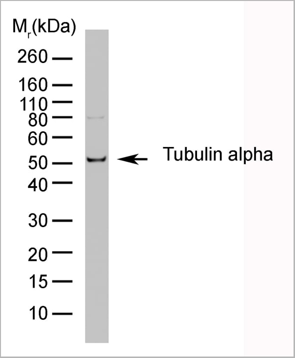
TUBULIN ALPHA, Monoclonal Antibody (Cat# AAA12009)
Full Name
RAT ANTI TUBULIN ALPHA
Applications
Immunohistochemistry, Immunofluorescence, Immunoprecipitation, Radioimmunoassay, Western Blot
Pricing
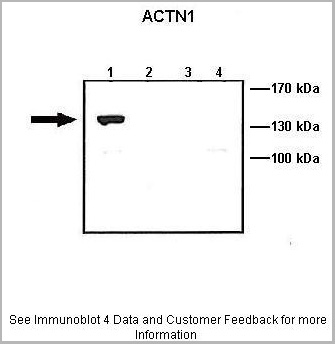
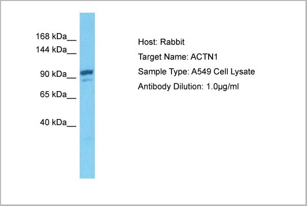
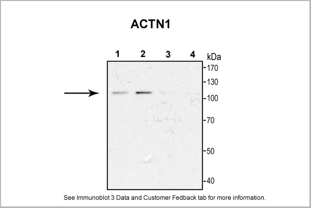
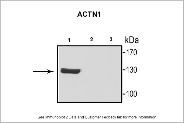
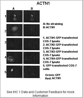
 WB (Western Blot)
(WB Suggested Anti-ACTN1 Antibody Titration: 0.2-1 ug/mlELISA Titer: 1:312500Positive Control: 293T cell lysate)
WB (Western Blot)
(WB Suggested Anti-ACTN1 Antibody Titration: 0.2-1 ug/mlELISA Titer: 1:312500Positive Control: 293T cell lysate)
ACTN1, Polyclonal Antibody (Cat# AAA23579)
Full Name
ACTN1 antibody - N-terminal region
Gene Names
ACTN1; BDPLT15
Reactivity
Cow, Dog, Guinea Pig, Horse, Human, Mouse, Rabbit, Rat, Zebrafish
Applications
Immunohistochemistry, Western Blot
Purity
Affinity Purified
Pricing
 WB (Western Blot)
(Western BlotPositive WB detected in: recombinant proteinAll lanes: csgA Antibody at 1:1000SecondaryGoat polyclonal to rabbit IgG at 1/50000 dilutionPredicted band size: 17 kDaObserved band size: 17 kDa)
WB (Western Blot)
(Western BlotPositive WB detected in: recombinant proteinAll lanes: csgA Antibody at 1:1000SecondaryGoat polyclonal to rabbit IgG at 1/50000 dilutionPredicted band size: 17 kDaObserved band size: 17 kDa)
csgA, Polyclonal Antibody (Cat# AAA27056)
Full Name
csgA Antibody
Gene Names
csgA; agfA; ECK1028; JW1025
Reactivity
E Coli (strain K12)
Applications
Western Blot
Purity
Affinity-chromatography
Pricing
 WB (Western Blot)
(Positive WB detected in Recombinant proteinAll lanes: L1R antibody at 1:2000Secondary Goat polyclonal to rabbit IgG at 1/50000 dilutionPredicted band size: 26.8 kDabserved band size: 29 kDa)
WB (Western Blot)
(Positive WB detected in Recombinant proteinAll lanes: L1R antibody at 1:2000Secondary Goat polyclonal to rabbit IgG at 1/50000 dilutionPredicted band size: 26.8 kDabserved band size: 29 kDa)
L1R, Polyclonal Antibody (Cat# AAA27054)
Full Name
Rabbit anti-Vaccinia virus (strain Copenhagen)(VACV) L1R Polyclonal Antibody
Reactivity
Vaccinia virus
Applications
Western Blot
Purity
Protein G
Pricing

 Application Data
(The extracellular domain is required for Sln1 kinase activation by wall perturbations. A, representative field showing localization of SLN1-GFP (pSS1881) or sln1ΔECD-GFP (pSS1904). BF, bright field. B, Western analysis showing reduced levels of sln1ΔECD compared with full-length SLN1. SLN1, JF2007 (sln1Δ) carrying full-length SLN1-myc (pCLM994); sln1ΔECD, JF2007 carrying sln1ΔECD-myc (pCLM1774). C, comparison of Hog1 phosphorylation kinetics in SLN1 (JF2008 (sln1Δ sho1Δ) carrying full-length (pCLM994)) and sln1ΔECD (JF2008 (sln1Δ sho1Δ) carrying sln1ΔECD (pCLM1774)) strains following addition of 0.4 M NaCl. D, Northern (RNA) hybridization analysis of RNA prepared from wild type cultures from the sln1Δ strain, JF2007, or sln1Δ ccw12Δ strain, JF2368, transformed with the SLN1 expression plasmids, pCLM994 (SLN1) or pCLM1774 (sln1ΔECD), and treated with 1 unit/ml zymolyase (+) where indicated. NCA3 expression was normalized to expression of the SLN1-SKN7-independent CDC33 gene. The extent of induction by zymolyase is shown relative to the untreated SLN1 or sln1ΔECD strains.)
Application Data
(The extracellular domain is required for Sln1 kinase activation by wall perturbations. A, representative field showing localization of SLN1-GFP (pSS1881) or sln1ΔECD-GFP (pSS1904). BF, bright field. B, Western analysis showing reduced levels of sln1ΔECD compared with full-length SLN1. SLN1, JF2007 (sln1Δ) carrying full-length SLN1-myc (pCLM994); sln1ΔECD, JF2007 carrying sln1ΔECD-myc (pCLM1774). C, comparison of Hog1 phosphorylation kinetics in SLN1 (JF2008 (sln1Δ sho1Δ) carrying full-length (pCLM994)) and sln1ΔECD (JF2008 (sln1Δ sho1Δ) carrying sln1ΔECD (pCLM1774)) strains following addition of 0.4 M NaCl. D, Northern (RNA) hybridization analysis of RNA prepared from wild type cultures from the sln1Δ strain, JF2007, or sln1Δ ccw12Δ strain, JF2368, transformed with the SLN1 expression plasmids, pCLM994 (SLN1) or pCLM1774 (sln1ΔECD), and treated with 1 unit/ml zymolyase (+) where indicated. NCA3 expression was normalized to expression of the SLN1-SKN7-independent CDC33 gene. The extent of induction by zymolyase is shown relative to the untreated SLN1 or sln1ΔECD strains.)
Zymolyase 100T Concentrate 10mg/ml, 0.1M Sorbitol, Enzyme (Cat# AAA14837)
Full Name
Zymolyase 100T Concentrate 10mg/ml, 0.1M Sorbitol (Lyticase, Yeast Lytic Enzyme)
Pricing