Filters
▼Clonality
▼Type
▼Reactivity
▼Gene Name
▼Isotype
▼Host
▼Application
▼Clone
▼Polyclonal Antibodies
At AAA Biotech also known as AAA Bio or AAABio, we provide a broad range of purified polyclonal antibodies (pAbs) that are able to all be browsed online through our website. Due to their high specificity and strong binding affinity, these antibodies are ideal for wide swathes of research and experimental applications.
Our polyclonal antibodies can easily support your work, whether you use them for Western Blotting, Immunocytochemistry (with or without Immunofluorescence used in conjunction), Immunohistochemistry, Immunoprecipitation, and ELISA tests. We highly encourage you to browse our range of pAbs and choose the one that best suits your experimental model.
Viewing 1-50 of 96805 product results
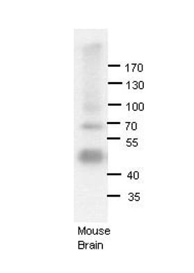
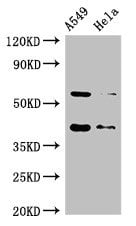
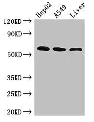
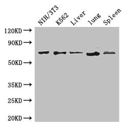
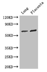
 WB (Western Blot)
(WB Suggested Anti-ADARB1 Antibody Titration: 1.25ug/mlELISA Titer: 1:1562500Positive Control: HepG2 cell lysate)
WB (Western Blot)
(WB Suggested Anti-ADARB1 Antibody Titration: 1.25ug/mlELISA Titer: 1:1562500Positive Control: HepG2 cell lysate)
ADARB1, Polyclonal Antibody (Cat# AAA198624)
mCherry, Polyclonal Antibody (Cat# AAA129121)
mPlum, Polyclonal Antibody (Cat# AAA129122)
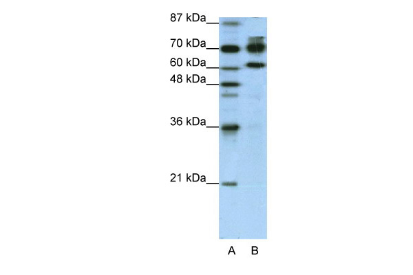
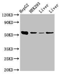
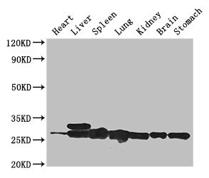
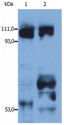 WB (Western Blot)
(Western blotting detection of kinesin in reduced lysates of human HEK cell line (1) and porcine brain (2) by rabbit polyclonal anti-kinesin.)
WB (Western Blot)
(Western blotting detection of kinesin in reduced lysates of human HEK cell line (1) and porcine brain (2) by rabbit polyclonal anti-kinesin.)
Kinesin, Polyclonal Antibody (Cat# AAA128932)
Dendra2, Polyclonal Antibody (Cat# AAA128935)
Ag85b, Polyclonal Antibody (Cat# AAA128931)
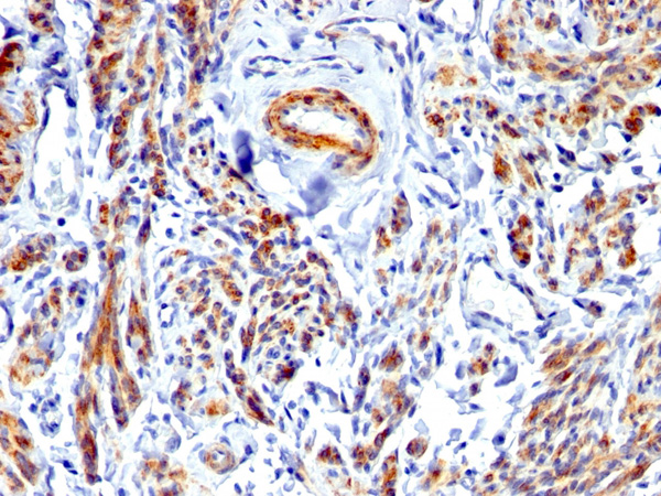
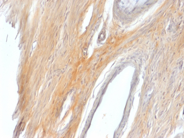 IHC (Immunohiostchemistry)
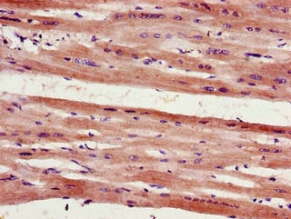
(Formalin-fixed, paraffin-embedded Rat Uterus stained with Caldesmon Rabbit Polyclonal Antibody.)
IHC (Immunohiostchemistry)
(Formalin-fixed, paraffin-embedded Rat Uterus stained with Caldesmon Rabbit Polyclonal Antibody.)
Caldesmon, HMW, Polyclonal Antibody (Cat# AAA215265)
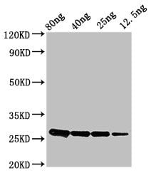 WB (Western Blot)
(Western BlotPositive WB detected in Recombinant proteinAll lanes: ptsH antibody at 2.8ug/mlSecondaryGoat polyclonal to rabbit IgG at 1/50000 dilutionpredicted band size: 26 KDaobserved band size: 26 KDa)
WB (Western Blot)
(Western BlotPositive WB detected in Recombinant proteinAll lanes: ptsH antibody at 2.8ug/mlSecondaryGoat polyclonal to rabbit IgG at 1/50000 dilutionpredicted band size: 26 KDaobserved band size: 26 KDa)
ptsH, Polyclonal Antibody (Cat# AAA232819)
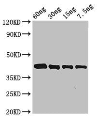 WB (Western Blot)
(Western BlotPositive WB detected in Recombinant proteinAll lanes: Arginine antibody at 3ug/mlSecondaryGoat polyclonal to rabbit IgG at 1/50000 dilutionpredicted band size: 42 KDaobserved band size: 42 KDa)
WB (Western Blot)
(Western BlotPositive WB detected in Recombinant proteinAll lanes: Arginine antibody at 3ug/mlSecondaryGoat polyclonal to rabbit IgG at 1/50000 dilutionpredicted band size: 42 KDaobserved band size: 42 KDa)
Arginine esterase, Polyclonal Antibody (Cat# AAA232820)
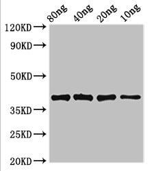 WB (Western Blot)
(Western BlotPositive WB detected in Recombinant proteinAll lanes: lexA antibody at 3.4ug/mlSecondaryGoat polyclonal to rabbit IgG at 1/50000 dilutionpredicted band size: 39 KDaobserved band size: 39 KDa)
WB (Western Blot)
(Western BlotPositive WB detected in Recombinant proteinAll lanes: lexA antibody at 3.4ug/mlSecondaryGoat polyclonal to rabbit IgG at 1/50000 dilutionpredicted band size: 39 KDaobserved band size: 39 KDa)
lexA, Polyclonal Antibody (Cat# AAA232822)
 WB (Western Blot)
(Western BlotPositive WB detected in Recombinant proteinAll lanes: AK antibody at 2.8ug/mlSecondaryGoat polyclonal to rabbit IgG at 1/50000 dilutionpredicted band size: 56 KDaobserved band size: 56 KDa)
WB (Western Blot)
(Western BlotPositive WB detected in Recombinant proteinAll lanes: AK antibody at 2.8ug/mlSecondaryGoat polyclonal to rabbit IgG at 1/50000 dilutionpredicted band size: 56 KDaobserved band size: 56 KDa)
AK, Polyclonal Antibody (Cat# AAA232825)
tir, Polyclonal Antibody (Cat# AAA232826)
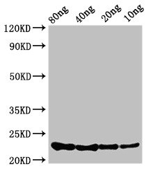 WB (Western Blot)
(Western BlotPositive WB detected in Recombinant proteinAll lanes: csrA antibody at 3.4ug/mlSecondaryGoat polyclonal to rabbit IgG at 1/50000 dilutionpredicted band size: 23 KDaobserved band size: 23 KDa)
WB (Western Blot)
(Western BlotPositive WB detected in Recombinant proteinAll lanes: csrA antibody at 3.4ug/mlSecondaryGoat polyclonal to rabbit IgG at 1/50000 dilutionpredicted band size: 23 KDaobserved band size: 23 KDa)
csrA, Polyclonal Antibody (Cat# AAA232829)
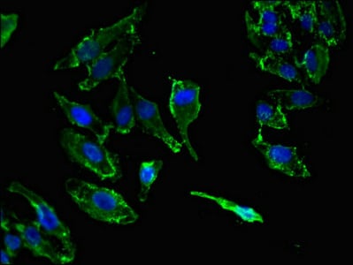
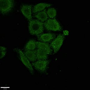
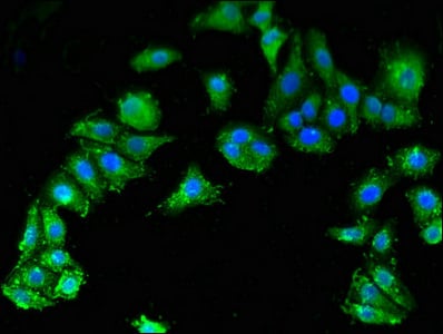
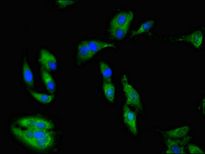
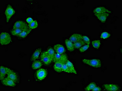
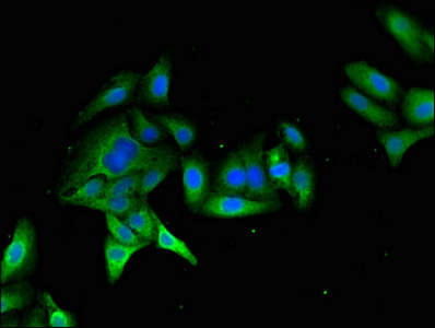
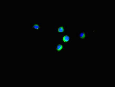
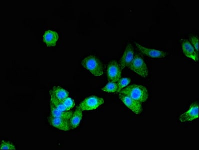
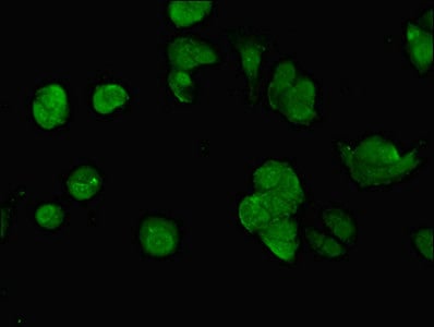
 IF (Immunofluorescence)
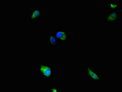
(Immunofluorescent analysis of Hela cells using AAA232834 at a dilution of 1:100 and Alexa Fluor 488-congugated AffiniPure Goat Anti-Rabbit IgG(H+L))
IF (Immunofluorescence)
(Immunofluorescent analysis of Hela cells using AAA232834 at a dilution of 1:100 and Alexa Fluor 488-congugated AffiniPure Goat Anti-Rabbit IgG(H+L))
OS9, Polyclonal Antibody (Cat# AAA232834)
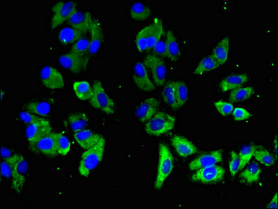
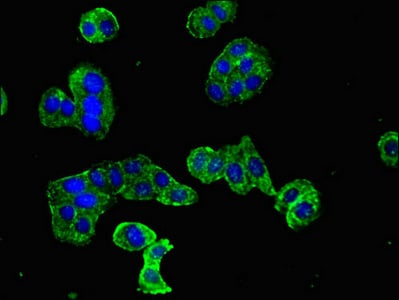
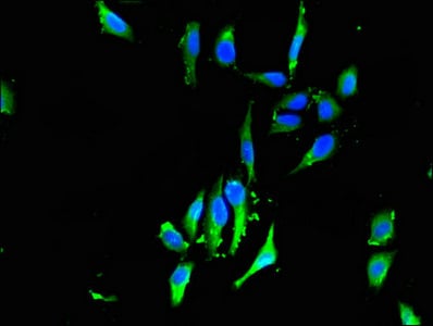
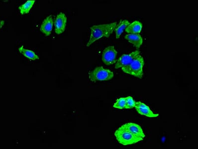
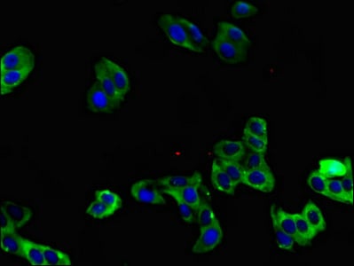
 IF (Immunofluorescence)
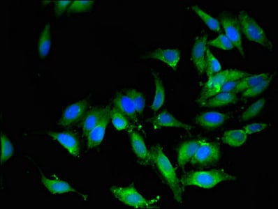
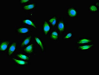
(Immunofluorescent analysis of HepG2 cells using AAA232836 at a dilution of 1:100 and Alexa Fluor 488-congugated AffiniPure Goat Anti-Rabbit IgG(H+L))
IF (Immunofluorescence)
(Immunofluorescent analysis of HepG2 cells using AAA232836 at a dilution of 1:100 and Alexa Fluor 488-congugated AffiniPure Goat Anti-Rabbit IgG(H+L))
BAAT, Polyclonal Antibody (Cat# AAA232836)
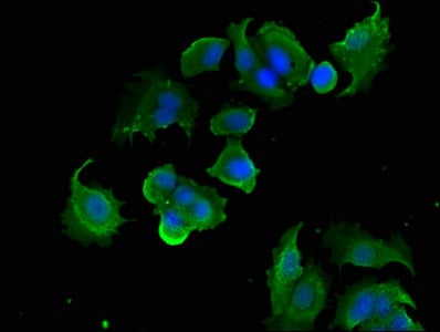
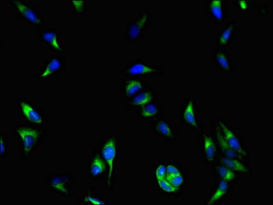
 IF (Immunofluorescence)
(Immunofluorescent analysis of MCF-7 cells using AAA232837 at a dilution of 1:100 and Alexa Fluor 488-congugated AffiniPure Goat Anti-Rabbit IgG(H+L))
IF (Immunofluorescence)
(Immunofluorescent analysis of MCF-7 cells using AAA232837 at a dilution of 1:100 and Alexa Fluor 488-congugated AffiniPure Goat Anti-Rabbit IgG(H+L))
DYNC1H1, Polyclonal Antibody (Cat# AAA232837)
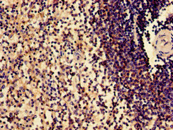
 IHC (Immunohistochemisry)
(Immunofluorescent analysis of HepG2 cells using AAA232841 at a dilution of 1:100 and Alexa Fluor 488-congugated AffiniPure Goat Anti-Rabbit IgG(H+L))
IHC (Immunohistochemisry)
(Immunofluorescent analysis of HepG2 cells using AAA232841 at a dilution of 1:100 and Alexa Fluor 488-congugated AffiniPure Goat Anti-Rabbit IgG(H+L))
KIF14, Polyclonal Antibody (Cat# AAA232841)
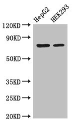
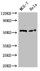
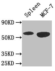
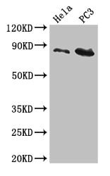
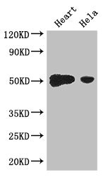
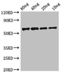
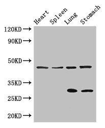
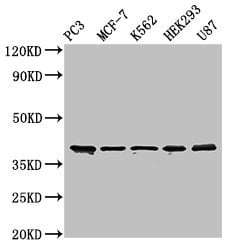 WB (Western Blot)
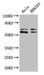
(Western BlotPositive WB detected in: PC3 whole cell lysate,MCF-7 whole cell lysate,K562 whole cell lysate,HEK293 whole cell lysate,U87 whole cell lysateAll lanes: SLAMF1 antibody at 3ug/mlSecondaryGoat polyclonal to rabbit IgG at 1/50000 dilutionPredicted band size: 38,34,41 KDaObserved band size: 38 KDa)
WB (Western Blot)
(Western BlotPositive WB detected in: PC3 whole cell lysate,MCF-7 whole cell lysate,K562 whole cell lysate,HEK293 whole cell lysate,U87 whole cell lysateAll lanes: SLAMF1 antibody at 3ug/mlSecondaryGoat polyclonal to rabbit IgG at 1/50000 dilutionPredicted band size: 38,34,41 KDaObserved band size: 38 KDa)
SLAMF1, Polyclonal Antibody (Cat# AAA232844)
PLD1, Polyclonal Antibody (Cat# AAA232845)
 IF (Immunofluorescence)
(Immunofluorescent analysis of MCF-7 cells using AAA232846 at a dilution of 1:100 and Alexa Fluor 488-congugated AffiniPure Goat Anti-Rabbit IgG(H+L))
IF (Immunofluorescence)
(Immunofluorescent analysis of MCF-7 cells using AAA232846 at a dilution of 1:100 and Alexa Fluor 488-congugated AffiniPure Goat Anti-Rabbit IgG(H+L))
PTK6, Polyclonal Antibody (Cat# AAA232846)
 IF (Immunofluorescence)
(Immunofluorescent analysis of PC3 cells using AAA232849 at a dilution of 1:100 and Alexa Fluor 488-congugated AffiniPure Goat Anti-Rabbit IgG(H+L))
IF (Immunofluorescence)
(Immunofluorescent analysis of PC3 cells using AAA232849 at a dilution of 1:100 and Alexa Fluor 488-congugated AffiniPure Goat Anti-Rabbit IgG(H+L))
SSX1, Polyclonal Antibody (Cat# AAA232849)
 IHC (Immunohistochemisry)
(Immunofluorescent analysis of HepG2 cells using AAA232851 at a dilution of 1:100 and Alexa Fluor 488-congugated AffiniPure Goat Anti-Rabbit IgG(H+L))
IHC (Immunohistochemisry)
(Immunofluorescent analysis of HepG2 cells using AAA232851 at a dilution of 1:100 and Alexa Fluor 488-congugated AffiniPure Goat Anti-Rabbit IgG(H+L))
SMN1, Polyclonal Antibody (Cat# AAA232851)
 IF (Immunofluorescence)
(Immunofluorescent analysis of Hela cells using AAA232852 at a dilution of 1:100 and Alexa Fluor 488-congugated AffiniPure Goat Anti-Rabbit IgG(H+L))
IF (Immunofluorescence)
(Immunofluorescent analysis of Hela cells using AAA232852 at a dilution of 1:100 and Alexa Fluor 488-congugated AffiniPure Goat Anti-Rabbit IgG(H+L))
MAP2K5, Polyclonal Antibody (Cat# AAA232852)
 IF (Immunofluorescence)
(Immunofluorescent analysis of Hela cells using AAA232854 at a dilution of 1:100 and Alexa Fluor 488-congugated AffiniPure Goat Anti-Rabbit IgG(H+L))
IF (Immunofluorescence)
(Immunofluorescent analysis of Hela cells using AAA232854 at a dilution of 1:100 and Alexa Fluor 488-congugated AffiniPure Goat Anti-Rabbit IgG(H+L))
FHL2, Polyclonal Antibody (Cat# AAA232854)
 IF (Immunofluorescence)
(Immunofluorescent analysis of Hela cells using AAA232863 at a dilution of 1:100 and Alexa Fluor 488-congugated AffiniPure Goat Anti-Rabbit IgG(H+L))
IF (Immunofluorescence)
(Immunofluorescent analysis of Hela cells using AAA232863 at a dilution of 1:100 and Alexa Fluor 488-congugated AffiniPure Goat Anti-Rabbit IgG(H+L))
COASY, Polyclonal Antibody (Cat# AAA232863)
 IF (Immunofluorescence)
(Immunofluorescent analysis of Hela cells using AAA232864 at a dilution of 1:100 and Alexa Fluor 488-congugated AffiniPure Goat Anti-Rabbit IgG(H+L))
IF (Immunofluorescence)
(Immunofluorescent analysis of Hela cells using AAA232864 at a dilution of 1:100 and Alexa Fluor 488-congugated AffiniPure Goat Anti-Rabbit IgG(H+L))
PRKAA1, Polyclonal Antibody (Cat# AAA232864)
 IF (Immunofluorescence)
(Immunofluorescent analysis of HepG2 cells using AAA232865 at a dilution of 1:100 and Alexa Fluor 488-congugated AffiniPure Goat Anti-Rabbit IgG(H+L))
IF (Immunofluorescence)
(Immunofluorescent analysis of HepG2 cells using AAA232865 at a dilution of 1:100 and Alexa Fluor 488-congugated AffiniPure Goat Anti-Rabbit IgG(H+L))
DOC2B, Polyclonal Antibody (Cat# AAA232865)
 IF (Immunofluorescence)
(Immunofluorescent analysis of Hela cells using AAA232868 at a dilution of 1:100 and Alexa Fluor 488-congugated AffiniPure Goat Anti-Rabbit IgG(H+L))
IF (Immunofluorescence)
(Immunofluorescent analysis of Hela cells using AAA232868 at a dilution of 1:100 and Alexa Fluor 488-congugated AffiniPure Goat Anti-Rabbit IgG(H+L))
FHL1, Polyclonal Antibody (Cat# AAA232868)
 IF (Immunofluorescence)
(Immunofluorescent analysis of HepG2 cells using AAA232869 at a dilution of 1:100 and Alexa Fluor 488-congugated AffiniPure Goat Anti-Rabbit IgG(H+L))
IF (Immunofluorescence)
(Immunofluorescent analysis of HepG2 cells using AAA232869 at a dilution of 1:100 and Alexa Fluor 488-congugated AffiniPure Goat Anti-Rabbit IgG(H+L))
SCARB2, Polyclonal Antibody (Cat# AAA232869)
 IHC (Immunohistochemisry)
(Immunofluorescent analysis of HepG2 cells using AAA232870 at a dilution of 1:100 and Alexa Fluor 488-congugated AffiniPure Goat Anti-Rabbit IgG(H+L))
IHC (Immunohistochemisry)
(Immunofluorescent analysis of HepG2 cells using AAA232870 at a dilution of 1:100 and Alexa Fluor 488-congugated AffiniPure Goat Anti-Rabbit IgG(H+L))
DDR2, Polyclonal Antibody (Cat# AAA232870)
 IHC (Immunohistochemisry)
(Immunofluorescent analysis of HepG2 cells using AAA232873 at a dilution of 1:100 and Alexa Fluor 488-congugated AffiniPure Goat Anti-Rabbit IgG(H+L))
IHC (Immunohistochemisry)
(Immunofluorescent analysis of HepG2 cells using AAA232873 at a dilution of 1:100 and Alexa Fluor 488-congugated AffiniPure Goat Anti-Rabbit IgG(H+L))
MYO7A, Polyclonal Antibody (Cat# AAA232873)
 IHC (Immunohistochemisry)
(Immunofluorescent analysis of Hela cells using AAA232876 at a dilution of 1:100 and Alexa Fluor 488-congugated AffiniPure Goat Anti-Rabbit IgG(H+L))
IHC (Immunohistochemisry)
(Immunofluorescent analysis of Hela cells using AAA232876 at a dilution of 1:100 and Alexa Fluor 488-congugated AffiniPure Goat Anti-Rabbit IgG(H+L))
EIF4EBP1, Polyclonal Antibody (Cat# AAA232876)
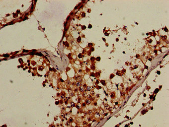
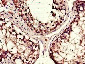
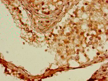
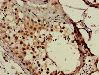
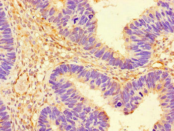
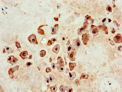
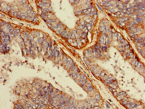
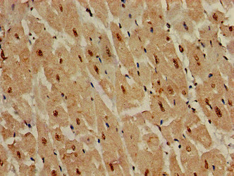
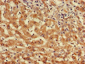
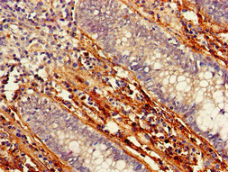
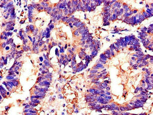
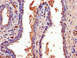
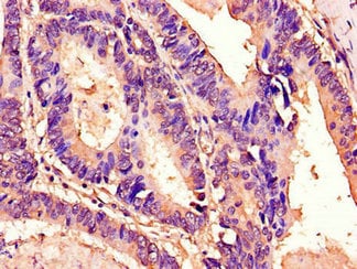
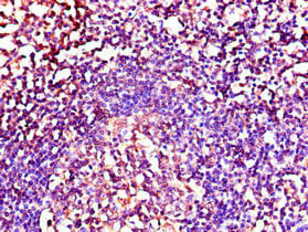
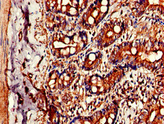
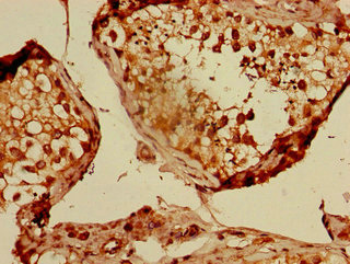 IHC (Immunohiostchemistry)
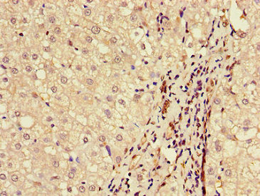
(Immunohistochemistry of paraffin-embedded human testis tissue using AAA232877 at dilution of 1:100)
IHC (Immunohiostchemistry)
(Immunohistochemistry of paraffin-embedded human testis tissue using AAA232877 at dilution of 1:100)
CUL2, Polyclonal Antibody (Cat# AAA232877)
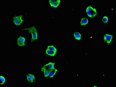
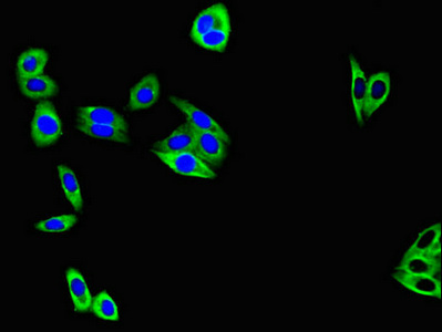
 IF (Immunofluorescence)
(Immunofluorescent analysis of HepG2 cells using AAA232878 at a dilution of 1:100 and Alexa Fluor 488-congugated AffiniPure Goat Anti-Rabbit IgG(H+L))
IF (Immunofluorescence)
(Immunofluorescent analysis of HepG2 cells using AAA232878 at a dilution of 1:100 and Alexa Fluor 488-congugated AffiniPure Goat Anti-Rabbit IgG(H+L))
PTCH1, Polyclonal Antibody (Cat# AAA232878)

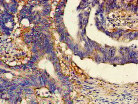
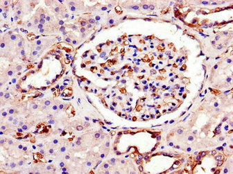
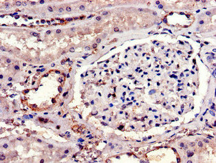
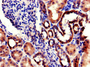
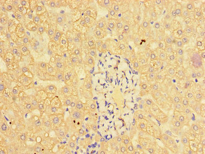
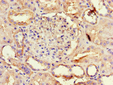
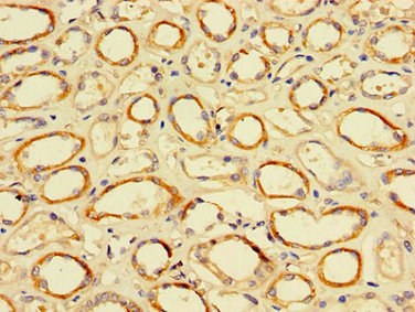 IHC (Immunohistochemisry)
(Immunohistochemistry of paraffin-embedded human kidney tissue using AAA232880 at dilution of 1:100)
IHC (Immunohistochemisry)
(Immunohistochemistry of paraffin-embedded human kidney tissue using AAA232880 at dilution of 1:100)
TFDP2, Polyclonal Antibody (Cat# AAA232880)
 IF (Immunofluorescence)
(Immunofluorescent analysis of MCF-7 cells using AAA232882 at a dilution of 1:100 and Alexa Fluor 488-congugated AffiniPure Goat Anti-Rabbit IgG(H+L))
IF (Immunofluorescence)
(Immunofluorescent analysis of MCF-7 cells using AAA232882 at a dilution of 1:100 and Alexa Fluor 488-congugated AffiniPure Goat Anti-Rabbit IgG(H+L))
SWI5, Polyclonal Antibody (Cat# AAA232882)
speB, Polyclonal Antibody (Cat# AAA232890)
 IHC (Immunohistochemisry)
(Immunofluorescent analysis of Hela cells using AAA232892 at a dilution of 1:100 and Alexa Fluor 488-congugated AffiniPure Goat Anti-Rabbit IgG(H+L))
IHC (Immunohistochemisry)
(Immunofluorescent analysis of Hela cells using AAA232892 at a dilution of 1:100 and Alexa Fluor 488-congugated AffiniPure Goat Anti-Rabbit IgG(H+L))
C1QTNF9B-AS1, Polyclonal Antibody (Cat# AAA232892)
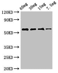
 WB (Western Blot)
(Western BlotPositive WB detected in Recombinant proteinAll lanes: FSIP2 antibody at 0.8ug/mlSecondaryGoat polyclonal to rabbit IgG at 1/50000 dilutionpredicted band size: 62 KDaobserved band size: 62 KDa)
WB (Western Blot)
(Western BlotPositive WB detected in Recombinant proteinAll lanes: FSIP2 antibody at 0.8ug/mlSecondaryGoat polyclonal to rabbit IgG at 1/50000 dilutionpredicted band size: 62 KDaobserved band size: 62 KDa)
FSIP2, Polyclonal Antibody (Cat# AAA232893)
nosZ, Polyclonal Antibody (Cat# AAA232897)
 IF (Immunofluorescence)
(Immunofluorescent analysis of HepG2 cells using AAA232900 at a dilution of 1:100 and Alexa Fluor 488-congugated AffiniPure Goat Anti-Rabbit IgG(H+L))
IF (Immunofluorescence)
(Immunofluorescent analysis of HepG2 cells using AAA232900 at a dilution of 1:100 and Alexa Fluor 488-congugated AffiniPure Goat Anti-Rabbit IgG(H+L))
TM4SF20, Polyclonal Antibody (Cat# AAA232900)
 IF (Immunofluorescence)
(Immunofluorescent analysis of MCF-7 cells using AAA232902 at a dilution of 1:100 and Alexa Fluor 488-congugated AffiniPure Goat Anti-Rabbit IgG(H+L))
IF (Immunofluorescence)
(Immunofluorescent analysis of MCF-7 cells using AAA232902 at a dilution of 1:100 and Alexa Fluor 488-congugated AffiniPure Goat Anti-Rabbit IgG(H+L))
IL23R, Polyclonal Antibody (Cat# AAA232902)
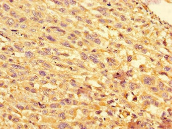
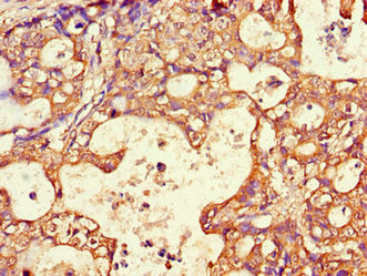
 IHC (Immunohistochemistry)
(Immunohistochemistry of paraffin-embedded human kidney tissue using AAA232904 at dilution of 1:100)
IHC (Immunohistochemistry)
(Immunohistochemistry of paraffin-embedded human kidney tissue using AAA232904 at dilution of 1:100)
RPL22L1, Polyclonal Antibody (Cat# AAA232904)
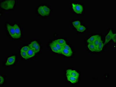
 IF (Immunofluorescence)
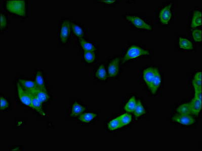
(Immunofluorescent analysis of HepG2 cells using AAA232755 at a dilution of 1:100 and Alexa Fluor 488-congugated AffiniPure Goat Anti-Rabbit IgG(H+L))
IF (Immunofluorescence)
(Immunofluorescent analysis of HepG2 cells using AAA232755 at a dilution of 1:100 and Alexa Fluor 488-congugated AffiniPure Goat Anti-Rabbit IgG(H+L))
NRG1, Polyclonal Antibody (Cat# AAA232755)
 IF (Immunofluorescence)
(Immunofluorescent analysis of HepG2 cells using AAA232762 at a dilution of 1:100 and Alexa Fluor 488-congugated AffiniPure Goat Anti-Rabbit IgG(H+L))
IF (Immunofluorescence)
(Immunofluorescent analysis of HepG2 cells using AAA232762 at a dilution of 1:100 and Alexa Fluor 488-congugated AffiniPure Goat Anti-Rabbit IgG(H+L))
CTSL, Polyclonal Antibody (Cat# AAA232762)
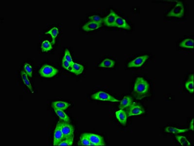
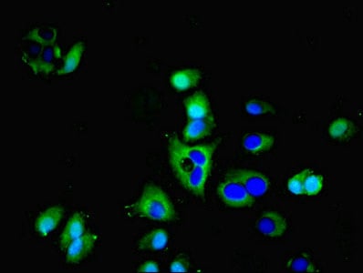
 IF (Immunofluorescence)
(Immunofluorescent analysis of HepG2 cells using AAA232763 at a dilution of 1:100 and Alexa Fluor 488-congugated AffiniPure Goat Anti-Rabbit IgG(H+L))
IF (Immunofluorescence)
(Immunofluorescent analysis of HepG2 cells using AAA232763 at a dilution of 1:100 and Alexa Fluor 488-congugated AffiniPure Goat Anti-Rabbit IgG(H+L))
REN, Polyclonal Antibody (Cat# AAA232763)
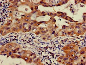
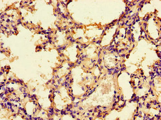
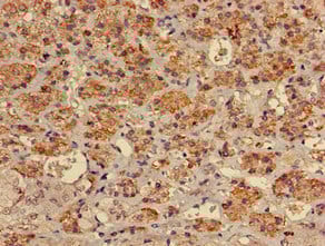
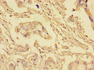
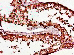
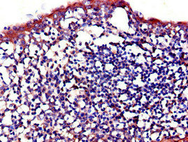
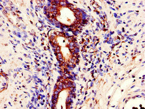
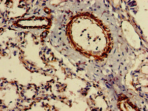 IHC (Immunohiostchemistry)
(Immunohistochemistry of paraffin-embedded human lung tissue using AAA232765 at dilution of 1:100)
IHC (Immunohiostchemistry)
(Immunohistochemistry of paraffin-embedded human lung tissue using AAA232765 at dilution of 1:100)
CIDEB, Polyclonal Antibody (Cat# AAA232765)
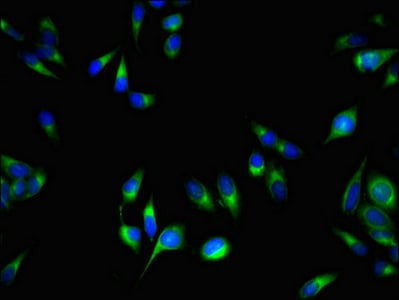
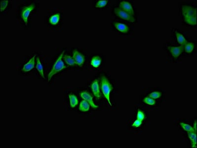
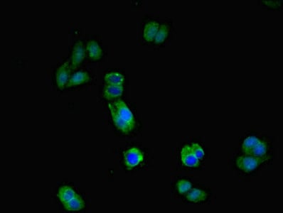 IF (Immunofluorescence)
(Immunofluorescent analysis of HepG2 cells using AAA232766 at a dilution of 1:100 and Alexa Fluor 488-congugated AffiniPure Goat Anti-Rabbit IgG(H+L))
IF (Immunofluorescence)
(Immunofluorescent analysis of HepG2 cells using AAA232766 at a dilution of 1:100 and Alexa Fluor 488-congugated AffiniPure Goat Anti-Rabbit IgG(H+L))
IL17RA, Polyclonal Antibody (Cat# AAA232766)
 IHC (Immunohistochemisry)
(Immunofluorescent analysis of HepG2 cells using AAA232771 at a dilution of 1:100 and Alexa Fluor 488-congugated AffiniPure Goat Anti-Rabbit IgG(H+L))
IHC (Immunohistochemisry)
(Immunofluorescent analysis of HepG2 cells using AAA232771 at a dilution of 1:100 and Alexa Fluor 488-congugated AffiniPure Goat Anti-Rabbit IgG(H+L))
STAU1, Polyclonal Antibody (Cat# AAA232771)
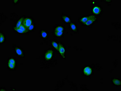
 IF (Immunofluorescence)
(Immunofluorescent analysis of MCF-7 cells using AAA232772 at a dilution of 1:100 and Alexa Fluor 488-congugated AffiniPure Goat Anti-Rabbit IgG(H+L))
IF (Immunofluorescence)
(Immunofluorescent analysis of MCF-7 cells using AAA232772 at a dilution of 1:100 and Alexa Fluor 488-congugated AffiniPure Goat Anti-Rabbit IgG(H+L))
AQP3, Polyclonal Antibody (Cat# AAA232772)
What are Polyclonal Antibodies?
Polyclonal antibodies are antibodies that come from multiple B cell clones of a host animal. The typical hosts used for the majority of polyclonal antibody production are rabbits, goats, sheep, and donkeys. These polyclonal antibodies, once having identified their target, will bind to different epitopes located at different regions or sequences on the same protein/antigen. As a result, they are ideal at locating and binding to the target, even if the target is in very low concentrations (due to many different antibodies being able to bind to the same target molecule, which allows for significant amplification of a downstream signal).
Polyclonal antibodies are typically produced by injecting an antigen into a host animal, which causes the animal’s immune system to attack the foreign antigen by mass generating antibodies against it. After a period of time, serum is collected from the animal and purified using physicochemical fractionation, class-specific affinity purification, and/or antigen-affinity purification.
Key Uses of Polyclonal Antibodies
- Western Blotting: This method is used to find specific proteins in biological samples after separating them by size.
- Immunohistochemistry: IHC helps visualize the location of proteins in tissue sections using various staining techniques.
- ELISA: (Enzyme-Linked Immunosorbent Assay) is typically used to identify specific protein quantities in a sample. ELISAs can be either “Quantitative” or “Qualitative”.
- Flow Cytometry: technique that identifies and measures the specific protein on the surface or inside the cells in a fluid suspension.
- Immunoprecipitation: IP isolates and studies a specific protein from a complex mixture using antibodies.
Why Buy Polyclonal Antibodies from AAA Biotech?
1. Ideal for Various Applications
Our antibodies are generally going to be validated for use in multiple types of assays, including ELISA, Western Blotting, Immunohistochemistry, Immunoprecipitation, amongst others. They are ideal for a wide range of research applications.
2. Rigorous Quality Control
All of the antibodies in our catalog undergo strict quality testing to ensure specificity, sensitivity, and consistent performance. We are confident in the ability of our antibodies to provide you with accurate results.
3. Wide Assortment of Antibodies
Antibodies in are catalog can be found for both common and exotic species, and these antibodies are also available in both conjugated and recombinant forms to suit many diverse experimental needs.
4. Highly Purified
Our antibodies are available in purified forms with over 85% purity, as confirmed by SDS-PAGE. They are also available with tags such as His, Flag, GST, or MBP. We cater to customers worldwide.
FAQ
1. How are polyclonal antibodies produced?
Traditionally, polyclonal antibodies are produced by injecting an antigen into a host animal (such as a rabbit or goat), which then triggers an immune response from the host animal. The animal’s B cells produce antibodies that will recognize different parts of the injected antigen. These antibodies are then collected from the animal’s blood and purified for use.
2. How do polyclonal antibodies differ from monoclonal antibodies?
Polyclonal antibodies are a mix of antibodies that bind to different locations (epitopes) of the same antigen, while monoclonal antibodies are identical and bind to just one specific epitope. This makes polyclonal antibodies more versatile and better at detecting proteins that may be present in low quantities or in altered/modified forms.
3. How should I store polyclonal antibodies?
Polyclonal antibodies should be stored at 4°C for short-term use (up to a few weeks) and at -20°C or -80°C for long-term storage. Avoid repeated freeze-thaw cycles by dividing them into small aliquots. Always check the datasheet for specific storage instructions.