
 Application Data
(Published customer image:Mouse anti Pig SLA Class I antibody, clone JM1E3 used for the evaluation of SLA Class I expression in cells over-expressing ?2-microglobulin by immunofluorescence.Image caption:Immunohistochemical analysis of the ?2M and SLA class I expression in cells over-expressing ?2M. The names of the transfected constructs are shown on top: pCMV-HA-B2M for the expression of HA-tagged ?2M, pCMV-HA for the HA tag without ?2M, and pEGFP-N1 for EGFP expression. The primary antibodies used for the analysis are shown on the left. A and B. Cells were stained with the HA tag-specific antibody. The cell nuclei were visualized using 4?,6-diamidino-2-phenylindole (DAPI; blue). C. pEGFP (green) plasmid was used to evaluate the transfection efficiency. D, E, and F. SLA class I heavy chains were stained with pan SLA class I-specific antibodies. The signals of primary antibodies were detected by Alexa 568-conjugated secondary antibody (red) in all cases.)
Application Data
(Published customer image:Mouse anti Pig SLA Class I antibody, clone JM1E3 used for the evaluation of SLA Class I expression in cells over-expressing ?2-microglobulin by immunofluorescence.Image caption:Immunohistochemical analysis of the ?2M and SLA class I expression in cells over-expressing ?2M. The names of the transfected constructs are shown on top: pCMV-HA-B2M for the expression of HA-tagged ?2M, pCMV-HA for the HA tag without ?2M, and pEGFP-N1 for EGFP expression. The primary antibodies used for the analysis are shown on the left. A and B. Cells were stained with the HA tag-specific antibody. The cell nuclei were visualized using 4?,6-diamidino-2-phenylindole (DAPI; blue). C. pEGFP (green) plasmid was used to evaluate the transfection efficiency. D, E, and F. SLA class I heavy chains were stained with pan SLA class I-specific antibodies. The signals of primary antibodies were detected by Alexa 568-conjugated secondary antibody (red) in all cases.)
SLA CLASS I (SLA) Antibody – Mouse Monoclonal
MOUSE ANTI PIG SLA CLASS I:RPE
Product Overview
ALEXA FLUOR 647, FITC, Low Endotoxin, Purified, RPE
Phosphate buffered saline
0.09% sodium azide (NaN3)
1% bovine serum albumin
Application Data
(Published customer image:Mouse anti Pig SLA Class I antibody, clone JM1E3 used for the evaluation of SLA Class I expression in cells over-expressing ?2-microglobulin by immunofluorescence.Image caption:Immunohistochemical analysis of the ?2M and SLA class I expression in cells over-expressing ?2M. The names of the transfected constructs are shown on top: pCMV-HA-B2M for the expression of HA-tagged ?2M, pCMV-HA for the HA tag without ?2M, and pEGFP-N1 for EGFP expression. The primary antibodies used for the analysis are shown on the left. A and B. Cells were stained with the HA tag-specific antibody. The cell nuclei were visualized using 4?,6-diamidino-2-phenylindole (DAPI; blue). C. pEGFP (green) plasmid was used to evaluate the transfection efficiency. D, E, and F. SLA class I heavy chains were stained with pan SLA class I-specific antibodies. The signals of primary antibodies were detected by Alexa 568-conjugated secondary antibody (red) in all cases.)
Application Data
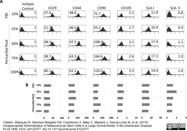
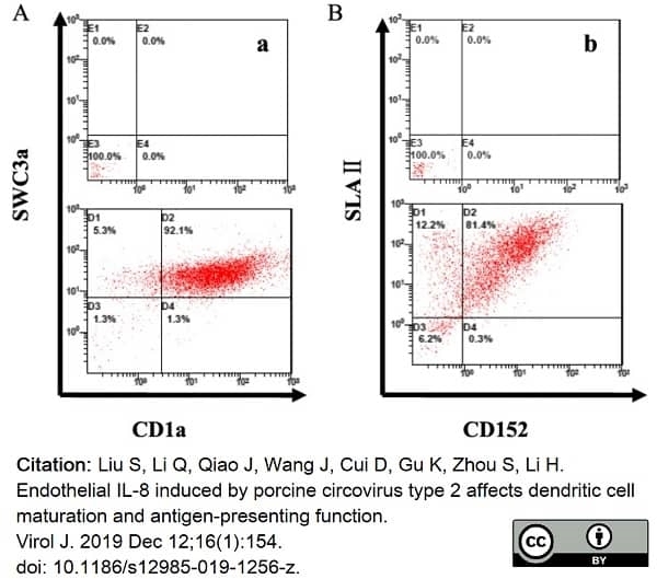
(Published customer image:Mouse anti Pig SLA Class I antibody, clone JM1E3 used for the evaluation of SLA Class I expression on bone marrow derived mesenchymal stem cells by flow cytometry.Image caption:Effect of pericardial fluid on pBM-MSCs phenotype. The phenotypic analysis was performed by multicolor flow cytometry. The cells were in vitro cultured for 7 days in the presence of Fetal Bovine Serum and in the presence of pericardial fluid at 25%, 50%, 75% and 100%. Representative histograms of four different experiments are shown (A). The expression level of Stem Cell Markers (CD29, CD31, CD44, CD90, CD105) and Swine Leukocyte Antigen Class-I and Class-II (SLA-I and SLA-II) is represented as Normalized Mean Relative Fluorescence Intensity which is calculated by dividing the Mean Fluorescent Intensity (MFI) by its isotype control. Graphic representation of mean ±SD for each marker is also provided (n = 4) (B). No significant differences were found between groups when ANOVA test was performed.)
Application Data
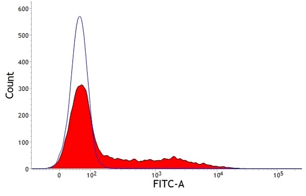
(Staining of porcine peripheral blood lymphocytes with Mouse anti Porcine SLA 1:FITC .)
Application Data
(Staining of porcine peripheral blood lymphocytes with Mouse anti Porcine SLA 1:FITC .)
Customer Reviews
Loading reviews...
Share Your Experience
Similar Products


Additional Details
Product Notes
The SLA (Catalog #AAA50479) is an Antibody produced from Mouse and is intended for research purposes only. The product is available for immediate purchase. The MOUSE ANTI PIG SLA CLASS I:RPE reacts with Pig, Human and may cross-react with other species as described in the data sheet. AAA Biotech's SLA CLASS I can be used in a range of immunoassay formats including, but not limited to, FCM/FACS (Flow Cytometry). Researchers should empirically determine the suitability of the SLA for an application not listed in the data sheet. Researchers commonly develop new applications and it is an integral, important part of the investigative research process. It is sometimes possible for the material contained within the vial of "SLA CLASS I, Monoclonal Antibody" to become dispersed throughout the inside of the vial, particularly around the seal of said vial, during shipment and storage. We always suggest centrifuging these vials to consolidate all of the liquid away from the lid and to the bottom of the vial prior to opening. Please be advised that certain products may require dry ice for shipping and that, if this is the case, an additional dry ice fee may also be required.Precautions
All products in the AAA Biotech catalog are strictly for research-use only, and are absolutely not suitable for use in any sort of medical, therapeutic, prophylactic, in-vivo, or diagnostic capacity. By purchasing a product from AAA Biotech, you are explicitly certifying that said products will be properly tested and used in line with industry standard. AAA Biotech and its authorized distribution partners reserve the right to refuse to fulfill any order if we have any indication that a purchaser may be intending to use a product outside of our accepted criteria.Disclaimer
Though we do strive to guarantee the information represented in this datasheet, AAA Biotech cannot be held responsible for any oversights or imprecisions. AAA Biotech reserves the right to adjust any aspect of this datasheet at any time and without notice. It is the responsibility of the customer to inform AAA Biotech of any product performance issues observed or experienced within 30 days of receipt of said product. To see additional details on this or any of our other policies, please see our Terms & Conditions page.Item has been added to Shopping Cart
If you are ready to order, navigate to Shopping Cart and get ready to checkout.