Filters
Clonality
Type
Reactivity
Gene Name
Isotype
Host
Application
Clone
13 results for "Primary Antibodies Rabbit Monoclonal Antibodies" - showing 1-13


 FCM (Flow Cytometry)
(Flow cytometric analysis of Jurkat cells with CD99 antibody at 1/50 dilution (red) compared with an unlabelled control (cells without incubation with primary antibody; black). Alexa Fluor 488-conjugated goat anti rabbit IgG was used as the secondary antibody.)
FCM (Flow Cytometry)
(Flow cytometric analysis of Jurkat cells with CD99 antibody at 1/50 dilution (red) compared with an unlabelled control (cells without incubation with primary antibody; black). Alexa Fluor 488-conjugated goat anti rabbit IgG was used as the secondary antibody.)
CD99, Monoclonal Antibody (Cat# AAA30274)
Full Name
CD99 Antibody
Gene Names
CD99; MIC2; HBA71; MIC2X; MIC2Y; MSK5X
Reactivity
Human
Applications
Western Blot, Immunocytochemistry, Immunofluorescence, Immunohistochemistry, Immunoprecipitation, Flow Cytometry
Purity
ProA affinity purified
Pricing


 ICC (Immunocytochemistry)
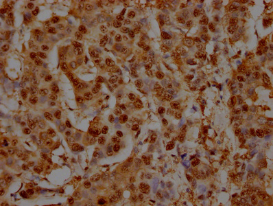
(Figure 10 Immunocytochemistry Validation of PD-L1Immunocytochemical analysis of 4% paraformaldehyde-fixed PD-L1 transfected 293 cells labeling PD-L1 with at 1 μg/ml, followed by Goat anti-mouse IgG secondary antibody at 1/250 dilution (red). Lower left: Use mouse IgG antibody at 1 μg/ml as control.)
ICC (Immunocytochemistry)
(Figure 10 Immunocytochemistry Validation of PD-L1Immunocytochemical analysis of 4% paraformaldehyde-fixed PD-L1 transfected 293 cells labeling PD-L1 with at 1 μg/ml, followed by Goat anti-mouse IgG secondary antibody at 1/250 dilution (red). Lower left: Use mouse IgG antibody at 1 μg/ml as control.)
PDL1, Monoclonal Antibody (Cat# AAA10992)
Full Name
PDL1 Antibody [6H10]
Gene Names
CD274; B7-H; B7H1; PDL1; PD-L1; PDCD1L1; PDCD1LG1
Reactivity
Human
Applications
Western Blot, Immunohistochemistry, Immunocytochemistry, Immunofluorescence
Purity
Protein A purified
Pricing


 Application Data
(Published customer image Infiltration of GFP+ BM-cells in infarct and peri-infarct regions. (A-B) Dot plots of viable macrophages/granulocytes (CD11b+CD45high, top right quadrants) and microglia (CD11b+CD45dim, bottom right quadrants) in cortex from BM-chimeric unmanipulated mice and mice exposed to pMCAO. (C) Bar graph showing mean numbers of CD11b+CD45dim microglia and CD11b+CD45high macrophages/granulocytes in BM-chimeric mice 24 hours after pMCAO, subdivided based on expression of GFP (n = 5). Approximately 92% of of the CD45high population were GFP+. (D) Estimation and comparison of mean numbers of CD11b+CD45dim microglia in non-chimeric (n = 10) versus BM-chimeric mice (n = 5) 24 hours after of pMCAO shows significantly fewer CD11b+CD45dim microglial cells in irradiated mice. (E) Overview, showing distribution of infiltrating GFP+ BM-derived cells into infarct (IF) and peri-infarct (P-IF) regions 24 hours after pMCAO. (E-G) By 24 hours, GFP+ single cells (F) and vessel-associated aggregates of GFP+ cells (arrows in G) were observed in infarct and peri-infarct regions. Some of the vessel-associated cells were round, leukocyte-like cells (arrows) while others were elongated cells lining the vasculature (arrow heads in G and in insert). (H) Bar graph showing mean numbers of single GFP+ cells and vessel-associated aggregates of GFP+ cells in ipsi- and contralateral cortex 24 hours after surgery (n = 10). (I-P) Immunohistochemical staining of CD45.1 (I, K), CD45.2 (J, L), IgG2a (M, O) and CD45 (N, P) in ischemic tissue in BM-chimeric (I, J, M, N) and non-chimeric mice (K, L, O, P) 24 hours after pMCAO. N.D, none detected. Scale bars: 200 um (A), 10 um (B, C). 50 um (I-P) *P < 0.05, **P < 0.01, and ***P < 0.001.From: Clausen BH, Lambertsen KL, Babcock AA, Holm TH, Dagnaes-Hansen F, Finsen B. Interleukin-1beta and tumor necrosis factor-alpha are expressed by different subsets of microglia and macrophages after ischemic stroke in mice. J Neuroinflammation. 2008 Oct 23;5:46.)
Application Data
(Published customer image Infiltration of GFP+ BM-cells in infarct and peri-infarct regions. (A-B) Dot plots of viable macrophages/granulocytes (CD11b+CD45high, top right quadrants) and microglia (CD11b+CD45dim, bottom right quadrants) in cortex from BM-chimeric unmanipulated mice and mice exposed to pMCAO. (C) Bar graph showing mean numbers of CD11b+CD45dim microglia and CD11b+CD45high macrophages/granulocytes in BM-chimeric mice 24 hours after pMCAO, subdivided based on expression of GFP (n = 5). Approximately 92% of of the CD45high population were GFP+. (D) Estimation and comparison of mean numbers of CD11b+CD45dim microglia in non-chimeric (n = 10) versus BM-chimeric mice (n = 5) 24 hours after of pMCAO shows significantly fewer CD11b+CD45dim microglial cells in irradiated mice. (E) Overview, showing distribution of infiltrating GFP+ BM-derived cells into infarct (IF) and peri-infarct (P-IF) regions 24 hours after pMCAO. (E-G) By 24 hours, GFP+ single cells (F) and vessel-associated aggregates of GFP+ cells (arrows in G) were observed in infarct and peri-infarct regions. Some of the vessel-associated cells were round, leukocyte-like cells (arrows) while others were elongated cells lining the vasculature (arrow heads in G and in insert). (H) Bar graph showing mean numbers of single GFP+ cells and vessel-associated aggregates of GFP+ cells in ipsi- and contralateral cortex 24 hours after surgery (n = 10). (I-P) Immunohistochemical staining of CD45.1 (I, K), CD45.2 (J, L), IgG2a (M, O) and CD45 (N, P) in ischemic tissue in BM-chimeric (I, J, M, N) and non-chimeric mice (K, L, O, P) 24 hours after pMCAO. N.D, none detected. Scale bars: 200 um (A), 10 um (B, C). 50 um (I-P) *P < 0.05, **P < 0.01, and ***P < 0.001.From: Clausen BH, Lambertsen KL, Babcock AA, Holm TH, Dagnaes-Hansen F, Finsen B. Interleukin-1beta and tumor necrosis factor-alpha are expressed by different subsets of microglia and macrophages after ischemic stroke in mice. J Neuroinflammation. 2008 Oct 23;5:46.)
CD11b, Monoclonal Antibody (Cat# AAA12182)
Full Name
RAT ANTI MOUSE CD11b:FITC
Gene Names
Itgam; CR3; CR3A; MAC1; Cd11b; Ly-40; Mac-1; Mac-1a; CD11b/CD18; F730045J24Rik
Applications
Flow Cytometry
Pricing


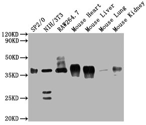
 IP (Immunoprecipitation)
(Immunoprecipitating GAPDH in Hela whole cell lysateLane 1: Mouse control IgG instead of in Hela whole cell lysate. Lane 2: (5ul) + Hela whole cell lysate (500ug)Lane 3: Hela whole cell lysate (10ug)For western blotting, the blot was detected at 1:5000, and a HRP-conjugated Protein G antibody was used as the secondary antibody at 1:2000)
IP (Immunoprecipitation)
(Immunoprecipitating GAPDH in Hela whole cell lysateLane 1: Mouse control IgG instead of in Hela whole cell lysate. Lane 2: (5ul) + Hela whole cell lysate (500ug)Lane 3: Hela whole cell lysate (10ug)For western blotting, the blot was detected at 1:5000, and a HRP-conjugated Protein G antibody was used as the secondary antibody at 1:2000)
GAPDH, Monoclonal Antibody (Cat# AAA27042)
Full Name
GAPDH Monoclonal Antibody
Reactivity
Human, Mouse, Rabbit
Applications
Western Blot, Immunohistochemistry, Immunoprecipitation, Immunofluorescence
Purity
>95%, Protein G purified
Pricing


 Application Data
(Published customer image Infiltration of GFP+ BM-cells in infarct and peri-infarct regions. (A-B) Dot plots of viable macrophages/granulocytes (CD11b+CD45high, top right quadrants) and microglia (CD11b+CD45dim, bottom right quadrants) in cortex from BM-chimeric unmanipulated mice and mice exposed to pMCAO. (C) Bar graph showing mean numbers of CD11b+CD45dim microglia and CD11b+CD45high macrophages/granulocytes in BM-chimeric mice 24 hours after pMCAO, subdivided based on expression of GFP (n = 5). Approximately 92% of of the CD45high population were GFP+. (D) Estimation and comparison of mean numbers of CD11b+CD45dim microglia in non-chimeric (n = 10) versus BM-chimeric mice (n = 5) 24 hours after of pMCAO shows significantly fewer CD11b+CD45dim microglial cells in irradiated mice. (E) Overview, showing distribution of infiltrating GFP+ BM-derived cells into infarct (IF) and peri-infarct (P-IF) regions 24 hours after pMCAO. (E-G) By 24 hours, GFP+ single cells (F) and vessel-associated aggregates of GFP+ cells (arrows in G) were observed in infarct and peri-infarct regions. Some of the vessel-associated cells were round, leukocyte-like cells (arrows) while others were elongated cells lining the vasculature (arrow heads in G and in insert). (H) Bar graph showing mean numbers of single GFP+ cells and vessel-associated aggregates of GFP+ cells in ipsi- and contralateral cortex 24 hours after surgery (n = 10). (I-P) Immunohistochemical staining of CD45.1 (I, K), CD45.2 (J, L), IgG2a (M, O) and CD45 (N, P) in ischemic tissue in BM-chimeric (I, J, M, N) and non-chimeric mice (K, L, O, P) 24 hours after pMCAO. N.D, none detected. Scale bars: 200 um (A), 10 um (B, C). 50 um (I-P) *P < 0.05, **P < 0.01, and ***P < 0.001.From: Clausen BH, Lambertsen KL, Babcock AA, Holm TH, Dagnaes-Hansen F, Finsen B. Interleukin-1beta and tumor necrosis factor-alpha are expressed by different subsets of microglia and macrophages after ischemic stroke in mice. J Neuroinflammation. 2008 Oct 23;5:46.)
Application Data
(Published customer image Infiltration of GFP+ BM-cells in infarct and peri-infarct regions. (A-B) Dot plots of viable macrophages/granulocytes (CD11b+CD45high, top right quadrants) and microglia (CD11b+CD45dim, bottom right quadrants) in cortex from BM-chimeric unmanipulated mice and mice exposed to pMCAO. (C) Bar graph showing mean numbers of CD11b+CD45dim microglia and CD11b+CD45high macrophages/granulocytes in BM-chimeric mice 24 hours after pMCAO, subdivided based on expression of GFP (n = 5). Approximately 92% of of the CD45high population were GFP+. (D) Estimation and comparison of mean numbers of CD11b+CD45dim microglia in non-chimeric (n = 10) versus BM-chimeric mice (n = 5) 24 hours after of pMCAO shows significantly fewer CD11b+CD45dim microglial cells in irradiated mice. (E) Overview, showing distribution of infiltrating GFP+ BM-derived cells into infarct (IF) and peri-infarct (P-IF) regions 24 hours after pMCAO. (E-G) By 24 hours, GFP+ single cells (F) and vessel-associated aggregates of GFP+ cells (arrows in G) were observed in infarct and peri-infarct regions. Some of the vessel-associated cells were round, leukocyte-like cells (arrows) while others were elongated cells lining the vasculature (arrow heads in G and in insert). (H) Bar graph showing mean numbers of single GFP+ cells and vessel-associated aggregates of GFP+ cells in ipsi- and contralateral cortex 24 hours after surgery (n = 10). (I-P) Immunohistochemical staining of CD45.1 (I, K), CD45.2 (J, L), IgG2a (M, O) and CD45 (N, P) in ischemic tissue in BM-chimeric (I, J, M, N) and non-chimeric mice (K, L, O, P) 24 hours after pMCAO. N.D, none detected. Scale bars: 200 um (A), 10 um (B, C). 50 um (I-P) *P < 0.05, **P < 0.01, and ***P < 0.001.From: Clausen BH, Lambertsen KL, Babcock AA, Holm TH, Dagnaes-Hansen F, Finsen B. Interleukin-1beta and tumor necrosis factor-alpha are expressed by different subsets of microglia and macrophages after ischemic stroke in mice. J Neuroinflammation. 2008 Oct 23;5:46.)
CD11b, Monoclonal Antibody (Cat# AAA12183)
Full Name
RAT ANTI MOUSE CD11b:FITC
Gene Names
Itgam; CR3; CR3A; MAC1; Cd11b; Ly-40; Mac-1; Mac-1a; CD11b/CD18; F730045J24Rik
Applications
Flow Cytometry
Pricing


 IP (Immunoprecipitation)
(Immunoprecipitating GAPDH in Hela whole cell lysate Lane 1: Mouse control IgG instead of AAA27043 in Hela whole cell lysate. Lane 2: AAA27043 (5ul) + Hela whole cell lysate (500ug) Lane 3: Hela whole cell lysate (10ug) For western blotting, the blot was detected with AAA27043 at 1:5000, and a HRPconjugated Protein G antibody was used as the secondary antibody at 1:2000)
IP (Immunoprecipitation)
(Immunoprecipitating GAPDH in Hela whole cell lysate Lane 1: Mouse control IgG instead of AAA27043 in Hela whole cell lysate. Lane 2: AAA27043 (5ul) + Hela whole cell lysate (500ug) Lane 3: Hela whole cell lysate (10ug) For western blotting, the blot was detected with AAA27043 at 1:5000, and a HRPconjugated Protein G antibody was used as the secondary antibody at 1:2000)
GAPDH, Monoclonal Antibody (Cat# AAA27043)
Full Name
GAPDH Monoclonal Antibody
Reactivity
Human, Rat, Rabbit, Mouse
Applications
Western Blot, Immunohistochemistry, Immunoprecipitation, Immunofluorescence
Purity
>95%, Protein G purified
Pricing


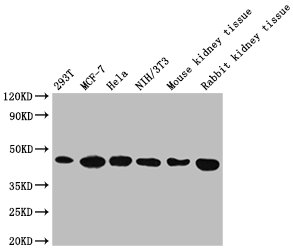

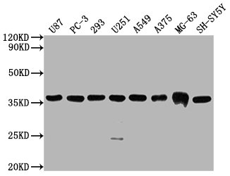
 FCM (Flow Cytometry)
(Overlay Peak curve showing Hela cells stained with AAA28066 (red line) at 1:200. The cells were fixed in 4% formaldehyde and permeated by 0.2% TritonX-100. Then 10% normal goat serum was Incubated to block non-specific protein-protein interactions followed by the antibody (1?g/1*106cells) for 1 h at 4 degree C. The secondary antibody used was FITC-conjugated Goat Anti-Mouse IgG(H+L) at 1/100 dilution for 30min at 4 degree C. Isotype control antibody (green line) was mouse IgG2b (1?g/1*106cells) used under the same conditions. Acquisition of >10,000 events was performed.)
FCM (Flow Cytometry)
(Overlay Peak curve showing Hela cells stained with AAA28066 (red line) at 1:200. The cells were fixed in 4% formaldehyde and permeated by 0.2% TritonX-100. Then 10% normal goat serum was Incubated to block non-specific protein-protein interactions followed by the antibody (1?g/1*106cells) for 1 h at 4 degree C. The secondary antibody used was FITC-conjugated Goat Anti-Mouse IgG(H+L) at 1/100 dilution for 30min at 4 degree C. Isotype control antibody (green line) was mouse IgG2b (1?g/1*106cells) used under the same conditions. Acquisition of >10,000 events was performed.)
ACTB, Monoclonal Antibody (Cat# AAA28066)
Full Name
ACTB Monoclonal Antibody
Reactivity
Human, Mouse, Rat, Rabbit
Applications
Western Blot, Immunohistochemistry, Immunofluorescence, Flow Cytometry, Immunoprecipitation
Purity
>95%, Protein A purified
Pricing


 Application Data
(Published customer image Infiltration of GFP+ BM-cells in infarct and peri-infarct regions. (A-B) Dot plots of viable macrophages/granulocytes (CD11b+CD45high, top right quadrants) and microglia (CD11b+CD45dim, bottom right quadrants) in cortex from BM-chimeric unmanipulated mice and mice exposed to pMCAO. (C) Bar graph showing mean numbers of CD11b+CD45dim microglia and CD11b+CD45high macrophages/granulocytes in BM-chimeric mice 24 hours after pMCAO, subdivided based on expression of GFP (n = 5). Approximately 92% of of the CD45high population were GFP+. (D) Estimation and comparison of mean numbers of CD11b+CD45dim microglia in non-chimeric (n = 10) versus BM-chimeric mice (n = 5) 24 hours after of pMCAO shows significantly fewer CD11b+CD45dim microglial cells in irradiated mice. (E) Overview, showing distribution of infiltrating GFP+ BM-derived cells into infarct (IF) and peri-infarct (P-IF) regions 24 hours after pMCAO. (E-G) By 24 hours, GFP+ single cells (F) and vessel-associated aggregates of GFP+ cells (arrows in G) were observed in infarct and peri-infarct regions. Some of the vessel-associated cells were round, leukocyte-like cells (arrows) while others were elongated cells lining the vasculature (arrow heads in G and in insert). (H) Bar graph showing mean numbers of single GFP+ cells and vessel-associated aggregates of GFP+ cells in ipsi- and contralateral cortex 24 hours after surgery (n = 10). (I-P) Immunohistochemical staining of CD45.1 (I, K), CD45.2 (J, L), IgG2a (M, O) and CD45 (N, P) in ischemic tissue in BM-chimeric (I, J, M, N) and non-chimeric mice (K, L, O, P) 24 hours after pMCAO. N.D, none detected. Scale bars: 200 um (A), 10 um (B, C). 50 um (I-P) *P < 0.05, **P < 0.01, and ***P < 0.001.From: Clausen BH, Lambertsen KL, Babcock AA, Holm TH, Dagnaes-Hansen F, Finsen B. Interleukin-1beta and tumor necrosis factor-alpha are expressed by different subsets of microglia and macrophages after ischemic stroke in mice. J Neuroinflammation. 2008 Oct 23;5:46.)
Application Data
(Published customer image Infiltration of GFP+ BM-cells in infarct and peri-infarct regions. (A-B) Dot plots of viable macrophages/granulocytes (CD11b+CD45high, top right quadrants) and microglia (CD11b+CD45dim, bottom right quadrants) in cortex from BM-chimeric unmanipulated mice and mice exposed to pMCAO. (C) Bar graph showing mean numbers of CD11b+CD45dim microglia and CD11b+CD45high macrophages/granulocytes in BM-chimeric mice 24 hours after pMCAO, subdivided based on expression of GFP (n = 5). Approximately 92% of of the CD45high population were GFP+. (D) Estimation and comparison of mean numbers of CD11b+CD45dim microglia in non-chimeric (n = 10) versus BM-chimeric mice (n = 5) 24 hours after of pMCAO shows significantly fewer CD11b+CD45dim microglial cells in irradiated mice. (E) Overview, showing distribution of infiltrating GFP+ BM-derived cells into infarct (IF) and peri-infarct (P-IF) regions 24 hours after pMCAO. (E-G) By 24 hours, GFP+ single cells (F) and vessel-associated aggregates of GFP+ cells (arrows in G) were observed in infarct and peri-infarct regions. Some of the vessel-associated cells were round, leukocyte-like cells (arrows) while others were elongated cells lining the vasculature (arrow heads in G and in insert). (H) Bar graph showing mean numbers of single GFP+ cells and vessel-associated aggregates of GFP+ cells in ipsi- and contralateral cortex 24 hours after surgery (n = 10). (I-P) Immunohistochemical staining of CD45.1 (I, K), CD45.2 (J, L), IgG2a (M, O) and CD45 (N, P) in ischemic tissue in BM-chimeric (I, J, M, N) and non-chimeric mice (K, L, O, P) 24 hours after pMCAO. N.D, none detected. Scale bars: 200 um (A), 10 um (B, C). 50 um (I-P) *P < 0.05, **P < 0.01, and ***P < 0.001.From: Clausen BH, Lambertsen KL, Babcock AA, Holm TH, Dagnaes-Hansen F, Finsen B. Interleukin-1beta and tumor necrosis factor-alpha are expressed by different subsets of microglia and macrophages after ischemic stroke in mice. J Neuroinflammation. 2008 Oct 23;5:46.)
CD11b, Monoclonal Antibody (Cat# AAA12186)
Full Name
RAT ANTI MOUSE CD11b:RPE
Gene Names
Itgam; CR3; CR3A; MAC1; Cd11b; Ly-40; Mac-1; Mac-1a; CD11b/CD18; F730045J24Rik
Applications
Flow Cytometry
Pricing
 Application Data
HIV-1 p24, Monoclonal Antibody (Cat# AAA13685)
Full Name
Anti-HIV-1 p24
Reactivity
Reacts with P24(HIC-1) proteins
Applications
Western Blot
Purity
Ion exchange column
Pricing


 IP (Immunoprecipitation)
(Figure 6. Immunoprecipitation analysis. Cellular overexpression lysates (made from HEK293F cells transfected with DYKDDDDK tagged human BCMA full length gene) were pre-incubated with 6 different rabbit DimAb clones and negative control IgG. The immunocomplexes were further pulled down by protein A beads, fractionated, and blotted with mouse anti-DYKDDDDK monoclonal antibody.)
IP (Immunoprecipitation)
(Figure 6. Immunoprecipitation analysis. Cellular overexpression lysates (made from HEK293F cells transfected with DYKDDDDK tagged human BCMA full length gene) were pre-incubated with 6 different rabbit DimAb clones and negative control IgG. The immunocomplexes were further pulled down by protein A beads, fractionated, and blotted with mouse anti-DYKDDDDK monoclonal antibody.)
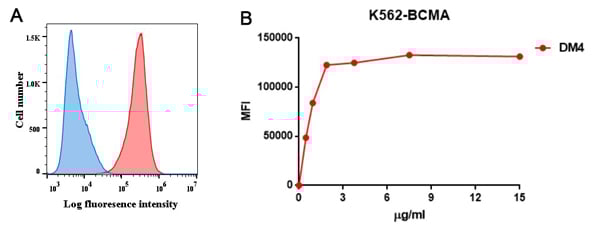
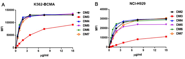
BCMA, Monoclonal Antibody (Cat# AAA11703)
Full Name
Anti-BCMA antibody (DM4), Rabbit mAb
Reactivity
Human
Applications
Flow Cytometry, Immunoprecipitation, Immunofluorescence
Purity
Purified from cell culture supernatant by affinity chromatography
Pricing


 IP (Immunoprecipitation)
(Figure 6. Immunoprecipitation analysis. Cellular overexpression lysates (made from HEK293F cells transfected with DYKDDDDK tagged human BCMA full length gene) were pre-incubated with 6 different rabbit DimAb clones and negative control IgG. The immunocomplexes were further pulled down by protein A beads, fractionated, and blotted with mouse anti-DYKDDDDK monoclonal antibody.)
IP (Immunoprecipitation)
(Figure 6. Immunoprecipitation analysis. Cellular overexpression lysates (made from HEK293F cells transfected with DYKDDDDK tagged human BCMA full length gene) were pre-incubated with 6 different rabbit DimAb clones and negative control IgG. The immunocomplexes were further pulled down by protein A beads, fractionated, and blotted with mouse anti-DYKDDDDK monoclonal antibody.)
BCMA, Monoclonal Antibody (Cat# AAA11704)
Full Name
Anti-BCMA antibody (DM6), Rabbit mAb
Reactivity
Human
Applications
Flow Cytometry, Immunofluorescence
Purity
Purified from cell culture supernatant by affinity chromatography
Pricing


 WB (Western Blot)
(FG Pancreatic Carcinoma Cell Lines stably expressing vector along (FG-V) the b3 integrin subunit (FG-b3) or a b3 truncation mutant (FG-759x). Src Mab (AAA28639) was diluted 1:500 in 1% BSA/TBST and incubated Overnight at 4 degree C. After washing 3x 5 min. with TBST the blots were incubated with 1:5000 Goat anti-mouse or Goat anti-rabbit secondary antibody for 1 hr at Room temperature. The blots were again washed 3x 5 min. with TBST and developed using ECL reagent.Data and protocol kindly provided by Dr. Weis of Cheresh Lab, UCSD.)
WB (Western Blot)
(FG Pancreatic Carcinoma Cell Lines stably expressing vector along (FG-V) the b3 integrin subunit (FG-b3) or a b3 truncation mutant (FG-759x). Src Mab (AAA28639) was diluted 1:500 in 1% BSA/TBST and incubated Overnight at 4 degree C. After washing 3x 5 min. with TBST the blots were incubated with 1:5000 Goat anti-mouse or Goat anti-rabbit secondary antibody for 1 hr at Room temperature. The blots were again washed 3x 5 min. with TBST and developed using ECL reagent.Data and protocol kindly provided by Dr. Weis of Cheresh Lab, UCSD.)
SRC, Monoclonal Antibody (Cat# AAA28639)
Full Name
SRC Antibody
Gene Names
SRC; ASV; SRC1; c-SRC; p60-Src
Reactivity
Human, mouse
Applications
WB, EIA, IF
Purity
This antibody is purified through a protein G column, followed by dialysis against PBS.
Pricing
 Application Data
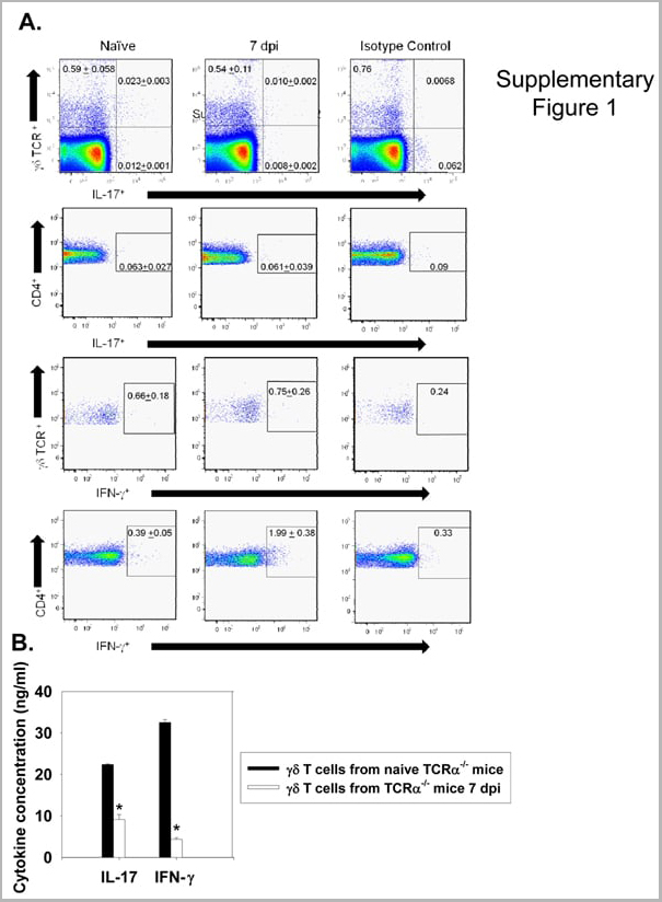
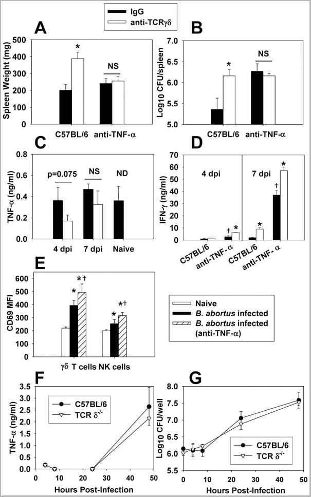
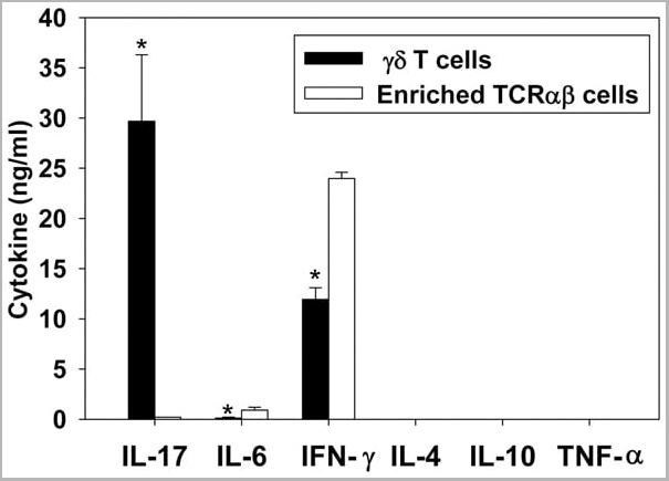
(Published customer image: gamma delta T cells are the primary source of IL-17 during B. abortus infection. C57BL/6 mice were infected i.p. with 5x10⁴ CFUs of B. abortus 2308, and two weeks later gamma delta T cells (>95% purity) and an enriched TCRalphabeta (~55% CD4+, 25% CD8+) cell fraction were isolated from the spleens of infected mice. Cells were stimulated with 500 ng/ml ionomycin and 50 ng/ml PMA for three days, and cell-free supernatants from triplicate wells were assayed for cytokine production via ELISA. The mean +/- SD is shown; * P)
Application Data
(Published customer image: gamma delta T cells are the primary source of IL-17 during B. abortus infection. C57BL/6 mice were infected i.p. with 5x10⁴ CFUs of B. abortus 2308, and two weeks later gamma delta T cells (>95% purity) and an enriched TCRalphabeta (~55% CD4+, 25% CD8+) cell fraction were isolated from the spleens of infected mice. Cells were stimulated with 500 ng/ml ionomycin and 50 ng/ml PMA for three days, and cell-free supernatants from triplicate wells were assayed for cytokine production via ELISA. The mean +/- SD is shown; * P)
IFN GAMMA, Monoclonal Antibody (Cat# AAA12093)
Full Name
MOUSE ANTI BOVINE INTERFERON GAMMA
Reactivity
Dog, Dolphin, Ferret, Fin Whale, Goat, Horse, Human, Mink, Rabbit, Pig, Sheep.
Based on sequence similarity, is expected to react with: Mustelid
N.B. Antibody reactivity and working conditions may vary between species.
Based on sequence similarity, is expected to react with: Mustelid
N.B. Antibody reactivity and working conditions may vary between species.
Applications
Flow Cytometry
Pricing