Filters
Clonality
Type
Reactivity
Gene Name
Isotype
Host
Application
Clone
24 results for "Negative Isotype Control" - showing 1-24

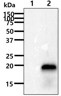 Western Blot (WB)
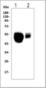
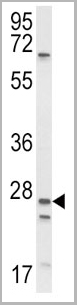
(The cell lysates(40ug) were resolved by SDS-PAGE, transferred to PVDF membrane and probed with anti-human TREM2 antibody (1:1000). Proteins were visualized using a goat anti-mouse secondary antibody conjugated to HRP and an ECL detection system.Lane 1 : 293T cell lysateLane 2 : TREM2 19-161aa Transfected 293T cell lysate)
Western Blot (WB)
(The cell lysates(40ug) were resolved by SDS-PAGE, transferred to PVDF membrane and probed with anti-human TREM2 antibody (1:1000). Proteins were visualized using a goat anti-mouse secondary antibody conjugated to HRP and an ECL detection system.Lane 1 : 293T cell lysateLane 2 : TREM2 19-161aa Transfected 293T cell lysate)
TREM2, Monoclonal Antibody (Cat# AAA332953)
Full Name
Human TREM2 antibody
Reactivity
Human
Applications
Western Blot, Immunocytochemistry, Flow Cytometry
Purity
By protein-A affinity chromatography
Pricing


 Application Data
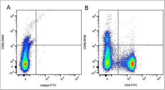
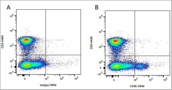
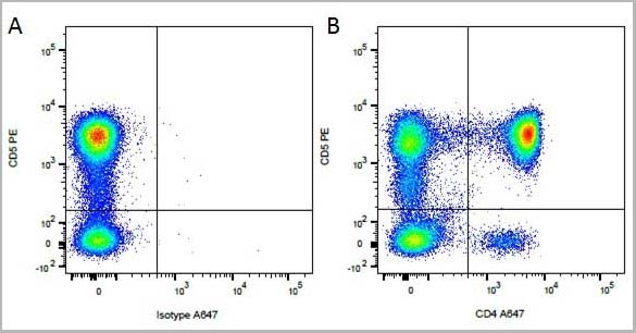
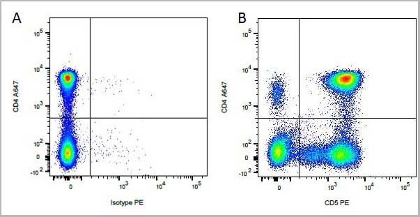
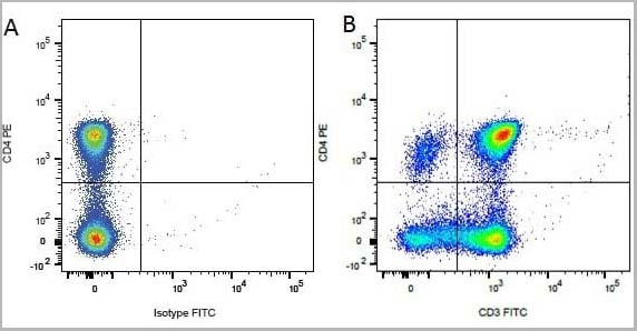
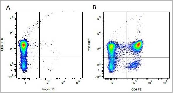
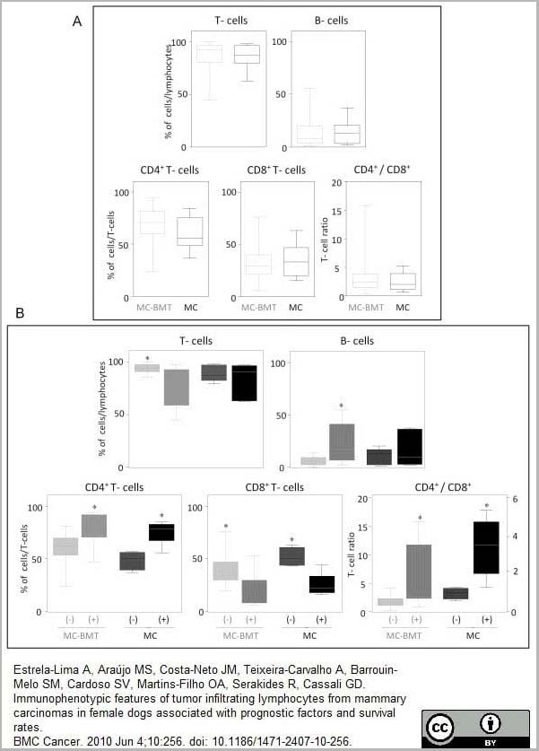
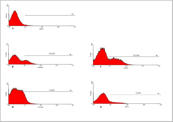
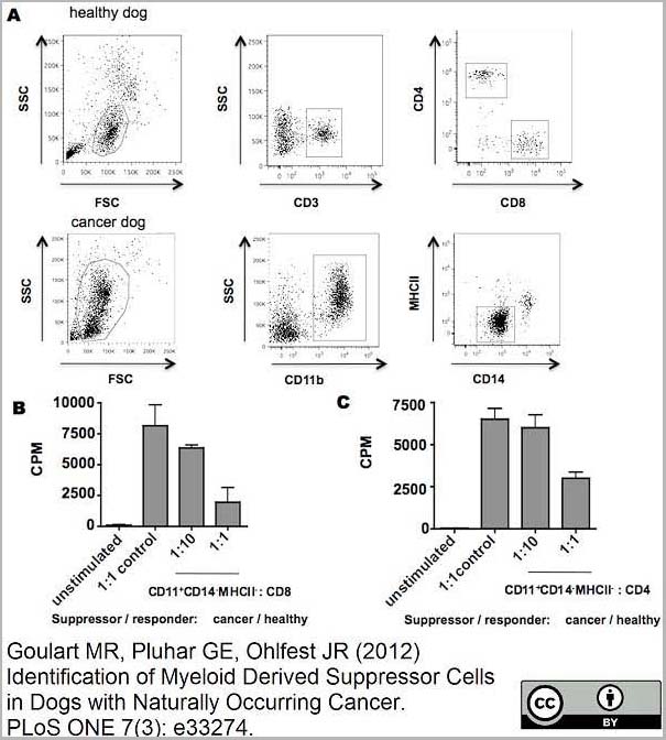
(Published customer image:RPE conjugated Mouse anti Canine CD4 antibody, clone YKIC302.9used for the assessment of CD4 levels on canine cells by flow cytometry.Image caption:Immunophenotypic profile of tumor infiltrating lymphocyte in canine mammary carcinomas. Analysis of tumor infiltrating T-cells, B-lymphocytes and T-cell subsets from MC-BMT or MC (A), further subcategorized according to the absence (-) or presence (+) of lymph node metastasis (-) (B). Lymphocyte populations and subsets were identified by flow cytometric immunostaining as described in Material and Methods. Data were expressed as percentage of positive cells within gated lymphocytes and CD4+/CD8+ T-cell ratio. Significant differences at p < 0.05 are highlighted by asterisk.)
Application Data
(Published customer image:RPE conjugated Mouse anti Canine CD4 antibody, clone YKIC302.9used for the assessment of CD4 levels on canine cells by flow cytometry.Image caption:Immunophenotypic profile of tumor infiltrating lymphocyte in canine mammary carcinomas. Analysis of tumor infiltrating T-cells, B-lymphocytes and T-cell subsets from MC-BMT or MC (A), further subcategorized according to the absence (-) or presence (+) of lymph node metastasis (-) (B). Lymphocyte populations and subsets were identified by flow cytometric immunostaining as described in Material and Methods. Data were expressed as percentage of positive cells within gated lymphocytes and CD4+/CD8+ T-cell ratio. Significant differences at p < 0.05 are highlighted by asterisk.)
CD4, Monoclonal Antibody (Cat# AAA12278)
Full Name
RAT ANTI DOG CD4: APC
Reactivity
Dog
Applications
Flow Cytometry
Pricing


 FCM (Flow Cytometry)
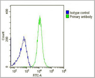
(HAS2 Antibody (Center) (AAA28714) flow cytometric analysis of K562 cells (bottom histogram) compared to a Rabbit IgG Isotype control (negative control-top histogram).FITC-conjugated goat-anti-rabbit secondary antibodies were used for the analysis.)
FCM (Flow Cytometry)
(HAS2 Antibody (Center) (AAA28714) flow cytometric analysis of K562 cells (bottom histogram) compared to a Rabbit IgG Isotype control (negative control-top histogram).FITC-conjugated goat-anti-rabbit secondary antibodies were used for the analysis.)
HAS2, Polyclonal Antibody (Cat# AAA28714)
Full Name
HAS2 Antibody (Center)
Reactivity
Human, mouse
Applications
Western Blot, Immunohistochemistry, Flow Cytometry
Purity
Peptide Affinity Purified Rabbit Polyclonal Antibody (Pab)
Pricing


 SDS-PAGE
(SDS Page analysis of purified S100B Mouse Monoclonal Antibody (S100B/1012).)
SDS-PAGE
(SDS Page analysis of purified S100B Mouse Monoclonal Antibody (S100B/1012).)
S100B, Monoclonal Antibody (Cat# AAA23893)
Full Name
S100B (Astrocyte and Melanoma Marker)
Gene Names
S100B; NEF; S100; S100-B; S100beta
Reactivity
Human, Mouse, Rat, Cow. Others not known.
Applications
Flow Cytometry, Immunofluorescence, Western Blot, Immunohistochemistry
Pricing


 Application Data
(Published customer imageSLAMF3 blockade in human hepatocytes is associated with lower susceptibility to HCV. (A) SLAMF3 was stained in primary human hepatocytes (PHHs) and cells from the Huh-7 human hepatoma cell line with a specific antibody (HLy9.1.25 clone; grey) and an isotype-matched control (empty). One of four independent experiments is shown. Huh-7 cells were transfected with scrambled control (sc) siRNA or three specific siRNAs (#1, #2 and #3) targeting SLAMF3, prior to infection with HCVcc; siRNA efficiency was checked by quantifying SLAMF3 mRNA (B) and the CD81 expression level (C) by flow cytometry analysis at 48 h post-transfection. Results are presented as the mean +/-SD (n = 3). Intracellular viral RNA was quantified at 72 h p.i. (D) and the infection was measured at 72 h p.i. by focus-forming units FFUs counting (E) (as inhibition percent; mean of three independent experiments; error bars: SD. **p)
Application Data
(Published customer imageSLAMF3 blockade in human hepatocytes is associated with lower susceptibility to HCV. (A) SLAMF3 was stained in primary human hepatocytes (PHHs) and cells from the Huh-7 human hepatoma cell line with a specific antibody (HLy9.1.25 clone; grey) and an isotype-matched control (empty). One of four independent experiments is shown. Huh-7 cells were transfected with scrambled control (sc) siRNA or three specific siRNAs (#1, #2 and #3) targeting SLAMF3, prior to infection with HCVcc; siRNA efficiency was checked by quantifying SLAMF3 mRNA (B) and the CD81 expression level (C) by flow cytometry analysis at 48 h post-transfection. Results are presented as the mean +/-SD (n = 3). Intracellular viral RNA was quantified at 72 h p.i. (D) and the infection was measured at 72 h p.i. by focus-forming units FFUs counting (E) (as inhibition percent; mean of three independent experiments; error bars: SD. **p)
CD229, Monoclonal Antibody (Cat# AAA11951)
Full Name
MOUSE ANTI HUMAN CD229
Gene Names
LY9; hly9; mLY9; CD229; SLAMF3
Applications
Flow Cytometry, Immunoprecipitation
Pricing


 IF (Immunofluorescence)
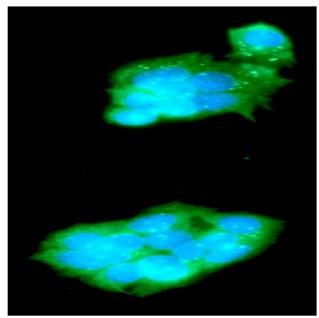
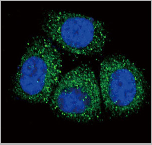
(Confocal Immunofluorescent analysis of SK-OV-3 cells using AF488-labeled Isotype Control Monoclonal Antibody (IgG1) (Green).DAPI was used to stain the cell nuclei (blue). (Negative Control))
IF (Immunofluorescence)
(Confocal Immunofluorescent analysis of SK-OV-3 cells using AF488-labeled Isotype Control Monoclonal Antibody (IgG1) (Green).DAPI was used to stain the cell nuclei (blue). (Negative Control))
Ep-CAM /CD326, Monoclonal Antibody (Cat# AAA13838)
Full Name
Ep-CAM /CD326 (Epithelial Marker) Mouse Monoclonal Antibody
Gene Names
EPCAM; ESA; KSA; M4S1; MK-1; DIAR5; EGP-2; EGP40; KS1/4; MIC18; TROP1; EGP314; HNPCC8; TACSTD1
Reactivity
Human.
Does not react with Mouse and Rat
Does not react with Mouse and Rat
Applications
Flow Cytometry, Immunofluorescence, Western Blot, Immunohistochemistry
Pricing
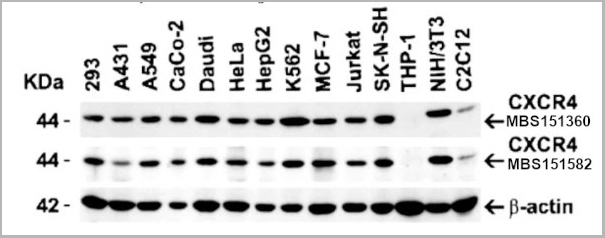
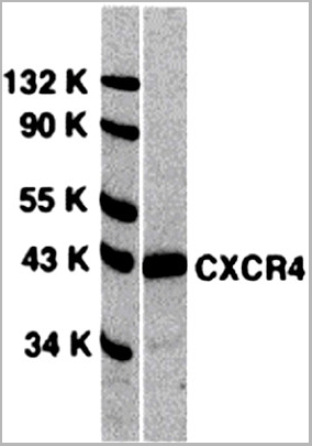
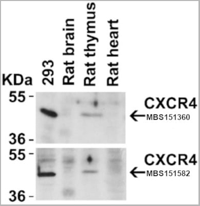 WB (Western Blot)
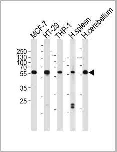
(Figure 4 Animal Species ReactivityLoading: Lysates/proteins at 20 μg per lane. Antibodies: 1009 (2 μg/mL) or 1012 (2 μg/mL). 1 h incubation at RT in 5% NFDM/TBST. Secondary: Goat anti-rabbit IgG HRP conjugate at 1:10000 dilution.)
WB (Western Blot)
(Figure 4 Animal Species ReactivityLoading: Lysates/proteins at 20 μg per lane. Antibodies: 1009 (2 μg/mL) or 1012 (2 μg/mL). 1 h incubation at RT in 5% NFDM/TBST. Secondary: Goat anti-rabbit IgG HRP conjugate at 1:10000 dilution.)
CXCR4, Polyclonal Antibody (Cat# AAA10951)
Full Name
CXCR4 Antibody
Gene Names
CXCR4; FB22; HM89; LAP3; LCR1; NPYR; WHIM; CD184; LAP-3; LESTR; NPY3R; NPYRL; HSY3RR; NPYY3R; D2S201E
Reactivity
Human, Mouse, Rat
Applications
Western Blot, Immunocytochemistry, Immunoprecipitation, Immunofluorescence, Immunohistochemistry, Flow Cytometry
Purity
CXCR4 Antibody is affinity chromatography purified via peptide column.
Pricing
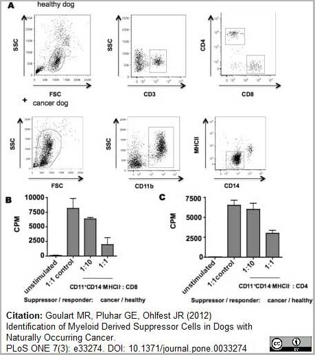
 Application Data
(Published customer image:RPE conjugated Mouse anti Canine CD4 antibody, clone YKIC302.9used for the assessment of CD4 levels on canine cells by flow cytometry.Image caption:Immunophenotypic profile of tumor infiltrating lymphocyte in canine mammary carcinomas. Analysis of tumor infiltrating T-cells, B-lymphocytes and T-cell subsets from MC-BMT or MC (A), further subcategorized according to the absence (-) or presence (+) of lymph node metastasis (-) (B). Lymphocyte populations and subsets were identified by flow cytometric immunostaining as described in Material and Methods. Data were expressed as percentage of positive cells within gated lymphocytes and CD4+/CD8+ T-cell ratio. Significant differences at p < 0.05 are highlighted by asterisk.From: From: Estrela-Lima et al.BMC Cancer 2010 10:256.)
Application Data
(Published customer image:RPE conjugated Mouse anti Canine CD4 antibody, clone YKIC302.9used for the assessment of CD4 levels on canine cells by flow cytometry.Image caption:Immunophenotypic profile of tumor infiltrating lymphocyte in canine mammary carcinomas. Analysis of tumor infiltrating T-cells, B-lymphocytes and T-cell subsets from MC-BMT or MC (A), further subcategorized according to the absence (-) or presence (+) of lymph node metastasis (-) (B). Lymphocyte populations and subsets were identified by flow cytometric immunostaining as described in Material and Methods. Data were expressed as percentage of positive cells within gated lymphocytes and CD4+/CD8+ T-cell ratio. Significant differences at p < 0.05 are highlighted by asterisk.From: From: Estrela-Lima et al.BMC Cancer 2010 10:256.)
CD4, Monoclonal Antibody (Cat# AAA12279)
Full Name
RAT ANTI DOG CD4: RPE-Cy7
Applications
Flow Cytometry
Purity
Purified IgG prepared by affinity chromatography on Protein G from tissue culture supernatant
Pricing


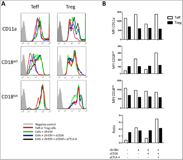
 Application Data
(Staining of human peripheral blood granulocytes with CD18: Alexa Fluor 647)
Application Data
(Staining of human peripheral blood granulocytes with CD18: Alexa Fluor 647)
CD18, Monoclonal Antibody (Cat# AAA12051)
Full Name
RAT ANTI HUMAN CD18:RPE
Gene Names
ITGB2; LAD; CD18; MF17; MFI7; LCAMB; LFA-1; MAC-1
Applications
Flow Cytometry
Pricing


 Application Data
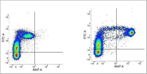
(Staining of human peripheral blood granulocytes with CD18: Alexa Fluor 647 (AAA11986A647))
Application Data
(Staining of human peripheral blood granulocytes with CD18: Alexa Fluor 647 (AAA11986A647))
CD18, Monoclonal Antibody (Cat# AAA11986)
Full Name
RAT ANTI HUMAN CD18
Gene Names
ITGB2; LAD; CD18; MF17; MFI7; LCAMB; LFA-1; MAC-1
Reactivity
Dog, Guinea Pig
Applications
Immunohistochemistry, Flow Cytometry, Immunoprecipitation
Pricing


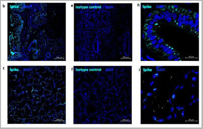
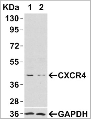
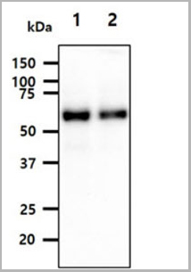
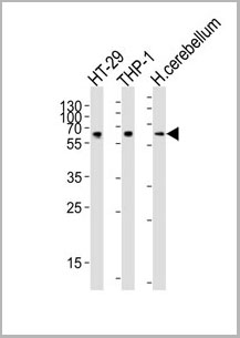
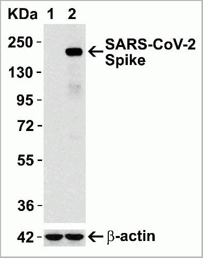 WB (Western Blot)
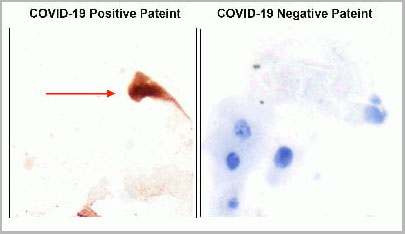
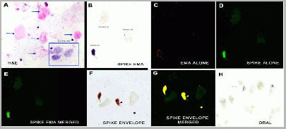
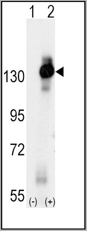
(Figure 12 Overexpression Validation in Spike Transfected 293 Cells Loading: 15 ug per lane of 293 cell lysate. Antibodies: SARS-CoV-2 (COVID-19) Spike, AAA10931 (1 ug/mL), 1h incubation at RT in 5% NFDM/TBST. Secondary: Goat anti rabbit IgG HRP conjugate at 1:10000 dilution. Lane 1: WT293 cells and Lane 2: SARS-CoV-2 Spike overexpressed 293 cells.)
WB (Western Blot)
(Figure 12 Overexpression Validation in Spike Transfected 293 Cells Loading: 15 ug per lane of 293 cell lysate. Antibodies: SARS-CoV-2 (COVID-19) Spike, AAA10931 (1 ug/mL), 1h incubation at RT in 5% NFDM/TBST. Secondary: Goat anti rabbit IgG HRP conjugate at 1:10000 dilution. Lane 1: WT293 cells and Lane 2: SARS-CoV-2 Spike overexpressed 293 cells.)
COVID 19 Spike Protein Coronavirus, Polyclonal Antibody (Cat# AAA10931)
Full Name
SARS-CoV-2 (COVID-19, 2019-nCoV) Spike Antibody
Reactivity
Virus
Applications
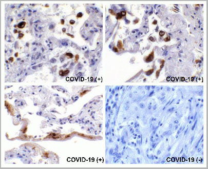
Immunofluorescence, Immunohistochemistry, Western Blot
Purity
SARS-CoV-2 (COVID-19, 2019-nCoV) Spike Antibody is affinity chromatography purified via peptide column.
Pricing


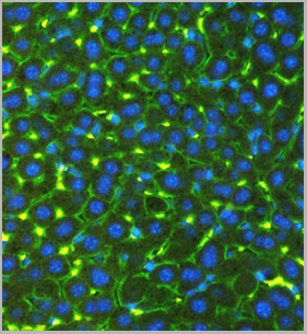
 Application Data
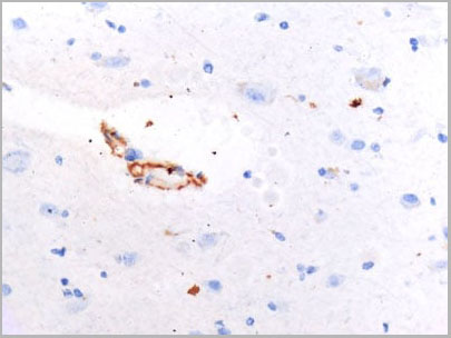
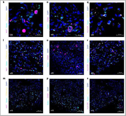
(Immunofluorescence staining of mouse lymph node cryosection with Rat anti Mouse CD11b, clone 5C6 , green in A and Rat anti Mouse CD8, clone YTS105.18 , red in B. C is the merged image with nuclei counterstained blue using DAPI. High power)
Application Data
(Immunofluorescence staining of mouse lymph node cryosection with Rat anti Mouse CD11b, clone 5C6 , green in A and Rat anti Mouse CD8, clone YTS105.18 , red in B. C is the merged image with nuclei counterstained blue using DAPI. High power)
CD8 ALPHA, Monoclonal Antibody (Cat# AAA12069)
Full Name
RAT ANTI MOUSE CD8
Gene Names
Cd8a; Ly-2; Ly-B; Ly-35; Lyt-2; BB154331
Reactivity
Mouse
Applications
Immunohistochemistry, Flow Cytometry, Immunofluorescence
Pricing


 Application Data
(Staining of human peripheral blood granulocytes with CD18: Alexa Fluor 647)
Application Data
(Staining of human peripheral blood granulocytes with CD18: Alexa Fluor 647)
CD18, Monoclonal Antibody (Cat# AAA11881)
Full Name
RAT ANTI HUMAN CD18:FITC
Gene Names
ITGB2; LAD; CD18; MF17; MFI7; LCAMB; LFA-1; MAC-1
Applications
Flow Cytometry
Pricing


 Application Data
(Staining of human peripheral blood granulocytes with CD18: Alexa Fluor 647)
Application Data
(Staining of human peripheral blood granulocytes with CD18: Alexa Fluor 647)
CD18, Monoclonal Antibody (Cat# AAA11987)
Full Name
RAT ANTI HUMAN CD18
Gene Names
ITGB2; LAD; CD18; MF17; MFI7; LCAMB; LFA-1; MAC-1
Applications
Immunohistochemistry, Flow Cytometry, Immunoprecipitation
Pricing
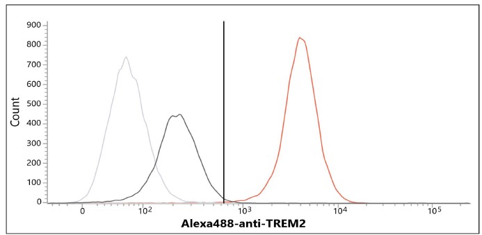
 FCM (Flow Cytometry)
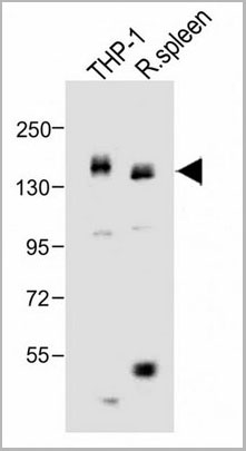
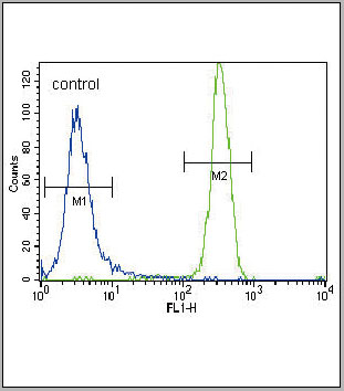
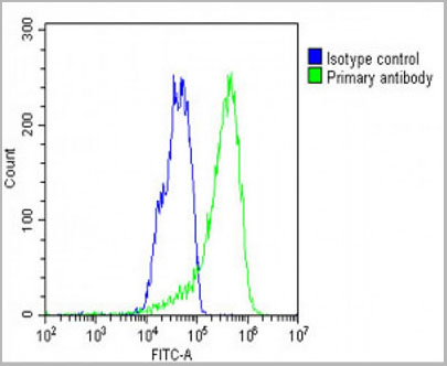
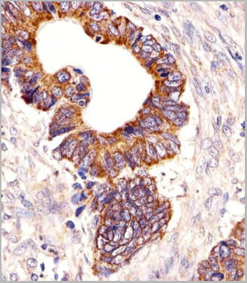
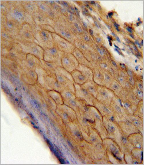
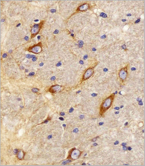
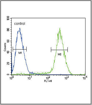
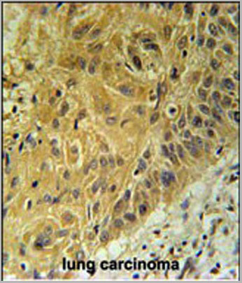
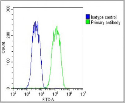
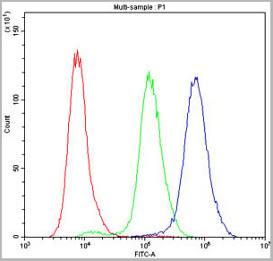
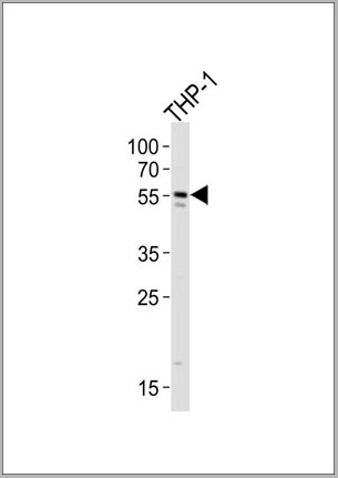
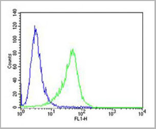
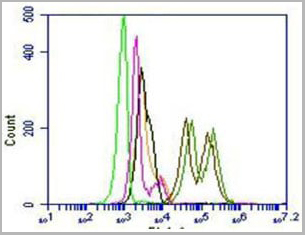
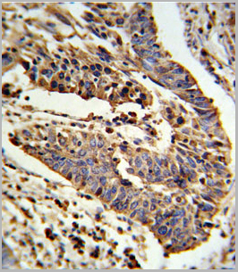
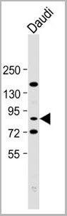
(WB - MCSF Receptor (CSF1R) Antibody (C-term) AAA28757 detail IHC-P - MCSF Receptor (CSF1R) Antibody (C-term) AAA28757 detail IHC-P - MCSF Receptor (CSF1R) Antibody (C-term) AAA28757 detail FC - MCSF Receptor (CSF1R) Antibody (C-term) AAA28757 detail Overlay histogram showing HepG2 cells stained with AAA28757(green line). The cells were fixed with 2% paraformaldehyde (10 min) and then permeabilized with 90% methanol for 10 min. The cells were then icubated in 2% bovine serum albumin to block non-specific protein-protein interactions followed by the antibody (AAA28757, 1:25 dilution) for 60 min at 37ºC. The secondary antibody used was Goat-Anti-Rabbit IgG, DyLight® 488 Conjugated Highly Cross-Adsorbed(1583138) at 1/200 dilution for 40 min at 37ºC. Isotype control antibody (blue line) was rabbit IgG1 (1ug/1x10^6 cells) used under the same conditions. Acquisition of >10, 000 events was performed.)
FCM (Flow Cytometry)
(WB - MCSF Receptor (CSF1R) Antibody (C-term) AAA28757 detail IHC-P - MCSF Receptor (CSF1R) Antibody (C-term) AAA28757 detail IHC-P - MCSF Receptor (CSF1R) Antibody (C-term) AAA28757 detail FC - MCSF Receptor (CSF1R) Antibody (C-term) AAA28757 detail Overlay histogram showing HepG2 cells stained with AAA28757(green line). The cells were fixed with 2% paraformaldehyde (10 min) and then permeabilized with 90% methanol for 10 min. The cells were then icubated in 2% bovine serum albumin to block non-specific protein-protein interactions followed by the antibody (AAA28757, 1:25 dilution) for 60 min at 37ºC. The secondary antibody used was Goat-Anti-Rabbit IgG, DyLight® 488 Conjugated Highly Cross-Adsorbed(1583138) at 1/200 dilution for 40 min at 37ºC. Isotype control antibody (blue line) was rabbit IgG1 (1ug/1x10^6 cells) used under the same conditions. Acquisition of >10, 000 events was performed.)
MCSF Receptor (CSF1R), Polyclonal Antibody (Cat# AAA28757)
Full Name
MCSF Receptor (CSF1R) Antibody (C-term)
Gene Names
CSF1R; FMS; CSFR; FIM2; HDLS; C-FMS; CD115; CSF-1R; M-CSF-R
Reactivity
Human
Applications
Western Blot, Flow Cytometry, Immunohistochemistry
Purity
Purified Rabbit Polyclonal Antibody (Pab)
Pricing
 FCM (Flow Cytometry)
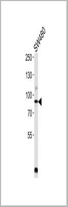
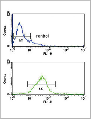
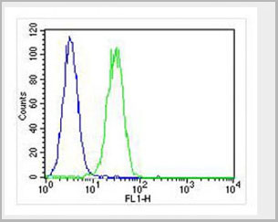
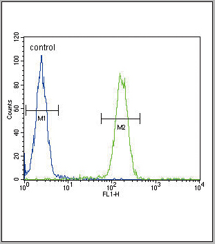
(PCSK9 Antibody (N-term) (Cat. #AAA28774) flow cytometry analysis of Jurkat cells (bottom histogram) compared to a negative control cell(top histogram).FITC-conjugated goat-anti-rabbit secondary antibodies were used for the analysis.)
FCM (Flow Cytometry)
(PCSK9 Antibody (N-term) (Cat. #AAA28774) flow cytometry analysis of Jurkat cells (bottom histogram) compared to a negative control cell(top histogram).FITC-conjugated goat-anti-rabbit secondary antibodies were used for the analysis.)
PCSK9, Polyclonal Antibody (Cat# AAA28774)
Full Name
PCSK9 Antibody (N-term)
Gene Names
PCSK9; FH3; PC9; NARC1; LDLCQ1; NARC-1; HCHOLA3
Reactivity
Human
Applications
Western Blot, Immunohistochemistry, Flow Cytometry
Pricing
 Application Data
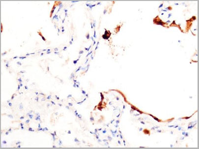
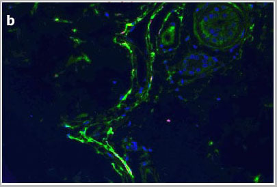
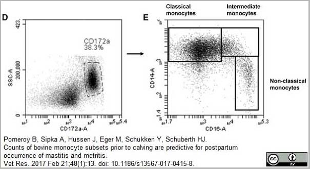
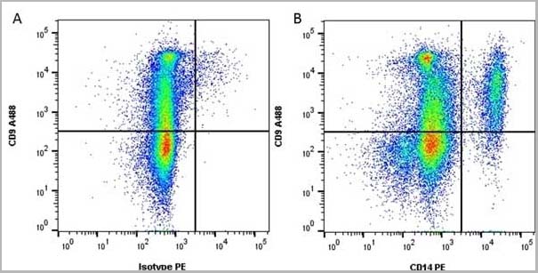
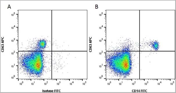
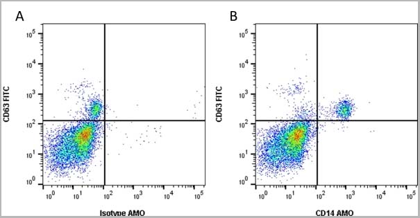
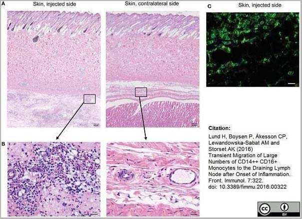
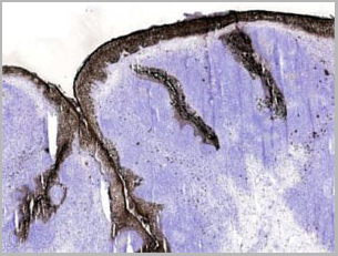
(Mouse anti Human CD14 antibody, clone Tük4 used to identify bovine monocytes in the skin and subcutaneous tissue of adjuvant injected calves by immunofluorescence.Image caption:Cellular recruitment to skin and subcutaneous tissues. (A) HE stained sections of skin with subcutaneous tissue from the side injected with adjuvant and the contralateral side, at 24 h post-injection. Scale bars: 200 ?m. (B) Enlargement of outlined areas in A, as indicated. Scale bars: 20 ?m. (C) Immunofluorescent labeling of subcutaneous tissue on the injected side with antibody against CD14 (green). Scale bar: 20 ?m.From: Lund H, Boysen P, Åkesson CP, Lewandowska-Sabat AM and Storset AK (2016)Transient Migration of Large Numbers of CD14++ CD16+ Monocytes to the Draining Lymph Node after Onset of Inflammation.Front. Immunol. 7:322.This is from an open access article distributed under the terms of the Creative Commons Attribution License.)
Application Data
(Mouse anti Human CD14 antibody, clone Tük4 used to identify bovine monocytes in the skin and subcutaneous tissue of adjuvant injected calves by immunofluorescence.Image caption:Cellular recruitment to skin and subcutaneous tissues. (A) HE stained sections of skin with subcutaneous tissue from the side injected with adjuvant and the contralateral side, at 24 h post-injection. Scale bars: 200 ?m. (B) Enlargement of outlined areas in A, as indicated. Scale bars: 20 ?m. (C) Immunofluorescent labeling of subcutaneous tissue on the injected side with antibody against CD14 (green). Scale bar: 20 ?m.From: Lund H, Boysen P, Åkesson CP, Lewandowska-Sabat AM and Storset AK (2016)Transient Migration of Large Numbers of CD14++ CD16+ Monocytes to the Draining Lymph Node after Onset of Inflammation.Front. Immunol. 7:322.This is from an open access article distributed under the terms of the Creative Commons Attribution License.)
CD14, Monoclonal Antibody (Cat# AAA12266)
Full Name
Mouse Anti Human CD14: Amethyst Orange
Reactivity
Human
Applications
Flow Cytometry
Purity
Purified IgG prepared by affinity chromatography on Protein A from tissue culture supernatant.
Pricing
 FCM (Flow Cytometry)
(CB2 Antibody (C-term) flow cytometric analysis of Jurkat cells (right histogram) compared to a negative control cell (left histogram).FITC-conjugated goat-anti-rabbit secondary antibodies were used for the analysis.)
FCM (Flow Cytometry)
(CB2 Antibody (C-term) flow cytometric analysis of Jurkat cells (right histogram) compared to a negative control cell (left histogram).FITC-conjugated goat-anti-rabbit secondary antibodies were used for the analysis.)
CB2, Polyclonal Antibody (Cat# AAA28672)
Full Name
CB2 Antibody (C-term)
Gene Names
CNR2; CB2; CX5; CB-2
Reactivity
Human
Applications
Immunohistochemistry, Flow Cytometry, Western Blot
Purity
This antibody is purified through a protein A column, followed by peptide affinity purification.
Pricing
 FCM (Flow Cytometry)
(Flow cytometry analysis of PARP2 in U87MG cells. The cell was stained with AAA11732 at 2-5ug for 1x10^6cells (red). A Goat anti mouse IgG (Alexa fluor 488) was used as the secondary antibody. Mouse monoclonal IgG was used as the isotype control (blue), cells without incubation with primary and secondary antibody was used as the negative control (black))
FCM (Flow Cytometry)
(Flow cytometry analysis of PARP2 in U87MG cells. The cell was stained with AAA11732 at 2-5ug for 1x10^6cells (red). A Goat anti mouse IgG (Alexa fluor 488) was used as the secondary antibody. Mouse monoclonal IgG was used as the isotype control (blue), cells without incubation with primary and secondary antibody was used as the negative control (black))
PARP2, Monoclonal Antibody (Cat# AAA11732)
Full Name
PARP2 antibody
Gene Names
PARP2; ARTD2; ADPRT2; PARP-2; ADPRTL2; ADPRTL3; pADPRT-2
Reactivity
Human
Applications
Western Blot, Flow Cytometry, Immunocytochemistry, Immunofluorescence
Purity
By protein-A affinity chromatography
Pricing
 FCM (Flow Cytometry)
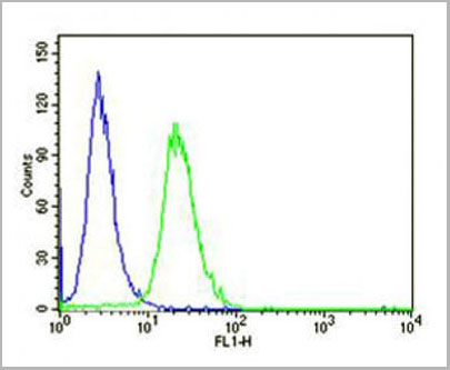
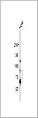
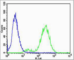
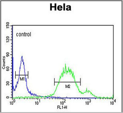
(Overlay histogram showing Hela cells stained with AAA28750 (green line). The cells were fixed with 2% paraformaldehyde (10 min) and then permeabilized with 90% methanol for 10 min. The cells were then icubated in 2% bovine serum albumin to block non-specific protein-protein interactions followed by the antibody (AAA28750, 1:25 dilution) for 60 min at 37ºC. The secondary antibody used was Goat-Anti-Rabbit IgG, DyLight® 488 Conjugated Highly Cross-Adsorbed (1583138) at 1/200 dilution for 40 min at 37°C. Isotype control antibody (blue line) was rabbit IgG1 (1ug/1x106 cells) used under the same conditions. Acquisition of >10, 000 events wasperformed.)
FCM (Flow Cytometry)
(Overlay histogram showing Hela cells stained with AAA28750 (green line). The cells were fixed with 2% paraformaldehyde (10 min) and then permeabilized with 90% methanol for 10 min. The cells were then icubated in 2% bovine serum albumin to block non-specific protein-protein interactions followed by the antibody (AAA28750, 1:25 dilution) for 60 min at 37ºC. The secondary antibody used was Goat-Anti-Rabbit IgG, DyLight® 488 Conjugated Highly Cross-Adsorbed (1583138) at 1/200 dilution for 40 min at 37°C. Isotype control antibody (blue line) was rabbit IgG1 (1ug/1x106 cells) used under the same conditions. Acquisition of >10, 000 events wasperformed.)
WNT5A, Polyclonal Antibody (Cat# AAA28750)
Full Name
WNT5A Antibody (Center)
Gene Names
WNT5A; hWNT5A
Reactivity
Human, Mouse, Rat
Predicted: Rabbit
Predicted: Rabbit
Applications
Immunohistochemistry, Immunohistochemistry, Flow Cytometry, Western Blot
Purity
This antibody is purified through a protein A column, followed by peptide affinity purification.
Pricing
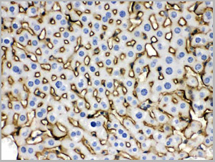
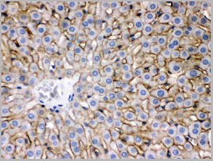
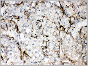
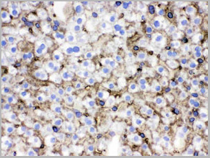
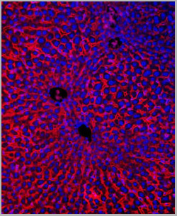
 WB (Western Blot)
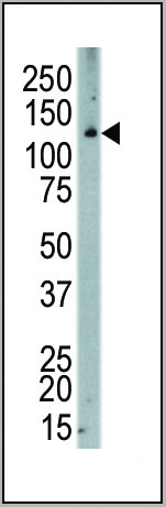
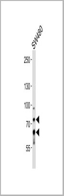
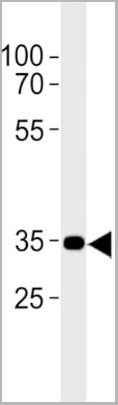
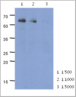
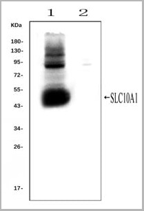
(Western blot analysis of SLC10A1 using anti- SLC10A1 antibody (AAA11653).Electrophoresis was performed on a 5-20% SDS-PAGE gel at 70V (Stacking gel) / 90V (Resolving gel) for 2-3 hours. The sample well of each lane was loaded with 50ug of sample under reducing conditions.Lane 1: rat liver tissue lysates (positive control), Lane 2: rat kidney tissue lysates, (negative control)After Electrophoresis, proteins were transferred to a Nitrocellulose membrane at 150mA for 50-90 minutes. Blocked the membrane with 5% Non-fat Milk/ TBS for 1.5 hour at RT. The membrane was incubated with rabbit anti-SLC10A1 antigen affinity purified polyclonal antibody (AAA11653) at 0.25ug/mL overnight at 4°C, then washed with TBS-0.1%Tween 3 times with 5 minutes each and probed with a goat anti-rabbit IgG-HRP secondary antibody at a dilution of 1:10000 for 1.5 hour at RT. The signal is developed using an Enhanced Chemiluminescent detection (ECL) kit with Tanon 5200 system. A specific band was detected for SLC10A1 at approximately 50KD. The expected band size for SLC10A1 is at 38KD.)
WB (Western Blot)
(Western blot analysis of SLC10A1 using anti- SLC10A1 antibody (AAA11653).Electrophoresis was performed on a 5-20% SDS-PAGE gel at 70V (Stacking gel) / 90V (Resolving gel) for 2-3 hours. The sample well of each lane was loaded with 50ug of sample under reducing conditions.Lane 1: rat liver tissue lysates (positive control), Lane 2: rat kidney tissue lysates, (negative control)After Electrophoresis, proteins were transferred to a Nitrocellulose membrane at 150mA for 50-90 minutes. Blocked the membrane with 5% Non-fat Milk/ TBS for 1.5 hour at RT. The membrane was incubated with rabbit anti-SLC10A1 antigen affinity purified polyclonal antibody (AAA11653) at 0.25ug/mL overnight at 4°C, then washed with TBS-0.1%Tween 3 times with 5 minutes each and probed with a goat anti-rabbit IgG-HRP secondary antibody at a dilution of 1:10000 for 1.5 hour at RT. The signal is developed using an Enhanced Chemiluminescent detection (ECL) kit with Tanon 5200 system. A specific band was detected for SLC10A1 at approximately 50KD. The expected band size for SLC10A1 is at 38KD.)
SLC10A1, Polyclonal Antibody (Cat# AAA11653)
Full Name
Anti-SLC10A1 Antibody
Gene Names
Slc10a1; Ntcp
Reactivity
Mouse, Rat
Applications
Flow Cytometry, Immunofluorescence, Immunohistochemistry, Immunohistochemistry, Immunocytochemistry, Western Blot
Purity
Immunogen affinity purified.
Pricing
 Application Data
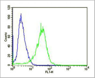
(Overlay histogram showing U-2OS cells stained with AP10510c (green line). The cells were fixed with 2% paraformaldehyde (10 min) and then permeabilized with 90% methanol for 10 min. The cells were then icubated n 2% bovine serum albumin to block non-specificprotein-protein interactions followed by the antibody (AP10510c, 1:25 dilution) for 60 min at 37ºC. The secondary antibody used wasGoat-Anti-Rabbit IgG, DyLight® 488 Conjugated Highly Cross-Adsorbed(NA168821) at 1/400 dilution for 40 min at 37ºC. Isotype control antibody (blue line) was rabbit IgG (1?g/1x10^6 cells) used under the same conditions. Acquisition of >10, 000 events wasperformed)
Application Data
(Overlay histogram showing U-2OS cells stained with AP10510c (green line). The cells were fixed with 2% paraformaldehyde (10 min) and then permeabilized with 90% methanol for 10 min. The cells were then icubated n 2% bovine serum albumin to block non-specificprotein-protein interactions followed by the antibody (AP10510c, 1:25 dilution) for 60 min at 37ºC. The secondary antibody used wasGoat-Anti-Rabbit IgG, DyLight® 488 Conjugated Highly Cross-Adsorbed(NA168821) at 1/400 dilution for 40 min at 37ºC. Isotype control antibody (blue line) was rabbit IgG (1?g/1x10^6 cells) used under the same conditions. Acquisition of >10, 000 events wasperformed)
CF150, Polyclonal Antibody (Cat# AAA28789)
Full Name
CF150 Antibody (Center)
Gene Names
MB21D1; cGAS; h-cGAS; C6orf150
Reactivity
Human
Applications
Flow Cytometry, Western Blot
Purity
Peptide Affinity Purified Rabbit Polyclonal Antibody (Pab)
Pricing
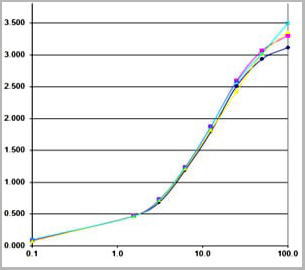 IA (Immuno Assay)
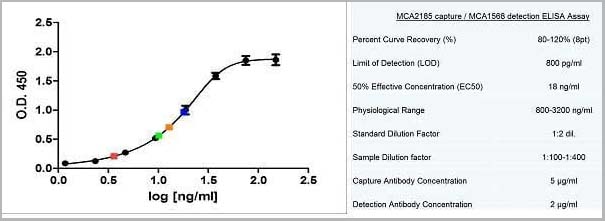
(used as capture antibody. Different concentrations were tested (0.5-2 ug/ml).)
IA (Immuno Assay)
(used as capture antibody. Different concentrations were tested (0.5-2 ug/ml).)
Calprotectin, Monoclonal Antibody (Cat# AAA14609)
Full Name
Calprotectin, Human, mAb 27E10, Biotin
Reactivity
Mouse: No; Rhesus Monkey: Yes (subpopulations of macrophages)
Applications
Immunohistochemistry, Immunofluorescence, Flow Cytometry, Immunoassay, Immunoprecipitation, Western Blot
Purity
Protein G
Pricing
 FCM (Flow Cytometry)
(SCNN1A Antibody (Center) flow cytometric analysis of WiDr cells (right histogram) compared to a negative control cell (left histogram).FITC-conjugated goat-anti-rabbit secondary antibodies were used for the analysis.)
FCM (Flow Cytometry)
(SCNN1A Antibody (Center) flow cytometric analysis of WiDr cells (right histogram) compared to a negative control cell (left histogram).FITC-conjugated goat-anti-rabbit secondary antibodies were used for the analysis.)
SCNN1A, Polyclonal Antibody (Cat# AAA28784)
Full Name
SCNN1A Antibody (Center)
Gene Names
SCNN1A; BESC2; ENaCa; SCNEA; SCNN1; ENaCalpha
Reactivity
Human
Applications
Western Blot, Immunohistochemistry, Immunofluorescence, Flow Cytometry
Purity
Peptide Affinity Purified Rabbit Polyclonal Antibody (Pab)
Pricing