Filters
Clonality
Type
Reactivity
Gene Name
Isotype
Host
Application
Clone
135 results for "Mouse Anti Rat IgG2b" - showing 1-50
 IF (Immunofluorescence)
(BALB/c mouse splenocytes were stained with Rat Anti-Mouse Lamda-PE and Rat Anti-Mouse CD119-FITC.)
IF (Immunofluorescence)
(BALB/c mouse splenocytes were stained with Rat Anti-Mouse Lamda-PE and Rat Anti-Mouse CD119-FITC.)
Rat Anti-Mouse Lambda (lambda light chain specific), Monoclonal Secondary Antibody (Cat# AAA14893)
Full Name
Rat Anti-Mouse Lambda-UNLB
Reactivity
Mouse
Applications
Flow Cytometry
Pricing


 Application Data
(Published customer image:RPE conjugated Mouse anti Canine CD4 antibody, clone YKIC302.9used for the assessment of CD4 levels on canine cells by flow cytometry.Image caption:Immunophenotypic profile of tumor infiltrating lymphocyte in canine mammary carcinomas. Analysis of tumor infiltrating T-cells, B-lymphocytes and T-cell subsets from MC-BMT or MC (A), further subcategorized according to the absence (-) or presence (+) of lymph node metastasis (-) (B). Lymphocyte populations and subsets were identified by flow cytometric immunostaining as described in Material and Methods. Data were expressed as percentage of positive cells within gated lymphocytes and CD4+/CD8+ T-cell ratio. Significant differences at p < 0.05 are highlighted by asterisk.)
Application Data
(Published customer image:RPE conjugated Mouse anti Canine CD4 antibody, clone YKIC302.9used for the assessment of CD4 levels on canine cells by flow cytometry.Image caption:Immunophenotypic profile of tumor infiltrating lymphocyte in canine mammary carcinomas. Analysis of tumor infiltrating T-cells, B-lymphocytes and T-cell subsets from MC-BMT or MC (A), further subcategorized according to the absence (-) or presence (+) of lymph node metastasis (-) (B). Lymphocyte populations and subsets were identified by flow cytometric immunostaining as described in Material and Methods. Data were expressed as percentage of positive cells within gated lymphocytes and CD4+/CD8+ T-cell ratio. Significant differences at p < 0.05 are highlighted by asterisk.)
CD4, Monoclonal Antibody (Cat# AAA12278)
Full Name
RAT ANTI DOG CD4: APC
Reactivity
Dog
Applications
Flow Cytometry
Pricing


 Application Data
(Staining of mouse peritoneal macrophages with RAT ANTI MOUSE F4/80 ANTIGEN:FITC)
Application Data
(Staining of mouse peritoneal macrophages with RAT ANTI MOUSE F4/80 ANTIGEN:FITC)
EMR1, Monoclonal Antibody (Cat# AAA26762)
Full Name
EMR1 (EGF-like Module Containing Mucin-like Hormone Receptor-like 1, EMR1 Hormone Receptor, Cell Surface Glycoprotein EMR1, Cell Surface Glycoprotein F4/80, DD7A5-7, EGF-TM7, F4/80, Gpf480, Lymphocyte Antigen 71, Ly71, TM7LN3) (HRP)
Reactivity
Mouse
Applications
Flow Cytometry, Immunohistochemistry, Immunoprecipitation, Radioimmunoassay, Western Blot
Purity
Purified by protein G affinity chromatography from tissue culture supernatant.
Pricing

 Application Data
(S100A8 in NIH-3T3 mouse cell line: S100A8 was detected in immersion fixed NIH-3T3 mouse embryonic fibroblast cell line using 10ug/mL AAA14670 for 3 hours at RT. Cells were stained (red) and counterstained with DAPI (blue).)
Application Data
(S100A8 in NIH-3T3 mouse cell line: S100A8 was detected in immersion fixed NIH-3T3 mouse embryonic fibroblast cell line using 10ug/mL AAA14670 for 3 hours at RT. Cells were stained (red) and counterstained with DAPI (blue).)
S100A8, Monoclonal Antibody (Cat# AAA14670)
Full Name
S100A8 (MRP-8)
Gene Names
S100A8; P8; MIF; NIF; CAGA; CFAG; CGLA; L1Ag; MRP8; CP-10; MA387; 60B8AG; S100A8
Reactivity
Mouse
Applications
Western Blot, Immunocytochemistry
Purity
Purified by Protein G affinity chromatography from hybridoma culture supernatant.
Pricing


 Application Data
(Staining of mouse peritoneal macrophages with Rat anti Mouse CD204: Alexa Fluor 488)
Application Data
(Staining of mouse peritoneal macrophages with Rat anti Mouse CD204: Alexa Fluor 488)
CD204, Monoclonal Antibody (Cat# AAA12080)
Full Name
RAT ANTI MOUSE CD204
Gene Names
Msr1; MSR; Scvr; MRS-A; MSR-A; SR-AI; SR-AII; Scara1
Applications
Immunohistochemistry, Flow Cytometry, Immunoprecipitation, Western Blot
Pricing


 Application Data
(Staining of mouse peritoneal macrophages with Rat anti Mouse CD204: Alexa Fluor 488)
Application Data
(Staining of mouse peritoneal macrophages with Rat anti Mouse CD204: Alexa Fluor 488)
CD204, Monoclonal Antibody (Cat# AAA12076)
Full Name
RAT ANTI MOUSE CD204:FITC
Gene Names
Msr1; MSR; Scvr; MRS-A; MSR-A; SR-AI; SR-AII; Scara1
Applications
Flow Cytometry
Pricing


 Application Data
(Staining of mouse spleen cells with Rat anti Mouse CD45 (Ly-5))
Application Data
(Staining of mouse spleen cells with Rat anti Mouse CD45 (Ly-5))
CD45, Monoclonal Antibody (Cat# AAA12021)
Full Name
RAT ANTI MOUSE CD45:RPE
Gene Names
Ptprc; loc; B220; Cd45; L-CA; Ly-5; T200; CD45R; Lyt-4
Applications
Flow Cytometry
Pricing


 Application Data
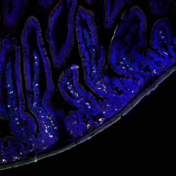
(Published Customer Image:Rat anti Mouse Gr-1 antibody, clone RB6-8C5 used for the identification of neutrophils by immunofluorescence.Image caption:S. typhimurium-Infected Macrophages Containing Phagocytosed Neutrophils and T Cells Confocal fluorescence microscopy of 50-mum-thick liver sections from 1-wk-infected Slc11a1 wild-type mice. (A-C) S.Typhimurium (O-antigen, arrows) are red, macrophages (F4-80 and MOMA-2) are blue, DNA (DAPI) is gray, phalloidin is green, and neutrophils (Gr-1/Ly-6G/RB6-8C5) are pink (arrowheads). (A) Collapsed image from a 40-mum Z-stack. Scale bar is 20 mum. (B and C) Sections from (A) that are 4 mum apart. The video from which (A-C) were derived (Video S2) is available online. (D-G) T cells within multinucleate macrophages.Macrophages (F4-80 and MOMA-2) are blue (D, G, and H), T cells (CD3zeta) are red (D, G, arrowheads), DAPI is gray (E, G), actin-bound phalloidin is green (F, G). (G) Is a composite of (D, E, and F). Scale bars are 16 mum. (H) An image from a different mouse stained and labeled as described for (D-G). Scale bar is 8 mum. A video showing a T cell inside of a macrophage is available online (Video S3).From: Nix RN, Altschuler SE, Henson PM, Detweiler CS (2007) Hemophagocytic Macrophages Harbor Salmonella enterica during Persistent Infection.PLoS Pathog 3(12): e193.)
Application Data
(Published Customer Image:Rat anti Mouse Gr-1 antibody, clone RB6-8C5 used for the identification of neutrophils by immunofluorescence.Image caption:S. typhimurium-Infected Macrophages Containing Phagocytosed Neutrophils and T Cells Confocal fluorescence microscopy of 50-mum-thick liver sections from 1-wk-infected Slc11a1 wild-type mice. (A-C) S.Typhimurium (O-antigen, arrows) are red, macrophages (F4-80 and MOMA-2) are blue, DNA (DAPI) is gray, phalloidin is green, and neutrophils (Gr-1/Ly-6G/RB6-8C5) are pink (arrowheads). (A) Collapsed image from a 40-mum Z-stack. Scale bar is 20 mum. (B and C) Sections from (A) that are 4 mum apart. The video from which (A-C) were derived (Video S2) is available online. (D-G) T cells within multinucleate macrophages.Macrophages (F4-80 and MOMA-2) are blue (D, G, and H), T cells (CD3zeta) are red (D, G, arrowheads), DAPI is gray (E, G), actin-bound phalloidin is green (F, G). (G) Is a composite of (D, E, and F). Scale bars are 16 mum. (H) An image from a different mouse stained and labeled as described for (D-G). Scale bar is 8 mum. A video showing a T cell inside of a macrophage is available online (Video S3).From: Nix RN, Altschuler SE, Henson PM, Detweiler CS (2007) Hemophagocytic Macrophages Harbor Salmonella enterica during Persistent Infection.PLoS Pathog 3(12): e193.)
Gr-1, Monoclonal Antibody (Cat# AAA12257)
Full Name
Rat Anti Mouse Gr-1: FITC
Reactivity
Mouse
Applications
Flow Cytometry
Purity
Purified IgG prepared by affinity chromatography on Protein G from tissue culture supernatant
Pricing


 Application Data
(Staining of mouse peritoneal macrophages with RAT ANTI MOUSE F4/80 ANTIGEN:FITC)
Application Data
(Staining of mouse peritoneal macrophages with RAT ANTI MOUSE F4/80 ANTIGEN:FITC)
EMR1, Monoclonal Antibody (Cat# AAA26737)
Full Name
EMR1 (EGF-like Module Containing Mucin-like Hormone Receptor-like 1, EMR1 Hormone Receptor, Cell Surface Glycoprotein EMR1, Cell Surface Glycoprotein F4/80, DD7A5-7, EGF-TM7, F4/80, Gpf480, Lymphocyte Antigen 71, Ly71, TM7LN3) (Biotin)
Reactivity
Mouse
Applications
Flow Cytometry, Immunohistochemistry, Immunoprecipitation, Radioimmunoassay, Western Blot
Purity
Purified by protein G affinity chromatography from tissue culture supernatant.
Pricing


 Application Data
(Staining of mouse peritoneal macrophages with Rat anti Mouse CD11b)
Application Data
(Staining of mouse peritoneal macrophages with Rat anti Mouse CD11b)
CD11b, Monoclonal Antibody (Cat# AAA12057)
Full Name
RAT ANTI MOUSE CD11b:RPE
Gene Names
Itgam; CR3; CR3A; MAC1; Cd11b; Ly-40; Mac-1; Mac-1a; CD11b/CD18; F730045J24Rik
Applications
Flow Cytometry
Pricing


 Application Data
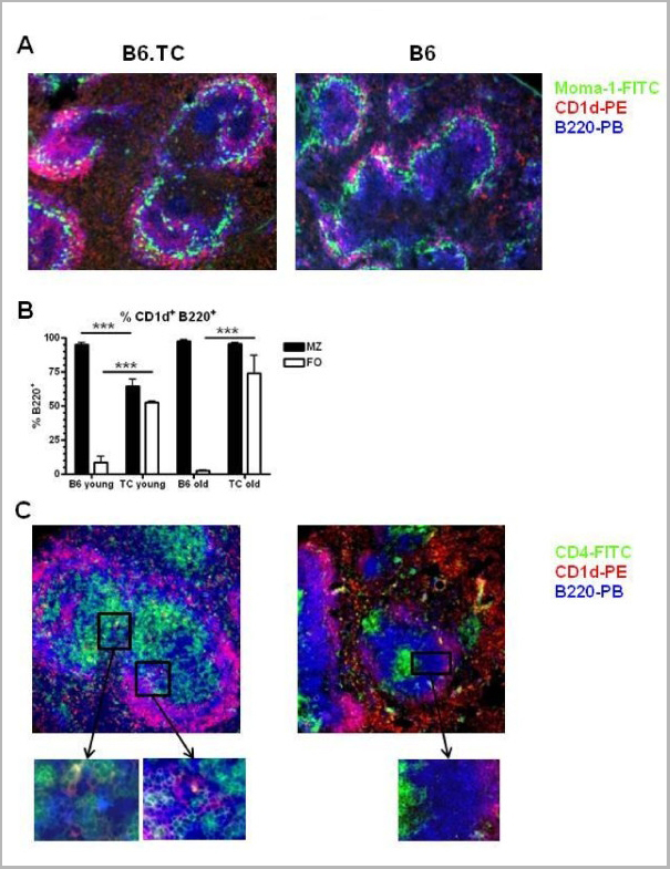
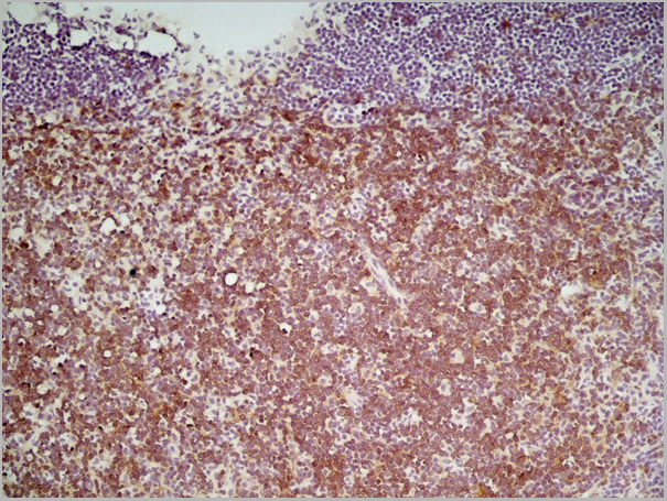
(Staining of mouse spleen with Rat anti Mouse CD4:RPE)
Application Data
(Staining of mouse spleen with Rat anti Mouse CD4:RPE)
CD4, Monoclonal Antibody (Cat# AAA12227)
Full Name
RAT ANTI MOUSE CD4
Gene Names
Cd4; L3T4; Ly-4
Applications
Immunohistochemistry, Flow Cytometry, Immunofluorescence, Immunoprecipitation, Western Blot
Pricing


 Application Data
(Staining of mouse spleen cells with Rat anti Mouse CD45 (Ly-5))
Application Data
(Staining of mouse spleen cells with Rat anti Mouse CD45 (Ly-5))
CD45, Monoclonal Antibody (Cat# AAA11856)
Full Name
RAT ANTI MOUSE CD45:FITC
Gene Names
Ptprc; loc; B220; Cd45; L-CA; Ly-5; T200; CD45R; Lyt-4
Applications
Flow Cytometry
Pricing


 Application Data
(Staining of J774 cells with Rat anti Mouse F4/80 antigen Biotin)
Application Data
(Staining of J774 cells with Rat anti Mouse F4/80 antigen Biotin)
F4/80, Monoclonal Antibody (Cat# AAA12165)
Full Name
RAT ANTI MOUSE F4/80:Low Endotoxin
Gene Names
Emr1; Ly71; F4/80; Gpf480; TM7LN3; DD7A5-7; EGF-TM7
Applications
Immunohistochemistry, Flow Cytometry, Functional Assay, Immunoprecipitation, Immunohistochemistry, Radioimmunoassay, Immunohistochemistry, Western Blot
Pricing


 IHC (Immunohistchemistry)
(DAB staining on IHC-P Samples:Human Prostate Gland Tissue))
IHC (Immunohistchemistry)
(DAB staining on IHC-P Samples:Human Prostate Gland Tissue))
Dickkopf Related Protein 1, Monoclonal Antibody (Cat# AAA20822)
Full Name
Dickkopf Related Protein 1 (DKK1)
Gene Names
DKK1; SK; DKK-1
Reactivity
Human, Mouse
Applications
Westen Blot, Immunohistochemistry, Immunocytochemistry, Immunoprecipitation
Purity
Purification: Protein A + Protein G affinity chromatography
Pricing


 Application Data
(Staining of mouse peritoneal macrophages with Rat anti Mouse CD11b)
Application Data
(Staining of mouse peritoneal macrophages with Rat anti Mouse CD11b)
CD11b, Monoclonal Antibody (Cat# AAA11889)
Full Name
RAT ANTI MOUSE CD11b:FITC
Gene Names
Itgam; CR3; CR3A; MAC1; Cd11b; Ly-40; Mac-1; Mac-1a; CD11b/CD18; F730045J24Rik
Applications
Flow Cytometry
Pricing


 Application Data
(Published customer image Infiltration of GFP+ BM-cells in infarct and peri-infarct regions. (A-B) Dot plots of viable macrophages/granulocytes (CD11b+CD45high, top right quadrants) and microglia (CD11b+CD45dim, bottom right quadrants) in cortex from BM-chimeric unmanipulated mice and mice exposed to pMCAO. (C) Bar graph showing mean numbers of CD11b+CD45dim microglia and CD11b+CD45high macrophages/granulocytes in BM-chimeric mice 24 hours after pMCAO, subdivided based on expression of GFP (n = 5). Approximately 92% of of the CD45high population were GFP+. (D) Estimation and comparison of mean numbers of CD11b+CD45dim microglia in non-chimeric (n = 10) versus BM-chimeric mice (n = 5) 24 hours after of pMCAO shows significantly fewer CD11b+CD45dim microglial cells in irradiated mice. (E) Overview, showing distribution of infiltrating GFP+ BM-derived cells into infarct (IF) and peri-infarct (P-IF) regions 24 hours after pMCAO. (E-G) By 24 hours, GFP+ single cells (F) and vessel-associated aggregates of GFP+ cells (arrows in G) were observed in infarct and peri-infarct regions. Some of the vessel-associated cells were round, leukocyte-like cells (arrows) while others were elongated cells lining the vasculature (arrow heads in G and in insert). (H) Bar graph showing mean numbers of single GFP+ cells and vessel-associated aggregates of GFP+ cells in ipsi- and contralateral cortex 24 hours after surgery (n = 10). (I-P) Immunohistochemical staining of CD45.1 (I, K), CD45.2 (J, L), IgG2a (M, O) and CD45 (N, P) in ischemic tissue in BM-chimeric (I, J, M, N) and non-chimeric mice (K, L, O, P) 24 hours after pMCAO. N.D, none detected. Scale bars: 200 um (A), 10 um (B, C). 50 um (I-P) *P < 0.05, **P < 0.01, and ***P < 0.001.From: Clausen BH, Lambertsen KL, Babcock AA, Holm TH, Dagnaes-Hansen F, Finsen B. Interleukin-1beta and tumor necrosis factor-alpha are expressed by different subsets of microglia and macrophages after ischemic stroke in mice. J Neuroinflammation. 2008 Oct 23;5:46.)
Application Data
(Published customer image Infiltration of GFP+ BM-cells in infarct and peri-infarct regions. (A-B) Dot plots of viable macrophages/granulocytes (CD11b+CD45high, top right quadrants) and microglia (CD11b+CD45dim, bottom right quadrants) in cortex from BM-chimeric unmanipulated mice and mice exposed to pMCAO. (C) Bar graph showing mean numbers of CD11b+CD45dim microglia and CD11b+CD45high macrophages/granulocytes in BM-chimeric mice 24 hours after pMCAO, subdivided based on expression of GFP (n = 5). Approximately 92% of of the CD45high population were GFP+. (D) Estimation and comparison of mean numbers of CD11b+CD45dim microglia in non-chimeric (n = 10) versus BM-chimeric mice (n = 5) 24 hours after of pMCAO shows significantly fewer CD11b+CD45dim microglial cells in irradiated mice. (E) Overview, showing distribution of infiltrating GFP+ BM-derived cells into infarct (IF) and peri-infarct (P-IF) regions 24 hours after pMCAO. (E-G) By 24 hours, GFP+ single cells (F) and vessel-associated aggregates of GFP+ cells (arrows in G) were observed in infarct and peri-infarct regions. Some of the vessel-associated cells were round, leukocyte-like cells (arrows) while others were elongated cells lining the vasculature (arrow heads in G and in insert). (H) Bar graph showing mean numbers of single GFP+ cells and vessel-associated aggregates of GFP+ cells in ipsi- and contralateral cortex 24 hours after surgery (n = 10). (I-P) Immunohistochemical staining of CD45.1 (I, K), CD45.2 (J, L), IgG2a (M, O) and CD45 (N, P) in ischemic tissue in BM-chimeric (I, J, M, N) and non-chimeric mice (K, L, O, P) 24 hours after pMCAO. N.D, none detected. Scale bars: 200 um (A), 10 um (B, C). 50 um (I-P) *P < 0.05, **P < 0.01, and ***P < 0.001.From: Clausen BH, Lambertsen KL, Babcock AA, Holm TH, Dagnaes-Hansen F, Finsen B. Interleukin-1beta and tumor necrosis factor-alpha are expressed by different subsets of microglia and macrophages after ischemic stroke in mice. J Neuroinflammation. 2008 Oct 23;5:46.)
CD11b, Monoclonal Antibody (Cat# AAA12182)
Full Name
RAT ANTI MOUSE CD11b:FITC
Gene Names
Itgam; CR3; CR3A; MAC1; Cd11b; Ly-40; Mac-1; Mac-1a; CD11b/CD18; F730045J24Rik
Applications
Flow Cytometry
Pricing


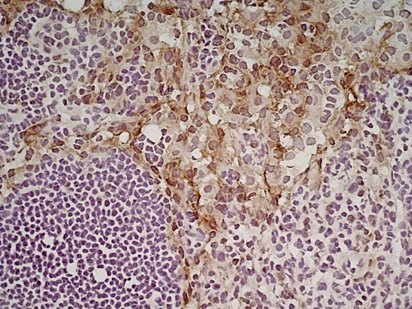
 IHC (Immunohistchemistry)
(Immunohistochemical analysis of paraffin-embedded colon cancer tissues using PBK mouse mAb with DAB staining.)
IHC (Immunohistchemistry)
(Immunohistochemical analysis of paraffin-embedded colon cancer tissues using PBK mouse mAb with DAB staining.)
PBK/TOPK, Monoclonal Antibody (Cat# AAA14113)
Full Name
Anti-PBK/TOPK Mouse mAb
Gene Names
PBK; SPK; CT84; TOPK; HEL164; Nori-3
Reactivity
Human, Rat
Applications
Western Blot
Pricing


 Application Data
(Staining of J774 cells with Rat anti Mouse F4/80 antigen Biotin)
Application Data
(Staining of J774 cells with Rat anti Mouse F4/80 antigen Biotin)
F4/80, Monoclonal Antibody (Cat# AAA12173)
Full Name
RAT ANTI MOUSE F4/80
Gene Names
Emr1; Ly71; F4/80; Gpf480; TM7LN3; DD7A5-7; EGF-TM7
Applications
Immunohistochemistry, Fluorescence Microscopy, Flow Cytometry, Immunofluorescence, Immunoprecipitation, Immunohistochemistry, Radioimmunoassay, Immunohistochemistry, Western Blot
Pricing


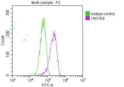 FCM (Flow Cytometry)
(Overlay histogram showing Hela cells stained with (red line) at 1:100. The cells were fixed in 4% formaldehyde and permeated by 0.2% TritonX-100. Then 10% normal goat serum was Incubated to block non-specific protein-protein interactions followed by the antibody (1ug/1*106cells) for 1 h at 4 degree C. The secondary antibody used was FITC-conjugated Goat Anti-Mouse IgG(H+L) at 1/100 dilution for 30min at 4 degree C. Isotype control antibody (green line) was mouse IgG2b (1ug/1*106cells) used under the same conditions. Acquisition of >10,000 events was performed.)
FCM (Flow Cytometry)
(Overlay histogram showing Hela cells stained with (red line) at 1:100. The cells were fixed in 4% formaldehyde and permeated by 0.2% TritonX-100. Then 10% normal goat serum was Incubated to block non-specific protein-protein interactions followed by the antibody (1ug/1*106cells) for 1 h at 4 degree C. The secondary antibody used was FITC-conjugated Goat Anti-Mouse IgG(H+L) at 1/100 dilution for 30min at 4 degree C. Isotype control antibody (green line) was mouse IgG2b (1ug/1*106cells) used under the same conditions. Acquisition of >10,000 events was performed.)
YWHAZ, Monoclonal Antibody (Cat# AAA27050)
Full Name
YWHAZ Monoclonal Antibody
Gene Names
YWHAZ; YWHAD; KCIP-1; 14-3-3-zeta
Reactivity
Human, Mouse, Rat
Applications
Western Blot, Immunohistochemistry, Immunofluorescence, Flow Cytometry
Purity
>95%, Protein A purified
Pricing


 Application Data
(Staining of J774 cells with Rat anti Mouse F4/80 antigen Biotin)
Application Data
(Staining of J774 cells with Rat anti Mouse F4/80 antigen Biotin)
F4/80, Monoclonal Antibody (Cat# AAA12170)
Full Name
RAT ANTI MOUSE F4/80
Gene Names
Emr1; Ly71; F4/80; Gpf480; TM7LN3; DD7A5-7; EGF-TM7
Applications
Immunohistochemistry, Fluorescence Microscopy, Flow Cytometry, Immunofluorescence, Immunoprecipitation, Immunohistochemistry, Radioimmunoassay, Immunohistochemistry, Western Blot
Pricing


 Application Data
(Published customer image Infiltration of GFP+ BM-cells in infarct and peri-infarct regions. (A-B) Dot plots of viable macrophages/granulocytes (CD11b+CD45high, top right quadrants) and microglia (CD11b+CD45dim, bottom right quadrants) in cortex from BM-chimeric unmanipulated mice and mice exposed to pMCAO. (C) Bar graph showing mean numbers of CD11b+CD45dim microglia and CD11b+CD45high macrophages/granulocytes in BM-chimeric mice 24 hours after pMCAO, subdivided based on expression of GFP (n = 5). Approximately 92% of of the CD45high population were GFP+. (D) Estimation and comparison of mean numbers of CD11b+CD45dim microglia in non-chimeric (n = 10) versus BM-chimeric mice (n = 5) 24 hours after of pMCAO shows significantly fewer CD11b+CD45dim microglial cells in irradiated mice. (E) Overview, showing distribution of infiltrating GFP+ BM-derived cells into infarct (IF) and peri-infarct (P-IF) regions 24 hours after pMCAO. (E-G) By 24 hours, GFP+ single cells (F) and vessel-associated aggregates of GFP+ cells (arrows in G) were observed in infarct and peri-infarct regions. Some of the vessel-associated cells were round, leukocyte-like cells (arrows) while others were elongated cells lining the vasculature (arrow heads in G and in insert). (H) Bar graph showing mean numbers of single GFP+ cells and vessel-associated aggregates of GFP+ cells in ipsi- and contralateral cortex 24 hours after surgery (n = 10). (I-P) Immunohistochemical staining of CD45.1 (I, K), CD45.2 (J, L), IgG2a (M, O) and CD45 (N, P) in ischemic tissue in BM-chimeric (I, J, M, N) and non-chimeric mice (K, L, O, P) 24 hours after pMCAO. N.D, none detected. Scale bars: 200 um (A), 10 um (B, C). 50 um (I-P) *P < 0.05, **P < 0.01, and ***P < 0.001.From: Clausen BH, Lambertsen KL, Babcock AA, Holm TH, Dagnaes-Hansen F, Finsen B. Interleukin-1beta and tumor necrosis factor-alpha are expressed by different subsets of microglia and macrophages after ischemic stroke in mice. J Neuroinflammation. 2008 Oct 23;5:46.)
Application Data
(Published customer image Infiltration of GFP+ BM-cells in infarct and peri-infarct regions. (A-B) Dot plots of viable macrophages/granulocytes (CD11b+CD45high, top right quadrants) and microglia (CD11b+CD45dim, bottom right quadrants) in cortex from BM-chimeric unmanipulated mice and mice exposed to pMCAO. (C) Bar graph showing mean numbers of CD11b+CD45dim microglia and CD11b+CD45high macrophages/granulocytes in BM-chimeric mice 24 hours after pMCAO, subdivided based on expression of GFP (n = 5). Approximately 92% of of the CD45high population were GFP+. (D) Estimation and comparison of mean numbers of CD11b+CD45dim microglia in non-chimeric (n = 10) versus BM-chimeric mice (n = 5) 24 hours after of pMCAO shows significantly fewer CD11b+CD45dim microglial cells in irradiated mice. (E) Overview, showing distribution of infiltrating GFP+ BM-derived cells into infarct (IF) and peri-infarct (P-IF) regions 24 hours after pMCAO. (E-G) By 24 hours, GFP+ single cells (F) and vessel-associated aggregates of GFP+ cells (arrows in G) were observed in infarct and peri-infarct regions. Some of the vessel-associated cells were round, leukocyte-like cells (arrows) while others were elongated cells lining the vasculature (arrow heads in G and in insert). (H) Bar graph showing mean numbers of single GFP+ cells and vessel-associated aggregates of GFP+ cells in ipsi- and contralateral cortex 24 hours after surgery (n = 10). (I-P) Immunohistochemical staining of CD45.1 (I, K), CD45.2 (J, L), IgG2a (M, O) and CD45 (N, P) in ischemic tissue in BM-chimeric (I, J, M, N) and non-chimeric mice (K, L, O, P) 24 hours after pMCAO. N.D, none detected. Scale bars: 200 um (A), 10 um (B, C). 50 um (I-P) *P < 0.05, **P < 0.01, and ***P < 0.001.From: Clausen BH, Lambertsen KL, Babcock AA, Holm TH, Dagnaes-Hansen F, Finsen B. Interleukin-1beta and tumor necrosis factor-alpha are expressed by different subsets of microglia and macrophages after ischemic stroke in mice. J Neuroinflammation. 2008 Oct 23;5:46.)
CD11b, Monoclonal Antibody (Cat# AAA12184)
Full Name
RAT ANTI MOUSE CD11b
Gene Names
Itgam; CR3; CR3A; MAC1; Cd11b; Ly-40; Mac-1; Mac-1a; CD11b/CD18; F730045J24Rik
Applications
Immunohistochemistry, Flow Cytometry, Immunofluorescence, Immunoprecipitation
Pricing


 Application Data
(Staining of J774 cells with Rat anti Mouse F4/80 antigen Biotin)
Application Data
(Staining of J774 cells with Rat anti Mouse F4/80 antigen Biotin)
F4/80, Monoclonal Antibody (Cat# AAA12171)
Full Name
RAT ANTI MOUSE F4/80:RPE
Gene Names
Emr1; Ly71; F4/80; Gpf480; TM7LN3; DD7A5-7; EGF-TM7
Applications
Flow Cytometry
Pricing


 Application Data
(Staining of mouse peritoneal macrophages with Rat anti Mouse CD11b)
Application Data
(Staining of mouse peritoneal macrophages with Rat anti Mouse CD11b)
CD11b, Monoclonal Antibody (Cat# AAA12219)
Full Name
RAT ANTI MOUSE CD11b:Low Endotoxin
Gene Names
Itgam; CR3; CR3A; MAC1; Cd11b; Ly-40; Mac-1; Mac-1a; CD11b/CD18; F730045J24Rik
Applications
Immunohistochemistry, Flow Cytometry, Functional Assay, Immunofluorescence, Immunoprecipitation, Immunohistochemistry
Pricing


 Application Data
(Staining of J774 cells with Rat anti Mouse F4/80 antigen Biotin)
Application Data
(Staining of J774 cells with Rat anti Mouse F4/80 antigen Biotin)
F4/80, Monoclonal Antibody (Cat# AAA12168)
Full Name
RAT ANTI MOUSE F4/80:FITC
Gene Names
Emr1; Ly71; F4/80; Gpf480; TM7LN3; DD7A5-7; EGF-TM7
Applications
Flow Cytometry, Immunofluorescence
Pricing


 Application Data
(Staining of J774 cells with Rat anti Mouse F4/80 antigen Biotin)
Application Data
(Staining of J774 cells with Rat anti Mouse F4/80 antigen Biotin)
F4/80, Monoclonal Antibody (Cat# AAA12166)
Full Name
RAT ANTI MOUSE F4/80:FITC
Gene Names
Emr1; Ly71; F4/80; Gpf480; TM7LN3; DD7A5-7; EGF-TM7
Applications
Flow Cytometry, Immunofluorescence
Pricing


 Application Data
(Staining of J774 cells with Rat anti Mouse F4/80 antigen Biotin)
Application Data
(Staining of J774 cells with Rat anti Mouse F4/80 antigen Biotin)
F4/80, Monoclonal Antibody (Cat# AAA12162)
Full Name
RAT ANTI MOUSE F4/80:Biotin
Gene Names
Emr1; Ly71; F4/80; Gpf480; TM7LN3; DD7A5-7; EGF-TM7
Applications
Flow Cytometry
Pricing


 Application Data
(Staining of J774 cells with Rat anti Mouse F4/80 antigen Biotin)
Application Data
(Staining of J774 cells with Rat anti Mouse F4/80 antigen Biotin)
F4/80, Monoclonal Antibody (Cat# AAA12161)
Full Name
RAT ANTI MOUSE F4/80:APC
Gene Names
Emr1; Ly71; F4/80; Gpf480; TM7LN3; DD7A5-7; EGF-TM7
Applications
Flow Cytometry
Pricing


 Application Data
(Staining of J774 cells with Rat anti Mouse F4/80 antigen Biotin)
Application Data
(Staining of J774 cells with Rat anti Mouse F4/80 antigen Biotin)
F4/80, Monoclonal Antibody (Cat# AAA12172)
Full Name
RAT ANTI MOUSE F4/80:RPE
Gene Names
Emr1; Ly71; F4/80; Gpf480; TM7LN3; DD7A5-7; EGF-TM7
Applications
Flow Cytometry, Immunofluorescence
Pricing


 Application Data
(Staining of J774 cells with Rat anti Mouse F4/80 antigen Biotin)
Application Data
(Staining of J774 cells with Rat anti Mouse F4/80 antigen Biotin)
F4/80, Monoclonal Antibody (Cat# AAA12217)
Full Name
RAT ANTI MOUSE F4/80:APC
Gene Names
Emr1; Ly71; F4/80; Gpf480; TM7LN3; DD7A5-7; EGF-TM7
Applications
Flow Cytometry
Pricing


 FCM (Flow Cytometry)
(Flow cytometry of Jurkat cells, using anti-BSG antibody, (Red), compared to a nonspecific negative control antibody, (Blue).)
FCM (Flow Cytometry)
(Flow cytometry of Jurkat cells, using anti-BSG antibody, (Red), compared to a nonspecific negative control antibody, (Blue).)
Basigin / Emmprin / CD147, Monoclonal Antibody (Cat# AAA12375)
Full Name
Anti-Basigin / Emmprin / CD147 Antibody (clone 10E10) IHC-plus
Gene Names
BSG; M6; OK; 5F7; TCSF; CD147; EMMPRIN
Reactivity
Dog, Rat, Human, Monkey
Applications
Immunohistochemistry, Immunofluorescence, Western Blot, Flow Cytometry
Purity
Protein A/G purified
Pricing


 Application Data
(Staining of mouse peritoneal macrophages with RAT ANTI MOUSE F4/80 ANTIGEN:FITC)
Application Data
(Staining of mouse peritoneal macrophages with RAT ANTI MOUSE F4/80 ANTIGEN:FITC)
EMR1, Monoclonal Antibody (Cat# AAA26723)
Full Name
EMR1 (EGF-like Module Containing Mucin-like Hormone Receptor-like 1, EMR1 Hormone Receptor, Cell Surface Glycoprotein EMR1, Cell Surface Glycoprotein F4/80, DD7A5-7, EGF-TM7, F4/80, Gpf480, Lymphocyte Antigen 71, Ly71, TM7LN3) (AP)
Reactivity
Mouse
Applications
Immunohistochemistry, Immunoprecipitation, Radioimmunoassay, Western Blot
Purity
Purified by protein G affinity chromatography from tissue culture supernatant.
Pricing


 Application Data
(Staining of mouse spleen cells with Rat anti Mouse CD4)
Application Data
(Staining of mouse spleen cells with Rat anti Mouse CD4)
CD4, Monoclonal Antibody (Cat# AAA11939)
Full Name
RAT ANTI MOUSE CD4
Gene Names
Cd4; L3T4; Ly-4
Applications
Immunohistochemistry, Flow Cytometry
Pricing


 WB (Western Blot)
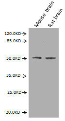
(MARCH7 monoclonal antibody. Western Blot analysis of 'MARCH7 expression in PC-12.)
WB (Western Blot)
(MARCH7 monoclonal antibody. Western Blot analysis of 'MARCH7 expression in PC-12.)
MARCH7, Monoclonal Antibody (Cat# AAA24075)
Full Name
MARCH7 (E3 Ubiquitin-protein Ligase MARCH7, Axotrophin, Membrane-associated RING Finger Protein 7, Membrane-associated RING-CH Protein VII, MARCH-VII, RING Finger Protein 177, AXOT, RNF177, DKFZp586F1122)
Reactivity
Human, Mouse, Rat
Applications
Western Blot, Immunofluorescence
Purity
Affinity Purified
Purified by Protein A affinity chromatography.
Purified by Protein A affinity chromatography.
Pricing


 Application Data
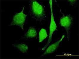
(Overlay histogram showing SY5Y cells stained with AAA27008 (red line). The cells were fixed with 70% Ethylalcohol (18h) and then permeabilized with 0.3% Triton X-100 for 2 min. The cells were then incubated in 1x PBS /10% normal goat serum to block non-specific protein-protein interactions followed by the antibody (10ug/1x10^6cells) for 1 h at 4 degree C. The secondary antibody used was FITC goat anti-mouse IgG (H+L) at 1/200 dilution for 1 h at 4 degree C. Isotype control antibody (green line) was mouse IgG2b (10ug/1x10^6cells) used under the same conditions. Acquisition of >10, 000 events was performed.)
Application Data
(Overlay histogram showing SY5Y cells stained with AAA27008 (red line). The cells were fixed with 70% Ethylalcohol (18h) and then permeabilized with 0.3% Triton X-100 for 2 min. The cells were then incubated in 1x PBS /10% normal goat serum to block non-specific protein-protein interactions followed by the antibody (10ug/1x10^6cells) for 1 h at 4 degree C. The secondary antibody used was FITC goat anti-mouse IgG (H+L) at 1/200 dilution for 1 h at 4 degree C. Isotype control antibody (green line) was mouse IgG2b (10ug/1x10^6cells) used under the same conditions. Acquisition of >10, 000 events was performed.)
GFAP, Monoclonal Antibody (Cat# AAA27008)
Full Name
GFAP Monoclonal Antibody
Gene Names
GFAP; ALXDRD
Reactivity
Human, Mouse, Rat
Applications
Western Blot, Immunohistochemistry, Immunofluorescence, Flow Cytometry
Purity
>95%, Protein G purified
Pricing


 Application Data
(Staining of mouse spleen cells with Rat anti Mouse CD45 (Ly-5))
Application Data
(Staining of mouse spleen cells with Rat anti Mouse CD45 (Ly-5))
CD45, Monoclonal Antibody (Cat# AAA11915)
Full Name
RAT ANTI MOUSE CD45
Gene Names
Ptprc; loc; B220; Cd45; L-CA; Ly-5; T200; CD45R; Lyt-4
Applications
Immunohistochemistry, Flow Cytometry, Immunofluorescence, Immunoprecipitation
Pricing


 Application Data
(Staining of mouse spleen with Rat anti Mouse CD4:RPE)
Application Data
(Staining of mouse spleen with Rat anti Mouse CD4:RPE)
CD4, Monoclonal Antibody (Cat# AAA12230)
Full Name
RAT ANTI MOUSE CD4
Gene Names
Cd4; L3T4; Ly-4
Applications
Immunohistochemistry, Flow Cytometry, Immunofluorescence, Immunoprecipitation, Western Blot
Pricing


 Application Data
(Published customer image Infiltration of GFP+ BM-cells in infarct and peri-infarct regions. (A-B) Dot plots of viable macrophages/granulocytes (CD11b+CD45high, top right quadrants) and microglia (CD11b+CD45dim, bottom right quadrants) in cortex from BM-chimeric unmanipulated mice and mice exposed to pMCAO. (C) Bar graph showing mean numbers of CD11b+CD45dim microglia and CD11b+CD45high macrophages/granulocytes in BM-chimeric mice 24 hours after pMCAO, subdivided based on expression of GFP (n = 5). Approximately 92% of of the CD45high population were GFP+. (D) Estimation and comparison of mean numbers of CD11b+CD45dim microglia in non-chimeric (n = 10) versus BM-chimeric mice (n = 5) 24 hours after of pMCAO shows significantly fewer CD11b+CD45dim microglial cells in irradiated mice. (E) Overview, showing distribution of infiltrating GFP+ BM-derived cells into infarct (IF) and peri-infarct (P-IF) regions 24 hours after pMCAO. (E-G) By 24 hours, GFP+ single cells (F) and vessel-associated aggregates of GFP+ cells (arrows in G) were observed in infarct and peri-infarct regions. Some of the vessel-associated cells were round, leukocyte-like cells (arrows) while others were elongated cells lining the vasculature (arrow heads in G and in insert). (H) Bar graph showing mean numbers of single GFP+ cells and vessel-associated aggregates of GFP+ cells in ipsi- and contralateral cortex 24 hours after surgery (n = 10). (I-P) Immunohistochemical staining of CD45.1 (I, K), CD45.2 (J, L), IgG2a (M, O) and CD45 (N, P) in ischemic tissue in BM-chimeric (I, J, M, N) and non-chimeric mice (K, L, O, P) 24 hours after pMCAO. N.D, none detected. Scale bars: 200 um (A), 10 um (B, C). 50 um (I-P) *P < 0.05, **P < 0.01, and ***P < 0.001.From: Clausen BH, Lambertsen KL, Babcock AA, Holm TH, Dagnaes-Hansen F, Finsen B. Interleukin-1beta and tumor necrosis factor-alpha are expressed by different subsets of microglia and macrophages after ischemic stroke in mice. J Neuroinflammation. 2008 Oct 23;5:46.)
Application Data
(Published customer image Infiltration of GFP+ BM-cells in infarct and peri-infarct regions. (A-B) Dot plots of viable macrophages/granulocytes (CD11b+CD45high, top right quadrants) and microglia (CD11b+CD45dim, bottom right quadrants) in cortex from BM-chimeric unmanipulated mice and mice exposed to pMCAO. (C) Bar graph showing mean numbers of CD11b+CD45dim microglia and CD11b+CD45high macrophages/granulocytes in BM-chimeric mice 24 hours after pMCAO, subdivided based on expression of GFP (n = 5). Approximately 92% of of the CD45high population were GFP+. (D) Estimation and comparison of mean numbers of CD11b+CD45dim microglia in non-chimeric (n = 10) versus BM-chimeric mice (n = 5) 24 hours after of pMCAO shows significantly fewer CD11b+CD45dim microglial cells in irradiated mice. (E) Overview, showing distribution of infiltrating GFP+ BM-derived cells into infarct (IF) and peri-infarct (P-IF) regions 24 hours after pMCAO. (E-G) By 24 hours, GFP+ single cells (F) and vessel-associated aggregates of GFP+ cells (arrows in G) were observed in infarct and peri-infarct regions. Some of the vessel-associated cells were round, leukocyte-like cells (arrows) while others were elongated cells lining the vasculature (arrow heads in G and in insert). (H) Bar graph showing mean numbers of single GFP+ cells and vessel-associated aggregates of GFP+ cells in ipsi- and contralateral cortex 24 hours after surgery (n = 10). (I-P) Immunohistochemical staining of CD45.1 (I, K), CD45.2 (J, L), IgG2a (M, O) and CD45 (N, P) in ischemic tissue in BM-chimeric (I, J, M, N) and non-chimeric mice (K, L, O, P) 24 hours after pMCAO. N.D, none detected. Scale bars: 200 um (A), 10 um (B, C). 50 um (I-P) *P < 0.05, **P < 0.01, and ***P < 0.001.From: Clausen BH, Lambertsen KL, Babcock AA, Holm TH, Dagnaes-Hansen F, Finsen B. Interleukin-1beta and tumor necrosis factor-alpha are expressed by different subsets of microglia and macrophages after ischemic stroke in mice. J Neuroinflammation. 2008 Oct 23;5:46.)
CD11b, Monoclonal Antibody (Cat# AAA12183)
Full Name
RAT ANTI MOUSE CD11b:FITC
Gene Names
Itgam; CR3; CR3A; MAC1; Cd11b; Ly-40; Mac-1; Mac-1a; CD11b/CD18; F730045J24Rik
Applications
Flow Cytometry
Pricing


 Application Data
(Published customer image Infiltration of GFP+ BM-cells in infarct and peri-infarct regions. (A-B) Dot plots of viable macrophages/granulocytes (CD11b+CD45high, top right quadrants) and microglia (CD11b+CD45dim, bottom right quadrants) in cortex from BM-chimeric unmanipulated mice and mice exposed to pMCAO. (C) Bar graph showing mean numbers of CD11b+CD45dim microglia and CD11b+CD45high macrophages/granulocytes in BM-chimeric mice 24 hours after pMCAO, subdivided based on expression of GFP (n = 5). Approximately 92% of of the CD45high population were GFP+. (D) Estimation and comparison of mean numbers of CD11b+CD45dim microglia in non-chimeric (n = 10) versus BM-chimeric mice (n = 5) 24 hours after of pMCAO shows significantly fewer CD11b+CD45dim microglial cells in irradiated mice. (E) Overview, showing distribution of infiltrating GFP+ BM-derived cells into infarct (IF) and peri-infarct (P-IF) regions 24 hours after pMCAO. (E-G) By 24 hours, GFP+ single cells (F) and vessel-associated aggregates of GFP+ cells (arrows in G) were observed in infarct and peri-infarct regions. Some of the vessel-associated cells were round, leukocyte-like cells (arrows) while others were elongated cells lining the vasculature (arrow heads in G and in insert). (H) Bar graph showing mean numbers of single GFP+ cells and vessel-associated aggregates of GFP+ cells in ipsi- and contralateral cortex 24 hours after surgery (n = 10). (I-P) Immunohistochemical staining of CD45.1 (I, K), CD45.2 (J, L), IgG2a (M, O) and CD45 (N, P) in ischemic tissue in BM-chimeric (I, J, M, N) and non-chimeric mice (K, L, O, P) 24 hours after pMCAO. N.D, none detected. Scale bars: 200 um (A), 10 um (B, C). 50 um (I-P) *P < 0.05, **P < 0.01, and ***P < 0.001.From: Clausen BH, Lambertsen KL, Babcock AA, Holm TH, Dagnaes-Hansen F, Finsen B. Interleukin-1beta and tumor necrosis factor-alpha are expressed by different subsets of microglia and macrophages after ischemic stroke in mice. J Neuroinflammation. 2008 Oct 23;5:46.)
Application Data
(Published customer image Infiltration of GFP+ BM-cells in infarct and peri-infarct regions. (A-B) Dot plots of viable macrophages/granulocytes (CD11b+CD45high, top right quadrants) and microglia (CD11b+CD45dim, bottom right quadrants) in cortex from BM-chimeric unmanipulated mice and mice exposed to pMCAO. (C) Bar graph showing mean numbers of CD11b+CD45dim microglia and CD11b+CD45high macrophages/granulocytes in BM-chimeric mice 24 hours after pMCAO, subdivided based on expression of GFP (n = 5). Approximately 92% of of the CD45high population were GFP+. (D) Estimation and comparison of mean numbers of CD11b+CD45dim microglia in non-chimeric (n = 10) versus BM-chimeric mice (n = 5) 24 hours after of pMCAO shows significantly fewer CD11b+CD45dim microglial cells in irradiated mice. (E) Overview, showing distribution of infiltrating GFP+ BM-derived cells into infarct (IF) and peri-infarct (P-IF) regions 24 hours after pMCAO. (E-G) By 24 hours, GFP+ single cells (F) and vessel-associated aggregates of GFP+ cells (arrows in G) were observed in infarct and peri-infarct regions. Some of the vessel-associated cells were round, leukocyte-like cells (arrows) while others were elongated cells lining the vasculature (arrow heads in G and in insert). (H) Bar graph showing mean numbers of single GFP+ cells and vessel-associated aggregates of GFP+ cells in ipsi- and contralateral cortex 24 hours after surgery (n = 10). (I-P) Immunohistochemical staining of CD45.1 (I, K), CD45.2 (J, L), IgG2a (M, O) and CD45 (N, P) in ischemic tissue in BM-chimeric (I, J, M, N) and non-chimeric mice (K, L, O, P) 24 hours after pMCAO. N.D, none detected. Scale bars: 200 um (A), 10 um (B, C). 50 um (I-P) *P < 0.05, **P < 0.01, and ***P < 0.001.From: Clausen BH, Lambertsen KL, Babcock AA, Holm TH, Dagnaes-Hansen F, Finsen B. Interleukin-1beta and tumor necrosis factor-alpha are expressed by different subsets of microglia and macrophages after ischemic stroke in mice. J Neuroinflammation. 2008 Oct 23;5:46.)
CD11b, Monoclonal Antibody (Cat# AAA12185)
Full Name
RAT ANTI MOUSE CD11b
Gene Names
Itgam; CR3; CR3A; MAC1; Cd11b; Ly-40; Mac-1; Mac-1a; CD11b/CD18; F730045J24Rik
Applications
Immunohistochemistry, Flow Cytometry, Immunofluorescence, Immunoprecipitation
Pricing


 WB (Western Blot)
GAA, Monoclonal Antibody (Cat# AAA19211)
Full Name
Anti-GAA Antibody (Monoclonal, 2G7)
Gene Names
GAA; LYAG
Reactivity
Human
Applications
Western Blot, Immunohistochemistry, Immunocytochemistry, Immunofluorescence, Flow Cytometry
Purity
Immunogen affinity purified.
Pricing


 Application Data
(Staining of mouse peritoneal macrophages with RAT ANTI MOUSE F4/80 ANTIGEN:FITC)
Application Data
(Staining of mouse peritoneal macrophages with RAT ANTI MOUSE F4/80 ANTIGEN:FITC)
EMR1, Monoclonal Antibody (Cat# AAA26786)
Full Name
EMR1 (EGF-like Module Containing Mucin-like Hormone Receptor-like 1, EMR1 Hormone Receptor, Cell Surface Glycoprotein EMR1, Cell Surface Glycoprotein F4/80, DD7A5-7, EGF-TM7, F4/80, Gpf480, Lymphocyte Antigen 71, Ly71, TM7LN3) (MaxLight 405)
Reactivity
Mouse
Applications
Flow Cytometry, Immunohistochemistry, Immunoprecipitation, Radioimmunoassay, Western Blot
Purity
Purified by protein G affinity chromatography from tissue culture supernatant.
Pricing


 Application Data
(Staining of mouse peritoneal macrophages with Rat anti Mouse CD11b)
Application Data
(Staining of mouse peritoneal macrophages with Rat anti Mouse CD11b)
CD11b, Monoclonal Antibody (Cat# AAA12187)
Full Name
RAT ANTI MOUSE CD11b:Biotin
Gene Names
Itgam; CR3; CR3A; MAC1; Cd11b; Ly-40; Mac-1; Mac-1a; CD11b/CD18; F730045J24Rik
Applications
Flow Cytometry
Pricing


 Application Data
(Staining of J774 cells with Rat anti Mouse F4/80 antigen Biotin)
Application Data
(Staining of J774 cells with Rat anti Mouse F4/80 antigen Biotin)
F4/80, Monoclonal Antibody (Cat# AAA12169)
Full Name
RAT ANTI MOUSE F4/80
Gene Names
Emr1; Ly71; F4/80; Gpf480; TM7LN3; DD7A5-7; EGF-TM7
Applications
Immunohistochemistry, Fluorescence Microscopy, Flow Cytometry, Immunofluorescence, Immunoprecipitation, Immunohistochemistry, Radioimmunoassay, Immunohistochemistry, Western Blot
Pricing


 IP (Immunoprecipitation)
(Immunoprecipitating HSPA8 in Hela whole cell lysate Lane 1: Mouse control IgG2b instead dilution for 30min at 4 degree C. Isotype control antibody (green line) was mouse IgG2b used under the same conditions. Acquisition of >10,000 events was performed.)
IP (Immunoprecipitation)
(Immunoprecipitating HSPA8 in Hela whole cell lysate Lane 1: Mouse control IgG2b instead dilution for 30min at 4 degree C. Isotype control antibody (green line) was mouse IgG2b used under the same conditions. Acquisition of >10,000 events was performed.)
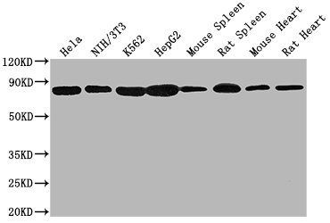
HSPA8, Monoclonal Antibody (Cat# AAA27045)
Full Name
HSPA8 Monoclonal Antibody
Gene Names
HSPA8; LAP1; HSC54; HSC70; HSC71; HSP71; HSP73; NIP71; HSPA10
Reactivity
Human, Rat, Mouse
Applications
Western Blot, Immunohistochemistry, Flow Cytometry, Immunoprecipitation
Purity
>95%, Protein A purified
Pricing


 Application Data
(Staining of J774 cells with Rat anti Mouse F4/80 antigen Biotin)
Application Data
(Staining of J774 cells with Rat anti Mouse F4/80 antigen Biotin)
F4/80, Monoclonal Antibody (Cat# AAA12163)
Full Name
RAT ANTI MOUSE F4/80:Biotin
Gene Names
Emr1; Ly71; F4/80; Gpf480; TM7LN3; DD7A5-7; EGF-TM7
Applications
Flow Cytometry
Pricing


 WB (Western Blot)
(Anti-Beta-Tubulin mouse monoclonal antibody at 1:2000 dilution Lane A: HepG2 Whole Cell Lysate Lane B: Daudi Whole Cell Lysate Lane C: MOLT-4 Whole Cell Lysate Lane D: A549 Whole Cell Lysate Lane E: 293T Whole Cell Lysate Lane F: HelaS3 Whole Cell Lysate Lysates/proteins at 30 ug per lane. Secondary Goat Anti-Mouse IgG H&L (Dylight800) at 1/15000 dilution. Developed using the Odyssey technique. Performed under reducing conditions. Predicted band size:50 kDa Observed band size:54 kDa)
WB (Western Blot)
(Anti-Beta-Tubulin mouse monoclonal antibody at 1:2000 dilution Lane A: HepG2 Whole Cell Lysate Lane B: Daudi Whole Cell Lysate Lane C: MOLT-4 Whole Cell Lysate Lane D: A549 Whole Cell Lysate Lane E: 293T Whole Cell Lysate Lane F: HelaS3 Whole Cell Lysate Lysates/proteins at 30 ug per lane. Secondary Goat Anti-Mouse IgG H&L (Dylight800) at 1/15000 dilution. Developed using the Odyssey technique. Performed under reducing conditions. Predicted band size:50 kDa Observed band size:54 kDa)
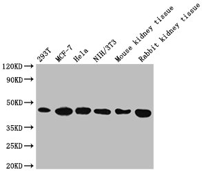
Beta-Tubulin, Monoclonal Antibody (Cat# AAA27713)
Full Name
Beta-Tubulin Loading Control Antibody, Mouse MAb
Reactivity
Human
Applications
Western Blot, Immunohistochemistry, Immunocytochemistry, Immunofluorescence, Immunoprecipitation
Purity
Protein A
Pricing


 Application Data
(Staining of J774 cells with Rat anti Mouse F4/80 antigen Biotin)
Application Data
(Staining of J774 cells with Rat anti Mouse F4/80 antigen Biotin)
F4/80, Monoclonal Antibody (Cat# AAA12174)
Full Name
RAT ANTI MOUSE F4/80
Gene Names
Emr1; Ly71; F4/80; Gpf480; TM7LN3; DD7A5-7; EGF-TM7
Applications
Immunohistochemistry, Fluorescence Microscopy, Flow Cytometry, Immunofluorescence, Immunoprecipitation, Immunohistochemistry, Radioimmunoassay, Immunohistochemistry, Western Blot
Pricing


 Application Data
(Staining of mouse spleen cells with Rat anti Mouse CD45 (Ly-5))
Application Data
(Staining of mouse spleen cells with Rat anti Mouse CD45 (Ly-5))
CD45, Monoclonal Antibody (Cat# AAA12063)
Full Name
RAT ANTI MOUSE CD45
Gene Names
Ptprc; loc; B220; Cd45; L-CA; Ly-5; T200; CD45R; Lyt-4
Applications
Immunohistochemistry, Flow Cytometry, Immunofluorescence, Immunoprecipitation
Pricing


 Application Data
(Staining of J774 cells with Rat anti Mouse F4/80 antigen Biotin)
Application Data
(Staining of J774 cells with Rat anti Mouse F4/80 antigen Biotin)
F4/80, Monoclonal Antibody (Cat# AAA12164)
Full Name
RAT ANTI MOUSE F4/80:Biotin
Gene Names
Emr1; Ly71; F4/80; Gpf480; TM7LN3; DD7A5-7; EGF-TM7
Applications
Flow Cytometry
Pricing


 Application Data
(Staining of mouse spleen cells with Rat anti Mouse CD45 (Ly-5))
Application Data
(Staining of mouse spleen cells with Rat anti Mouse CD45 (Ly-5))
CD45, Monoclonal Antibody (Cat# AAA11914)
Full Name
RAT ANTI MOUSE CD45
Gene Names
Ptprc; loc; B220; Cd45; L-CA; Ly-5; T200; CD45R; Lyt-4
Applications
Immunohistochemistry, Flow Cytometry, Immunofluorescence, Immunoprecipitation
Pricing


 FCM (Flow Cytometry)
(Overlay Peak curve showing Hela cells stained with AAA28066 (red line) at 1:200. The cells were fixed in 4% formaldehyde and permeated by 0.2% TritonX-100. Then 10% normal goat serum was Incubated to block non-specific protein-protein interactions followed by the antibody (1?g/1*106cells) for 1 h at 4 degree C. The secondary antibody used was FITC-conjugated Goat Anti-Mouse IgG(H+L) at 1/100 dilution for 30min at 4 degree C. Isotype control antibody (green line) was mouse IgG2b (1?g/1*106cells) used under the same conditions. Acquisition of >10,000 events was performed.)
FCM (Flow Cytometry)
(Overlay Peak curve showing Hela cells stained with AAA28066 (red line) at 1:200. The cells were fixed in 4% formaldehyde and permeated by 0.2% TritonX-100. Then 10% normal goat serum was Incubated to block non-specific protein-protein interactions followed by the antibody (1?g/1*106cells) for 1 h at 4 degree C. The secondary antibody used was FITC-conjugated Goat Anti-Mouse IgG(H+L) at 1/100 dilution for 30min at 4 degree C. Isotype control antibody (green line) was mouse IgG2b (1?g/1*106cells) used under the same conditions. Acquisition of >10,000 events was performed.)
ACTB, Monoclonal Antibody (Cat# AAA28066)
Full Name
ACTB Monoclonal Antibody
Reactivity
Human, Mouse, Rat, Rabbit
Applications
Western Blot, Immunohistochemistry, Immunofluorescence, Flow Cytometry, Immunoprecipitation
Purity
>95%, Protein A purified
Pricing