Filters
Clonality
Type
Reactivity
Gene Name
Isotype
Host
Application
Clone
20 results for " Ubiquitin Ligases" - showing 1-20


 WB (Western Blot)
(SMURF1 monoclonal antibody, Western Blot analysis of SMURF1 expression in HeLa.)
WB (Western Blot)
(SMURF1 monoclonal antibody, Western Blot analysis of SMURF1 expression in HeLa.)
SMURF1, Monoclonal Antibody (Cat# AAA25845)
Full Name
SMURF1 (E3 Ubiquitin-protein Ligase SMURF1, hSMURF1, SMAD Ubiquitination Regulatory Factor 1, KIAA1625, SMAD-specific E3 Ubiquitin-protein Ligase 1) (PE)
Reactivity
Human, Mouse
Applications
EIA, IF, IHC, IP, WB
Purity
Purified by Protein A Affinity Chromatography.
Pricing


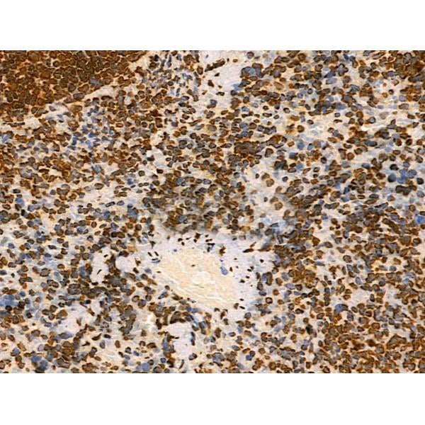
 IHC (Immunohistochemistry)
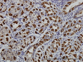
(At 1/100 staining Human colorectal cancer by IHC-P. The sample was formaldehyde fixed and a heat mediated antigen retrieval step in citrate buffer was performed. The sample was then blocked and incubated with the primary antibody at 4 degree C overnight. An HRP conjugated anti-Rabbit antibody was used as the secondary antibody.)
IHC (Immunohistochemistry)
(At 1/100 staining Human colorectal cancer by IHC-P. The sample was formaldehyde fixed and a heat mediated antigen retrieval step in citrate buffer was performed. The sample was then blocked and incubated with the primary antibody at 4 degree C overnight. An HRP conjugated anti-Rabbit antibody was used as the secondary antibody.)
Histone H2A.X, Polyclonal Antibody (Cat# AAA31320)
Full Name
Acetyl-Histone H2A.X (Lys9) Antibody
Gene Names
H2AFX; H2AX; H2A.X; H2A/X
Reactivity
Human, Mouse, Rat
Applications
Immunohistochemistry, Peptide ELISA
Purity
The antiserum was purified by peptide affinity chromatography using SulfoLink Coupling Resin
Pricing


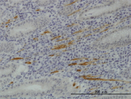
 IHC (Immunohistochemistry)
(At 1/100 staining Mouse lung tissue by IHC-P. The sample was formaldehyde fixed and a heat mediated antigen retrieval step in citrate buffer was performed. The sample was then blocked and incubated with the primary antibody at 4 degree C overnight. An HRP conjugated anti-Rabbit antibody was used as the secondary antibody.)
IHC (Immunohistochemistry)
(At 1/100 staining Mouse lung tissue by IHC-P. The sample was formaldehyde fixed and a heat mediated antigen retrieval step in citrate buffer was performed. The sample was then blocked and incubated with the primary antibody at 4 degree C overnight. An HRP conjugated anti-Rabbit antibody was used as the secondary antibody.)
Histone H2A, Polyclonal Antibody (Cat# AAA31347)
Full Name
Acetyl-Histone H2A (Lys5) Antibody
Gene Names
HIST1H2AB; H2A/m; H2AFM
Reactivity
Human, Mouse, Rat
Applications
Western Blot, Immunohistochemistry, Peptide ELISA
Purity
The antiserum was purified by peptide affinity chromatography using SulfoLink Coupling Resin
Pricing


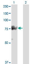
 WB (Western Blot)
(SMURF1 monoclonal antibody, Western Blot analysis of SMURF1 expression in HeLa.)
WB (Western Blot)
(SMURF1 monoclonal antibody, Western Blot analysis of SMURF1 expression in HeLa.)
SMURF1, Monoclonal Antibody (Cat# AAA24367)
Full Name
SMURF1 (E3 Ubiquitin-protein Ligase SMURF1, hSMURF1, SMAD Ubiquitination Regulatory Factor 1, KIAA1625, SMAD-specific E3 Ubiquitin-protein Ligase 1) (AP)
Reactivity
Human, Mouse
Applications
Immunohistochemistry, Immunoprecipitation, Western Blot
Purity
Purified by Protein A Affinity Chromatography.
Pricing


 Application Data
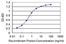
(Detection limit for recombinant GST tagged WWP1 is 0.03ng/ml as a capture antibody.)
Application Data
(Detection limit for recombinant GST tagged WWP1 is 0.03ng/ml as a capture antibody.)
WWP1, Monoclonal Antibody (Cat# AAA24097)
Full Name
WWP1 (WW Domain-containing Protein 1, Atrophin-1-interacting Protein 5, AIP5, NEDD4-like E3 Ubiquitin-protein Ligase WWP1, TGIF-interacting Ubiquitin Ligase 1, Tiul1, hSDRP1)
Gene Names
WWP1; AIP5; Tiul1; hSDRP1
Reactivity
Human
Applications
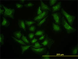
Western Blot, Immunoprecipitation, Immunohistochemistry, Immunofluorescence
Purity
Affinity Purified
Purified by Protein A affinity chromatography.
Purified by Protein A affinity chromatography.
Pricing


 WB (Western Blot)
(SMURF1 monoclonal antibody, Western Blot analysis of SMURF1 expression in HeLa.)
WB (Western Blot)
(SMURF1 monoclonal antibody, Western Blot analysis of SMURF1 expression in HeLa.)
SMURF1, Monoclonal Antibody (Cat# AAA24663)
Full Name
SMURF1 (E3 Ubiquitin-protein Ligase SMURF1, hSMURF1, SMAD Ubiquitination Regulatory Factor 1, KIAA1625, SMAD-specific E3 Ubiquitin-protein Ligase 1) APC
Reactivity
Human, Mouse
Applications
Immunofluorescence, Immunohistochemistry, Immunoprecipitation, Western Blot
Purity
Purified by Protein A Affinity Chromatography.
Pricing


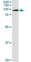


 WB (Western Blot)
(SMURF1 monoclonal antibody, Western Blot analysis of SMURF1 expression in HeLa.)
WB (Western Blot)
(SMURF1 monoclonal antibody, Western Blot analysis of SMURF1 expression in HeLa.)
SMURF1, Monoclonal Antibody (Cat# AAA24959)
Full Name
SMURF1 (E3 Ubiquitin-protein Ligase SMURF1, hSMURF1, SMAD Ubiquitination Regulatory Factor 1, KIAA1625, SMAD-specific E3 Ubiquitin-protein Ligase 1) (Biotin)
Reactivity
Human, Mouse
Applications
Immunofluorescence, Immunohistochemistry, Immunoprecipitation, Western Blot
Purity
Purified by Protein A Affinity Chromatography.
Pricing


 Application Data
(Detection limit for recombinant GST tagged WWP1 is 0.03ng/ml as a capture antibody.)
Application Data
(Detection limit for recombinant GST tagged WWP1 is 0.03ng/ml as a capture antibody.)
WWP1, Monoclonal Antibody (Cat# AAA24407)
Full Name
WWP1 (WW Domain-containing Protein 1, Atrophin-1-interacting Protein 5, AIP5, NEDD4-like E3 Ubiquitin-protein Ligase WWP1, TGIF-interacting Ubiquitin Ligase 1, Tiul1, hSDRP1) (AP)
Gene Names
WWP1; AIP5; Tiul1; hSDRP1
Reactivity
Human
Applications
Immunohistochemistry, Immunoprecipitation, Western Blot
Purity
Purified by Protein A Affinity Chromatography.
Pricing


 Application Data
(Detection limit for recombinant GST tagged WWP1 is 0.03ng/ml as a capture antibody.)
Application Data
(Detection limit for recombinant GST tagged WWP1 is 0.03ng/ml as a capture antibody.)
WWP1, Monoclonal Antibody (Cat# AAA24702)
Full Name
WWP1 (WW Domain-containing Protein 1, Atrophin-1-interacting Protein 5, AIP5, NEDD4-like E3 Ubiquitin-protein Ligase WWP1, TGIF-interacting Ubiquitin Ligase 1, Tiul1, hSDRP1) APC
Gene Names
WWP1; AIP5; Tiul1; hSDRP1
Reactivity
Human
Applications
Immunofluorescence, Immunohistochemistry, Immunoprecipitation, Western Blot
Purity
Purified by Protein A Affinity Chromatography.
Pricing


 Application Data
(Detection limit for recombinant GST tagged WWP1 is 0.03ng/ml as a capture antibody.)
Application Data
(Detection limit for recombinant GST tagged WWP1 is 0.03ng/ml as a capture antibody.)
WWP1, Monoclonal Antibody (Cat# AAA25886)
Full Name
WWP1 (WW Domain-containing Protein 1, Atrophin-1-interacting Protein 5, AIP5, NEDD4-like E3 Ubiquitin-protein Ligase WWP1, TGIF-interacting Ubiquitin Ligase 1, Tiul1, hSDRP1) (PE)
Gene Names
WWP1; AIP5; Tiul1; hSDRP1
Reactivity
Human
Applications
Immunofluorescence, Immunohistochemistry, Immunoprecipitation, Western Blot
Purity
Purified by Protein A Affinity Chromatography.
Pricing


 Application Data
(Detection limit for recombinant GST tagged WWP1 is 0.03ng/ml as a capture antibody.)
Application Data
(Detection limit for recombinant GST tagged WWP1 is 0.03ng/ml as a capture antibody.)
WWP1, Monoclonal Antibody (Cat# AAA24999)
Full Name
WWP1 (WW Domain-containing Protein 1, Atrophin-1-interacting Protein 5, AIP5, NEDD4-like E3 Ubiquitin-protein Ligase WWP1, TGIF-interacting Ubiquitin Ligase 1, Tiul1, hSDRP1) (Biotin)
Gene Names
WWP1; AIP5; Tiul1; hSDRP1
Reactivity
Human
Applications
EIA, IF, IHC, IP, WB
Purity
Purified by Protein A Affinity Chromatography.
Pricing


 WB (Western Blot)
(SMURF1 monoclonal antibody, Western Blot analysis of SMURF1 expression in HeLa.)
WB (Western Blot)
(SMURF1 monoclonal antibody, Western Blot analysis of SMURF1 expression in HeLa.)
SMURF1, Monoclonal Antibody (Cat# AAA25549)
Full Name
SMURF1 (E3 Ubiquitin-protein Ligase SMURF1, hSMURF1, SMAD Ubiquitination Regulatory Factor 1, KIAA1625, SMAD-specific E3 Ubiquitin-protein Ligase 1) (HRP)
Reactivity
Human, Mouse
Applications
EIA, IHC, IP, WB
Purity
Purified by Protein A Affinity Chromatography.
Pricing


 Application Data
(Detection limit for recombinant GST tagged WWP1 is 0.03ng/ml as a capture antibody.)
Application Data
(Detection limit for recombinant GST tagged WWP1 is 0.03ng/ml as a capture antibody.)
WWP1, Monoclonal Antibody (Cat# AAA25295)
Full Name
WWP1 (WW Domain-containing Protein 1, Atrophin-1-interacting Protein 5, AIP5, NEDD4-like E3 Ubiquitin-protein Ligase WWP1, TGIF-interacting Ubiquitin Ligase 1, Tiul1, hSDRP1) (FITC)
Gene Names
WWP1; AIP5; Tiul1; hSDRP1
Reactivity
Human
Applications
EIA, IF, IHC, IP, WB
Purity
Purified by Protein A Affinity Chromatography.
Pricing


 Application Data
(Detection limit for recombinant GST tagged WWP1 is 0.03ng/ml as a capture antibody.)
Application Data
(Detection limit for recombinant GST tagged WWP1 is 0.03ng/ml as a capture antibody.)
WWP1, Monoclonal Antibody (Cat# AAA25588)
Full Name
WWP1 (WW Domain-containing Protein 1, Atrophin-1-interacting Protein 5, AIP5, NEDD4-like E3 Ubiquitin-protein Ligase WWP1, TGIF-interacting Ubiquitin Ligase 1, Tiul1, hSDRP1) (HRP)
Gene Names
WWP1; AIP5; Tiul1; hSDRP1
Reactivity
Human
Applications
EIA, IHC, IP, WB
Purity
Purified by Protein A Affinity Chromatography.
Pricing


 WB (Western Blot)
(SMURF1 monoclonal antibody, Western Blot analysis of SMURF1 expression in HeLa.)
WB (Western Blot)
(SMURF1 monoclonal antibody, Western Blot analysis of SMURF1 expression in HeLa.)
SMURF1, Monoclonal Antibody (Cat# AAA25255)
Full Name
SMURF1 (E3 Ubiquitin-protein Ligase SMURF1, hSMURF1, SMAD Ubiquitination Regulatory Factor 1, KIAA1625, SMAD-specific E3 Ubiquitin-protein Ligase 1) (FITC)
Reactivity
Human, Mouse
Applications
EIA, IF, IHC, IP, WB
Purity
Purified by Protein A Affinity Chromatography.
Pricing


 Application Data
(Published customer image: Mouse anti V5 tag antibody, clone SV5-Pk1 used for the detection of V5 tagged WEEV_nsP3 protein by western blotting and immunofluorescenceImage caption: WEEV nsP3 interaction with host IKKbeta. A) U87MGs were transfected in a 6-well plate with 5 ug of pUC19 and WEEV_nsP3_HA for 24 hours. Cell lysates were resolved using SDS-PAGE and subsequently immunoblotted with V5 antibody and beta-actin served as a loading control. B) U87MGs were transfected with WEEV_nsP3_V5; cells were fixed after 24 hours and stained with antibodies against the endogenous IKKbeta and the V5 tag. Cells were incubated with appropriate secondary Alexa Fluor antibodies and the nuclei stained with DAPI. Co-localization of IKKbeta with WEEV_nsP3_V5 (yellow) was observed as shown by the arrows. B) Panels E -H serve as an example of transfected cells in a given field of view that show co-localization of IKKbeta and WEEV_nsP3_V5 24 hours post transfection. Panels I-L represent magnified images of other cells showing co-localization of IKKbeta and WEEV_nsP3_V5. Panel M is a magnified image of panel L. The co-localization was confirmed by Z-stack analysis. Co-localization was calculated to be approximately in 61% of cells (163 cells were counted of which 44% demonstrated expression of nsP3. Of those cells that expressed nsP3, 61% showed co-localization of both proteins). Images were taken using Nikon Eclipse TE2000-U at 60x magnification and are representative of 2 independent experiments.From: Amaya M, Voss K, Sampey G, Senina S, de la Fuente C, et al. (2014) The Role of IKKbeta in Venezuelan Equine Encephalitis Virus Infection. PLoS ONE 9(2): e86745.)
Application Data
(Published customer image: Mouse anti V5 tag antibody, clone SV5-Pk1 used for the detection of V5 tagged WEEV_nsP3 protein by western blotting and immunofluorescenceImage caption: WEEV nsP3 interaction with host IKKbeta. A) U87MGs were transfected in a 6-well plate with 5 ug of pUC19 and WEEV_nsP3_HA for 24 hours. Cell lysates were resolved using SDS-PAGE and subsequently immunoblotted with V5 antibody and beta-actin served as a loading control. B) U87MGs were transfected with WEEV_nsP3_V5; cells were fixed after 24 hours and stained with antibodies against the endogenous IKKbeta and the V5 tag. Cells were incubated with appropriate secondary Alexa Fluor antibodies and the nuclei stained with DAPI. Co-localization of IKKbeta with WEEV_nsP3_V5 (yellow) was observed as shown by the arrows. B) Panels E -H serve as an example of transfected cells in a given field of view that show co-localization of IKKbeta and WEEV_nsP3_V5 24 hours post transfection. Panels I-L represent magnified images of other cells showing co-localization of IKKbeta and WEEV_nsP3_V5. Panel M is a magnified image of panel L. The co-localization was confirmed by Z-stack analysis. Co-localization was calculated to be approximately in 61% of cells (163 cells were counted of which 44% demonstrated expression of nsP3. Of those cells that expressed nsP3, 61% showed co-localization of both proteins). Images were taken using Nikon Eclipse TE2000-U at 60x magnification and are representative of 2 independent experiments.From: Amaya M, Voss K, Sampey G, Senina S, de la Fuente C, et al. (2014) The Role of IKKbeta in Venezuelan Equine Encephalitis Virus Infection. PLoS ONE 9(2): e86745.)
V5-TAG, Monoclonal Antibody (Cat# AAA12081)
Full Name
MOUSE ANTI V5-TAG:HRP
Applications
Western Blot
Pricing


 Application Data
(Published customer image: Mouse anti V5 tag antibody, clone SV5-Pk1 used for the detection of V5 tagged WEEV_nsP3 protein by western blotting and immunofluorescenceImage caption: WEEV nsP3 interaction with host IKKbeta. A) U87MGs were transfected in a 6-well plate with 5 ug of pUC19 and WEEV_nsP3_HA for 24 hours. Cell lysates were resolved using SDS-PAGE and subsequently immunoblotted with V5 antibody and beta-actin served as a loading control. B) U87MGs were transfected with WEEV_nsP3_V5; cells were fixed after 24 hours and stained with antibodies against the endogenous IKKbeta and the V5 tag. Cells were incubated with appropriate secondary Alexa Fluor antibodies and the nuclei stained with DAPI. Co-localization of IKKbeta with WEEV_nsP3_V5 (yellow) was observed as shown by the arrows. B) Panels E -H serve as an example of transfected cells in a given field of view that show co-localization of IKKbeta and WEEV_nsP3_V5 24 hours post transfection. Panels I-L represent magnified images of other cells showing co-localization of IKKbeta and WEEV_nsP3_V5. Panel M is a magnified image of panel L. The co-localization was confirmed by Z-stack analysis. Co-localization was calculated to be approximately in 61% of cells (163 cells were counted of which 44% demonstrated expression of nsP3. Of those cells that expressed nsP3, 61% showed co-localization of both proteins). Images were taken using Nikon Eclipse TE2000-U at 60x magnification and are representative of 2 independent experiments.From: Amaya M, Voss K, Sampey G, Senina S, de la Fuente C, et al. (2014) The Role of IKKbeta in Venezuelan Equine Encephalitis Virus Infection. PLoS ONE 9(2): e86745.)
Application Data
(Published customer image: Mouse anti V5 tag antibody, clone SV5-Pk1 used for the detection of V5 tagged WEEV_nsP3 protein by western blotting and immunofluorescenceImage caption: WEEV nsP3 interaction with host IKKbeta. A) U87MGs were transfected in a 6-well plate with 5 ug of pUC19 and WEEV_nsP3_HA for 24 hours. Cell lysates were resolved using SDS-PAGE and subsequently immunoblotted with V5 antibody and beta-actin served as a loading control. B) U87MGs were transfected with WEEV_nsP3_V5; cells were fixed after 24 hours and stained with antibodies against the endogenous IKKbeta and the V5 tag. Cells were incubated with appropriate secondary Alexa Fluor antibodies and the nuclei stained with DAPI. Co-localization of IKKbeta with WEEV_nsP3_V5 (yellow) was observed as shown by the arrows. B) Panels E -H serve as an example of transfected cells in a given field of view that show co-localization of IKKbeta and WEEV_nsP3_V5 24 hours post transfection. Panels I-L represent magnified images of other cells showing co-localization of IKKbeta and WEEV_nsP3_V5. Panel M is a magnified image of panel L. The co-localization was confirmed by Z-stack analysis. Co-localization was calculated to be approximately in 61% of cells (163 cells were counted of which 44% demonstrated expression of nsP3. Of those cells that expressed nsP3, 61% showed co-localization of both proteins). Images were taken using Nikon Eclipse TE2000-U at 60x magnification and are representative of 2 independent experiments.From: Amaya M, Voss K, Sampey G, Senina S, de la Fuente C, et al. (2014) The Role of IKKbeta in Venezuelan Equine Encephalitis Virus Infection. PLoS ONE 9(2): e86745.)
V5-TAG, Monoclonal Antibody (Cat# AAA11864)
Full Name
MOUSE ANTI V5-TAG:FITC
Applications
Immunofluorescence
Pricing


 Application Data
(Published customer image: Mouse anti V5 tag antibody, clone SV5-Pk1 used for the detection of V5 tagged WEEV_nsP3 protein by western blotting and immunofluorescenceImage caption: WEEV nsP3 interaction with host IKKbeta. A) U87MGs were transfected in a 6-well plate with 5 ug of pUC19 and WEEV_nsP3_HA for 24 hours. Cell lysates were resolved using SDS-PAGE and subsequently immunoblotted with V5 antibody and beta-actin served as a loading control. B) U87MGs were transfected with WEEV_nsP3_V5; cells were fixed after 24 hours and stained with antibodies against the endogenous IKKbeta and the V5 tag. Cells were incubated with appropriate secondary Alexa Fluor antibodies and the nuclei stained with DAPI. Co-localization of IKKbeta with WEEV_nsP3_V5 (yellow) was observed as shown by the arrows. B) Panels E -H serve as an example of transfected cells in a given field of view that show co-localization of IKKbeta and WEEV_nsP3_V5 24 hours post transfection. Panels I-L represent magnified images of other cells showing co-localization of IKKbeta and WEEV_nsP3_V5. Panel M is a magnified image of panel L. The co-localization was confirmed by Z-stack analysis. Co-localization was calculated to be approximately in 61% of cells (163 cells were counted of which 44% demonstrated expression of nsP3. Of those cells that expressed nsP3, 61% showed co-localization of both proteins). Images were taken using Nikon Eclipse TE2000-U at 60x magnification and are representative of 2 independent experiments.From: Amaya M, Voss K, Sampey G, Senina S, de la Fuente C, et al. (2014) The Role of IKKbeta in Venezuelan Equine Encephalitis Virus Infection. PLoS ONE 9(2): e86745.)
Application Data
(Published customer image: Mouse anti V5 tag antibody, clone SV5-Pk1 used for the detection of V5 tagged WEEV_nsP3 protein by western blotting and immunofluorescenceImage caption: WEEV nsP3 interaction with host IKKbeta. A) U87MGs were transfected in a 6-well plate with 5 ug of pUC19 and WEEV_nsP3_HA for 24 hours. Cell lysates were resolved using SDS-PAGE and subsequently immunoblotted with V5 antibody and beta-actin served as a loading control. B) U87MGs were transfected with WEEV_nsP3_V5; cells were fixed after 24 hours and stained with antibodies against the endogenous IKKbeta and the V5 tag. Cells were incubated with appropriate secondary Alexa Fluor antibodies and the nuclei stained with DAPI. Co-localization of IKKbeta with WEEV_nsP3_V5 (yellow) was observed as shown by the arrows. B) Panels E -H serve as an example of transfected cells in a given field of view that show co-localization of IKKbeta and WEEV_nsP3_V5 24 hours post transfection. Panels I-L represent magnified images of other cells showing co-localization of IKKbeta and WEEV_nsP3_V5. Panel M is a magnified image of panel L. The co-localization was confirmed by Z-stack analysis. Co-localization was calculated to be approximately in 61% of cells (163 cells were counted of which 44% demonstrated expression of nsP3. Of those cells that expressed nsP3, 61% showed co-localization of both proteins). Images were taken using Nikon Eclipse TE2000-U at 60x magnification and are representative of 2 independent experiments.From: Amaya M, Voss K, Sampey G, Senina S, de la Fuente C, et al. (2014) The Role of IKKbeta in Venezuelan Equine Encephalitis Virus Infection. PLoS ONE 9(2): e86745.)
V5-TAG, Monoclonal Antibody (Cat# AAA11930)
Full Name
MOUSE ANTI V5-TAG
Applications
Immunohistochemistry, Flow Cytometry, Immunofluorescence, Immunoprecipitation, Western Blot, Radioimmunoassay
Pricing


 Application Data
(Published customer image: Mouse anti V5 tag antibody, clone SV5-Pk1 used for the detection of V5 tagged WEEV_nsP3 protein by western blotting and immunofluorescenceImage caption: WEEV nsP3 interaction with host IKKbeta. A) U87MGs were transfected in a 6-well plate with 5 ug of pUC19 and WEEV_nsP3_HA for 24 hours. Cell lysates were resolved using SDS-PAGE and subsequently immunoblotted with V5 antibody and beta-actin served as a loading control. B) U87MGs were transfected with WEEV_nsP3_V5; cells were fixed after 24 hours and stained with antibodies against the endogenous IKKbeta and the V5 tag. Cells were incubated with appropriate secondary Alexa Fluor antibodies and the nuclei stained with DAPI. Co-localization of IKKbeta with WEEV_nsP3_V5 (yellow) was observed as shown by the arrows. B) Panels E -H serve as an example of transfected cells in a given field of view that show co-localization of IKKbeta and WEEV_nsP3_V5 24 hours post transfection. Panels I-L represent magnified images of other cells showing co-localization of IKKbeta and WEEV_nsP3_V5. Panel M is a magnified image of panel L. The co-localization was confirmed by Z-stack analysis. Co-localization was calculated to be approximately in 61% of cells (163 cells were counted of which 44% demonstrated expression of nsP3. Of those cells that expressed nsP3, 61% showed co-localization of both proteins). Images were taken using Nikon Eclipse TE2000-U at 60x magnification and are representative of 2 independent experiments.From: Amaya M, Voss K, Sampey G, Senina S, de la Fuente C, et al. (2014) The Role of IKKbeta in Venezuelan Equine Encephalitis Virus Infection. PLoS ONE 9(2): e86745.)
Application Data
(Published customer image: Mouse anti V5 tag antibody, clone SV5-Pk1 used for the detection of V5 tagged WEEV_nsP3 protein by western blotting and immunofluorescenceImage caption: WEEV nsP3 interaction with host IKKbeta. A) U87MGs were transfected in a 6-well plate with 5 ug of pUC19 and WEEV_nsP3_HA for 24 hours. Cell lysates were resolved using SDS-PAGE and subsequently immunoblotted with V5 antibody and beta-actin served as a loading control. B) U87MGs were transfected with WEEV_nsP3_V5; cells were fixed after 24 hours and stained with antibodies against the endogenous IKKbeta and the V5 tag. Cells were incubated with appropriate secondary Alexa Fluor antibodies and the nuclei stained with DAPI. Co-localization of IKKbeta with WEEV_nsP3_V5 (yellow) was observed as shown by the arrows. B) Panels E -H serve as an example of transfected cells in a given field of view that show co-localization of IKKbeta and WEEV_nsP3_V5 24 hours post transfection. Panels I-L represent magnified images of other cells showing co-localization of IKKbeta and WEEV_nsP3_V5. Panel M is a magnified image of panel L. The co-localization was confirmed by Z-stack analysis. Co-localization was calculated to be approximately in 61% of cells (163 cells were counted of which 44% demonstrated expression of nsP3. Of those cells that expressed nsP3, 61% showed co-localization of both proteins). Images were taken using Nikon Eclipse TE2000-U at 60x magnification and are representative of 2 independent experiments.From: Amaya M, Voss K, Sampey G, Senina S, de la Fuente C, et al. (2014) The Role of IKKbeta in Venezuelan Equine Encephalitis Virus Infection. PLoS ONE 9(2): e86745.)
V5-TAG, Monoclonal Antibody (Cat# AAA12211)
Full Name
MOUSE ANTI V5-TAG
Applications
Immunohistochemistry, Flow Cytometry, Immunofluorescence, Immunoprecipitation, Western Blot, Radioimmunoassay
Pricing


 Application Data
(Published customer image: Mouse anti V5 tag antibody, clone SV5-Pk1 used for the detection of V5 tagged WEEV_nsP3 protein by western blotting and immunofluorescenceImage caption: WEEV nsP3 interaction with host IKKbeta. A) U87MGs were transfected in a 6-well plate with 5 ug of pUC19 and WEEV_nsP3_HA for 24 hours. Cell lysates were resolved using SDS-PAGE and subsequently immunoblotted with V5 antibody and beta-actin served as a loading control. B) U87MGs were transfected with WEEV_nsP3_V5; cells were fixed after 24 hours and stained with antibodies against the endogenous IKKbeta and the V5 tag. Cells were incubated with appropriate secondary Alexa Fluor antibodies and the nuclei stained with DAPI. Co-localization of IKKbeta with WEEV_nsP3_V5 (yellow) was observed as shown by the arrows. B) Panels E -H serve as an example of transfected cells in a given field of view that show co-localization of IKKbeta and WEEV_nsP3_V5 24 hours post transfection. Panels I-L represent magnified images of other cells showing co-localization of IKKbeta and WEEV_nsP3_V5. Panel M is a magnified image of panel L. The co-localization was confirmed by Z-stack analysis. Co-localization was calculated to be approximately in 61% of cells (163 cells were counted of which 44% demonstrated expression of nsP3. Of those cells that expressed nsP3, 61% showed co-localization of both proteins). Images were taken using Nikon Eclipse TE2000-U at 60x magnification and are representative of 2 independent experiments.From: Amaya M, Voss K, Sampey G, Senina S, de la Fuente C, et al. (2014) The Role of IKKbeta in Venezuelan Equine Encephalitis Virus Infection. PLoS ONE 9(2): e86745.)
Application Data
(Published customer image: Mouse anti V5 tag antibody, clone SV5-Pk1 used for the detection of V5 tagged WEEV_nsP3 protein by western blotting and immunofluorescenceImage caption: WEEV nsP3 interaction with host IKKbeta. A) U87MGs were transfected in a 6-well plate with 5 ug of pUC19 and WEEV_nsP3_HA for 24 hours. Cell lysates were resolved using SDS-PAGE and subsequently immunoblotted with V5 antibody and beta-actin served as a loading control. B) U87MGs were transfected with WEEV_nsP3_V5; cells were fixed after 24 hours and stained with antibodies against the endogenous IKKbeta and the V5 tag. Cells were incubated with appropriate secondary Alexa Fluor antibodies and the nuclei stained with DAPI. Co-localization of IKKbeta with WEEV_nsP3_V5 (yellow) was observed as shown by the arrows. B) Panels E -H serve as an example of transfected cells in a given field of view that show co-localization of IKKbeta and WEEV_nsP3_V5 24 hours post transfection. Panels I-L represent magnified images of other cells showing co-localization of IKKbeta and WEEV_nsP3_V5. Panel M is a magnified image of panel L. The co-localization was confirmed by Z-stack analysis. Co-localization was calculated to be approximately in 61% of cells (163 cells were counted of which 44% demonstrated expression of nsP3. Of those cells that expressed nsP3, 61% showed co-localization of both proteins). Images were taken using Nikon Eclipse TE2000-U at 60x magnification and are representative of 2 independent experiments.From: Amaya M, Voss K, Sampey G, Senina S, de la Fuente C, et al. (2014) The Role of IKKbeta in Venezuelan Equine Encephalitis Virus Infection. PLoS ONE 9(2): e86745.)
V5-TAG, Monoclonal Antibody (Cat# AAA11850)
Full Name
MOUSE ANTI V5-TAG:Biotin
Applications
Immunohistochemistry, Western Blot
Pricing