Filters
Clonality
Type
Reactivity
Gene Name
Isotype
Host
Application
Clone
18 results for " HIV infection" - showing 1-18


 IHC (Immunohistochemistry)
(At 1/100 staining Human mammary cancer by IHC-P. The sample was formaldehyde fixed and a heat mediated antigen retrieval step in citrate buffer was performed. The sample was then blocked and incubated with the primary antibody at 4 degree C overnight. An HRP conjugated anti-Rabbit antibody was used as the secondary antibody.)
IHC (Immunohistochemistry)
(At 1/100 staining Human mammary cancer by IHC-P. The sample was formaldehyde fixed and a heat mediated antigen retrieval step in citrate buffer was performed. The sample was then blocked and incubated with the primary antibody at 4 degree C overnight. An HRP conjugated anti-Rabbit antibody was used as the secondary antibody.)
SAMHD1, Polyclonal Antibody (Cat# AAA31278)
Full Name
Phospho-SAMHD1 (Thr592) Antibody
Gene Names
SAMHD1; DCIP; CHBL2; HDDC1; MOP-5; SBBI88
Reactivity
Human, Mouse, Rat, Monkey
Applications
Western Blot, Immunohistochemistry, Peptide ELISA
Purity
The antibody is from purified rabbit serum by affinity purification via sequential chromatography on phospho-peptide and non-phospho-peptide affinity columns.
Pricing
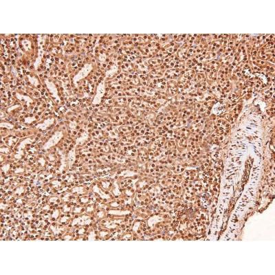


 IHC (Immunohistochemistry)
(At 1/200 staining Human lung cancer tissue sections by IHC-P. The tissue was formaldehyde fixed and a heat mediated antigen retrieval step in citrate buffer was performed. The tissue was then blocked and incubated with the antibody for 1.5 hours at 22 degree C. An HRP conjugated goat anti-rabbit antibody was used as the secondary antibody.)
IHC (Immunohistochemistry)
(At 1/200 staining Human lung cancer tissue sections by IHC-P. The tissue was formaldehyde fixed and a heat mediated antigen retrieval step in citrate buffer was performed. The tissue was then blocked and incubated with the antibody for 1.5 hours at 22 degree C. An HRP conjugated goat anti-rabbit antibody was used as the secondary antibody.)
ERK1/2, Polyclonal Antibody (Cat# AAA31416)
Full Name
Phospho-ERK1/2 (Thr202+Tyr204/Thr185+Tyr187) Antibody
Gene Names
MAPK3; ERK1; ERT2; ERK-1; PRKM3; P44ERK1; P44MAPK; HS44KDAP; HUMKER1A; p44-ERK1; p44-MAPK
Reactivity
Human, Mouse, Rat, Monkey
Predicted Reactivity: Pig (100%), Zebrafish (100%), Bovine (100%), Horse (100%), Sheep (100%), Rabbit (100%)
Predicted Reactivity: Pig (100%), Zebrafish (100%), Bovine (100%), Horse (100%), Sheep (100%), Rabbit (100%)
Applications
Western Blot, Immunohistochemistry, Peptide ELISA
Purity
The antibody is from purified rabbit serum by affinity purification via sequential chromatography on phospho-peptide and non-phospho-peptide affinity columns.
Pricing


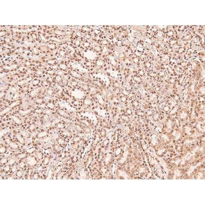
 IHC (Immunohistchemistry)
(At 1/200 staining Human breast tissue sections by IHC-P. The tissue was formaldehyde fixed and a heat mediated antigen retrieval step in citrate buffer was performed. The tissue was then blocked and incubated with the antibody for 1.5 hours at 22 degree C. An HRP conjugated goat anti-rabbit antibody was used as the secondary antibody.)
IHC (Immunohistchemistry)
(At 1/200 staining Human breast tissue sections by IHC-P. The tissue was formaldehyde fixed and a heat mediated antigen retrieval step in citrate buffer was performed. The tissue was then blocked and incubated with the antibody for 1.5 hours at 22 degree C. An HRP conjugated goat anti-rabbit antibody was used as the secondary antibody.)
KAT3B/p300, Polyclonal Antibody (Cat# AAA31393)
Full Name
Phospho-KAT3B/p300 (Ser1834) Antibody
Gene Names
EP300; p300; KAT3B; RSTS2
Reactivity
Human, Mouse, Rat
Predicted Reactivity: Pig (100%), Bovine (100%), Horse (100%), Sheep (100%), Rabbit (100%), Chicken (100%), Xenopus (100%)
Predicted Reactivity: Pig (100%), Bovine (100%), Horse (100%), Sheep (100%), Rabbit (100%), Chicken (100%), Xenopus (100%)
Applications
Western Blot, Immunohistochemistry, Peptide ELISA
Purity
The antibody is from purified rabbit serum by affinity purification via sequential chromatography on phospho-peptide and non-phospho-peptide affinity columns.
Pricing


 Application Data
(At 25 degree C. Samples were then incubated with primary Ab(At 37 degree C. An AlexaFluor594 conjugated goat anti-rabbit IgG(H+L) Ab(Red) and an AlexaFluor488 conjugated goat anti-mouse IgG(H+L) Ab(Green) were used as the secondary antibody.The nuclear counter stain is DAPI (blue).)
Application Data
(At 25 degree C. Samples were then incubated with primary Ab(At 37 degree C. An AlexaFluor594 conjugated goat anti-rabbit IgG(H+L) Ab(Red) and an AlexaFluor488 conjugated goat anti-mouse IgG(H+L) Ab(Green) were used as the secondary antibody.The nuclear counter stain is DAPI (blue).)
ERK1/2, Polyclonal Antibody (Cat# AAA31300)
Full Name
Phospho-ERK1/2 (Thr177/Thr160) Antibody
Gene Names
MAPK3; ERK1; ERT2; ERK-1; PRKM3; P44ERK1; P44MAPK; HS44KDAP; HUMKER1A; p44-ERK1; p44-MAPK
Reactivity
Human, Mouse, Rat
Applications
Immunohistochemistry, Immunofluorescence, Immunocytochemistry, Peptide ELISA
Purity
The antibody is from purified rabbit serum by affinity purification via sequential chromatography on phospho-peptide and non-phospho-peptide affinity columns.
Pricing

 Application Data
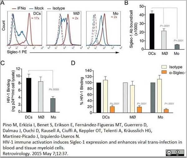
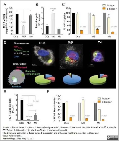
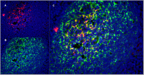
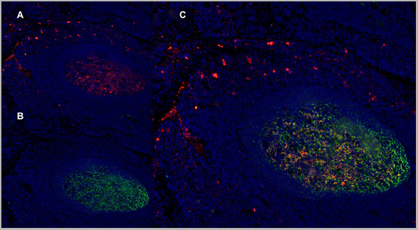
(PE conjugatedMouse anti Human CD169 antibody, clone 7-239 used to block CD169 function on myeloid cells.Image caption:Siglec-1 mediates HIV-1 uptake into a storage compartment and enhances HIV-1 trans-infection specially in IFN?-treated monocytes and DCs. A. Uptake of HIV-1NL4–3 by different myeloid cells exposed to IFN?. Cells were cultured with HIV-1 to measure p24Gag by ELISA. Mean values and SEM from four experiments include cells from 12 donors. B. Fold change in HIV-1NL4–3 uptake of cells treated with bafilomycin A1 compared to untreated cells. Mean values and SEM include cells from three donors. C. Relative uptake of HIV-1NL4–3 by IFN?-treated myeloid cells pre-incubated with the indicated mAbs. Values are normalized to the level of HIV-1 uptake by mock-treated cells (set at 100%). Mean values and SEM from two experiments include cells from six donors. D. Confocal microscopy analysis of different IFN?-treated myeloid cells pulsed with HIV-1Cherry and stained for Siglec-1 (Alexa 488), HLA-DR (Alexa 647) and DAPI. (Top) Representative viral pattern for each kind of myeloid cell analyzed, showing maximum fluorescence intensity of four channels. (Bottom) Percentage of myeloid cells with distinct viral patterns: random distribution, polarized accumulation, and sac-like compartment formation, as illustrated in the left drawing. Mean values of 50 cells from two different donors are shown. E. HIV-1 transmission from IFN?-treated myeloid cells to a luciferase reporter CD4+ cell line. HIV-1 infection was determined by induced luciferase activity in relative light units (RLUs). Mean values and SEM from four experiments include cells from 12 donors. F. Relative HIV-1 transmission from IFN?-treated myeloid cells pre-incubated with the indicated mAbs. Values are normalized to the level of HIV-1 trans-infected by mock-treated cells. Mean values and SEM from two experiments include cells from six donors. Statistical differences were assessed with a paired t test in A and E, and with a one sample t-test in B, C and F.From: Pino M, Erkizia I, Benet S, Erikson E, Fernández-Figueras MT, Guerrero D, Dalmau J, Ouchi D, Rausell A, Ciuffi A, Keppler OT, Telenti A, Kräusslich HG, Martinez-Picado J, Izquierdo-Useros N.HIV-1 immune activation induces Siglec-1 expression and enhances viral trans-infection in blood and tissue myeloid cells.Retrovirology. 2015 May 7;12:37.This image is from an open access article distributed under the terms of the Creative Commons Attribution License.)
Application Data
(PE conjugatedMouse anti Human CD169 antibody, clone 7-239 used to block CD169 function on myeloid cells.Image caption:Siglec-1 mediates HIV-1 uptake into a storage compartment and enhances HIV-1 trans-infection specially in IFN?-treated monocytes and DCs. A. Uptake of HIV-1NL4–3 by different myeloid cells exposed to IFN?. Cells were cultured with HIV-1 to measure p24Gag by ELISA. Mean values and SEM from four experiments include cells from 12 donors. B. Fold change in HIV-1NL4–3 uptake of cells treated with bafilomycin A1 compared to untreated cells. Mean values and SEM include cells from three donors. C. Relative uptake of HIV-1NL4–3 by IFN?-treated myeloid cells pre-incubated with the indicated mAbs. Values are normalized to the level of HIV-1 uptake by mock-treated cells (set at 100%). Mean values and SEM from two experiments include cells from six donors. D. Confocal microscopy analysis of different IFN?-treated myeloid cells pulsed with HIV-1Cherry and stained for Siglec-1 (Alexa 488), HLA-DR (Alexa 647) and DAPI. (Top) Representative viral pattern for each kind of myeloid cell analyzed, showing maximum fluorescence intensity of four channels. (Bottom) Percentage of myeloid cells with distinct viral patterns: random distribution, polarized accumulation, and sac-like compartment formation, as illustrated in the left drawing. Mean values of 50 cells from two different donors are shown. E. HIV-1 transmission from IFN?-treated myeloid cells to a luciferase reporter CD4+ cell line. HIV-1 infection was determined by induced luciferase activity in relative light units (RLUs). Mean values and SEM from four experiments include cells from 12 donors. F. Relative HIV-1 transmission from IFN?-treated myeloid cells pre-incubated with the indicated mAbs. Values are normalized to the level of HIV-1 trans-infected by mock-treated cells. Mean values and SEM from two experiments include cells from six donors. Statistical differences were assessed with a paired t test in A and E, and with a one sample t-test in B, C and F.From: Pino M, Erkizia I, Benet S, Erikson E, Fernández-Figueras MT, Guerrero D, Dalmau J, Ouchi D, Rausell A, Ciuffi A, Keppler OT, Telenti A, Kräusslich HG, Martinez-Picado J, Izquierdo-Useros N.HIV-1 immune activation induces Siglec-1 expression and enhances viral trans-infection in blood and tissue myeloid cells.Retrovirology. 2015 May 7;12:37.This image is from an open access article distributed under the terms of the Creative Commons Attribution License.)
CD169, Monoclonal Antibody (Cat# AAA12264)
Full Name
Mouse Anti Human CD169
Gene Names
SIGLEC1; SN; CD169; SIGLEC-1
Reactivity
Human
Applications
Immunohistochemistry, Flow Cytometry, Functional Assay, Immunoprecipitation, Western Blot
Purity
>95% by SDS PAGE
Purified IgG prepared by affinity chromatography on Protein A from tissue culture supernatant.
Purified IgG prepared by affinity chromatography on Protein A from tissue culture supernatant.
Pricing
 SDS-PAGE
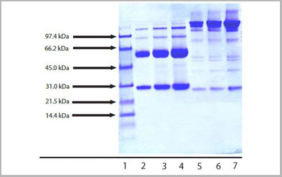
(4-12% Bis-Tris NuPAGE gel showing the following lanes:1- MW standard;2- IgG3 (5 ug reduced/heated);3- IgG3 (10 ug reduced/heated);4- IgG3 (20 ug reduced/heated);5- IgG3 (5 ug non-reduced/no heat);6- IgG3 (10 ug non-reduced/no heat);7- IgG3 (20 ug non-reduced/no heat))
SDS-PAGE
(4-12% Bis-Tris NuPAGE gel showing the following lanes:1- MW standard;2- IgG3 (5 ug reduced/heated);3- IgG3 (10 ug reduced/heated);4- IgG3 (20 ug reduced/heated);5- IgG3 (5 ug non-reduced/no heat);6- IgG3 (10 ug non-reduced/no heat);7- IgG3 (20 ug non-reduced/no heat))
Human IgG3 protein, Immunoglobulin (Cat# AAA14399)
Full Name
Human IgG3 protein
Purity
> 95% pure by SDS-PAGE
Pricing


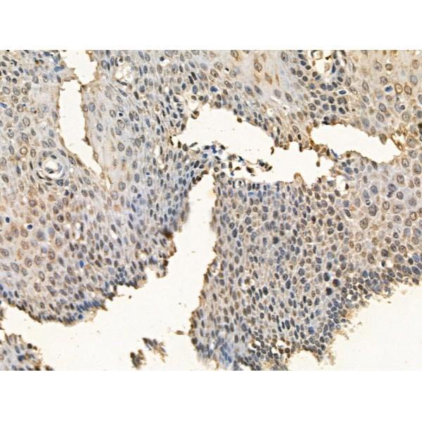
 Application Data
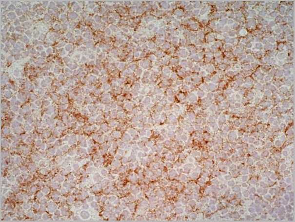
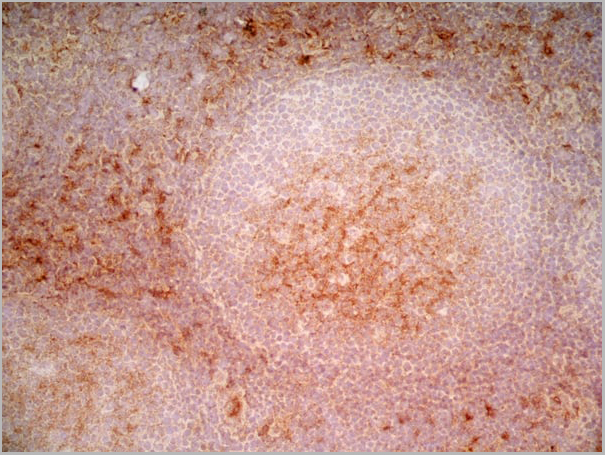
(Immunoperoxidase staining of human tonsil cryosection using Mouse anti Human CD1a antibody followed by HISTAR detection system. Medium power)
Application Data
(Immunoperoxidase staining of human tonsil cryosection using Mouse anti Human CD1a antibody followed by HISTAR detection system. Medium power)
CD1a, Monoclonal Antibody (Cat# AAA12012)
Full Name
MOUSE ANTI HUMAN CD1a
Gene Names
CD1A; R4; T6; CD1; FCB6; HTA1
Applications
Immunohistochemistry, Flow Cytometry
Pricing


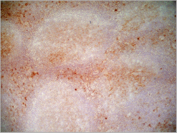
 Application Data
(Immunoperoxidase staining of human tonsil cryosection using Mouse anti Human CD1a antibody followed by HISTAR detection system. Medium power)
Application Data
(Immunoperoxidase staining of human tonsil cryosection using Mouse anti Human CD1a antibody followed by HISTAR detection system. Medium power)
CD1a, Monoclonal Antibody (Cat# AAA12058)
Full Name
MOUSE ANTI HUMAN CD1a:RPE
Gene Names
CD1A; R4; T6; CD1; FCB6; HTA1
Applications
Flow Cytometry
Pricing


 Application Data
(Immunoperoxidase staining of human tonsil cryosection using Mouse anti Human CD1a antibody followed by HISTAR detection system. Medium power)
Application Data
(Immunoperoxidase staining of human tonsil cryosection using Mouse anti Human CD1a antibody followed by HISTAR detection system. Medium power)
CD1a, Monoclonal Antibody (Cat# AAA12200)
Full Name
MOUSE ANTI HUMAN CD1a:RPE
Gene Names
CD1A; R4; T6; CD1; FCB6; HTA1
Applications
Flow Cytometry
Pricing


 Application Data
(Immunoperoxidase staining of human tonsil cryosection using Mouse anti Human CD1a antibody followed by HISTAR detection system. Medium power)
Application Data
(Immunoperoxidase staining of human tonsil cryosection using Mouse anti Human CD1a antibody followed by HISTAR detection system. Medium power)
CD1a, Monoclonal Antibody (Cat# AAA12011)
Full Name
MOUSE ANTI HUMAN CD1a
Gene Names
CD1A; R4; T6; CD1; FCB6; HTA1
Reactivity
Cynomolgus monkey, Dog
Applications
Immunohistochemistry, Flow Cytometry
Pricing


 Application Data
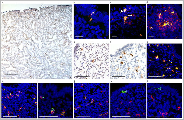
(Published customer image: Mouse anti V5 tag antibody, clone SV5-Pk1 used for the detection of V5 tagged WEEV_nsP3 protein by western blotting and immunofluorescenceImage caption: WEEV nsP3 interaction with host IKKbeta. A) U87MGs were transfected in a 6-well plate with 5 ug of pUC19 and WEEV_nsP3_HA for 24 hours. Cell lysates were resolved using SDS-PAGE and subsequently immunoblotted with V5 antibody and beta-actin served as a loading control. B) U87MGs were transfected with WEEV_nsP3_V5; cells were fixed after 24 hours and stained with antibodies against the endogenous IKKbeta and the V5 tag. Cells were incubated with appropriate secondary Alexa Fluor antibodies and the nuclei stained with DAPI. Co-localization of IKKbeta with WEEV_nsP3_V5 (yellow) was observed as shown by the arrows. B) Panels E -H serve as an example of transfected cells in a given field of view that show co-localization of IKKbeta and WEEV_nsP3_V5 24 hours post transfection. Panels I-L represent magnified images of other cells showing co-localization of IKKbeta and WEEV_nsP3_V5. Panel M is a magnified image of panel L. The co-localization was confirmed by Z-stack analysis. Co-localization was calculated to be approximately in 61% of cells (163 cells were counted of which 44% demonstrated expression of nsP3. Of those cells that expressed nsP3, 61% showed co-localization of both proteins). Images were taken using Nikon Eclipse TE2000-U at 60x magnification and are representative of 2 independent experiments.From: Amaya M, Voss K, Sampey G, Senina S, de la Fuente C, et al. (2014) The Role of IKKbeta in Venezuelan Equine Encephalitis Virus Infection. PLoS ONE 9(2): e86745.)
Application Data
(Published customer image: Mouse anti V5 tag antibody, clone SV5-Pk1 used for the detection of V5 tagged WEEV_nsP3 protein by western blotting and immunofluorescenceImage caption: WEEV nsP3 interaction with host IKKbeta. A) U87MGs were transfected in a 6-well plate with 5 ug of pUC19 and WEEV_nsP3_HA for 24 hours. Cell lysates were resolved using SDS-PAGE and subsequently immunoblotted with V5 antibody and beta-actin served as a loading control. B) U87MGs were transfected with WEEV_nsP3_V5; cells were fixed after 24 hours and stained with antibodies against the endogenous IKKbeta and the V5 tag. Cells were incubated with appropriate secondary Alexa Fluor antibodies and the nuclei stained with DAPI. Co-localization of IKKbeta with WEEV_nsP3_V5 (yellow) was observed as shown by the arrows. B) Panels E -H serve as an example of transfected cells in a given field of view that show co-localization of IKKbeta and WEEV_nsP3_V5 24 hours post transfection. Panels I-L represent magnified images of other cells showing co-localization of IKKbeta and WEEV_nsP3_V5. Panel M is a magnified image of panel L. The co-localization was confirmed by Z-stack analysis. Co-localization was calculated to be approximately in 61% of cells (163 cells were counted of which 44% demonstrated expression of nsP3. Of those cells that expressed nsP3, 61% showed co-localization of both proteins). Images were taken using Nikon Eclipse TE2000-U at 60x magnification and are representative of 2 independent experiments.From: Amaya M, Voss K, Sampey G, Senina S, de la Fuente C, et al. (2014) The Role of IKKbeta in Venezuelan Equine Encephalitis Virus Infection. PLoS ONE 9(2): e86745.)
V5-TAG, Monoclonal Antibody (Cat# AAA12081)
Full Name
MOUSE ANTI V5-TAG:HRP
Applications
Western Blot
Pricing


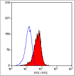
 Application Data
(PE conjugatedMouse anti Human CD169 antibody, clone 7-239 used to block CD169 function on myeloid cells.Image caption:Siglec-1 mediates HIV-1 uptake into a storage compartment and enhances HIV-1 trans-infection specially in IFN?-treated monocytes and DCs. A. Uptake of HIV-1NL4–3 by different myeloid cells exposed to IFN?. Cells were cultured with HIV-1 to measure p24Gag by ELISA. Mean values and SEM from four experiments include cells from 12 donors. B. Fold change in HIV-1NL4–3 uptake of cells treated with bafilomycin A1 compared to untreated cells. Mean values and SEM include cells from three donors. C. Relative uptake of HIV-1NL4–3 by IFN?-treated myeloid cells pre-incubated with the indicated mAbs. Values are normalized to the level of HIV-1 uptake by mock-treated cells (set at 100%). Mean values and SEM from two experiments include cells from six donors. D. Confocal microscopy analysis of different IFN?-treated myeloid cells pulsed with HIV-1Cherry and stained for Siglec-1 (Alexa 488), HLA-DR (Alexa 647) and DAPI. (Top) Representative viral pattern for each kind of myeloid cell analyzed, showing maximum fluorescence intensity of four channels. (Bottom) Percentage of myeloid cells with distinct viral patterns: random distribution, polarized accumulation, and sac-like compartment formation, as illustrated in the left drawing. Mean values of 50 cells from two different donors are shown. E. HIV-1 transmission from IFN?-treated myeloid cells to a luciferase reporter CD4+ cell line. HIV-1 infection was determined by induced luciferase activity in relative light units (RLUs). Mean values and SEM from four experiments include cells from 12 donors. F. Relative HIV-1 transmission from IFN?-treated myeloid cells pre-incubated with the indicated mAbs. Values are normalized to the level of HIV-1 trans-infected by mock-treated cells. Mean values and SEM from two experiments include cells from six donors. Statistical differences were assessed with a paired t test in A and E, and with a one sample t-test in B, C and F.From: Pino M, Erkizia I, Benet S, Erikson E, Fernández-Figueras MT, Guerrero D, Dalmau J, Ouchi D, Rausell A, Ciuffi A, Keppler OT, Telenti A, Kräusslich HG, Martinez-Picado J, Izquierdo-Useros N.HIV-1 immune activation induces Siglec-1 expression and enhances viral trans-infection in blood and tissue myeloid cells.Retrovirology. 2015 May 7;12:37.This image is from an open access article distributed under the terms of the Creative Commons Attribution License.)
Application Data
(PE conjugatedMouse anti Human CD169 antibody, clone 7-239 used to block CD169 function on myeloid cells.Image caption:Siglec-1 mediates HIV-1 uptake into a storage compartment and enhances HIV-1 trans-infection specially in IFN?-treated monocytes and DCs. A. Uptake of HIV-1NL4–3 by different myeloid cells exposed to IFN?. Cells were cultured with HIV-1 to measure p24Gag by ELISA. Mean values and SEM from four experiments include cells from 12 donors. B. Fold change in HIV-1NL4–3 uptake of cells treated with bafilomycin A1 compared to untreated cells. Mean values and SEM include cells from three donors. C. Relative uptake of HIV-1NL4–3 by IFN?-treated myeloid cells pre-incubated with the indicated mAbs. Values are normalized to the level of HIV-1 uptake by mock-treated cells (set at 100%). Mean values and SEM from two experiments include cells from six donors. D. Confocal microscopy analysis of different IFN?-treated myeloid cells pulsed with HIV-1Cherry and stained for Siglec-1 (Alexa 488), HLA-DR (Alexa 647) and DAPI. (Top) Representative viral pattern for each kind of myeloid cell analyzed, showing maximum fluorescence intensity of four channels. (Bottom) Percentage of myeloid cells with distinct viral patterns: random distribution, polarized accumulation, and sac-like compartment formation, as illustrated in the left drawing. Mean values of 50 cells from two different donors are shown. E. HIV-1 transmission from IFN?-treated myeloid cells to a luciferase reporter CD4+ cell line. HIV-1 infection was determined by induced luciferase activity in relative light units (RLUs). Mean values and SEM from four experiments include cells from 12 donors. F. Relative HIV-1 transmission from IFN?-treated myeloid cells pre-incubated with the indicated mAbs. Values are normalized to the level of HIV-1 trans-infected by mock-treated cells. Mean values and SEM from two experiments include cells from six donors. Statistical differences were assessed with a paired t test in A and E, and with a one sample t-test in B, C and F.From: Pino M, Erkizia I, Benet S, Erikson E, Fernández-Figueras MT, Guerrero D, Dalmau J, Ouchi D, Rausell A, Ciuffi A, Keppler OT, Telenti A, Kräusslich HG, Martinez-Picado J, Izquierdo-Useros N.HIV-1 immune activation induces Siglec-1 expression and enhances viral trans-infection in blood and tissue myeloid cells.Retrovirology. 2015 May 7;12:37.This image is from an open access article distributed under the terms of the Creative Commons Attribution License.)
CD169, Monoclonal Antibody (Cat# AAA12265)
Full Name
Mouse Anti Human CD169: RPE
Gene Names
SIGLEC1; SN; CD169; SIGLEC-1
Reactivity
Human
Applications
Flow Cytometry
Purity
>95% by SDS PAGE
Purified IgG prepared by affinity chromatography on Protein A from tissue culture supernatant.
Purified IgG prepared by affinity chromatography on Protein A from tissue culture supernatant.
Pricing


 Application Data
(Immunoperoxidase staining of human tonsil cryosection using Mouse anti Human CD1a antibody followed by HISTAR detection system. Medium power)
Application Data
(Immunoperoxidase staining of human tonsil cryosection using Mouse anti Human CD1a antibody followed by HISTAR detection system. Medium power)
CD1a, Monoclonal Antibody (Cat# AAA11890)
Full Name
MOUSE ANTI HUMAN CD1a:FITC
Gene Names
CD1A; R4; T6; CD1; FCB6; HTA1
Applications
Flow Cytometry
Pricing


 Application Data
(Published customer image: Mouse anti V5 tag antibody, clone SV5-Pk1 used for the detection of V5 tagged WEEV_nsP3 protein by western blotting and immunofluorescenceImage caption: WEEV nsP3 interaction with host IKKbeta. A) U87MGs were transfected in a 6-well plate with 5 ug of pUC19 and WEEV_nsP3_HA for 24 hours. Cell lysates were resolved using SDS-PAGE and subsequently immunoblotted with V5 antibody and beta-actin served as a loading control. B) U87MGs were transfected with WEEV_nsP3_V5; cells were fixed after 24 hours and stained with antibodies against the endogenous IKKbeta and the V5 tag. Cells were incubated with appropriate secondary Alexa Fluor antibodies and the nuclei stained with DAPI. Co-localization of IKKbeta with WEEV_nsP3_V5 (yellow) was observed as shown by the arrows. B) Panels E -H serve as an example of transfected cells in a given field of view that show co-localization of IKKbeta and WEEV_nsP3_V5 24 hours post transfection. Panels I-L represent magnified images of other cells showing co-localization of IKKbeta and WEEV_nsP3_V5. Panel M is a magnified image of panel L. The co-localization was confirmed by Z-stack analysis. Co-localization was calculated to be approximately in 61% of cells (163 cells were counted of which 44% demonstrated expression of nsP3. Of those cells that expressed nsP3, 61% showed co-localization of both proteins). Images were taken using Nikon Eclipse TE2000-U at 60x magnification and are representative of 2 independent experiments.From: Amaya M, Voss K, Sampey G, Senina S, de la Fuente C, et al. (2014) The Role of IKKbeta in Venezuelan Equine Encephalitis Virus Infection. PLoS ONE 9(2): e86745.)
Application Data
(Published customer image: Mouse anti V5 tag antibody, clone SV5-Pk1 used for the detection of V5 tagged WEEV_nsP3 protein by western blotting and immunofluorescenceImage caption: WEEV nsP3 interaction with host IKKbeta. A) U87MGs were transfected in a 6-well plate with 5 ug of pUC19 and WEEV_nsP3_HA for 24 hours. Cell lysates were resolved using SDS-PAGE and subsequently immunoblotted with V5 antibody and beta-actin served as a loading control. B) U87MGs were transfected with WEEV_nsP3_V5; cells were fixed after 24 hours and stained with antibodies against the endogenous IKKbeta and the V5 tag. Cells were incubated with appropriate secondary Alexa Fluor antibodies and the nuclei stained with DAPI. Co-localization of IKKbeta with WEEV_nsP3_V5 (yellow) was observed as shown by the arrows. B) Panels E -H serve as an example of transfected cells in a given field of view that show co-localization of IKKbeta and WEEV_nsP3_V5 24 hours post transfection. Panels I-L represent magnified images of other cells showing co-localization of IKKbeta and WEEV_nsP3_V5. Panel M is a magnified image of panel L. The co-localization was confirmed by Z-stack analysis. Co-localization was calculated to be approximately in 61% of cells (163 cells were counted of which 44% demonstrated expression of nsP3. Of those cells that expressed nsP3, 61% showed co-localization of both proteins). Images were taken using Nikon Eclipse TE2000-U at 60x magnification and are representative of 2 independent experiments.From: Amaya M, Voss K, Sampey G, Senina S, de la Fuente C, et al. (2014) The Role of IKKbeta in Venezuelan Equine Encephalitis Virus Infection. PLoS ONE 9(2): e86745.)
V5-TAG, Monoclonal Antibody (Cat# AAA11864)
Full Name
MOUSE ANTI V5-TAG:FITC
Applications
Immunofluorescence
Pricing


 Application Data
(Published customer image: Mouse anti V5 tag antibody, clone SV5-Pk1 used for the detection of V5 tagged WEEV_nsP3 protein by western blotting and immunofluorescenceImage caption: WEEV nsP3 interaction with host IKKbeta. A) U87MGs were transfected in a 6-well plate with 5 ug of pUC19 and WEEV_nsP3_HA for 24 hours. Cell lysates were resolved using SDS-PAGE and subsequently immunoblotted with V5 antibody and beta-actin served as a loading control. B) U87MGs were transfected with WEEV_nsP3_V5; cells were fixed after 24 hours and stained with antibodies against the endogenous IKKbeta and the V5 tag. Cells were incubated with appropriate secondary Alexa Fluor antibodies and the nuclei stained with DAPI. Co-localization of IKKbeta with WEEV_nsP3_V5 (yellow) was observed as shown by the arrows. B) Panels E -H serve as an example of transfected cells in a given field of view that show co-localization of IKKbeta and WEEV_nsP3_V5 24 hours post transfection. Panels I-L represent magnified images of other cells showing co-localization of IKKbeta and WEEV_nsP3_V5. Panel M is a magnified image of panel L. The co-localization was confirmed by Z-stack analysis. Co-localization was calculated to be approximately in 61% of cells (163 cells were counted of which 44% demonstrated expression of nsP3. Of those cells that expressed nsP3, 61% showed co-localization of both proteins). Images were taken using Nikon Eclipse TE2000-U at 60x magnification and are representative of 2 independent experiments.From: Amaya M, Voss K, Sampey G, Senina S, de la Fuente C, et al. (2014) The Role of IKKbeta in Venezuelan Equine Encephalitis Virus Infection. PLoS ONE 9(2): e86745.)
Application Data
(Published customer image: Mouse anti V5 tag antibody, clone SV5-Pk1 used for the detection of V5 tagged WEEV_nsP3 protein by western blotting and immunofluorescenceImage caption: WEEV nsP3 interaction with host IKKbeta. A) U87MGs were transfected in a 6-well plate with 5 ug of pUC19 and WEEV_nsP3_HA for 24 hours. Cell lysates were resolved using SDS-PAGE and subsequently immunoblotted with V5 antibody and beta-actin served as a loading control. B) U87MGs were transfected with WEEV_nsP3_V5; cells were fixed after 24 hours and stained with antibodies against the endogenous IKKbeta and the V5 tag. Cells were incubated with appropriate secondary Alexa Fluor antibodies and the nuclei stained with DAPI. Co-localization of IKKbeta with WEEV_nsP3_V5 (yellow) was observed as shown by the arrows. B) Panels E -H serve as an example of transfected cells in a given field of view that show co-localization of IKKbeta and WEEV_nsP3_V5 24 hours post transfection. Panels I-L represent magnified images of other cells showing co-localization of IKKbeta and WEEV_nsP3_V5. Panel M is a magnified image of panel L. The co-localization was confirmed by Z-stack analysis. Co-localization was calculated to be approximately in 61% of cells (163 cells were counted of which 44% demonstrated expression of nsP3. Of those cells that expressed nsP3, 61% showed co-localization of both proteins). Images were taken using Nikon Eclipse TE2000-U at 60x magnification and are representative of 2 independent experiments.From: Amaya M, Voss K, Sampey G, Senina S, de la Fuente C, et al. (2014) The Role of IKKbeta in Venezuelan Equine Encephalitis Virus Infection. PLoS ONE 9(2): e86745.)
V5-TAG, Monoclonal Antibody (Cat# AAA11930)
Full Name
MOUSE ANTI V5-TAG
Applications
Immunohistochemistry, Flow Cytometry, Immunofluorescence, Immunoprecipitation, Western Blot, Radioimmunoassay
Pricing


 Application Data
(Published customer image: Mouse anti V5 tag antibody, clone SV5-Pk1 used for the detection of V5 tagged WEEV_nsP3 protein by western blotting and immunofluorescenceImage caption: WEEV nsP3 interaction with host IKKbeta. A) U87MGs were transfected in a 6-well plate with 5 ug of pUC19 and WEEV_nsP3_HA for 24 hours. Cell lysates were resolved using SDS-PAGE and subsequently immunoblotted with V5 antibody and beta-actin served as a loading control. B) U87MGs were transfected with WEEV_nsP3_V5; cells were fixed after 24 hours and stained with antibodies against the endogenous IKKbeta and the V5 tag. Cells were incubated with appropriate secondary Alexa Fluor antibodies and the nuclei stained with DAPI. Co-localization of IKKbeta with WEEV_nsP3_V5 (yellow) was observed as shown by the arrows. B) Panels E -H serve as an example of transfected cells in a given field of view that show co-localization of IKKbeta and WEEV_nsP3_V5 24 hours post transfection. Panels I-L represent magnified images of other cells showing co-localization of IKKbeta and WEEV_nsP3_V5. Panel M is a magnified image of panel L. The co-localization was confirmed by Z-stack analysis. Co-localization was calculated to be approximately in 61% of cells (163 cells were counted of which 44% demonstrated expression of nsP3. Of those cells that expressed nsP3, 61% showed co-localization of both proteins). Images were taken using Nikon Eclipse TE2000-U at 60x magnification and are representative of 2 independent experiments.From: Amaya M, Voss K, Sampey G, Senina S, de la Fuente C, et al. (2014) The Role of IKKbeta in Venezuelan Equine Encephalitis Virus Infection. PLoS ONE 9(2): e86745.)
Application Data
(Published customer image: Mouse anti V5 tag antibody, clone SV5-Pk1 used for the detection of V5 tagged WEEV_nsP3 protein by western blotting and immunofluorescenceImage caption: WEEV nsP3 interaction with host IKKbeta. A) U87MGs were transfected in a 6-well plate with 5 ug of pUC19 and WEEV_nsP3_HA for 24 hours. Cell lysates were resolved using SDS-PAGE and subsequently immunoblotted with V5 antibody and beta-actin served as a loading control. B) U87MGs were transfected with WEEV_nsP3_V5; cells were fixed after 24 hours and stained with antibodies against the endogenous IKKbeta and the V5 tag. Cells were incubated with appropriate secondary Alexa Fluor antibodies and the nuclei stained with DAPI. Co-localization of IKKbeta with WEEV_nsP3_V5 (yellow) was observed as shown by the arrows. B) Panels E -H serve as an example of transfected cells in a given field of view that show co-localization of IKKbeta and WEEV_nsP3_V5 24 hours post transfection. Panels I-L represent magnified images of other cells showing co-localization of IKKbeta and WEEV_nsP3_V5. Panel M is a magnified image of panel L. The co-localization was confirmed by Z-stack analysis. Co-localization was calculated to be approximately in 61% of cells (163 cells were counted of which 44% demonstrated expression of nsP3. Of those cells that expressed nsP3, 61% showed co-localization of both proteins). Images were taken using Nikon Eclipse TE2000-U at 60x magnification and are representative of 2 independent experiments.From: Amaya M, Voss K, Sampey G, Senina S, de la Fuente C, et al. (2014) The Role of IKKbeta in Venezuelan Equine Encephalitis Virus Infection. PLoS ONE 9(2): e86745.)
V5-TAG, Monoclonal Antibody (Cat# AAA12211)
Full Name
MOUSE ANTI V5-TAG
Applications
Immunohistochemistry, Flow Cytometry, Immunofluorescence, Immunoprecipitation, Western Blot, Radioimmunoassay
Pricing


 Application Data
(Published customer image: Mouse anti V5 tag antibody, clone SV5-Pk1 used for the detection of V5 tagged WEEV_nsP3 protein by western blotting and immunofluorescenceImage caption: WEEV nsP3 interaction with host IKKbeta. A) U87MGs were transfected in a 6-well plate with 5 ug of pUC19 and WEEV_nsP3_HA for 24 hours. Cell lysates were resolved using SDS-PAGE and subsequently immunoblotted with V5 antibody and beta-actin served as a loading control. B) U87MGs were transfected with WEEV_nsP3_V5; cells were fixed after 24 hours and stained with antibodies against the endogenous IKKbeta and the V5 tag. Cells were incubated with appropriate secondary Alexa Fluor antibodies and the nuclei stained with DAPI. Co-localization of IKKbeta with WEEV_nsP3_V5 (yellow) was observed as shown by the arrows. B) Panels E -H serve as an example of transfected cells in a given field of view that show co-localization of IKKbeta and WEEV_nsP3_V5 24 hours post transfection. Panels I-L represent magnified images of other cells showing co-localization of IKKbeta and WEEV_nsP3_V5. Panel M is a magnified image of panel L. The co-localization was confirmed by Z-stack analysis. Co-localization was calculated to be approximately in 61% of cells (163 cells were counted of which 44% demonstrated expression of nsP3. Of those cells that expressed nsP3, 61% showed co-localization of both proteins). Images were taken using Nikon Eclipse TE2000-U at 60x magnification and are representative of 2 independent experiments.From: Amaya M, Voss K, Sampey G, Senina S, de la Fuente C, et al. (2014) The Role of IKKbeta in Venezuelan Equine Encephalitis Virus Infection. PLoS ONE 9(2): e86745.)
Application Data
(Published customer image: Mouse anti V5 tag antibody, clone SV5-Pk1 used for the detection of V5 tagged WEEV_nsP3 protein by western blotting and immunofluorescenceImage caption: WEEV nsP3 interaction with host IKKbeta. A) U87MGs were transfected in a 6-well plate with 5 ug of pUC19 and WEEV_nsP3_HA for 24 hours. Cell lysates were resolved using SDS-PAGE and subsequently immunoblotted with V5 antibody and beta-actin served as a loading control. B) U87MGs were transfected with WEEV_nsP3_V5; cells were fixed after 24 hours and stained with antibodies against the endogenous IKKbeta and the V5 tag. Cells were incubated with appropriate secondary Alexa Fluor antibodies and the nuclei stained with DAPI. Co-localization of IKKbeta with WEEV_nsP3_V5 (yellow) was observed as shown by the arrows. B) Panels E -H serve as an example of transfected cells in a given field of view that show co-localization of IKKbeta and WEEV_nsP3_V5 24 hours post transfection. Panels I-L represent magnified images of other cells showing co-localization of IKKbeta and WEEV_nsP3_V5. Panel M is a magnified image of panel L. The co-localization was confirmed by Z-stack analysis. Co-localization was calculated to be approximately in 61% of cells (163 cells were counted of which 44% demonstrated expression of nsP3. Of those cells that expressed nsP3, 61% showed co-localization of both proteins). Images were taken using Nikon Eclipse TE2000-U at 60x magnification and are representative of 2 independent experiments.From: Amaya M, Voss K, Sampey G, Senina S, de la Fuente C, et al. (2014) The Role of IKKbeta in Venezuelan Equine Encephalitis Virus Infection. PLoS ONE 9(2): e86745.)
V5-TAG, Monoclonal Antibody (Cat# AAA11850)
Full Name
MOUSE ANTI V5-TAG:Biotin
Applications
Immunohistochemistry, Western Blot
Pricing
HIV1 p24, Recombinant Protein (Cat# AAA14383)
Full Name
HIV1 p24 protein
Purity
> 90% pure by SDS-PAGE
Purification: Agrose Chromatography
Purification: Agrose Chromatography
Pricing