Filters
Clonality
Type
Reactivity
Gene Name
Isotype
Host
Application
Clone
48 results for " GFAP Antibody" - showing 1-48

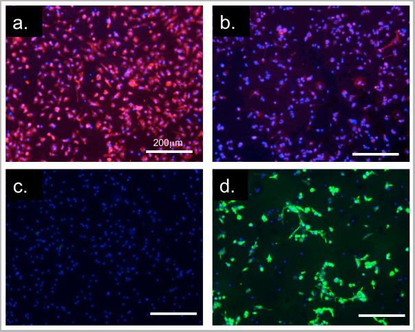
 IF (Immunofluorescence)
(Immunostaining of mixed neuron/glial cultures stained with anti-UCHL1 antibody (AAA14227), green, 1:500) and rabbit anti-GFAP antibody , red, 1:1000. The blue stains nuclear DNA. The anti-UCHL1 stains strongly the cell body and dendrites of neurons, while anti-GFAP specifically labels astrocytes.)
IF (Immunofluorescence)
(Immunostaining of mixed neuron/glial cultures stained with anti-UCHL1 antibody (AAA14227), green, 1:500) and rabbit anti-GFAP antibody , red, 1:1000. The blue stains nuclear DNA. The anti-UCHL1 stains strongly the cell body and dendrites of neurons, while anti-GFAP specifically labels astrocytes.)
Ubiquitin C-terminal Hydrolase 1 (UCHL1) ck, Polyclonal Antibody (Cat# AAA14227)
Full Name
Anti-Ubiquitin C Terminal Hydrolase 1 (UCHL1)
Gene Names
UCHL1; NDGOA; PARK5; PGP95; PGP9.5; Uch-L1; HEL-117; PGP 9.5
Reactivity
Bovine, Human, Mouse, Pig, Rat
Expected Reactivity: Canine, Feline, Goat, Guinea Pig, Hamster, Horse, Non-Human Primate, Rabbit, Sheep, Vole
Expected Reactivity: Canine, Feline, Goat, Guinea Pig, Hamster, Horse, Non-Human Primate, Rabbit, Sheep, Vole
Applications
Western Blot, Immunofluorescence
Purity
Total IgY fraction
Pricing


 Application Data
(Staining of mouse peritoneal macrophages with Rat anti Mouse CD11b)
Application Data
(Staining of mouse peritoneal macrophages with Rat anti Mouse CD11b)
CD11b, Monoclonal Antibody (Cat# AAA12057)
Full Name
RAT ANTI MOUSE CD11b:RPE
Gene Names
Itgam; CR3; CR3A; MAC1; Cd11b; Ly-40; Mac-1; Mac-1a; CD11b/CD18; F730045J24Rik
Applications
Flow Cytometry
Pricing


 IF (Immunofluorescence)
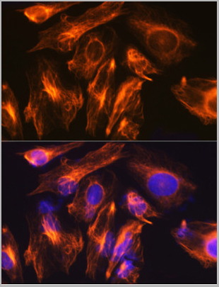
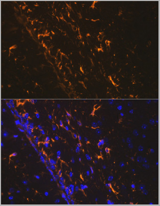
(Fig.6. Immunofluorescence analysis of rat brain tissue. 1, GFAP Monoclonal Antibody (red) was diluted at 1:200 (4 degree C, overnight). 2, Cy3 Labeled secondary antibody was diluted at 1:300 (room temperature, 50min). 3, Picture B: DAPI (blue) 10min. Picture A: Target. Picture B: DAPI. Picture C: merge of A+B.)
IF (Immunofluorescence)
(Fig.6. Immunofluorescence analysis of rat brain tissue. 1, GFAP Monoclonal Antibody (red) was diluted at 1:200 (4 degree C, overnight). 2, Cy3 Labeled secondary antibody was diluted at 1:300 (room temperature, 50min). 3, Picture B: DAPI (blue) 10min. Picture A: Target. Picture B: DAPI. Picture C: merge of A+B.)
GFAP, Monoclonal Antibody (Cat# AAA31476)
Full Name
GFAP Monoclonal Antibody
Gene Names
GFAP; ALXDRD
Reactivity
Mouse, Rat
Applications
Western Blot, Immunofluorescence, Immunohistochemistry
Purity
Affinity-purified from mouse ascites by affinity-chromatography using specific immunogen.
Pricing


 Application Data
(Staining of J774 cells with Rat anti Mouse F4/80 antigen Biotin)
Application Data
(Staining of J774 cells with Rat anti Mouse F4/80 antigen Biotin)
F4/80, Monoclonal Antibody (Cat# AAA12165)
Full Name
RAT ANTI MOUSE F4/80:Low Endotoxin
Gene Names
Emr1; Ly71; F4/80; Gpf480; TM7LN3; DD7A5-7; EGF-TM7
Applications
Immunohistochemistry, Flow Cytometry, Functional Assay, Immunoprecipitation, Immunohistochemistry, Radioimmunoassay, Immunohistochemistry, Western Blot
Pricing


 Application Data
(Staining of mouse peritoneal macrophages with Rat anti Mouse CD11b)
Application Data
(Staining of mouse peritoneal macrophages with Rat anti Mouse CD11b)
CD11b, Monoclonal Antibody (Cat# AAA11889)
Full Name
RAT ANTI MOUSE CD11b:FITC
Gene Names
Itgam; CR3; CR3A; MAC1; Cd11b; Ly-40; Mac-1; Mac-1a; CD11b/CD18; F730045J24Rik
Applications
Flow Cytometry
Pricing


 Application Data
(Published customer image Infiltration of GFP+ BM-cells in infarct and peri-infarct regions. (A-B) Dot plots of viable macrophages/granulocytes (CD11b+CD45high, top right quadrants) and microglia (CD11b+CD45dim, bottom right quadrants) in cortex from BM-chimeric unmanipulated mice and mice exposed to pMCAO. (C) Bar graph showing mean numbers of CD11b+CD45dim microglia and CD11b+CD45high macrophages/granulocytes in BM-chimeric mice 24 hours after pMCAO, subdivided based on expression of GFP (n = 5). Approximately 92% of of the CD45high population were GFP+. (D) Estimation and comparison of mean numbers of CD11b+CD45dim microglia in non-chimeric (n = 10) versus BM-chimeric mice (n = 5) 24 hours after of pMCAO shows significantly fewer CD11b+CD45dim microglial cells in irradiated mice. (E) Overview, showing distribution of infiltrating GFP+ BM-derived cells into infarct (IF) and peri-infarct (P-IF) regions 24 hours after pMCAO. (E-G) By 24 hours, GFP+ single cells (F) and vessel-associated aggregates of GFP+ cells (arrows in G) were observed in infarct and peri-infarct regions. Some of the vessel-associated cells were round, leukocyte-like cells (arrows) while others were elongated cells lining the vasculature (arrow heads in G and in insert). (H) Bar graph showing mean numbers of single GFP+ cells and vessel-associated aggregates of GFP+ cells in ipsi- and contralateral cortex 24 hours after surgery (n = 10). (I-P) Immunohistochemical staining of CD45.1 (I, K), CD45.2 (J, L), IgG2a (M, O) and CD45 (N, P) in ischemic tissue in BM-chimeric (I, J, M, N) and non-chimeric mice (K, L, O, P) 24 hours after pMCAO. N.D, none detected. Scale bars: 200 um (A), 10 um (B, C). 50 um (I-P) *P < 0.05, **P < 0.01, and ***P < 0.001.From: Clausen BH, Lambertsen KL, Babcock AA, Holm TH, Dagnaes-Hansen F, Finsen B. Interleukin-1beta and tumor necrosis factor-alpha are expressed by different subsets of microglia and macrophages after ischemic stroke in mice. J Neuroinflammation. 2008 Oct 23;5:46.)
Application Data
(Published customer image Infiltration of GFP+ BM-cells in infarct and peri-infarct regions. (A-B) Dot plots of viable macrophages/granulocytes (CD11b+CD45high, top right quadrants) and microglia (CD11b+CD45dim, bottom right quadrants) in cortex from BM-chimeric unmanipulated mice and mice exposed to pMCAO. (C) Bar graph showing mean numbers of CD11b+CD45dim microglia and CD11b+CD45high macrophages/granulocytes in BM-chimeric mice 24 hours after pMCAO, subdivided based on expression of GFP (n = 5). Approximately 92% of of the CD45high population were GFP+. (D) Estimation and comparison of mean numbers of CD11b+CD45dim microglia in non-chimeric (n = 10) versus BM-chimeric mice (n = 5) 24 hours after of pMCAO shows significantly fewer CD11b+CD45dim microglial cells in irradiated mice. (E) Overview, showing distribution of infiltrating GFP+ BM-derived cells into infarct (IF) and peri-infarct (P-IF) regions 24 hours after pMCAO. (E-G) By 24 hours, GFP+ single cells (F) and vessel-associated aggregates of GFP+ cells (arrows in G) were observed in infarct and peri-infarct regions. Some of the vessel-associated cells were round, leukocyte-like cells (arrows) while others were elongated cells lining the vasculature (arrow heads in G and in insert). (H) Bar graph showing mean numbers of single GFP+ cells and vessel-associated aggregates of GFP+ cells in ipsi- and contralateral cortex 24 hours after surgery (n = 10). (I-P) Immunohistochemical staining of CD45.1 (I, K), CD45.2 (J, L), IgG2a (M, O) and CD45 (N, P) in ischemic tissue in BM-chimeric (I, J, M, N) and non-chimeric mice (K, L, O, P) 24 hours after pMCAO. N.D, none detected. Scale bars: 200 um (A), 10 um (B, C). 50 um (I-P) *P < 0.05, **P < 0.01, and ***P < 0.001.From: Clausen BH, Lambertsen KL, Babcock AA, Holm TH, Dagnaes-Hansen F, Finsen B. Interleukin-1beta and tumor necrosis factor-alpha are expressed by different subsets of microglia and macrophages after ischemic stroke in mice. J Neuroinflammation. 2008 Oct 23;5:46.)
CD11b, Monoclonal Antibody (Cat# AAA12182)
Full Name
RAT ANTI MOUSE CD11b:FITC
Gene Names
Itgam; CR3; CR3A; MAC1; Cd11b; Ly-40; Mac-1; Mac-1a; CD11b/CD18; F730045J24Rik
Applications
Flow Cytometry
Pricing


 Application Data
(Staining of J774 cells with Rat anti Mouse F4/80 antigen Biotin)
Application Data
(Staining of J774 cells with Rat anti Mouse F4/80 antigen Biotin)
F4/80, Monoclonal Antibody (Cat# AAA12173)
Full Name
RAT ANTI MOUSE F4/80
Gene Names
Emr1; Ly71; F4/80; Gpf480; TM7LN3; DD7A5-7; EGF-TM7
Applications
Immunohistochemistry, Fluorescence Microscopy, Flow Cytometry, Immunofluorescence, Immunoprecipitation, Immunohistochemistry, Radioimmunoassay, Immunohistochemistry, Western Blot
Pricing


 Application Data
(Staining of J774 cells with Rat anti Mouse F4/80 antigen Biotin)
Application Data
(Staining of J774 cells with Rat anti Mouse F4/80 antigen Biotin)
F4/80, Monoclonal Antibody (Cat# AAA12170)
Full Name
RAT ANTI MOUSE F4/80
Gene Names
Emr1; Ly71; F4/80; Gpf480; TM7LN3; DD7A5-7; EGF-TM7
Applications
Immunohistochemistry, Fluorescence Microscopy, Flow Cytometry, Immunofluorescence, Immunoprecipitation, Immunohistochemistry, Radioimmunoassay, Immunohistochemistry, Western Blot
Pricing


 Application Data
(Published customer image Infiltration of GFP+ BM-cells in infarct and peri-infarct regions. (A-B) Dot plots of viable macrophages/granulocytes (CD11b+CD45high, top right quadrants) and microglia (CD11b+CD45dim, bottom right quadrants) in cortex from BM-chimeric unmanipulated mice and mice exposed to pMCAO. (C) Bar graph showing mean numbers of CD11b+CD45dim microglia and CD11b+CD45high macrophages/granulocytes in BM-chimeric mice 24 hours after pMCAO, subdivided based on expression of GFP (n = 5). Approximately 92% of of the CD45high population were GFP+. (D) Estimation and comparison of mean numbers of CD11b+CD45dim microglia in non-chimeric (n = 10) versus BM-chimeric mice (n = 5) 24 hours after of pMCAO shows significantly fewer CD11b+CD45dim microglial cells in irradiated mice. (E) Overview, showing distribution of infiltrating GFP+ BM-derived cells into infarct (IF) and peri-infarct (P-IF) regions 24 hours after pMCAO. (E-G) By 24 hours, GFP+ single cells (F) and vessel-associated aggregates of GFP+ cells (arrows in G) were observed in infarct and peri-infarct regions. Some of the vessel-associated cells were round, leukocyte-like cells (arrows) while others were elongated cells lining the vasculature (arrow heads in G and in insert). (H) Bar graph showing mean numbers of single GFP+ cells and vessel-associated aggregates of GFP+ cells in ipsi- and contralateral cortex 24 hours after surgery (n = 10). (I-P) Immunohistochemical staining of CD45.1 (I, K), CD45.2 (J, L), IgG2a (M, O) and CD45 (N, P) in ischemic tissue in BM-chimeric (I, J, M, N) and non-chimeric mice (K, L, O, P) 24 hours after pMCAO. N.D, none detected. Scale bars: 200 um (A), 10 um (B, C). 50 um (I-P) *P < 0.05, **P < 0.01, and ***P < 0.001.From: Clausen BH, Lambertsen KL, Babcock AA, Holm TH, Dagnaes-Hansen F, Finsen B. Interleukin-1beta and tumor necrosis factor-alpha are expressed by different subsets of microglia and macrophages after ischemic stroke in mice. J Neuroinflammation. 2008 Oct 23;5:46.)
Application Data
(Published customer image Infiltration of GFP+ BM-cells in infarct and peri-infarct regions. (A-B) Dot plots of viable macrophages/granulocytes (CD11b+CD45high, top right quadrants) and microglia (CD11b+CD45dim, bottom right quadrants) in cortex from BM-chimeric unmanipulated mice and mice exposed to pMCAO. (C) Bar graph showing mean numbers of CD11b+CD45dim microglia and CD11b+CD45high macrophages/granulocytes in BM-chimeric mice 24 hours after pMCAO, subdivided based on expression of GFP (n = 5). Approximately 92% of of the CD45high population were GFP+. (D) Estimation and comparison of mean numbers of CD11b+CD45dim microglia in non-chimeric (n = 10) versus BM-chimeric mice (n = 5) 24 hours after of pMCAO shows significantly fewer CD11b+CD45dim microglial cells in irradiated mice. (E) Overview, showing distribution of infiltrating GFP+ BM-derived cells into infarct (IF) and peri-infarct (P-IF) regions 24 hours after pMCAO. (E-G) By 24 hours, GFP+ single cells (F) and vessel-associated aggregates of GFP+ cells (arrows in G) were observed in infarct and peri-infarct regions. Some of the vessel-associated cells were round, leukocyte-like cells (arrows) while others were elongated cells lining the vasculature (arrow heads in G and in insert). (H) Bar graph showing mean numbers of single GFP+ cells and vessel-associated aggregates of GFP+ cells in ipsi- and contralateral cortex 24 hours after surgery (n = 10). (I-P) Immunohistochemical staining of CD45.1 (I, K), CD45.2 (J, L), IgG2a (M, O) and CD45 (N, P) in ischemic tissue in BM-chimeric (I, J, M, N) and non-chimeric mice (K, L, O, P) 24 hours after pMCAO. N.D, none detected. Scale bars: 200 um (A), 10 um (B, C). 50 um (I-P) *P < 0.05, **P < 0.01, and ***P < 0.001.From: Clausen BH, Lambertsen KL, Babcock AA, Holm TH, Dagnaes-Hansen F, Finsen B. Interleukin-1beta and tumor necrosis factor-alpha are expressed by different subsets of microglia and macrophages after ischemic stroke in mice. J Neuroinflammation. 2008 Oct 23;5:46.)
CD11b, Monoclonal Antibody (Cat# AAA12184)
Full Name
RAT ANTI MOUSE CD11b
Gene Names
Itgam; CR3; CR3A; MAC1; Cd11b; Ly-40; Mac-1; Mac-1a; CD11b/CD18; F730045J24Rik
Applications
Immunohistochemistry, Flow Cytometry, Immunofluorescence, Immunoprecipitation
Pricing


 Application Data
(Staining of J774 cells with Rat anti Mouse F4/80 antigen Biotin)
Application Data
(Staining of J774 cells with Rat anti Mouse F4/80 antigen Biotin)
F4/80, Monoclonal Antibody (Cat# AAA12171)
Full Name
RAT ANTI MOUSE F4/80:RPE
Gene Names
Emr1; Ly71; F4/80; Gpf480; TM7LN3; DD7A5-7; EGF-TM7
Applications
Flow Cytometry
Pricing


 Application Data
(Staining of mouse peritoneal macrophages with Rat anti Mouse CD11b)
Application Data
(Staining of mouse peritoneal macrophages with Rat anti Mouse CD11b)
CD11b, Monoclonal Antibody (Cat# AAA12219)
Full Name
RAT ANTI MOUSE CD11b:Low Endotoxin
Gene Names
Itgam; CR3; CR3A; MAC1; Cd11b; Ly-40; Mac-1; Mac-1a; CD11b/CD18; F730045J24Rik
Applications
Immunohistochemistry, Flow Cytometry, Functional Assay, Immunofluorescence, Immunoprecipitation, Immunohistochemistry
Pricing


 Application Data
(Staining of J774 cells with Rat anti Mouse F4/80 antigen Biotin)
Application Data
(Staining of J774 cells with Rat anti Mouse F4/80 antigen Biotin)
F4/80, Monoclonal Antibody (Cat# AAA12168)
Full Name
RAT ANTI MOUSE F4/80:FITC
Gene Names
Emr1; Ly71; F4/80; Gpf480; TM7LN3; DD7A5-7; EGF-TM7
Applications
Flow Cytometry, Immunofluorescence
Pricing


 Application Data
(Staining of J774 cells with Rat anti Mouse F4/80 antigen Biotin)
Application Data
(Staining of J774 cells with Rat anti Mouse F4/80 antigen Biotin)
F4/80, Monoclonal Antibody (Cat# AAA12166)
Full Name
RAT ANTI MOUSE F4/80:FITC
Gene Names
Emr1; Ly71; F4/80; Gpf480; TM7LN3; DD7A5-7; EGF-TM7
Applications
Flow Cytometry, Immunofluorescence
Pricing


 Application Data
BrdU, Monoclonal Antibody (Cat# AAA12259)
Full Name
Mouse Anti BrdU
Applications
Flow Cytometry, Immunocytochemistry, Immunofluorescence, Immunohistochemistry
Purity
Purified IgG prepared by affinity chromatography on Protein A from tissue culture supernatant
Pricing


 Application Data
(Staining of J774 cells with Rat anti Mouse F4/80 antigen Biotin)
Application Data
(Staining of J774 cells with Rat anti Mouse F4/80 antigen Biotin)
F4/80, Monoclonal Antibody (Cat# AAA12162)
Full Name
RAT ANTI MOUSE F4/80:Biotin
Gene Names
Emr1; Ly71; F4/80; Gpf480; TM7LN3; DD7A5-7; EGF-TM7
Applications
Flow Cytometry
Pricing


 Application Data
(Staining of J774 cells with Rat anti Mouse F4/80 antigen Biotin)
Application Data
(Staining of J774 cells with Rat anti Mouse F4/80 antigen Biotin)
F4/80, Monoclonal Antibody (Cat# AAA12217)
Full Name
RAT ANTI MOUSE F4/80:APC
Gene Names
Emr1; Ly71; F4/80; Gpf480; TM7LN3; DD7A5-7; EGF-TM7
Applications
Flow Cytometry
Pricing


 Application Data
(Staining of J774 cells with Rat anti Mouse F4/80 antigen Biotin)
Application Data
(Staining of J774 cells with Rat anti Mouse F4/80 antigen Biotin)
F4/80, Monoclonal Antibody (Cat# AAA12161)
Full Name
RAT ANTI MOUSE F4/80:APC
Gene Names
Emr1; Ly71; F4/80; Gpf480; TM7LN3; DD7A5-7; EGF-TM7
Applications
Flow Cytometry
Pricing


 Application Data
(Staining of J774 cells with Rat anti Mouse F4/80 antigen Biotin)
Application Data
(Staining of J774 cells with Rat anti Mouse F4/80 antigen Biotin)
F4/80, Monoclonal Antibody (Cat# AAA12172)
Full Name
RAT ANTI MOUSE F4/80:RPE
Gene Names
Emr1; Ly71; F4/80; Gpf480; TM7LN3; DD7A5-7; EGF-TM7
Applications
Flow Cytometry, Immunofluorescence
Pricing


 Application Data
(Overlay histogram showing SY5Y cells stained with AAA27008 (red line). The cells were fixed with 70% Ethylalcohol (18h) and then permeabilized with 0.3% Triton X-100 for 2 min. The cells were then incubated in 1x PBS /10% normal goat serum to block non-specific protein-protein interactions followed by the antibody (10ug/1x10^6cells) for 1 h at 4 degree C. The secondary antibody used was FITC goat anti-mouse IgG (H+L) at 1/200 dilution for 1 h at 4 degree C. Isotype control antibody (green line) was mouse IgG2b (10ug/1x10^6cells) used under the same conditions. Acquisition of >10, 000 events was performed.)
Application Data
(Overlay histogram showing SY5Y cells stained with AAA27008 (red line). The cells were fixed with 70% Ethylalcohol (18h) and then permeabilized with 0.3% Triton X-100 for 2 min. The cells were then incubated in 1x PBS /10% normal goat serum to block non-specific protein-protein interactions followed by the antibody (10ug/1x10^6cells) for 1 h at 4 degree C. The secondary antibody used was FITC goat anti-mouse IgG (H+L) at 1/200 dilution for 1 h at 4 degree C. Isotype control antibody (green line) was mouse IgG2b (10ug/1x10^6cells) used under the same conditions. Acquisition of >10, 000 events was performed.)
GFAP, Monoclonal Antibody (Cat# AAA27008)
Full Name
GFAP Monoclonal Antibody
Gene Names
GFAP; ALXDRD
Reactivity
Human, Mouse, Rat
Applications
Western Blot, Immunohistochemistry, Immunofluorescence, Flow Cytometry
Purity
>95%, Protein G purified
Pricing


 Application Data
(Published customer image Infiltration of GFP+ BM-cells in infarct and peri-infarct regions. (A-B) Dot plots of viable macrophages/granulocytes (CD11b+CD45high, top right quadrants) and microglia (CD11b+CD45dim, bottom right quadrants) in cortex from BM-chimeric unmanipulated mice and mice exposed to pMCAO. (C) Bar graph showing mean numbers of CD11b+CD45dim microglia and CD11b+CD45high macrophages/granulocytes in BM-chimeric mice 24 hours after pMCAO, subdivided based on expression of GFP (n = 5). Approximately 92% of of the CD45high population were GFP+. (D) Estimation and comparison of mean numbers of CD11b+CD45dim microglia in non-chimeric (n = 10) versus BM-chimeric mice (n = 5) 24 hours after of pMCAO shows significantly fewer CD11b+CD45dim microglial cells in irradiated mice. (E) Overview, showing distribution of infiltrating GFP+ BM-derived cells into infarct (IF) and peri-infarct (P-IF) regions 24 hours after pMCAO. (E-G) By 24 hours, GFP+ single cells (F) and vessel-associated aggregates of GFP+ cells (arrows in G) were observed in infarct and peri-infarct regions. Some of the vessel-associated cells were round, leukocyte-like cells (arrows) while others were elongated cells lining the vasculature (arrow heads in G and in insert). (H) Bar graph showing mean numbers of single GFP+ cells and vessel-associated aggregates of GFP+ cells in ipsi- and contralateral cortex 24 hours after surgery (n = 10). (I-P) Immunohistochemical staining of CD45.1 (I, K), CD45.2 (J, L), IgG2a (M, O) and CD45 (N, P) in ischemic tissue in BM-chimeric (I, J, M, N) and non-chimeric mice (K, L, O, P) 24 hours after pMCAO. N.D, none detected. Scale bars: 200 um (A), 10 um (B, C). 50 um (I-P) *P < 0.05, **P < 0.01, and ***P < 0.001.From: Clausen BH, Lambertsen KL, Babcock AA, Holm TH, Dagnaes-Hansen F, Finsen B. Interleukin-1beta and tumor necrosis factor-alpha are expressed by different subsets of microglia and macrophages after ischemic stroke in mice. J Neuroinflammation. 2008 Oct 23;5:46.)
Application Data
(Published customer image Infiltration of GFP+ BM-cells in infarct and peri-infarct regions. (A-B) Dot plots of viable macrophages/granulocytes (CD11b+CD45high, top right quadrants) and microglia (CD11b+CD45dim, bottom right quadrants) in cortex from BM-chimeric unmanipulated mice and mice exposed to pMCAO. (C) Bar graph showing mean numbers of CD11b+CD45dim microglia and CD11b+CD45high macrophages/granulocytes in BM-chimeric mice 24 hours after pMCAO, subdivided based on expression of GFP (n = 5). Approximately 92% of of the CD45high population were GFP+. (D) Estimation and comparison of mean numbers of CD11b+CD45dim microglia in non-chimeric (n = 10) versus BM-chimeric mice (n = 5) 24 hours after of pMCAO shows significantly fewer CD11b+CD45dim microglial cells in irradiated mice. (E) Overview, showing distribution of infiltrating GFP+ BM-derived cells into infarct (IF) and peri-infarct (P-IF) regions 24 hours after pMCAO. (E-G) By 24 hours, GFP+ single cells (F) and vessel-associated aggregates of GFP+ cells (arrows in G) were observed in infarct and peri-infarct regions. Some of the vessel-associated cells were round, leukocyte-like cells (arrows) while others were elongated cells lining the vasculature (arrow heads in G and in insert). (H) Bar graph showing mean numbers of single GFP+ cells and vessel-associated aggregates of GFP+ cells in ipsi- and contralateral cortex 24 hours after surgery (n = 10). (I-P) Immunohistochemical staining of CD45.1 (I, K), CD45.2 (J, L), IgG2a (M, O) and CD45 (N, P) in ischemic tissue in BM-chimeric (I, J, M, N) and non-chimeric mice (K, L, O, P) 24 hours after pMCAO. N.D, none detected. Scale bars: 200 um (A), 10 um (B, C). 50 um (I-P) *P < 0.05, **P < 0.01, and ***P < 0.001.From: Clausen BH, Lambertsen KL, Babcock AA, Holm TH, Dagnaes-Hansen F, Finsen B. Interleukin-1beta and tumor necrosis factor-alpha are expressed by different subsets of microglia and macrophages after ischemic stroke in mice. J Neuroinflammation. 2008 Oct 23;5:46.)
CD11b, Monoclonal Antibody (Cat# AAA12183)
Full Name
RAT ANTI MOUSE CD11b:FITC
Gene Names
Itgam; CR3; CR3A; MAC1; Cd11b; Ly-40; Mac-1; Mac-1a; CD11b/CD18; F730045J24Rik
Applications
Flow Cytometry
Pricing


 Application Data
(Published customer image Infiltration of GFP+ BM-cells in infarct and peri-infarct regions. (A-B) Dot plots of viable macrophages/granulocytes (CD11b+CD45high, top right quadrants) and microglia (CD11b+CD45dim, bottom right quadrants) in cortex from BM-chimeric unmanipulated mice and mice exposed to pMCAO. (C) Bar graph showing mean numbers of CD11b+CD45dim microglia and CD11b+CD45high macrophages/granulocytes in BM-chimeric mice 24 hours after pMCAO, subdivided based on expression of GFP (n = 5). Approximately 92% of of the CD45high population were GFP+. (D) Estimation and comparison of mean numbers of CD11b+CD45dim microglia in non-chimeric (n = 10) versus BM-chimeric mice (n = 5) 24 hours after of pMCAO shows significantly fewer CD11b+CD45dim microglial cells in irradiated mice. (E) Overview, showing distribution of infiltrating GFP+ BM-derived cells into infarct (IF) and peri-infarct (P-IF) regions 24 hours after pMCAO. (E-G) By 24 hours, GFP+ single cells (F) and vessel-associated aggregates of GFP+ cells (arrows in G) were observed in infarct and peri-infarct regions. Some of the vessel-associated cells were round, leukocyte-like cells (arrows) while others were elongated cells lining the vasculature (arrow heads in G and in insert). (H) Bar graph showing mean numbers of single GFP+ cells and vessel-associated aggregates of GFP+ cells in ipsi- and contralateral cortex 24 hours after surgery (n = 10). (I-P) Immunohistochemical staining of CD45.1 (I, K), CD45.2 (J, L), IgG2a (M, O) and CD45 (N, P) in ischemic tissue in BM-chimeric (I, J, M, N) and non-chimeric mice (K, L, O, P) 24 hours after pMCAO. N.D, none detected. Scale bars: 200 um (A), 10 um (B, C). 50 um (I-P) *P < 0.05, **P < 0.01, and ***P < 0.001.From: Clausen BH, Lambertsen KL, Babcock AA, Holm TH, Dagnaes-Hansen F, Finsen B. Interleukin-1beta and tumor necrosis factor-alpha are expressed by different subsets of microglia and macrophages after ischemic stroke in mice. J Neuroinflammation. 2008 Oct 23;5:46.)
Application Data
(Published customer image Infiltration of GFP+ BM-cells in infarct and peri-infarct regions. (A-B) Dot plots of viable macrophages/granulocytes (CD11b+CD45high, top right quadrants) and microglia (CD11b+CD45dim, bottom right quadrants) in cortex from BM-chimeric unmanipulated mice and mice exposed to pMCAO. (C) Bar graph showing mean numbers of CD11b+CD45dim microglia and CD11b+CD45high macrophages/granulocytes in BM-chimeric mice 24 hours after pMCAO, subdivided based on expression of GFP (n = 5). Approximately 92% of of the CD45high population were GFP+. (D) Estimation and comparison of mean numbers of CD11b+CD45dim microglia in non-chimeric (n = 10) versus BM-chimeric mice (n = 5) 24 hours after of pMCAO shows significantly fewer CD11b+CD45dim microglial cells in irradiated mice. (E) Overview, showing distribution of infiltrating GFP+ BM-derived cells into infarct (IF) and peri-infarct (P-IF) regions 24 hours after pMCAO. (E-G) By 24 hours, GFP+ single cells (F) and vessel-associated aggregates of GFP+ cells (arrows in G) were observed in infarct and peri-infarct regions. Some of the vessel-associated cells were round, leukocyte-like cells (arrows) while others were elongated cells lining the vasculature (arrow heads in G and in insert). (H) Bar graph showing mean numbers of single GFP+ cells and vessel-associated aggregates of GFP+ cells in ipsi- and contralateral cortex 24 hours after surgery (n = 10). (I-P) Immunohistochemical staining of CD45.1 (I, K), CD45.2 (J, L), IgG2a (M, O) and CD45 (N, P) in ischemic tissue in BM-chimeric (I, J, M, N) and non-chimeric mice (K, L, O, P) 24 hours after pMCAO. N.D, none detected. Scale bars: 200 um (A), 10 um (B, C). 50 um (I-P) *P < 0.05, **P < 0.01, and ***P < 0.001.From: Clausen BH, Lambertsen KL, Babcock AA, Holm TH, Dagnaes-Hansen F, Finsen B. Interleukin-1beta and tumor necrosis factor-alpha are expressed by different subsets of microglia and macrophages after ischemic stroke in mice. J Neuroinflammation. 2008 Oct 23;5:46.)
CD11b, Monoclonal Antibody (Cat# AAA12185)
Full Name
RAT ANTI MOUSE CD11b
Gene Names
Itgam; CR3; CR3A; MAC1; Cd11b; Ly-40; Mac-1; Mac-1a; CD11b/CD18; F730045J24Rik
Applications
Immunohistochemistry, Flow Cytometry, Immunofluorescence, Immunoprecipitation
Pricing


 Application Data
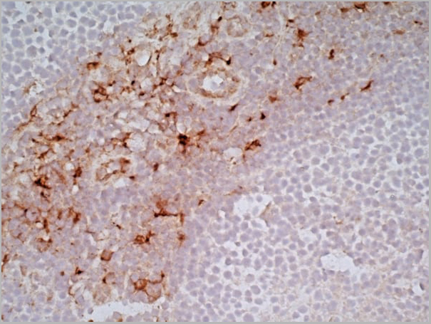
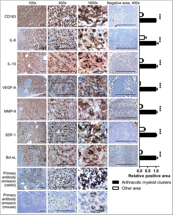
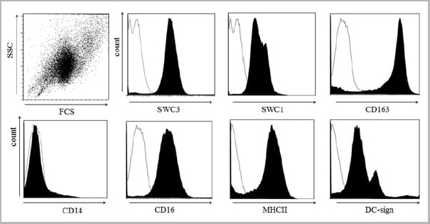
(Immunoperoxidase staining of a human tonsil cryosection with Mouse anti Human CD163 antibody, clone EDHu-1 followed by the Histar detection system . Low power)
Application Data
(Immunoperoxidase staining of a human tonsil cryosection with Mouse anti Human CD163 antibody, clone EDHu-1 followed by the Histar detection system . Low power)
CD163, Monoclonal Antibody (Cat# AAA12100)
Full Name
MOUSE ANTI HUMAN CD163
Gene Names
CD163; M130; MM130
Applications
Immunohistochemistry, Flow Cytometry, Immunofluorescence, Immunoassay, Immunohistochemistry, Western Blot
Pricing


 Application Data
(Staining of mouse peritoneal macrophages with Rat anti Mouse CD11b)
Application Data
(Staining of mouse peritoneal macrophages with Rat anti Mouse CD11b)
CD11b, Monoclonal Antibody (Cat# AAA12187)
Full Name
RAT ANTI MOUSE CD11b:Biotin
Gene Names
Itgam; CR3; CR3A; MAC1; Cd11b; Ly-40; Mac-1; Mac-1a; CD11b/CD18; F730045J24Rik
Applications
Flow Cytometry
Pricing


 Application Data
(Staining of J774 cells with Rat anti Mouse F4/80 antigen Biotin)
Application Data
(Staining of J774 cells with Rat anti Mouse F4/80 antigen Biotin)
F4/80, Monoclonal Antibody (Cat# AAA12169)
Full Name
RAT ANTI MOUSE F4/80
Gene Names
Emr1; Ly71; F4/80; Gpf480; TM7LN3; DD7A5-7; EGF-TM7
Applications
Immunohistochemistry, Fluorescence Microscopy, Flow Cytometry, Immunofluorescence, Immunoprecipitation, Immunohistochemistry, Radioimmunoassay, Immunohistochemistry, Western Blot
Pricing


 Application Data
(Staining of J774 cells with Rat anti Mouse F4/80 antigen Biotin)
Application Data
(Staining of J774 cells with Rat anti Mouse F4/80 antigen Biotin)
F4/80, Monoclonal Antibody (Cat# AAA12163)
Full Name
RAT ANTI MOUSE F4/80:Biotin
Gene Names
Emr1; Ly71; F4/80; Gpf480; TM7LN3; DD7A5-7; EGF-TM7
Applications
Flow Cytometry
Pricing


 Application Data
(Staining of J774 cells with Rat anti Mouse F4/80 antigen Biotin)
Application Data
(Staining of J774 cells with Rat anti Mouse F4/80 antigen Biotin)
F4/80, Monoclonal Antibody (Cat# AAA12174)
Full Name
RAT ANTI MOUSE F4/80
Gene Names
Emr1; Ly71; F4/80; Gpf480; TM7LN3; DD7A5-7; EGF-TM7
Applications
Immunohistochemistry, Fluorescence Microscopy, Flow Cytometry, Immunofluorescence, Immunoprecipitation, Immunohistochemistry, Radioimmunoassay, Immunohistochemistry, Western Blot
Pricing


 Application Data
(Staining of J774 cells with Rat anti Mouse F4/80 antigen Biotin)
Application Data
(Staining of J774 cells with Rat anti Mouse F4/80 antigen Biotin)
F4/80, Monoclonal Antibody (Cat# AAA12164)
Full Name
RAT ANTI MOUSE F4/80:Biotin
Gene Names
Emr1; Ly71; F4/80; Gpf480; TM7LN3; DD7A5-7; EGF-TM7
Applications
Flow Cytometry
Pricing


 Application Data
(Staining of J774 cells with Rat anti Mouse F4/80 antigen Biotin)
Application Data
(Staining of J774 cells with Rat anti Mouse F4/80 antigen Biotin)
F4/80, Monoclonal Antibody (Cat# AAA12167)
Full Name
RAT ANTI MOUSE F4/80:FITC
Gene Names
Emr1; Ly71; F4/80; Gpf480; TM7LN3; DD7A5-7; EGF-TM7
Applications
Flow Cytometry, Immunofluorescence
Pricing


 Application Data
(Published customer image Infiltration of GFP+ BM-cells in infarct and peri-infarct regions. (A-B) Dot plots of viable macrophages/granulocytes (CD11b+CD45high, top right quadrants) and microglia (CD11b+CD45dim, bottom right quadrants) in cortex from BM-chimeric unmanipulated mice and mice exposed to pMCAO. (C) Bar graph showing mean numbers of CD11b+CD45dim microglia and CD11b+CD45high macrophages/granulocytes in BM-chimeric mice 24 hours after pMCAO, subdivided based on expression of GFP (n = 5). Approximately 92% of of the CD45high population were GFP+. (D) Estimation and comparison of mean numbers of CD11b+CD45dim microglia in non-chimeric (n = 10) versus BM-chimeric mice (n = 5) 24 hours after of pMCAO shows significantly fewer CD11b+CD45dim microglial cells in irradiated mice. (E) Overview, showing distribution of infiltrating GFP+ BM-derived cells into infarct (IF) and peri-infarct (P-IF) regions 24 hours after pMCAO. (E-G) By 24 hours, GFP+ single cells (F) and vessel-associated aggregates of GFP+ cells (arrows in G) were observed in infarct and peri-infarct regions. Some of the vessel-associated cells were round, leukocyte-like cells (arrows) while others were elongated cells lining the vasculature (arrow heads in G and in insert). (H) Bar graph showing mean numbers of single GFP+ cells and vessel-associated aggregates of GFP+ cells in ipsi- and contralateral cortex 24 hours after surgery (n = 10). (I-P) Immunohistochemical staining of CD45.1 (I, K), CD45.2 (J, L), IgG2a (M, O) and CD45 (N, P) in ischemic tissue in BM-chimeric (I, J, M, N) and non-chimeric mice (K, L, O, P) 24 hours after pMCAO. N.D, none detected. Scale bars: 200 um (A), 10 um (B, C). 50 um (I-P) *P < 0.05, **P < 0.01, and ***P < 0.001.From: Clausen BH, Lambertsen KL, Babcock AA, Holm TH, Dagnaes-Hansen F, Finsen B. Interleukin-1beta and tumor necrosis factor-alpha are expressed by different subsets of microglia and macrophages after ischemic stroke in mice. J Neuroinflammation. 2008 Oct 23;5:46.)
Application Data
(Published customer image Infiltration of GFP+ BM-cells in infarct and peri-infarct regions. (A-B) Dot plots of viable macrophages/granulocytes (CD11b+CD45high, top right quadrants) and microglia (CD11b+CD45dim, bottom right quadrants) in cortex from BM-chimeric unmanipulated mice and mice exposed to pMCAO. (C) Bar graph showing mean numbers of CD11b+CD45dim microglia and CD11b+CD45high macrophages/granulocytes in BM-chimeric mice 24 hours after pMCAO, subdivided based on expression of GFP (n = 5). Approximately 92% of of the CD45high population were GFP+. (D) Estimation and comparison of mean numbers of CD11b+CD45dim microglia in non-chimeric (n = 10) versus BM-chimeric mice (n = 5) 24 hours after of pMCAO shows significantly fewer CD11b+CD45dim microglial cells in irradiated mice. (E) Overview, showing distribution of infiltrating GFP+ BM-derived cells into infarct (IF) and peri-infarct (P-IF) regions 24 hours after pMCAO. (E-G) By 24 hours, GFP+ single cells (F) and vessel-associated aggregates of GFP+ cells (arrows in G) were observed in infarct and peri-infarct regions. Some of the vessel-associated cells were round, leukocyte-like cells (arrows) while others were elongated cells lining the vasculature (arrow heads in G and in insert). (H) Bar graph showing mean numbers of single GFP+ cells and vessel-associated aggregates of GFP+ cells in ipsi- and contralateral cortex 24 hours after surgery (n = 10). (I-P) Immunohistochemical staining of CD45.1 (I, K), CD45.2 (J, L), IgG2a (M, O) and CD45 (N, P) in ischemic tissue in BM-chimeric (I, J, M, N) and non-chimeric mice (K, L, O, P) 24 hours after pMCAO. N.D, none detected. Scale bars: 200 um (A), 10 um (B, C). 50 um (I-P) *P < 0.05, **P < 0.01, and ***P < 0.001.From: Clausen BH, Lambertsen KL, Babcock AA, Holm TH, Dagnaes-Hansen F, Finsen B. Interleukin-1beta and tumor necrosis factor-alpha are expressed by different subsets of microglia and macrophages after ischemic stroke in mice. J Neuroinflammation. 2008 Oct 23;5:46.)
CD11b, Monoclonal Antibody (Cat# AAA12186)
Full Name
RAT ANTI MOUSE CD11b:RPE
Gene Names
Itgam; CR3; CR3A; MAC1; Cd11b; Ly-40; Mac-1; Mac-1a; CD11b/CD18; F730045J24Rik
Applications
Flow Cytometry
Pricing


 Application Data
(Published customer image Infiltration of GFP+ BM-cells in infarct and peri-infarct regions. (A-B) Dot plots of viable macrophages/granulocytes (CD11b+CD45high, top right quadrants) and microglia (CD11b+CD45dim, bottom right quadrants) in cortex from BM-chimeric unmanipulated mice and mice exposed to pMCAO. (C) Bar graph showing mean numbers of CD11b+CD45dim microglia and CD11b+CD45high macrophages/granulocytes in BM-chimeric mice 24 hours after pMCAO, subdivided based on expression of GFP (n = 5). Approximately 92% of of the CD45high population were GFP+. (D) Estimation and comparison of mean numbers of CD11b+CD45dim microglia in non-chimeric (n = 10) versus BM-chimeric mice (n = 5) 24 hours after of pMCAO shows significantly fewer CD11b+CD45dim microglial cells in irradiated mice. (E) Overview, showing distribution of infiltrating GFP+ BM-derived cells into infarct (IF) and peri-infarct (P-IF) regions 24 hours after pMCAO. (E-G) By 24 hours, GFP+ single cells (F) and vessel-associated aggregates of GFP+ cells (arrows in G) were observed in infarct and peri-infarct regions. Some of the vessel-associated cells were round, leukocyte-like cells (arrows) while others were elongated cells lining the vasculature (arrow heads in G and in insert). (H) Bar graph showing mean numbers of single GFP+ cells and vessel-associated aggregates of GFP+ cells in ipsi- and contralateral cortex 24 hours after surgery (n = 10). (I-P) Immunohistochemical staining of CD45.1 (I, K), CD45.2 (J, L), IgG2a (M, O) and CD45 (N, P) in ischemic tissue in BM-chimeric (I, J, M, N) and non-chimeric mice (K, L, O, P) 24 hours after pMCAO. N.D, none detected. Scale bars: 200 um (A), 10 um (B, C). 50 um (I-P) *P < 0.05, **P < 0.01, and ***P < 0.001.From: Clausen BH, Lambertsen KL, Babcock AA, Holm TH, Dagnaes-Hansen F, Finsen B. Interleukin-1beta and tumor necrosis factor-alpha are expressed by different subsets of microglia and macrophages after ischemic stroke in mice. J Neuroinflammation. 2008 Oct 23;5:46.)
Application Data
(Published customer image Infiltration of GFP+ BM-cells in infarct and peri-infarct regions. (A-B) Dot plots of viable macrophages/granulocytes (CD11b+CD45high, top right quadrants) and microglia (CD11b+CD45dim, bottom right quadrants) in cortex from BM-chimeric unmanipulated mice and mice exposed to pMCAO. (C) Bar graph showing mean numbers of CD11b+CD45dim microglia and CD11b+CD45high macrophages/granulocytes in BM-chimeric mice 24 hours after pMCAO, subdivided based on expression of GFP (n = 5). Approximately 92% of of the CD45high population were GFP+. (D) Estimation and comparison of mean numbers of CD11b+CD45dim microglia in non-chimeric (n = 10) versus BM-chimeric mice (n = 5) 24 hours after of pMCAO shows significantly fewer CD11b+CD45dim microglial cells in irradiated mice. (E) Overview, showing distribution of infiltrating GFP+ BM-derived cells into infarct (IF) and peri-infarct (P-IF) regions 24 hours after pMCAO. (E-G) By 24 hours, GFP+ single cells (F) and vessel-associated aggregates of GFP+ cells (arrows in G) were observed in infarct and peri-infarct regions. Some of the vessel-associated cells were round, leukocyte-like cells (arrows) while others were elongated cells lining the vasculature (arrow heads in G and in insert). (H) Bar graph showing mean numbers of single GFP+ cells and vessel-associated aggregates of GFP+ cells in ipsi- and contralateral cortex 24 hours after surgery (n = 10). (I-P) Immunohistochemical staining of CD45.1 (I, K), CD45.2 (J, L), IgG2a (M, O) and CD45 (N, P) in ischemic tissue in BM-chimeric (I, J, M, N) and non-chimeric mice (K, L, O, P) 24 hours after pMCAO. N.D, none detected. Scale bars: 200 um (A), 10 um (B, C). 50 um (I-P) *P < 0.05, **P < 0.01, and ***P < 0.001.From: Clausen BH, Lambertsen KL, Babcock AA, Holm TH, Dagnaes-Hansen F, Finsen B. Interleukin-1beta and tumor necrosis factor-alpha are expressed by different subsets of microglia and macrophages after ischemic stroke in mice. J Neuroinflammation. 2008 Oct 23;5:46.)
CD11b, Monoclonal Antibody (Cat# AAA12231)
Full Name
RAT ANTI MOUSE CD11b:Low Endotoxin
Gene Names
Itgam; CR3; CR3A; MAC1; Cd11b; Ly-40; Mac-1; Mac-1a; CD11b/CD18; F730045J24Rik
Applications
Immunohistochemistry, Flow Cytometry, Functional Assay, Immunofluorescence, Immunoprecipitation
Pricing


 Application Data
(Published customer image Infiltration of GFP+ BM-cells in infarct and peri-infarct regions. (A-B) Dot plots of viable macrophages/granulocytes (CD11b+CD45high, top right quadrants) and microglia (CD11b+CD45dim, bottom right quadrants) in cortex from BM-chimeric unmanipulated mice and mice exposed to pMCAO. (C) Bar graph showing mean numbers of CD11b+CD45dim microglia and CD11b+CD45high macrophages/granulocytes in BM-chimeric mice 24 hours after pMCAO, subdivided based on expression of GFP (n = 5). Approximately 92% of of the CD45high population were GFP+. (D) Estimation and comparison of mean numbers of CD11b+CD45dim microglia in non-chimeric (n = 10) versus BM-chimeric mice (n = 5) 24 hours after of pMCAO shows significantly fewer CD11b+CD45dim microglial cells in irradiated mice. (E) Overview, showing distribution of infiltrating GFP+ BM-derived cells into infarct (IF) and peri-infarct (P-IF) regions 24 hours after pMCAO. (E-G) By 24 hours, GFP+ single cells (F) and vessel-associated aggregates of GFP+ cells (arrows in G) were observed in infarct and peri-infarct regions. Some of the vessel-associated cells were round, leukocyte-like cells (arrows) while others were elongated cells lining the vasculature (arrow heads in G and in insert). (H) Bar graph showing mean numbers of single GFP+ cells and vessel-associated aggregates of GFP+ cells in ipsi- and contralateral cortex 24 hours after surgery (n = 10). (I-P) Immunohistochemical staining of CD45.1 (I, K), CD45.2 (J, L), IgG2a (M, O) and CD45 (N, P) in ischemic tissue in BM-chimeric (I, J, M, N) and non-chimeric mice (K, L, O, P) 24 hours after pMCAO. N.D, none detected. Scale bars: 200 um (A), 10 um (B, C). 50 um (I-P) *P < 0.05, **P < 0.01, and ***P < 0.001.From: Clausen BH, Lambertsen KL, Babcock AA, Holm TH, Dagnaes-Hansen F, Finsen B. Interleukin-1beta and tumor necrosis factor-alpha are expressed by different subsets of microglia and macrophages after ischemic stroke in mice. J Neuroinflammation. 2008 Oct 23;5:46.)
Application Data
(Published customer image Infiltration of GFP+ BM-cells in infarct and peri-infarct regions. (A-B) Dot plots of viable macrophages/granulocytes (CD11b+CD45high, top right quadrants) and microglia (CD11b+CD45dim, bottom right quadrants) in cortex from BM-chimeric unmanipulated mice and mice exposed to pMCAO. (C) Bar graph showing mean numbers of CD11b+CD45dim microglia and CD11b+CD45high macrophages/granulocytes in BM-chimeric mice 24 hours after pMCAO, subdivided based on expression of GFP (n = 5). Approximately 92% of of the CD45high population were GFP+. (D) Estimation and comparison of mean numbers of CD11b+CD45dim microglia in non-chimeric (n = 10) versus BM-chimeric mice (n = 5) 24 hours after of pMCAO shows significantly fewer CD11b+CD45dim microglial cells in irradiated mice. (E) Overview, showing distribution of infiltrating GFP+ BM-derived cells into infarct (IF) and peri-infarct (P-IF) regions 24 hours after pMCAO. (E-G) By 24 hours, GFP+ single cells (F) and vessel-associated aggregates of GFP+ cells (arrows in G) were observed in infarct and peri-infarct regions. Some of the vessel-associated cells were round, leukocyte-like cells (arrows) while others were elongated cells lining the vasculature (arrow heads in G and in insert). (H) Bar graph showing mean numbers of single GFP+ cells and vessel-associated aggregates of GFP+ cells in ipsi- and contralateral cortex 24 hours after surgery (n = 10). (I-P) Immunohistochemical staining of CD45.1 (I, K), CD45.2 (J, L), IgG2a (M, O) and CD45 (N, P) in ischemic tissue in BM-chimeric (I, J, M, N) and non-chimeric mice (K, L, O, P) 24 hours after pMCAO. N.D, none detected. Scale bars: 200 um (A), 10 um (B, C). 50 um (I-P) *P < 0.05, **P < 0.01, and ***P < 0.001.From: Clausen BH, Lambertsen KL, Babcock AA, Holm TH, Dagnaes-Hansen F, Finsen B. Interleukin-1beta and tumor necrosis factor-alpha are expressed by different subsets of microglia and macrophages after ischemic stroke in mice. J Neuroinflammation. 2008 Oct 23;5:46.)
CD11b, Monoclonal Antibody (Cat# AAA12181)
Full Name
RAT ANTI MOUSE CD11b
Gene Names
Itgam; CR3; CR3A; MAC1; Cd11b; Ly-40; Mac-1; Mac-1a; CD11b/CD18; F730045J24Rik
Reactivity
Human
Applications
Immunohistochemistry, Flow Cytometry, Immunofluorescence, Immunoprecipitation
Pricing


 Application Data
(Staining of mouse peritoneal macrophages with Rat anti Mouse CD11b)
Application Data
(Staining of mouse peritoneal macrophages with Rat anti Mouse CD11b)
CD11b, Monoclonal Antibody (Cat# AAA12008)
Full Name
RAT ANTI MOUSE CD11b
Gene Names
Itgam; CR3; CR3A; MAC1; Cd11b; Ly-40; Mac-1; Mac-1a; CD11b/CD18; F730045J24Rik
Applications
Immunohistochemistry, Flow Cytometry, Immunofluorescence, Immunoprecipitation, Immunohistochemistry
Pricing


 Application Data
(Staining of mouse peritoneal macrophages with Rat anti Mouse CD11b)
Application Data
(Staining of mouse peritoneal macrophages with Rat anti Mouse CD11b)
CD11b, Monoclonal Antibody (Cat# AAA12007)
Full Name
RAT ANTI MOUSE CD11b
Gene Names
Itgam; CR3; CR3A; MAC1; Cd11b; Ly-40; Mac-1; Mac-1a; CD11b/CD18; F730045J24Rik
Applications
Immunohistochemistry, Flow Cytometry, Immunofluorescence, Immunoprecipitation, Immunohistochemistry
Pricing
 Standard Curve (Sample)
Glial Fibrillary Acidic Protein (GFAP), Antibody Pair Kit (Cat# AAA21084)
Full Name
Mouse Glial Fibrillary Acidic Protein (GFAP) Antibody Pair Kit (with Standard)
Gene Names
GFAP; ALXDRD
Reactivity
Mouse
Applications
EIA, CLIA
Pricing


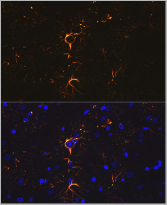
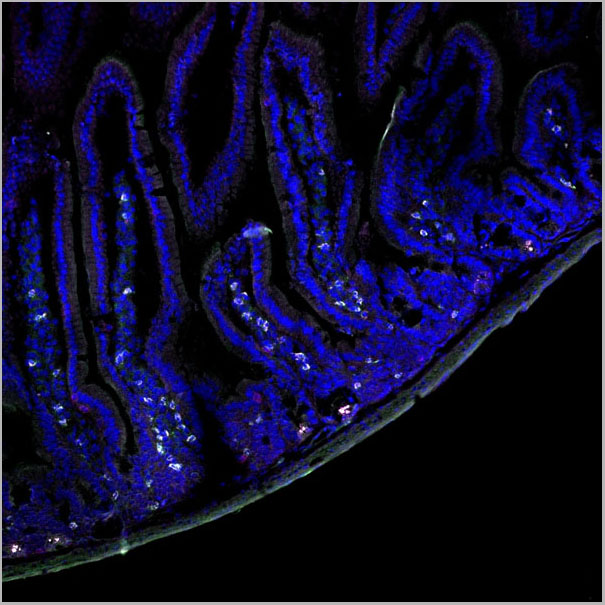
 IF (Immunofluorescence)
(Immunofluorescence analysis of rat brain using GFAP Rabbit mAb at dilution of 1:100 (40x lens). Blue: DAPI for nuclear staining.)
IF (Immunofluorescence)
(Immunofluorescence analysis of rat brain using GFAP Rabbit mAb at dilution of 1:100 (40x lens). Blue: DAPI for nuclear staining.)
GFAP, Monoclonal Antibody (Cat# AAA28378)
Full Name
GFAP Rabbit mAb
Reactivity
Human, Mouse, Rat
Applications
WB, IHC, IF
Purity
Affinity purification
Pricing
CDw17, Monoclonal Antibody (Cat# AAA13832)
Full Name
CDw17 (Lactosylceramide or LacCer) Mouse Monoclonal Antibody
Reactivity
Human
Applications
Flow Cytometry, Immunofluorescence
Pricing
 IF (Immunofluorescence)
(Immunofluorescence analysis of mouse brain cells using GFAP Rabbit pAb (AAA10625) at dilution of 1:100 (40x lens). Blue: DAPI for nuclear staining.)
IF (Immunofluorescence)
(Immunofluorescence analysis of mouse brain cells using GFAP Rabbit pAb (AAA10625) at dilution of 1:100 (40x lens). Blue: DAPI for nuclear staining.)
GFAP, Polyclonal Antibody (Cat# AAA10625)
Full Name
GFAP Polyclonal Antibody
Applications
Western Blot, Immunohistochemistry, Immunofluorescence, Immunocytochemistry
Purity
Affinity Purification
Pricing
 Application Data
(Staining of J774 cells with Rat anti Mouse F4/80 antigen Biotin)
Application Data
(Staining of J774 cells with Rat anti Mouse F4/80 antigen Biotin)
F4/80, Monoclonal Antibody (Cat# AAA12225)
Full Name
RAT ANTI MOUSE F4/80
Gene Names
Emr1; Ly71; F4/80; Gpf480; TM7LN3; DD7A5-7; EGF-TM7
Applications
Flow Cytometry, Immunohistochemistry, Immunohistochemistry, Immunohistochemistry, Immunoprecipitation, Immunofluorescence, Radioimmunoassay, Fluorescence Microscopy
Pricing

 IF (Immunofluorescence)
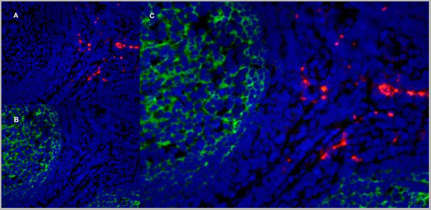
(Figure 10 Immunofluorescence Validation of TACE in Rat Brain (Pradillo et al, 2005)Cellular localization of TACE. Double immunofluorescence staining of brain sections from sham-operated (SHAM; A, C, E) and IPC-exposed animals (IPC; B, D, F) of TACE (red) and the cellular markers (green) NeuN (neurons; A, B), GFAP (astrocytes; C, D) and L. esculentum lectin (microglia and endothelium; E, F). White arrows indicate TACE-positive cells.)
IF (Immunofluorescence)
(Figure 10 Immunofluorescence Validation of TACE in Rat Brain (Pradillo et al, 2005)Cellular localization of TACE. Double immunofluorescence staining of brain sections from sham-operated (SHAM; A, C, E) and IPC-exposed animals (IPC; B, D, F) of TACE (red) and the cellular markers (green) NeuN (neurons; A, B), GFAP (astrocytes; C, D) and L. esculentum lectin (microglia and endothelium; E, F). White arrows indicate TACE-positive cells.)
TACE, Polyclonal Antibody (Cat# AAA10912)
Full Name
TACE Antibody
Gene Names
ADAM17; CSVP; TACE; NISBD; ADAM18; CD156B; NISBD1
Reactivity
Human, Mouse, Rat
Applications
Western Blot, Immunocytochemistry, Immunofluorescence, Flow Cytometry
Purity
TACE Antibody is affinity chromatography purified via peptide column.
Pricing
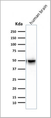
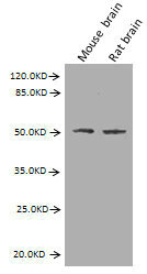
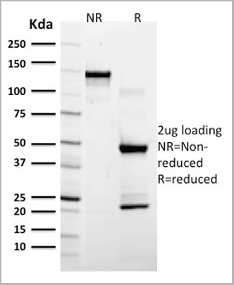 SDS-PAGE
(SDS-PAGE Analysis Purified EGFR Mouse Monoclonal Antibody (GFR/2596).Confirmation of Integrity and Purity of Antibody)
SDS-PAGE
(SDS-PAGE Analysis Purified EGFR Mouse Monoclonal Antibody (GFR/2596).Confirmation of Integrity and Purity of Antibody)
GFAP, Monoclonal Antibody (Cat# AAA13808)
Full Name
GFAP (Astrocyte & Neural Stem Cell Marker) Mouse Monoclonal Antibody
Gene Names
GFAP; ALXDRD
Reactivity
Human, Mouse, Rat, Cow, Pig, Rabbit, Chicken
Applications
Flow Cytometry, Immunofluorescence, Western Blot, Immunohistochemistry
Purity
200ug/ml of Ab purified from Bioreactor Concentrate by Protein A/G.
Pricing
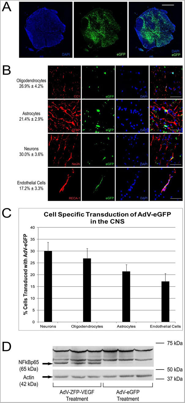


 Application Data
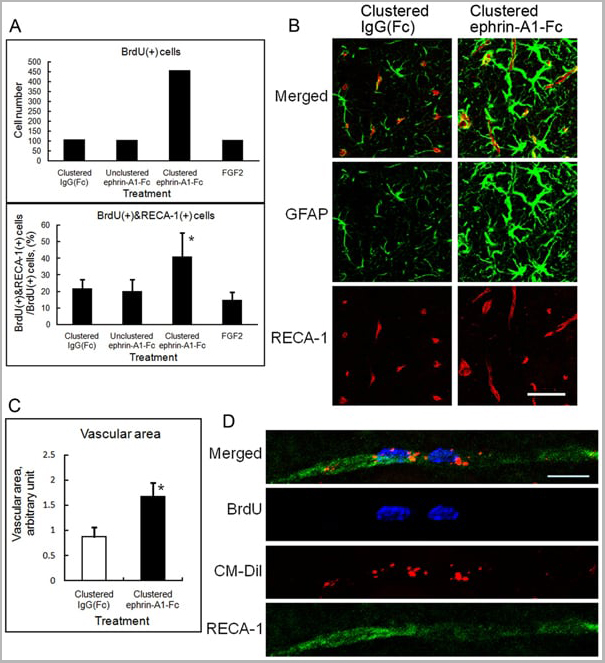
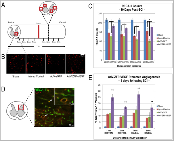
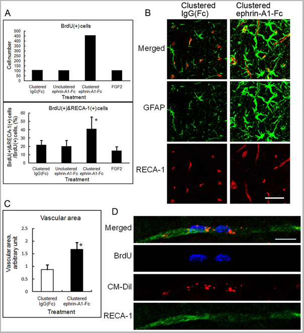
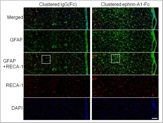
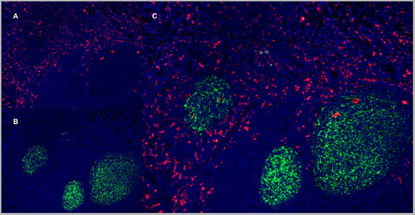
(Published customer image: Effect of clustered ephrin-A1-Fc on vascular formation in the rat striatum. Clustered ephrin-A1-Fc was injected into the lesioned side of the lateral ventricle in the unilaterally lesioned rats. Brains taken 6 weeks after injection were sectioned coronally and stained for GFAP (green) and RECA-1 (red) and with DAPI (nuclei; blue). The rectangular insets are shown in Fig. 8B. Scale bar: 100 um.From: Jing X, Miwa H, Sawada T, Nakanishi I, Kondo T, et al. (2012) Ephrin-A1-Mediated Dopaminergic Neurogenesis and Angiogenesis in a Rat Model of Parkinson's Disease. PLoS ONE 7(2): e32019.)
Application Data
(Published customer image: Effect of clustered ephrin-A1-Fc on vascular formation in the rat striatum. Clustered ephrin-A1-Fc was injected into the lesioned side of the lateral ventricle in the unilaterally lesioned rats. Brains taken 6 weeks after injection were sectioned coronally and stained for GFAP (green) and RECA-1 (red) and with DAPI (nuclei; blue). The rectangular insets are shown in Fig. 8B. Scale bar: 100 um.From: Jing X, Miwa H, Sawada T, Nakanishi I, Kondo T, et al. (2012) Ephrin-A1-Mediated Dopaminergic Neurogenesis and Angiogenesis in a Rat Model of Parkinson's Disease. PLoS ONE 7(2): e32019.)
RECA-1, Monoclonal Antibody (Cat# AAA12018)
Full Name
MOUSE ANTI RAT RECA-1
Applications
Immunohistochemistry, Immunofluorescence
Pricing
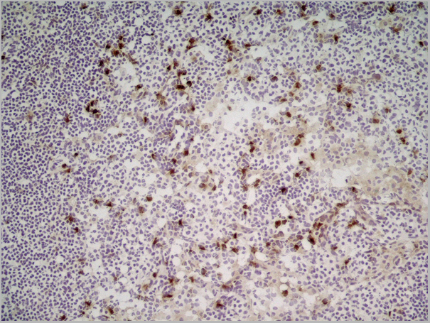
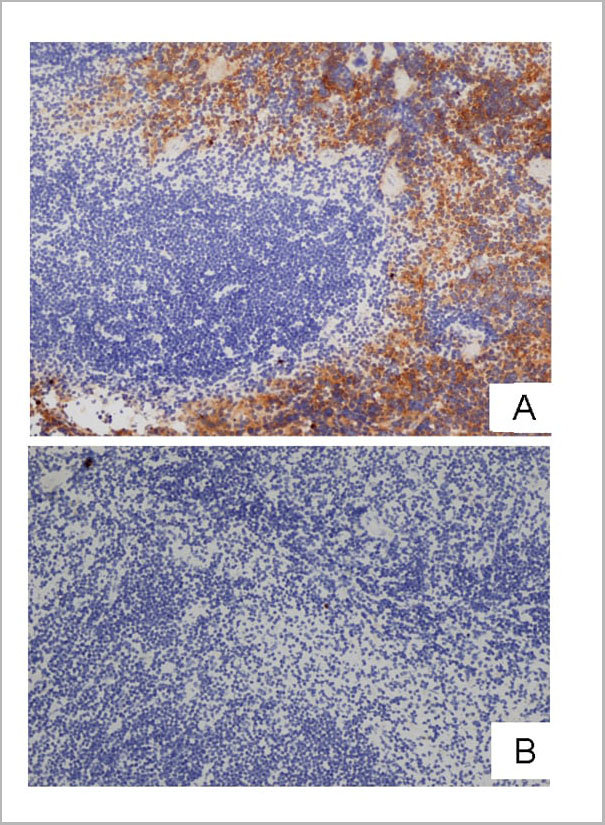
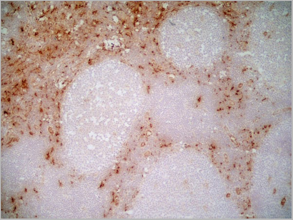
 IHC (Immunohistochemistry)
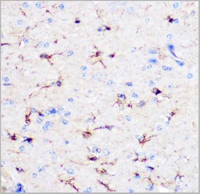
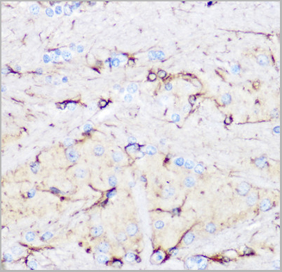
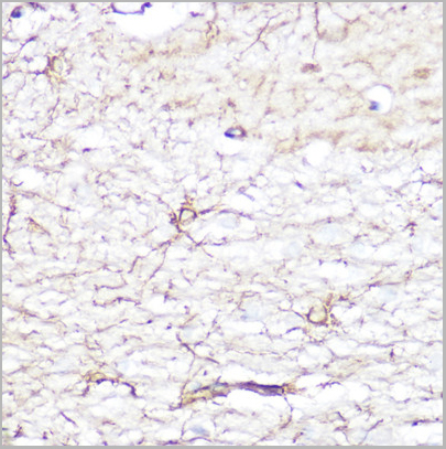
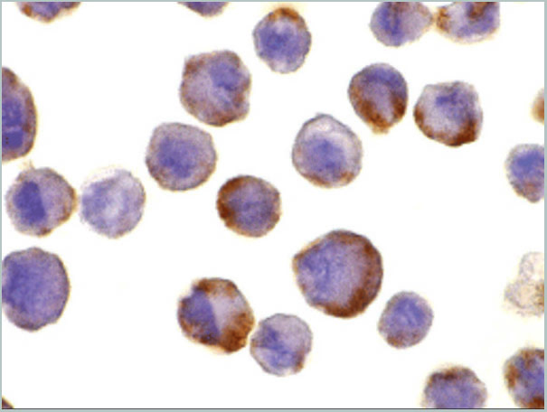
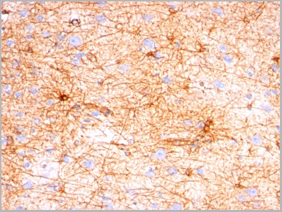
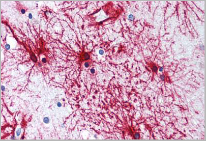
(Anti-GFAP antibody IHC of human brain, cortex, astrocytes. Immunohistochemistry of formalin-fixed, paraffin-embedded tissue after heat-induced antigen retrieval. Antibody concentration 5 ug/ml.)
IHC (Immunohistochemistry)
(Anti-GFAP antibody IHC of human brain, cortex, astrocytes. Immunohistochemistry of formalin-fixed, paraffin-embedded tissue after heat-induced antigen retrieval. Antibody concentration 5 ug/ml.)
GFAP, Polyclonal Antibody (Cat# AAA12316)
Full Name
Goat Polyclonal to Human GFAP
Reactivity
Human, Monkey, Rat, Bat, Bovine, Horse, Rabbit
Applications
Immunohistochemistry, Western Blot
Purity
Purified from goat serum by ammonium sulphate precipitacion followed by antigen affinity chromatography using the immunizing peptide.
Pricing


 Application Data
(Published customer image: Effect of clustered ephrin-A1-Fc on vascular formation in the rat striatum. Clustered ephrin-A1-Fc was injected into the lesioned side of the lateral ventricle in the unilaterally lesioned rats. Brains taken 6 weeks after injection were sectioned coronally and stained for GFAP (green) and RECA-1 (red) and with DAPI (nuclei; blue). The rectangular insets are shown in Fig. 8B. Scale bar: 100 um.From: Jing X, Miwa H, Sawada T, Nakanishi I, Kondo T, et al. (2012) Ephrin-A1-Mediated Dopaminergic Neurogenesis and Angiogenesis in a Rat Model of Parkinson's Disease. PLoS ONE 7(2): e32019.)
Application Data
(Published customer image: Effect of clustered ephrin-A1-Fc on vascular formation in the rat striatum. Clustered ephrin-A1-Fc was injected into the lesioned side of the lateral ventricle in the unilaterally lesioned rats. Brains taken 6 weeks after injection were sectioned coronally and stained for GFAP (green) and RECA-1 (red) and with DAPI (nuclei; blue). The rectangular insets are shown in Fig. 8B. Scale bar: 100 um.From: Jing X, Miwa H, Sawada T, Nakanishi I, Kondo T, et al. (2012) Ephrin-A1-Mediated Dopaminergic Neurogenesis and Angiogenesis in a Rat Model of Parkinson's Disease. PLoS ONE 7(2): e32019.)
RECA-1, Monoclonal Antibody (Cat# AAA12019)
Full Name
MOUSE ANTI RAT RECA-1
Applications
Immunohistochemistry, Immunofluorescence
Pricing
 Application Data
(Immunoperoxidase staining of a human tonsil cryosection with Mouse anti Human CD163 antibody, clone EDHu-1 followed by the Histar detection system . Low power)
Application Data
(Immunoperoxidase staining of a human tonsil cryosection with Mouse anti Human CD163 antibody, clone EDHu-1 followed by the Histar detection system . Low power)
CD163, Monoclonal Antibody (Cat# AAA11943)
Full Name
MOUSE ANTI HUMAN CD163
Gene Names
CD163; M130; MM130
Reactivity
Guinea Pig, Pig, Rhesus Monkey, Sheep
Applications
Immunohistochemistry, Immunohistochemistry, Flow Cytometry, Immunofluorescence, Immunoassay, Western Blot
Pricing
GFAP, Monoclonal Antibody (Cat# AAA14892)
Full Name
Mouse Anti-Human GFAP-UNLB
Gene Names
GFAP; FLJ45472; GFAP
Reactivity
Human
Applications
ELISA
Pricing
GFAP, Antibody (Cat# AAA13651)
Full Name
Goat anti-GFAP Antibody
Gene Names
GFAP; ALXDRD
Reactivity
Tested: Human, Rat
Expected from sequence similarity: Human, Mouse, Rat, Dog
Expected from sequence similarity: Human, Mouse, Rat, Dog
Applications
Peptide ELISA, Western Blot
Purity
Purified from goat serum by ammonium sulphate precipitation followed by antigen affinity chromatography using the immunizing peptide.
Pricing
anti-apolipoprotein A1 (Apo-A1) antibody, ELISA Kit (Cat# AAA15460)
Full Name
Human anti-apolipoprotein A1, Apo-A1antibody ELISA Kit
Reactivity
Human
Pricing
GFAP, Polyclonal Antibody (Cat# AAA17932)
Full Name
GFAP antibody
Gene Names
GFAP; ALXDRD
Applications
Immunohistochemistry, Immunohistochemistry, Western Blot
Pricing