Filters
Clonality
Type
Reactivity
Gene Name
Isotype
Host
Application
Clone
110 results for " G protein signaling" - showing 1-50


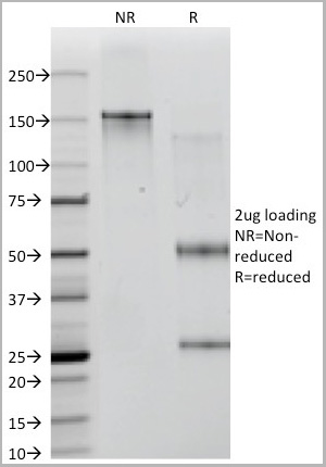
 WB (Western Blot)
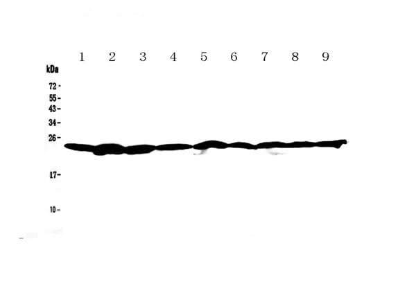
(WB Suggested Anti-GNAS Antibody Titration: 1 ug/mlPositive Control: MCF-7 whole cell lysatesGNAS is strongly supported by BioGPS gene expression data to be expressed in Human MCF7 cells)
WB (Western Blot)
(WB Suggested Anti-GNAS Antibody Titration: 1 ug/mlPositive Control: MCF-7 whole cell lysatesGNAS is strongly supported by BioGPS gene expression data to be expressed in Human MCF7 cells)
GNAS, Polyclonal Antibody (Cat# AAA23503)
Full Name
GNAS antibody - N-terminal region
Gene Names
GNAS; AHO; GSA; GSP; POH; GPSA; NESP; SCG6; SgVI; GNAS1; PITA3; C20orf45
Reactivity
Cow, Dog, Guinea Pig, Horse, Human, Mouse, Rabbit, Rat
Applications
IHC, WB
Purity
Affinity Purified
Pricing


 Application Data
(Published Customer Image:Rat anti Mouse Gr-1 antibody, clone RB6-8C5 used for the identification of neutrophils by immunofluorescence.Image caption:S. typhimurium-Infected Macrophages Containing Phagocytosed Neutrophils and T Cells Confocal fluorescence microscopy of 50-mum-thick liver sections from 1-wk-infected Slc11a1 wild-type mice. (A-C) S.Typhimurium (O-antigen, arrows) are red, macrophages (F4-80 and MOMA-2) are blue, DNA (DAPI) is gray, phalloidin is green, and neutrophils (Gr-1/Ly-6G/RB6-8C5) are pink (arrowheads). (A) Collapsed image from a 40-mum Z-stack. Scale bar is 20 mum. (B and C) Sections from (A) that are 4 mum apart. The video from which (A-C) were derived (Video S2) is available online. (D-G) T cells within multinucleate macrophages.Macrophages (F4-80 and MOMA-2) are blue (D, G, and H), T cells (CD3zeta) are red (D, G, arrowheads), DAPI is gray (E, G), actin-bound phalloidin is green (F, G). (G) Is a composite of (D, E, and F). Scale bars are 16 mum. (H) An image from a different mouse stained and labeled as described for (D-G). Scale bar is 8 mum. A video showing a T cell inside of a macrophage is available online (Video S3).From: Nix RN, Altschuler SE, Henson PM, Detweiler CS (2007) Hemophagocytic Macrophages Harbor Salmonella enterica during Persistent Infection.PLoS Pathog 3(12): e193.)
Application Data
(Published Customer Image:Rat anti Mouse Gr-1 antibody, clone RB6-8C5 used for the identification of neutrophils by immunofluorescence.Image caption:S. typhimurium-Infected Macrophages Containing Phagocytosed Neutrophils and T Cells Confocal fluorescence microscopy of 50-mum-thick liver sections from 1-wk-infected Slc11a1 wild-type mice. (A-C) S.Typhimurium (O-antigen, arrows) are red, macrophages (F4-80 and MOMA-2) are blue, DNA (DAPI) is gray, phalloidin is green, and neutrophils (Gr-1/Ly-6G/RB6-8C5) are pink (arrowheads). (A) Collapsed image from a 40-mum Z-stack. Scale bar is 20 mum. (B and C) Sections from (A) that are 4 mum apart. The video from which (A-C) were derived (Video S2) is available online. (D-G) T cells within multinucleate macrophages.Macrophages (F4-80 and MOMA-2) are blue (D, G, and H), T cells (CD3zeta) are red (D, G, arrowheads), DAPI is gray (E, G), actin-bound phalloidin is green (F, G). (G) Is a composite of (D, E, and F). Scale bars are 16 mum. (H) An image from a different mouse stained and labeled as described for (D-G). Scale bar is 8 mum. A video showing a T cell inside of a macrophage is available online (Video S3).From: Nix RN, Altschuler SE, Henson PM, Detweiler CS (2007) Hemophagocytic Macrophages Harbor Salmonella enterica during Persistent Infection.PLoS Pathog 3(12): e193.)
Gr-1, Monoclonal Antibody (Cat# AAA12257)
Full Name
Rat Anti Mouse Gr-1: FITC
Reactivity
Mouse
Applications
Flow Cytometry
Purity
Purified IgG prepared by affinity chromatography on Protein G from tissue culture supernatant
Pricing
 Application Data
(FASN antibody at 1/2000 dilution interacts with recombinant FASN protein with a DsbA tag. Predicted band size: 50Kda. Observed band size: 50Kda.)
Application Data
(FASN antibody at 1/2000 dilution interacts with recombinant FASN protein with a DsbA tag. Predicted band size: 50Kda. Observed band size: 50Kda.)
FASN, Antibody (Cat# AAA17951)
Full Name
FASN Antibody
Gene Names
FASN; FAS; OA-519; SDR27X1
Reactivity
Human
Applications
Western Blot
Purity
Protein G-Sepharose affinity purified
Pricing


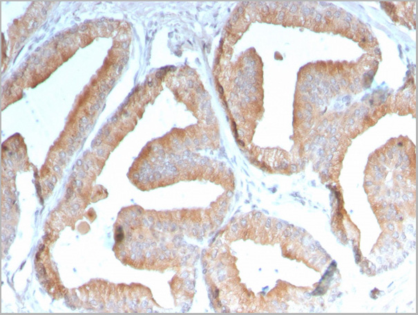
 IHC (Immunohistchemistry)
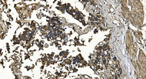
(DAB staining on IHC-P Samples:Human Prostate Gland Tissue))
IHC (Immunohistchemistry)
(DAB staining on IHC-P Samples:Human Prostate Gland Tissue))
Dickkopf Related Protein 1, Monoclonal Antibody (Cat# AAA20822)
Full Name
Dickkopf Related Protein 1 (DKK1)
Gene Names
DKK1; SK; DKK-1
Reactivity
Human, Mouse
Applications
Westen Blot, Immunohistochemistry, Immunocytochemistry, Immunoprecipitation
Purity
Purification: Protein A + Protein G affinity chromatography
Pricing


 WB (Western Blot)
(Western blot analysis of Human Lysates showing detection of Hsp90 protein using Mouse Anti-Hsp90 Monoclonal Antibody, Clone H9010 . Primary Antibody: Mouse Anti-Hsp90 Monoclonal Antibody at 1:1000. Comparison of clone H9010 behavior with Hsp90 human beta (1) and Hsp90 human alpha (2). Courtesy of: David Toft, Mayo Clinic.)
WB (Western Blot)
(Western blot analysis of Human Lysates showing detection of Hsp90 protein using Mouse Anti-Hsp90 Monoclonal Antibody, Clone H9010 . Primary Antibody: Mouse Anti-Hsp90 Monoclonal Antibody at 1:1000. Comparison of clone H9010 behavior with Hsp90 human beta (1) and Hsp90 human alpha (2). Courtesy of: David Toft, Mayo Clinic.)
HSP90, Monoclonal Antibody (Cat# AAA27660)
Full Name
HSP90 Antibody
Gene Names
HSP90AB1; HSP84; HSPC2; HSPCB; D6S182; HSP90B
Reactivity
Human, Mouse, Rat, Rabbit, Chicken, Dog, Fish, Shark, Hamster
Applications
Western Blot, Immunohistochemistry, Immunocytochemistry, Immunofluorescence, Immunoprecipitation, Antibody Microarray
Purity
Protein G Purified
Pricing

 Application Data
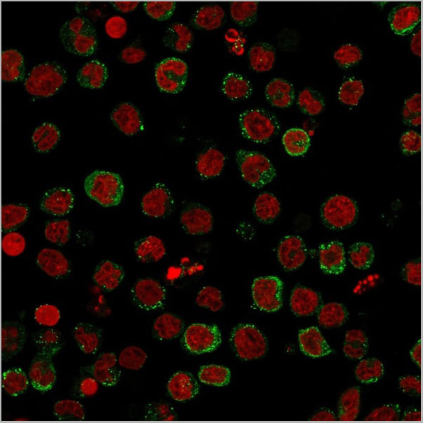
(ICC image of LGR5 antibody stained NCCIT cells. The secondary antibody (green) was goat anti-rabbit IgG (H+L) FITC conjugated.)
Application Data
(ICC image of LGR5 antibody stained NCCIT cells. The secondary antibody (green) was goat anti-rabbit IgG (H+L) FITC conjugated.)
LGR5, Polyclonal Antibody (Cat# AAA17985)
Full Name
LGR5 Antibody
Gene Names
LGR5; FEX; HG38; GPR49; GPR67; GRP49
Reactivity
Human, Mouse
Applications
Western Blot, Immunocytochemistry
Purity
Immunogen affinity purified
Pricing


 IP (Immunoprecipitation)
(HSPA1A was immunoprecipitated using:Lane A:0.5 mg Hela Whole Cell Lysate2 uL anti-HSPA1A rabbit monoclonal antibody and 15 ul of 50 % Protein G agarose.Primary antibody:Anti-HSPA1A rabbit monoclonal antibody,at 1:200 dilution Secondary antibody:Dylight 800-labeled antibody to rabbit IgG (H+L), at 1:5000 dilution Developed using the odssey technique.Performed under reducing conditions.Predicted band size: 70 kDaObserved band size: 70 kDa)
IP (Immunoprecipitation)
(HSPA1A was immunoprecipitated using:Lane A:0.5 mg Hela Whole Cell Lysate2 uL anti-HSPA1A rabbit monoclonal antibody and 15 ul of 50 % Protein G agarose.Primary antibody:Anti-HSPA1A rabbit monoclonal antibody,at 1:200 dilution Secondary antibody:Dylight 800-labeled antibody to rabbit IgG (H+L), at 1:5000 dilution Developed using the odssey technique.Performed under reducing conditions.Predicted band size: 70 kDaObserved band size: 70 kDa)
HSP70, Monoclonal Antibody (Cat# AAA27747)
Full Name
Recombinant Anti-HSP70 Antibody, Rabbit Monoclonal
Gene Names
HSPA1B; HSP72; HSPA1; HSX70; HSP70-1; HSP70-2; HSP70.1; HSP70.2; HSP70-1B
Reactivity
Human
Applications
Western Blot, Immunohistochemistry, Flow Cytometry, Immunocytochemistry, Immunofluorescence, Immunoprecipitation
Purity
Protein A
Pricing


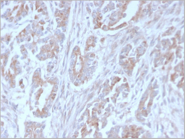
 IHC (Immunohistochemistry)
(Immunohistochemistry of paraffin-embedded human prostate cancer using AAA26967 at dilution of 1:100)
IHC (Immunohistochemistry)
(Immunohistochemistry of paraffin-embedded human prostate cancer using AAA26967 at dilution of 1:100)
SPDEF, Polyclonal Antibody (Cat# AAA26967)
Full Name
SPDEF Antibody
Gene Names
SPDEF; PDEF; bA375E1.3
Reactivity
Human, Mouse
Applications
Western Blot, Immunohistochemistry
Purity
>95%, Protein G purified
Pricing


 ICC (Immunocytochemistry)
(Immunostaining analysis in HeLa cells. HeLa cells were fixed with 4% paraformaldehyde and permeabilized with 0.1% Triton X-100 in PBS. The cells were immunostained with anti-SMAD1 mAb. [Lot No. 913C1b-1])
ICC (Immunocytochemistry)
(Immunostaining analysis in HeLa cells. HeLa cells were fixed with 4% paraformaldehyde and permeabilized with 0.1% Triton X-100 in PBS. The cells were immunostained with anti-SMAD1 mAb. [Lot No. 913C1b-1])
SMAD1, Monoclonal Antibody (Cat# AAA10611)
Full Name
Mouse monoclonal antibody Anti-Human SMAD1
Gene Names
SMAD1; BSP1; JV41; BSP-1; JV4-1; MADH1; MADR1
Reactivity
Human
Applications
Dot Blot, Immunocytochemistry, Immunoprecipitation, Western Blot, Flow Cytometry
Pricing


 Application Data
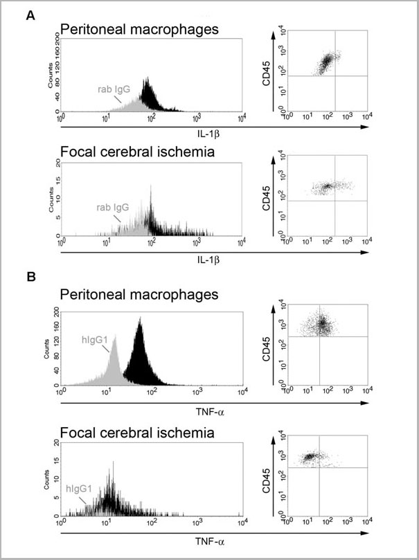
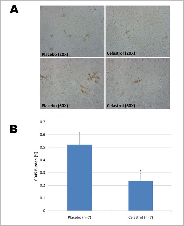
(Published customer image: Leukocyte infiltration in COX-2-M/-M and COX-2+/+ mice. MPO enzymatic activity (panel A) was statistically similar in COX-2-M/-M and COX-2+/+ livers at 6 h and 24 h post-IRI. Ly-6G+ neutrophil (panel B) and granulocyte (panel C) infiltration were also comparable in COX-2-M/-M and COX-2+/+ livers after IRI. Mac-1+ (panel D) and CD68 (panel E) infiltrating macrophages were significantly reduced in COX-2-M/-M livers at 24 h post-reperfusion, but were statistically indistinguishable in COX-2-M/-M and COX-2+/+ livers at 6 h after IRI. No statistical differences in MMP-9 expression (panel F) could be demonstrated in livers of COX-2-M/-M and COX-2+/+ mice post-IRI. Representative immunostaining (panel G) of infiltrating Ly-6G+ (a,b,e,f) and Mac-1+ (c,d,g,h) leukocytes in livers of COX-2+/+ (a,c,e,g) and COX-2-M/-M (b,d,f,h) mice at 6 h (a to d) and 24 h (e to h) post IRI; (n = 5 -6/group; * indicates p)
Application Data
(Published customer image: Leukocyte infiltration in COX-2-M/-M and COX-2+/+ mice. MPO enzymatic activity (panel A) was statistically similar in COX-2-M/-M and COX-2+/+ livers at 6 h and 24 h post-IRI. Ly-6G+ neutrophil (panel B) and granulocyte (panel C) infiltration were also comparable in COX-2-M/-M and COX-2+/+ livers after IRI. Mac-1+ (panel D) and CD68 (panel E) infiltrating macrophages were significantly reduced in COX-2-M/-M livers at 24 h post-reperfusion, but were statistically indistinguishable in COX-2-M/-M and COX-2+/+ livers at 6 h after IRI. No statistical differences in MMP-9 expression (panel F) could be demonstrated in livers of COX-2-M/-M and COX-2+/+ mice post-IRI. Representative immunostaining (panel G) of infiltrating Ly-6G+ (a,b,e,f) and Mac-1+ (c,d,g,h) leukocytes in livers of COX-2+/+ (a,c,e,g) and COX-2-M/-M (b,d,f,h) mice at 6 h (a to d) and 24 h (e to h) post IRI; (n = 5 -6/group; * indicates p)
CD68, Monoclonal Antibody (Cat# AAA12105)
Full Name
RAT ANTI MOUSE CD68:FITC
Gene Names
Cd68; Lamp4; gp110; Scard1
Applications
Flow Cytometry
Pricing


 Application Data
(Staining of mouse thymus with Rat anti Mouse CD150: RPE)
Application Data
(Staining of mouse thymus with Rat anti Mouse CD150: RPE)
CD150, Monoclonal Antibody (Cat# AAA12038)
Full Name
RAT ANTI MOUSE CD150:RPE
Gene Names
Slamf1; Slam; CD150; IPO-3; CDw150; ESTM51; AA177906; 4933415F16
Applications
Flow Cytometry
Pricing


 IF (Immunofluorescence)
(Immunofluorescence of IRE1p in rat small intestine tissue with IRE1p antibody at 20 μg/ml.Green: IRE1p Antibody (3655)Blue: DAPI staining)
IF (Immunofluorescence)
(Immunofluorescence of IRE1p in rat small intestine tissue with IRE1p antibody at 20 μg/ml.Green: IRE1p Antibody (3655)Blue: DAPI staining)
IRE1p, Polyclonal Antibody (Cat# AAA10921)
Full Name
IRE1p Antibody
Gene Names
ERN1; IRE1; IRE1P; IRE1a; hIRE1p
Reactivity
Human, Mouse, Rat
Applications
Western Blot, Immunocytochemistry, Immunofluorescence
Purity
IRE1p Antibody is affinity chromatography purified via peptide column.
Pricing


 Application Data
(Published customer image: Leukocyte infiltration in COX-2-M/-M and COX-2+/+ mice. MPO enzymatic activity (panel A) was statistically similar in COX-2-M/-M and COX-2+/+ livers at 6 h and 24 h post-IRI. Ly-6G+ neutrophil (panel B) and granulocyte (panel C) infiltration were also comparable in COX-2-M/-M and COX-2+/+ livers after IRI. Mac-1+ (panel D) and CD68 (panel E) infiltrating macrophages were significantly reduced in COX-2-M/-M livers at 24 h post-reperfusion, but were statistically indistinguishable in COX-2-M/-M and COX-2+/+ livers at 6 h after IRI. No statistical differences in MMP-9 expression (panel F) could be demonstrated in livers of COX-2-M/-M and COX-2+/+ mice post-IRI. Representative immunostaining (panel G) of infiltrating Ly-6G+ (a,b,e,f) and Mac-1+ (c,d,g,h) leukocytes in livers of COX-2+/+ (a,c,e,g) and COX-2-M/-M (b,d,f,h) mice at 6 h (a to d) and 24 h (e to h) post IRI; (n = 5 -6/group; * indicates p)
Application Data
(Published customer image: Leukocyte infiltration in COX-2-M/-M and COX-2+/+ mice. MPO enzymatic activity (panel A) was statistically similar in COX-2-M/-M and COX-2+/+ livers at 6 h and 24 h post-IRI. Ly-6G+ neutrophil (panel B) and granulocyte (panel C) infiltration were also comparable in COX-2-M/-M and COX-2+/+ livers after IRI. Mac-1+ (panel D) and CD68 (panel E) infiltrating macrophages were significantly reduced in COX-2-M/-M livers at 24 h post-reperfusion, but were statistically indistinguishable in COX-2-M/-M and COX-2+/+ livers at 6 h after IRI. No statistical differences in MMP-9 expression (panel F) could be demonstrated in livers of COX-2-M/-M and COX-2+/+ mice post-IRI. Representative immunostaining (panel G) of infiltrating Ly-6G+ (a,b,e,f) and Mac-1+ (c,d,g,h) leukocytes in livers of COX-2+/+ (a,c,e,g) and COX-2-M/-M (b,d,f,h) mice at 6 h (a to d) and 24 h (e to h) post IRI; (n = 5 -6/group; * indicates p)
CD68, Monoclonal Antibody (Cat# AAA12103)
Full Name
RAT ANTI MOUSE CD68:Biotin
Gene Names
Cd68; Lamp4; gp110; Scard1
Applications
Flow Cytometry
Pricing


 Application Data
(Published customer imageSLAMF3 blockade in human hepatocytes is associated with lower susceptibility to HCV. (A) SLAMF3 was stained in primary human hepatocytes (PHHs) and cells from the Huh-7 human hepatoma cell line with a specific antibody (HLy9.1.25 clone; grey) and an isotype-matched control (empty). One of four independent experiments is shown. Huh-7 cells were transfected with scrambled control (sc) siRNA or three specific siRNAs (#1, #2 and #3) targeting SLAMF3, prior to infection with HCVcc; siRNA efficiency was checked by quantifying SLAMF3 mRNA (B) and the CD81 expression level (C) by flow cytometry analysis at 48 h post-transfection. Results are presented as the mean +/-SD (n = 3). Intracellular viral RNA was quantified at 72 h p.i. (D) and the infection was measured at 72 h p.i. by focus-forming units FFUs counting (E) (as inhibition percent; mean of three independent experiments; error bars: SD. **p)
Application Data
(Published customer imageSLAMF3 blockade in human hepatocytes is associated with lower susceptibility to HCV. (A) SLAMF3 was stained in primary human hepatocytes (PHHs) and cells from the Huh-7 human hepatoma cell line with a specific antibody (HLy9.1.25 clone; grey) and an isotype-matched control (empty). One of four independent experiments is shown. Huh-7 cells were transfected with scrambled control (sc) siRNA or three specific siRNAs (#1, #2 and #3) targeting SLAMF3, prior to infection with HCVcc; siRNA efficiency was checked by quantifying SLAMF3 mRNA (B) and the CD81 expression level (C) by flow cytometry analysis at 48 h post-transfection. Results are presented as the mean +/-SD (n = 3). Intracellular viral RNA was quantified at 72 h p.i. (D) and the infection was measured at 72 h p.i. by focus-forming units FFUs counting (E) (as inhibition percent; mean of three independent experiments; error bars: SD. **p)
CD229, Monoclonal Antibody (Cat# AAA11951)
Full Name
MOUSE ANTI HUMAN CD229
Gene Names
LY9; hly9; mLY9; CD229; SLAMF3
Applications
Flow Cytometry, Immunoprecipitation
Pricing


 Application Data
(Published customer image: Leukocyte infiltration in COX-2-M/-M and COX-2+/+ mice. MPO enzymatic activity (panel A) was statistically similar in COX-2-M/-M and COX-2+/+ livers at 6 h and 24 h post-IRI. Ly-6G+ neutrophil (panel B) and granulocyte (panel C) infiltration were also comparable in COX-2-M/-M and COX-2+/+ livers after IRI. Mac-1+ (panel D) and CD68 (panel E) infiltrating macrophages were significantly reduced in COX-2-M/-M livers at 24 h post-reperfusion, but were statistically indistinguishable in COX-2-M/-M and COX-2+/+ livers at 6 h after IRI. No statistical differences in MMP-9 expression (panel F) could be demonstrated in livers of COX-2-M/-M and COX-2+/+ mice post-IRI. Representative immunostaining (panel G) of infiltrating Ly-6G+ (a,b,e,f) and Mac-1+ (c,d,g,h) leukocytes in livers of COX-2+/+ (a,c,e,g) and COX-2-M/-M (b,d,f,h) mice at 6 h (a to d) and 24 h (e to h) post IRI; (n = 5 -6/group; * indicates p)
Application Data
(Published customer image: Leukocyte infiltration in COX-2-M/-M and COX-2+/+ mice. MPO enzymatic activity (panel A) was statistically similar in COX-2-M/-M and COX-2+/+ livers at 6 h and 24 h post-IRI. Ly-6G+ neutrophil (panel B) and granulocyte (panel C) infiltration were also comparable in COX-2-M/-M and COX-2+/+ livers after IRI. Mac-1+ (panel D) and CD68 (panel E) infiltrating macrophages were significantly reduced in COX-2-M/-M livers at 24 h post-reperfusion, but were statistically indistinguishable in COX-2-M/-M and COX-2+/+ livers at 6 h after IRI. No statistical differences in MMP-9 expression (panel F) could be demonstrated in livers of COX-2-M/-M and COX-2+/+ mice post-IRI. Representative immunostaining (panel G) of infiltrating Ly-6G+ (a,b,e,f) and Mac-1+ (c,d,g,h) leukocytes in livers of COX-2+/+ (a,c,e,g) and COX-2-M/-M (b,d,f,h) mice at 6 h (a to d) and 24 h (e to h) post IRI; (n = 5 -6/group; * indicates p)
CD68, Monoclonal Antibody (Cat# AAA12110)
Full Name
RAT ANTI MOUSE CD68
Gene Names
Cd68; Lamp4; gp110; Scard1
Applications
Immunohistochemistry, Flow Cytometry, Immunofluorescence, Immunoprecipitation, Immunohistochemistry, Western Blot
Pricing


 IP (Immunoprecipitation)
(HDAC2 was immunoprecipitated using:Lane A:0.5 mg Jurkat Whole Cell LysateLane B:0.5 mg NIH-3T3 Whole Cell LysateLane C:0.5 mg Hela Whole Cell Lysate4 uL anti-HDAC2 rabbit polyclonal antibody and 15 ul of 50 % Protein G agarose.Primary antibody:Anti-HDAC2 rabbit polyclonal antibody,at 1:100 dilution Secondary antibody:Dylight 800-labeled antibody to rabbit IgG (H+L), at 1:5000 dilution Developed using the odssey technique.Performed under reducing conditions.Predicted band size: 60 kDaObserved band size: 60 kDa)
IP (Immunoprecipitation)
(HDAC2 was immunoprecipitated using:Lane A:0.5 mg Jurkat Whole Cell LysateLane B:0.5 mg NIH-3T3 Whole Cell LysateLane C:0.5 mg Hela Whole Cell Lysate4 uL anti-HDAC2 rabbit polyclonal antibody and 15 ul of 50 % Protein G agarose.Primary antibody:Anti-HDAC2 rabbit polyclonal antibody,at 1:100 dilution Secondary antibody:Dylight 800-labeled antibody to rabbit IgG (H+L), at 1:5000 dilution Developed using the odssey technique.Performed under reducing conditions.Predicted band size: 60 kDaObserved band size: 60 kDa)
HDAC2, Polyclonal Antibody (Cat# AAA27718)
Full Name
Anti-HDAC2 Antibody, Rabbit Polyclonal
Gene Names
HDAC2; HD2; RPD3; YAF1
Reactivity
Human
Applications
Western Blot, Immunohistochemistry, Immunocytochemistry, Immunofluorescence, Immunoprecipitation
Purity
Protein A & Antigen Affinity
Pricing


 Application Data
(Staining of mouse thymus with RAT ANTI MOUSE CD150: RPE)
Application Data
(Staining of mouse thymus with RAT ANTI MOUSE CD150: RPE)
CD150, Monoclonal Antibody (Cat# AAA26741)
Full Name
CD150 (CD150 Antigen, Cdw150, 4933415F16, Estm51, Ipo 3, Ipo-3, OTTHUMP00000060252, Signaling Lymphocytic Activation Molecule, SLAM, Signaling Lymphocytic Activation Molecule Family Member 1, SLAMF1 Protein) (FITC)
Reactivity
Mouse
Applications
Flow Cytometry
Purity
Purified by Protein G Affinity Chromatography
Pricing


 Application Data
(Staining of mouse thymus with RAT ANTI MOUSE CD150: RPE)
Application Data
(Staining of mouse thymus with RAT ANTI MOUSE CD150: RPE)
CD150, Monoclonal Antibody (Cat# AAA26752)
Full Name
CD150 (CD150 Antigen, Cdw150, 4933415F16, Estm51, Ipo 3, Ipo-3, OTTHUMP00000060252, Signaling Lymphocytic Activation Molecule, SLAM, Signaling Lymphocytic Activation Molecule Family Member 1, SLAMF1 Protein) (HRP)
Reactivity
Mouse
Applications
Flow Cytometry
Purity
Purified by Protein G Affinity Chromatography
Pricing


 IHC (Immunohistchemistry)
(Immunohistochemistry analysis using Mouse Anti-Hsp70 Monoclonal Antibody, Clone BB70 . Tissue: hepatocytes. Species: Rat. Fixation: Paraffin Embedded. Primary Antibody: Mouse Anti-Hsp70 Monoclonal Antibody at 1:200. Liver sections were paraffin embedded. First pictures in series show two hours after exposure to stress, the second shows the control. Courtesy of: G. Matic, University of Belgrade, Serbia.)
IHC (Immunohistchemistry)
(Immunohistochemistry analysis using Mouse Anti-Hsp70 Monoclonal Antibody, Clone BB70 . Tissue: hepatocytes. Species: Rat. Fixation: Paraffin Embedded. Primary Antibody: Mouse Anti-Hsp70 Monoclonal Antibody at 1:200. Liver sections were paraffin embedded. First pictures in series show two hours after exposure to stress, the second shows the control. Courtesy of: G. Matic, University of Belgrade, Serbia.)
HSP70/HSC70, Monoclonal Antibody (Cat# AAA27659)
Full Name
HSP70/HSC70 Antibody
Gene Names
HSPA2; HSP70
Reactivity
Human, Mouse, Rat, Bovine, Sheep, Dog, Beluga, Fish, Guinea Pig, Pig, Hamster, Rabbit, Chicken, African Clawed Frog, Fruit Fly, Yeast
Applications
Western Blot, Immunocytochemistry, Immunofluorescence, Immunohistochemistry, Immunoprecipitation
Purity
Protein G Purified
Pricing


 IP (Immunoprecipitation)
(MAP2K3 was immunoprecipitated using: Lane A:0.5 mg Jurkat Whole Cell Lysate Lane B:0.5 mg HepG2 Whole Cell Lysate Lane C:0.5 mg HeLa Whole Cell Lysate 2 uL anti-MAP2K3 rabbit polyclonal antibody and 60 ug of Immunomagnetic beads Protein A/G. Primary antibody: Anti-MAP2K3 rabbit polyclonal antibody,at 1:100 dilution Secondary antibody: Clean-Blot IP Detection Reagent (HRP) at 1:1000dilution Developed using the ECL technique. Performed under reducing conditions. Predicted band size: 39 kDa Observed band size :39 kDa)
IP (Immunoprecipitation)
(MAP2K3 was immunoprecipitated using: Lane A:0.5 mg Jurkat Whole Cell Lysate Lane B:0.5 mg HepG2 Whole Cell Lysate Lane C:0.5 mg HeLa Whole Cell Lysate 2 uL anti-MAP2K3 rabbit polyclonal antibody and 60 ug of Immunomagnetic beads Protein A/G. Primary antibody: Anti-MAP2K3 rabbit polyclonal antibody,at 1:100 dilution Secondary antibody: Clean-Blot IP Detection Reagent (HRP) at 1:1000dilution Developed using the ECL technique. Performed under reducing conditions. Predicted band size: 39 kDa Observed band size :39 kDa)
MEK3/MKK3, Polyclonal Antibody (Cat# AAA27720)
Full Name
Anti-MEK3/MKK3 Antibody, Rabbit Polyclonal
Gene Names
MAP2K3; MEK3; MKK3; MAPKK3; PRKMK3; SAPKK2; SAPKK-2
Reactivity
Human, Mouse
Applications
Western Blot, Immunohistochemistry, Immunocytochemistry, Immunofluorescence, Immunoprecipitation
Purity
Protein A & Antigen Affinity
Pricing


 Application Data
(Published customer image: Leukocyte infiltration in COX-2-M/-M and COX-2+/+ mice. MPO enzymatic activity (panel A) was statistically similar in COX-2-M/-M and COX-2+/+ livers at 6 h and 24 h post-IRI. Ly-6G+ neutrophil (panel B) and granulocyte (panel C) infiltration were also comparable in COX-2-M/-M and COX-2+/+ livers after IRI. Mac-1+ (panel D) and CD68 (panel E) infiltrating macrophages were significantly reduced in COX-2-M/-M livers at 24 h post-reperfusion, but were statistically indistinguishable in COX-2-M/-M and COX-2+/+ livers at 6 h after IRI. No statistical differences in MMP-9 expression (panel F) could be demonstrated in livers of COX-2-M/-M and COX-2+/+ mice post-IRI. Representative immunostaining (panel G) of infiltrating Ly-6G+ (a,b,e,f) and Mac-1+ (c,d,g,h) leukocytes in livers of COX-2+/+ (a,c,e,g) and COX-2-M/-M (b,d,f,h) mice at 6 h (a to d) and 24 h (e to h) post IRI; (n = 5 -6/group; * indicates p)
Application Data
(Published customer image: Leukocyte infiltration in COX-2-M/-M and COX-2+/+ mice. MPO enzymatic activity (panel A) was statistically similar in COX-2-M/-M and COX-2+/+ livers at 6 h and 24 h post-IRI. Ly-6G+ neutrophil (panel B) and granulocyte (panel C) infiltration were also comparable in COX-2-M/-M and COX-2+/+ livers after IRI. Mac-1+ (panel D) and CD68 (panel E) infiltrating macrophages were significantly reduced in COX-2-M/-M livers at 24 h post-reperfusion, but were statistically indistinguishable in COX-2-M/-M and COX-2+/+ livers at 6 h after IRI. No statistical differences in MMP-9 expression (panel F) could be demonstrated in livers of COX-2-M/-M and COX-2+/+ mice post-IRI. Representative immunostaining (panel G) of infiltrating Ly-6G+ (a,b,e,f) and Mac-1+ (c,d,g,h) leukocytes in livers of COX-2+/+ (a,c,e,g) and COX-2-M/-M (b,d,f,h) mice at 6 h (a to d) and 24 h (e to h) post IRI; (n = 5 -6/group; * indicates p)
CD68, Monoclonal Antibody (Cat# AAA12104)
Full Name
RAT ANTI MOUSE CD68:Biotin
Gene Names
Cd68; Lamp4; gp110; Scard1
Applications
Flow Cytometry
Pricing


 Application Data
(Staining of mouse thymus with Rat anti Mouse CD150: RPE)
Application Data
(Staining of mouse thymus with Rat anti Mouse CD150: RPE)
CD150, Monoclonal Antibody (Cat# AAA11956)
Full Name
RAT ANTI MOUSE CD150
Gene Names
Slamf1; Slam; CD150; IPO-3; CDw150; ESTM51; AA177906; 4933415F16
Applications
Flow Cytometry
Pricing


 Application Data
(Staining of mouse thymus with RAT ANTI MOUSE CD150: RPE)
Application Data
(Staining of mouse thymus with RAT ANTI MOUSE CD150: RPE)
CD150, Monoclonal Antibody (Cat# AAA26790)
Full Name
CD150 (CD150 Antigen, Cdw150, 4933415F16, Estm51, Ipo 3, Ipo-3, OTTHUMP00000060252, Signaling Lymphocytic Activation Molecule, SLAM, Signaling Lymphocytic Activation Molecule Family Member 1, SLAMF1 Protein) (MaxLight 490)
Reactivity
Mouse
Applications
Flow Cytometry
Purity
Purified by Protein G Affinity Chromatography
Pricing


 Application Data
(Published customer image: Leukocyte infiltration in COX-2-M/-M and COX-2+/+ mice. MPO enzymatic activity (panel A) was statistically similar in COX-2-M/-M and COX-2+/+ livers at 6 h and 24 h post-IRI. Ly-6G+ neutrophil (panel B) and granulocyte (panel C) infiltration were also comparable in COX-2-M/-M and COX-2+/+ livers after IRI. Mac-1+ (panel D) and CD68 (panel E) infiltrating macrophages were significantly reduced in COX-2-M/-M livers at 24 h post-reperfusion, but were statistically indistinguishable in COX-2-M/-M and COX-2+/+ livers at 6 h after IRI. No statistical differences in MMP-9 expression (panel F) could be demonstrated in livers of COX-2-M/-M and COX-2+/+ mice post-IRI. Representative immunostaining (panel G) of infiltrating Ly-6G+ (a,b,e,f) and Mac-1+ (c,d,g,h) leukocytes in livers of COX-2+/+ (a,c,e,g) and COX-2-M/-M (b,d,f,h) mice at 6 h (a to d) and 24 h (e to h) post IRI; (n = 5 -6/group; * indicates p)
Application Data
(Published customer image: Leukocyte infiltration in COX-2-M/-M and COX-2+/+ mice. MPO enzymatic activity (panel A) was statistically similar in COX-2-M/-M and COX-2+/+ livers at 6 h and 24 h post-IRI. Ly-6G+ neutrophil (panel B) and granulocyte (panel C) infiltration were also comparable in COX-2-M/-M and COX-2+/+ livers after IRI. Mac-1+ (panel D) and CD68 (panel E) infiltrating macrophages were significantly reduced in COX-2-M/-M livers at 24 h post-reperfusion, but were statistically indistinguishable in COX-2-M/-M and COX-2+/+ livers at 6 h after IRI. No statistical differences in MMP-9 expression (panel F) could be demonstrated in livers of COX-2-M/-M and COX-2+/+ mice post-IRI. Representative immunostaining (panel G) of infiltrating Ly-6G+ (a,b,e,f) and Mac-1+ (c,d,g,h) leukocytes in livers of COX-2+/+ (a,c,e,g) and COX-2-M/-M (b,d,f,h) mice at 6 h (a to d) and 24 h (e to h) post IRI; (n = 5 -6/group; * indicates p)
CD68, Monoclonal Antibody (Cat# AAA12107)
Full Name
RAT ANTI MOUSE CD68
Gene Names
Cd68; Lamp4; gp110; Scard1
Applications
Immunohistochemistry, Flow Cytometry, Immunofluorescence, Immunoprecipitation, Immunohistochemistry, Western Blot
Purity
Purified
Purified IgG - liquid
Purified IgG - liquid
Pricing


 Application Data
(Staining of mouse thymus with RAT ANTI MOUSE CD150: RPE)
Application Data
(Staining of mouse thymus with RAT ANTI MOUSE CD150: RPE)
CD150, Monoclonal Antibody (Cat# AAA26776)
Full Name
CD150 (CD150 Antigen, Cdw150, 4933415F16, Estm51, Ipo 3, Ipo-3, OTTHUMP00000060252, Signaling Lymphocytic Activation Molecule, SLAM, Signaling Lymphocytic Activation Molecule Family Member 1, SLAMF1 Protein) (MaxLight 405)
Reactivity
Mouse
Applications
Flow Cytometry
Purity
Purified by Protein G Affinity Chromatography
Pricing


 Application Data
(Published customer image: Leukocyte infiltration in COX-2-M/-M and COX-2+/+ mice. MPO enzymatic activity (panel A) was statistically similar in COX-2-M/-M and COX-2+/+ livers at 6 h and 24 h post-IRI. Ly-6G+ neutrophil (panel B) and granulocyte (panel C) infiltration were also comparable in COX-2-M/-M and COX-2+/+ livers after IRI. Mac-1+ (panel D) and CD68 (panel E) infiltrating macrophages were significantly reduced in COX-2-M/-M livers at 24 h post-reperfusion, but were statistically indistinguishable in COX-2-M/-M and COX-2+/+ livers at 6 h after IRI. No statistical differences in MMP-9 expression (panel F) could be demonstrated in livers of COX-2-M/-M and COX-2+/+ mice post-IRI. Representative immunostaining (panel G) of infiltrating Ly-6G+ (a,b,e,f) and Mac-1+ (c,d,g,h) leukocytes in livers of COX-2+/+ (a,c,e,g) and COX-2-M/-M (b,d,f,h) mice at 6 h (a to d) and 24 h (e to h) post IRI; (n = 5 -6/group; * indicates p)
Application Data
(Published customer image: Leukocyte infiltration in COX-2-M/-M and COX-2+/+ mice. MPO enzymatic activity (panel A) was statistically similar in COX-2-M/-M and COX-2+/+ livers at 6 h and 24 h post-IRI. Ly-6G+ neutrophil (panel B) and granulocyte (panel C) infiltration were also comparable in COX-2-M/-M and COX-2+/+ livers after IRI. Mac-1+ (panel D) and CD68 (panel E) infiltrating macrophages were significantly reduced in COX-2-M/-M livers at 24 h post-reperfusion, but were statistically indistinguishable in COX-2-M/-M and COX-2+/+ livers at 6 h after IRI. No statistical differences in MMP-9 expression (panel F) could be demonstrated in livers of COX-2-M/-M and COX-2+/+ mice post-IRI. Representative immunostaining (panel G) of infiltrating Ly-6G+ (a,b,e,f) and Mac-1+ (c,d,g,h) leukocytes in livers of COX-2+/+ (a,c,e,g) and COX-2-M/-M (b,d,f,h) mice at 6 h (a to d) and 24 h (e to h) post IRI; (n = 5 -6/group; * indicates p)
CD68, Monoclonal Antibody (Cat# AAA12108)
Full Name
RAT ANTI MOUSE CD68:RPE
Gene Names
Cd68; Lamp4; gp110; Scard1
Applications
Flow Cytometry
Pricing


 Application Data
(Staining of mouse thymus with RAT ANTI MOUSE CD150: RPE)
Application Data
(Staining of mouse thymus with RAT ANTI MOUSE CD150: RPE)
CD150, Monoclonal Antibody (Cat# AAA26727)
Full Name
CD150 (CD150 Antigen, Cdw150, 4933415F16, Estm51, Ipo 3, Ipo-3, OTTHUMP00000060252, Signaling Lymphocytic Activation Molecule, SLAM, Signaling Lymphocytic Activation Molecule Family Member 1, SLAMF1 Protein) (Biotin)
Reactivity
Mouse
Applications
Flow Cytometry
Purity
Purified by Protein G Affinity Chromatography
Pricing


 Application Data
(Published customer image: Leukocyte infiltration in COX-2-M/-M and COX-2+/+ mice. MPO enzymatic activity (panel A) was statistically similar in COX-2-M/-M and COX-2+/+ livers at 6 h and 24 h post-IRI. Ly-6G+ neutrophil (panel B) and granulocyte (panel C) infiltration were also comparable in COX-2-M/-M and COX-2+/+ livers after IRI. Mac-1+ (panel D) and CD68 (panel E) infiltrating macrophages were significantly reduced in COX-2-M/-M livers at 24 h post-reperfusion, but were statistically indistinguishable in COX-2-M/-M and COX-2+/+ livers at 6 h after IRI. No statistical differences in MMP-9 expression (panel F) could be demonstrated in livers of COX-2-M/-M and COX-2+/+ mice post-IRI. Representative immunostaining (panel G) of infiltrating Ly-6G+ (a,b,e,f) and Mac-1+ (c,d,g,h) leukocytes in livers of COX-2+/+ (a,c,e,g) and COX-2-M/-M (b,d,f,h) mice at 6 h (a to d) and 24 h (e to h) post IRI; (n = 5 -6/group; * indicates p)
Application Data
(Published customer image: Leukocyte infiltration in COX-2-M/-M and COX-2+/+ mice. MPO enzymatic activity (panel A) was statistically similar in COX-2-M/-M and COX-2+/+ livers at 6 h and 24 h post-IRI. Ly-6G+ neutrophil (panel B) and granulocyte (panel C) infiltration were also comparable in COX-2-M/-M and COX-2+/+ livers after IRI. Mac-1+ (panel D) and CD68 (panel E) infiltrating macrophages were significantly reduced in COX-2-M/-M livers at 24 h post-reperfusion, but were statistically indistinguishable in COX-2-M/-M and COX-2+/+ livers at 6 h after IRI. No statistical differences in MMP-9 expression (panel F) could be demonstrated in livers of COX-2-M/-M and COX-2+/+ mice post-IRI. Representative immunostaining (panel G) of infiltrating Ly-6G+ (a,b,e,f) and Mac-1+ (c,d,g,h) leukocytes in livers of COX-2+/+ (a,c,e,g) and COX-2-M/-M (b,d,f,h) mice at 6 h (a to d) and 24 h (e to h) post IRI; (n = 5 -6/group; * indicates p)
CD68, Monoclonal Antibody (Cat# AAA12102)
Full Name
RAT ANTI MOUSE CD68
Gene Names
Cd68; Lamp4; gp110; Scard1
Applications
Immunohistochemistry, Flow Cytometry, Immunofluorescence, Immunoprecipitation, Immunohistochemistry, Western Blot
Pricing


 Application Data
(Staining of mouse thymus with RAT ANTI MOUSE CD150: RPE)
Application Data
(Staining of mouse thymus with RAT ANTI MOUSE CD150: RPE)
CD150, Monoclonal Antibody (Cat# AAA26804)
Full Name
CD150 (CD150 Antigen, Cdw150, 4933415F16, Estm51, Ipo 3, Ipo-3, OTTHUMP00000060252, Signaling Lymphocytic Activation Molecule, SLAM, Signaling Lymphocytic Activation Molecule Family Member 1, SLAMF1 Protein) (MaxLight 550)
Reactivity
Mouse
Applications
Flow Cytometry
Purity
Purified by Protein G Affinity Chromatography
Pricing


 IF (Immunofluorescence)
(Immunofluorescence analysis of U-2 OS cells using RALGDS antibody at dilution of 1:100. Blue: DAPI for nuclear staining.)
IF (Immunofluorescence)
(Immunofluorescence analysis of U-2 OS cells using RALGDS antibody at dilution of 1:100. Blue: DAPI for nuclear staining.)
RALGDS, Polyclonal Antibody (Cat# AAA28359)
Full Name
RALGDS Rabbit pAb
Gene Names
RALGDS; RGF; RGDS; RalGEF
Reactivity
Human, Mouse, Rat
Applications
Western Blot, Immunohistochemistry, Immunofluorescence
Purity
Affinity purification
Pricing


 Application Data
(Staining of mouse thymus with RAT ANTI MOUSE CD150: RPE)
Application Data
(Staining of mouse thymus with RAT ANTI MOUSE CD150: RPE)
CD150, Monoclonal Antibody (Cat# AAA26714)
Full Name
CD150 (CD150 Antigen, Cdw150, 4933415F16, Estm51, Ipo 3, Ipo-3, OTTHUMP00000060252, Signaling Lymphocytic Activation Molecule, SLAM, Signaling Lymphocytic Activation Molecule Family Member 1, SLAMF1 Protein) (AP)
Reactivity
Mouse
Purity
Purified by Protein G Affinity Chromatography
Pricing
 Application Data
(DsbA antibody (AAA17946) at 1/3000 dilution interacts with recombinant DsbA protein. Predicted band size: 24KDa. Observed band size: 24KDa.)
Application Data
(DsbA antibody (AAA17946) at 1/3000 dilution interacts with recombinant DsbA protein. Predicted band size: 24KDa. Observed band size: 24KDa.)
DsbA, Antibody (Cat# AAA17946)
Full Name
DsbA Antibody
Reactivity
Escherichia coli
Applications
Western Blot
Purity
Protein G-Sepharose affinity purified
Pricing


 WB (Western Blot)
(WB Suggested Anti-GNAI1 Antibody Titration: 0.2-1 ug/mlELISA Titer: 1:12500Positive Control: Human brain)
WB (Western Blot)
(WB Suggested Anti-GNAI1 Antibody Titration: 0.2-1 ug/mlELISA Titer: 1:12500Positive Control: Human brain)
GNAI1, Polyclonal Antibody (Cat# AAA23558)
Full Name
GNAI1 antibody - middle region
Gene Names
GNAI1; Gi
Reactivity
Cow, Dog, Goat, Guinea Pig, Horse, Human, Mouse, Pig, Rabbit, Rat, Sheep, Yeast, Zebrafish
Applications
Western Blot
Purity
Affinity Purified
Pricing


 IHC (Immunohistchemistry)
(Formalin-fixed, paraffin-embedded human Breast Carcinoma stained with MUC1 / EMA Monoclonal Antibody (MUC1/845).)
IHC (Immunohistchemistry)
(Formalin-fixed, paraffin-embedded human Breast Carcinoma stained with MUC1 / EMA Monoclonal Antibody (MUC1/845).)
MUC1 / EMA /CD227, Monoclonal Antibody (Cat# AAA13841)
Full Name
MUC1 / EMA /CD227 (Epithelial Marker) Mouse Monoclonal Antibody
Gene Names
MUC1; EMA; MCD; PEM; PUM; KL-6; MAM6; MCKD; PEMT; CD227; H23AG; MCKD1; MUC-1; ADMCKD; ADMCKD1; CA 15-3; MUC-1/X; MUC1/ZD; MUC-1/SEC
Reactivity
Human
Applications
Flow Cytometry, Immunofluorescence, Immunohistochemistry
Pricing
 WB (Western Blot)
(Western BlotPositive WB detected in: HepG2 whole cell lysate, Mouse liver tissue, Mouse kidney tissueAll lanes: NAPRT antibody at 4ug/mlSecondaryGoat polyclonal to rabbit IgG at 1/10000 dilutionPredicted band size: 58,61,57 kDaObserved band size: 58 kDa)
WB (Western Blot)
(Western BlotPositive WB detected in: HepG2 whole cell lysate, Mouse liver tissue, Mouse kidney tissueAll lanes: NAPRT antibody at 4ug/mlSecondaryGoat polyclonal to rabbit IgG at 1/10000 dilutionPredicted band size: 58,61,57 kDaObserved band size: 58 kDa)
Nicotinate phosphoribosyltransferase, Polyclonal Antibody (Cat# AAA18824)
Full Name
Rabbit anti-human Nicotinate phosphoribosyltransferase polyclonal Antibody
Gene Names
NAPRT; NAPRT1; PP3856
Reactivity
Human, Mouse
Applications
EIA, WB
Purity
>95%, Protein G Purified
Pricing


 Application Data
(Staining of mouse thymus with RAT ANTI MOUSE CD150: RPE)
Application Data
(Staining of mouse thymus with RAT ANTI MOUSE CD150: RPE)
CD150, Monoclonal Antibody (Cat# AAA26832)
Full Name
CD150 (CD150 Antigen, Cdw150, 4933415F16, Estm51, Ipo 3, Ipo-3, OTTHUMP00000060252, Signaling Lymphocytic Activation Molecule, SLAM, Signaling Lymphocytic Activation Molecule Family Member 1, SLAMF1 Protein) (MaxLight 750)
Reactivity
Mouse
Applications
FC/FACS
Purity
Purified by Protein G Affinity Chromatography
Pricing


 Application Data
(Staining of mouse thymus with RAT ANTI MOUSE CD150: RPE)
Application Data
(Staining of mouse thymus with RAT ANTI MOUSE CD150: RPE)
CD150, Monoclonal Antibody (Cat# AAA26818)
Full Name
CD150 (CD150 Antigen, Cdw150, 4933415F16, Estm51, Ipo 3, Ipo-3, OTTHUMP00000060252, Signaling Lymphocytic Activation Molecule, SLAM, Signaling Lymphocytic Activation Molecule Family Member 1, SLAMF1 Protein) (MaxLight 650)
Reactivity
Mouse
Applications
FC/FACS
Purity
Purified by Protein G Affinity Chromatography
Pricing


 WB (Western Blot)
(FG Pancreatic Carcinoma Cell Lines stably expressing vector along (FG-V) the b3 integrin subunit (FG-b3) or a b3 truncation mutant (FG-759x). Src Mab (AAA28639) was diluted 1:500 in 1% BSA/TBST and incubated Overnight at 4 degree C. After washing 3x 5 min. with TBST the blots were incubated with 1:5000 Goat anti-mouse or Goat anti-rabbit secondary antibody for 1 hr at Room temperature. The blots were again washed 3x 5 min. with TBST and developed using ECL reagent.Data and protocol kindly provided by Dr. Weis of Cheresh Lab, UCSD.)
WB (Western Blot)
(FG Pancreatic Carcinoma Cell Lines stably expressing vector along (FG-V) the b3 integrin subunit (FG-b3) or a b3 truncation mutant (FG-759x). Src Mab (AAA28639) was diluted 1:500 in 1% BSA/TBST and incubated Overnight at 4 degree C. After washing 3x 5 min. with TBST the blots were incubated with 1:5000 Goat anti-mouse or Goat anti-rabbit secondary antibody for 1 hr at Room temperature. The blots were again washed 3x 5 min. with TBST and developed using ECL reagent.Data and protocol kindly provided by Dr. Weis of Cheresh Lab, UCSD.)
SRC, Monoclonal Antibody (Cat# AAA28639)
Full Name
SRC Antibody
Gene Names
SRC; ASV; SRC1; c-SRC; p60-Src
Reactivity
Human, mouse
Applications
WB, EIA, IF
Purity
This antibody is purified through a protein G column, followed by dialysis against PBS.
Pricing


 IF (Immunofluorescence)
(Immunofluorescence analysis of L929 cells using GRSF1 antibody at dilution of 1:100. Blue: DAPI for nuclear staining.)
IF (Immunofluorescence)
(Immunofluorescence analysis of L929 cells using GRSF1 antibody at dilution of 1:100. Blue: DAPI for nuclear staining.)
GRSF1, Polyclonal Antibody (Cat# AAA28367)
Full Name
GRSF1 Rabbit pAb
Reactivity
Human, Mouse, Rat
Applications
WB, IHC, IF
Purity
Affinity purification
Pricing


 WB (Western Blot)
(Figure 1. Western blot analysis of Ran using anti-Ran antibody (AAA19131). Electrophoresis was performed on a 5-20% SDS-PAGE gel at 70V (Stacking gel) / 90V (Resolving gel) for 2-3 hours. The sample well of each lane was loaded with 50ug of sample under reducing conditions. Lane 1: rat brain tissue lysate,Lane 2: rat testis tissue lysate,Lane 3: rat thymus tissue lysate,Lane 4: mouse brain tissue lysate,Lane 5: mouse testis tissue lysate,Lane 6: mouse thymus tissue lysate,Lane 7: human A549 whole cell lysate,Lane 8: human 22RV1 whole cell lysate,Lane 9: human Hela whole cell lysate. After Electrophoresis, proteins were transferred to a Nitrocellulose membrane at 150mA for 50-90 minutes. Blocked the membrane with 5% Non-fat Milk/ TBS for 1.5 hour at RT. The membrane was incubated with rabbit anti-Ran antigen affinity purified polyclonal antibody at 0.5ug/mL overnight at 4 degree C, then washed with TBS-0.1%Tween 3 times with 5 minutes each and probed with a goat anti-rabbit IgG-HRP secondary antibody at a dilution of 1:10000 for 1.5 hour at RT. The signal is developed using an Enhanced Chemiluminescent detection (ECL) kit with Tanon 5200 system. A specific band was detected for Ran at approximately 24KD. The expected band size for Ran is at 24KD.)
WB (Western Blot)
(Figure 1. Western blot analysis of Ran using anti-Ran antibody (AAA19131). Electrophoresis was performed on a 5-20% SDS-PAGE gel at 70V (Stacking gel) / 90V (Resolving gel) for 2-3 hours. The sample well of each lane was loaded with 50ug of sample under reducing conditions. Lane 1: rat brain tissue lysate,Lane 2: rat testis tissue lysate,Lane 3: rat thymus tissue lysate,Lane 4: mouse brain tissue lysate,Lane 5: mouse testis tissue lysate,Lane 6: mouse thymus tissue lysate,Lane 7: human A549 whole cell lysate,Lane 8: human 22RV1 whole cell lysate,Lane 9: human Hela whole cell lysate. After Electrophoresis, proteins were transferred to a Nitrocellulose membrane at 150mA for 50-90 minutes. Blocked the membrane with 5% Non-fat Milk/ TBS for 1.5 hour at RT. The membrane was incubated with rabbit anti-Ran antigen affinity purified polyclonal antibody at 0.5ug/mL overnight at 4 degree C, then washed with TBS-0.1%Tween 3 times with 5 minutes each and probed with a goat anti-rabbit IgG-HRP secondary antibody at a dilution of 1:10000 for 1.5 hour at RT. The signal is developed using an Enhanced Chemiluminescent detection (ECL) kit with Tanon 5200 system. A specific band was detected for Ran at approximately 24KD. The expected band size for Ran is at 24KD.)
Ran, Polyclonal Antibody (Cat# AAA19131)
Full Name
Anti-Ran Picoband Antibody
Gene Names
RAN; TC4; Gsp1; ARA24
Reactivity
Human, Mouse, Rat
No cross reactivity with other proteins.
No cross reactivity with other proteins.
Applications
IHC, WB
Purity
Immunogen affinity purified
Pricing


 FCM (Flow Cytometry)
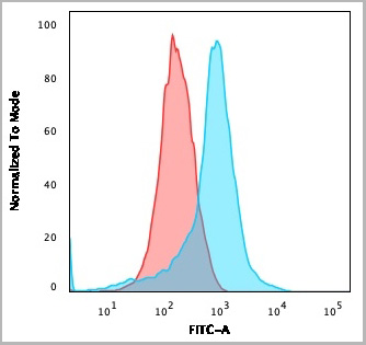
(Figure 7. Flow Cytometry analysis of MCF-7 cells using anti-RGS6 antibody (AAA19308).Overlay histogram showing MCF-7 cells stained with AAA19308 (Blue line). The cells were blocked with 10% normal goat serum. And then incubated with rabbit anti-RGS6 Antibody (AAA19308, 1μg/1x106 cells) for 30 min at 20 degree C. DyLight®488 conjugated goat anti-rabbit IgG (5-10μg/1x106 cells) was used as secondary antibody for 30 minutes at 20 degree C. Isotype control antibody (Green line) was rabbit IgG (1μg/1x106) used under the same conditions. Unlabelled sample (Red line) was also used as a control.)
FCM (Flow Cytometry)
(Figure 7. Flow Cytometry analysis of MCF-7 cells using anti-RGS6 antibody (AAA19308).Overlay histogram showing MCF-7 cells stained with AAA19308 (Blue line). The cells were blocked with 10% normal goat serum. And then incubated with rabbit anti-RGS6 Antibody (AAA19308, 1μg/1x106 cells) for 30 min at 20 degree C. DyLight®488 conjugated goat anti-rabbit IgG (5-10μg/1x106 cells) was used as secondary antibody for 30 minutes at 20 degree C. Isotype control antibody (Green line) was rabbit IgG (1μg/1x106) used under the same conditions. Unlabelled sample (Red line) was also used as a control.)
RGS6, Polyclonal Antibody (Cat# AAA19308)
Full Name
Anti-RGS6 Antibody
Gene Names
RGS6; GAP
Reactivity
Human, Mouse, Rat
Applications
WB, IHC-P, FC/FACS/FCM, EIA
Purity
Immunogen affinity purified.
Pricing
 Application Data
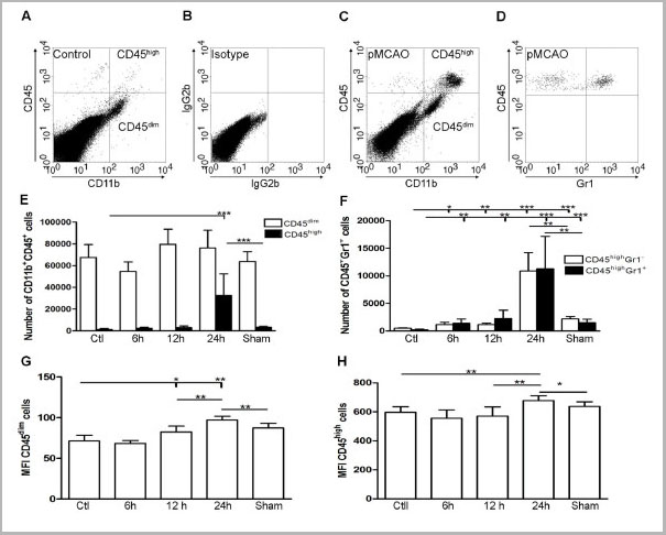
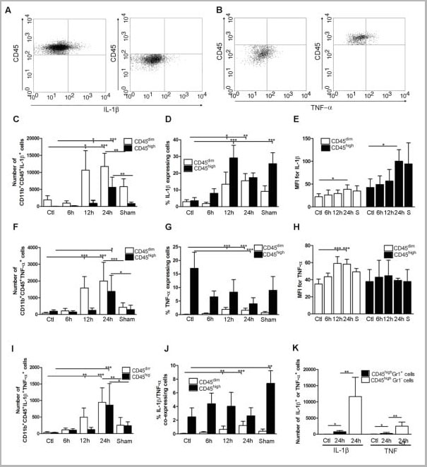
(Published customer image: Cytokine expression in segregated populations of cells following stroke. (A, B) Dot plots showing CD11b+CD45high macrophages/granulocytes (upper right quadrants) and CD11b+CD45dim microglia (bottom right quadrants) expressing IL-1beta (A) or TNF-a (B). (C-J) Bar graphs showing numbers and proportions of IL-1beta (C, D), TNF-a (F, G) and IL-1beta/TNF-a co-expressing (I, J) CD11b+CD45dim microglia and CD11b+CD45high macrophages/granulocytes in unmanipulated control mice (n = 10), in mice 6 (n = 7), 12 (n = 7), or 24 hours after pMCAO (n = 10), and in sham-operated mice 24 hours after pMCAO (n = 7). (E, H) Comparison of the MFI values for IL-1beta (E) and TNF-a (H) in viable CD11b+CD45dim microglia and CD11b+CD45high macrophages/granulocytes in unmanipulated mice, in mice 6, 12, or 24 hours after pMCAO, and in sham-operated mice 24 hours after pMCAO. Macrophages/granulocytes express significantly more IL-1beta than do microglial in unmanipulated mice, in mice 6, 12, or 24 hours after pMCAO, and in sham-operated mice 24 hours after pMCAO (E), whereas microglial cells express significantly higher levels of TNF-a than do macrophages/granulocytes at 12 h and 24 hours, and in sham-operated mice 24 hours after pMCAO (H). (K) CD11b+CD45highGr1- macrophages and not CD11b+CD45highGr1+ granulocytes are the main producers of IL-1beta and TNF-a 24 hours after pMCAO. *P < 0.05, **P < 0.01, and ***P < 0.001.From: http://www.jneuroinflammation.com/content/5/1/46.)
Application Data
(Published customer image: Cytokine expression in segregated populations of cells following stroke. (A, B) Dot plots showing CD11b+CD45high macrophages/granulocytes (upper right quadrants) and CD11b+CD45dim microglia (bottom right quadrants) expressing IL-1beta (A) or TNF-a (B). (C-J) Bar graphs showing numbers and proportions of IL-1beta (C, D), TNF-a (F, G) and IL-1beta/TNF-a co-expressing (I, J) CD11b+CD45dim microglia and CD11b+CD45high macrophages/granulocytes in unmanipulated control mice (n = 10), in mice 6 (n = 7), 12 (n = 7), or 24 hours after pMCAO (n = 10), and in sham-operated mice 24 hours after pMCAO (n = 7). (E, H) Comparison of the MFI values for IL-1beta (E) and TNF-a (H) in viable CD11b+CD45dim microglia and CD11b+CD45high macrophages/granulocytes in unmanipulated mice, in mice 6, 12, or 24 hours after pMCAO, and in sham-operated mice 24 hours after pMCAO. Macrophages/granulocytes express significantly more IL-1beta than do microglial in unmanipulated mice, in mice 6, 12, or 24 hours after pMCAO, and in sham-operated mice 24 hours after pMCAO (E), whereas microglial cells express significantly higher levels of TNF-a than do macrophages/granulocytes at 12 h and 24 hours, and in sham-operated mice 24 hours after pMCAO (H). (K) CD11b+CD45highGr1- macrophages and not CD11b+CD45highGr1+ granulocytes are the main producers of IL-1beta and TNF-a 24 hours after pMCAO. *P < 0.05, **P < 0.01, and ***P < 0.001.From: http://www.jneuroinflammation.com/content/5/1/46.)
CD45, Monoclonal Antibody (Cat# AAA11896)
Full Name
RAT ANTI MOUSE CD45
Gene Names
Ptprc; loc; B220; Cd45; L-CA; Ly-5; T200; CD45R; Lyt-4
Applications
Immunohistochemistry, Flow Cytometry, Immunofluorescence, Immunoprecipitation
Pricing
 SDS-PAGE
(SDS-PAGE analysis of Prod.(AAA14516): Lane 1: MW marker, Lane 2: 1µg , Lane 3: 2µg BCMA (mouse) monoclonal antibody (Vicky-2), integrity of antibody is shown as both light chain and heavy chain are present.)
SDS-PAGE
(SDS-PAGE analysis of Prod.(AAA14516): Lane 1: MW marker, Lane 2: 1µg , Lane 3: 2µg BCMA (mouse) monoclonal antibody (Vicky-2), integrity of antibody is shown as both light chain and heavy chain are present.)
BCMA, Monoclonal Antibody (Cat# AAA14516)
Full Name
BCMA (mouse) monoclonal antibody (Vicky-2)
Gene Names
Tnfrsf17; BCM; BCMA; Tnfrsf13; Tnfrsf13a
Reactivity
Mouse
Applications
Flow Cytometry, Immunoprecipitation, Western Blot
Purity
Protein G-affinity purified.
Pricing
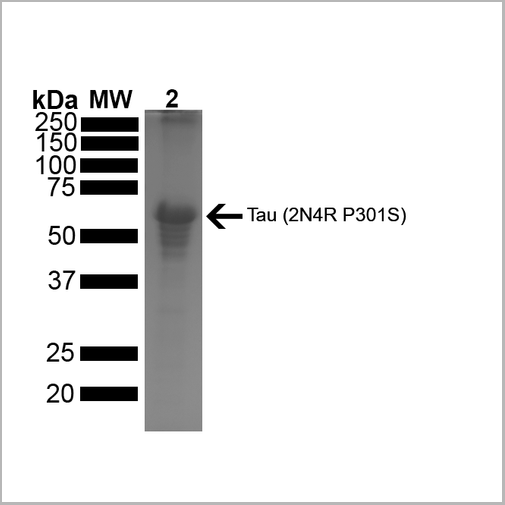 SDS-PAGE
(SDS-PAGE of ~67 kDa Human Tau Protein 2N4R P301S Preformed Fibrils. Lane 1: MW Ladder. Lane 2: Tau Protein Preformed Fibrils)
SDS-PAGE
(SDS-PAGE of ~67 kDa Human Tau Protein 2N4R P301S Preformed Fibrils. Lane 1: MW Ladder. Lane 2: Tau Protein Preformed Fibrils)
Tau441, Active Protein (Cat# AAA27663)
Full Name
Active Human Recombinant Tau441 (2N4R), P301S Mutant Protein Preformed Fibrils
Gene Names
MAPT; TAU; MSTD; PPND; DDPAC; MAPTL; MTBT1; MTBT2; FTDP-17; PPP1R103
Applications
Western Blot
Purity
>95%
Ion-Exchange Purified
Ion-Exchange Purified
Pricing


 IP (Immunoprecipitation)
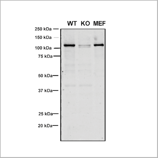
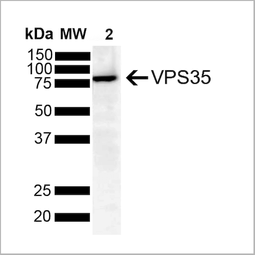
(Immunoprecipitation analysis using Mouse Anti-VPS35 Monoclonal Antibody, Clone 11H10. Tissue: embryonic fibroblast. Species: Mouse. Primary Antibody: Mouse Anti-VPS35 Monoclonal Antibody. Three amounts of (3, 1 and 0.3 ug) were non-covalently coupled to 10uL of A/G sepharose beads for 1 hour at 4 degree C and next incubated with 250ug of MEF lysate for 2 hours at 4 degree C.)
IP (Immunoprecipitation)
(Immunoprecipitation analysis using Mouse Anti-VPS35 Monoclonal Antibody, Clone 11H10. Tissue: embryonic fibroblast. Species: Mouse. Primary Antibody: Mouse Anti-VPS35 Monoclonal Antibody. Three amounts of (3, 1 and 0.3 ug) were non-covalently coupled to 10uL of A/G sepharose beads for 1 hour at 4 degree C and next incubated with 250ug of MEF lysate for 2 hours at 4 degree C.)
VPS35, Monoclonal Antibody (Cat# AAA27707)
Full Name
VPS35 Antibody, Clone 11H10: PerCP
Gene Names
VPS35; MEM3; PARK17
Reactivity
Human, Mouse, Rat
Applications
Western Blot, Immunoprecipitation
Purity
Protein G Purified
Pricing


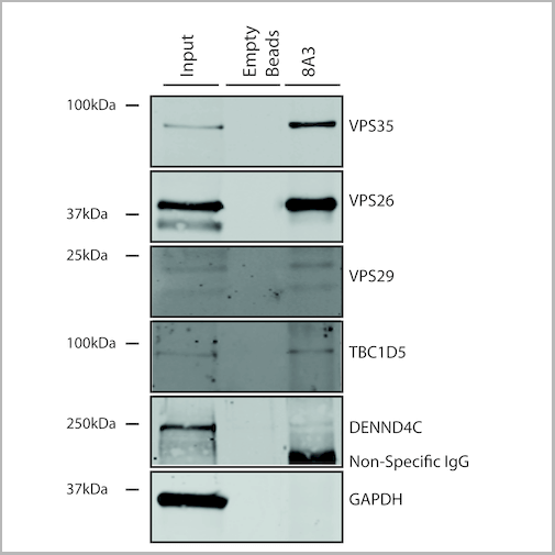
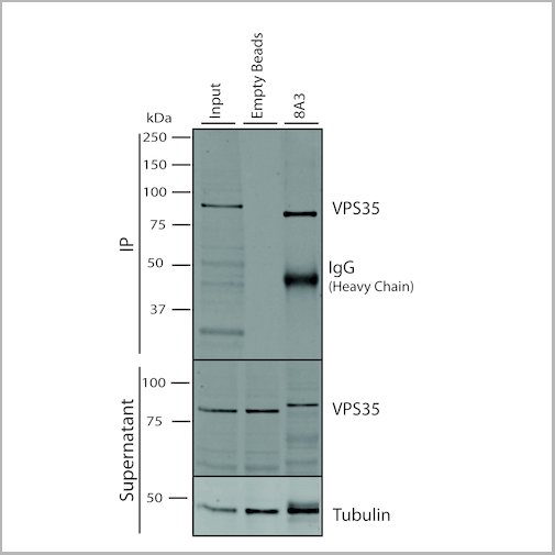

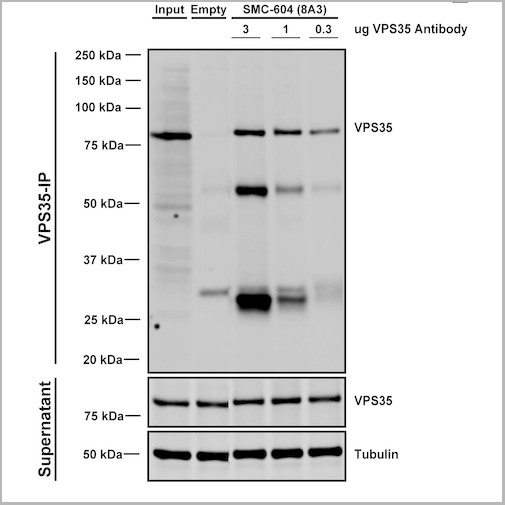
 IP (Immunoprecipitation)
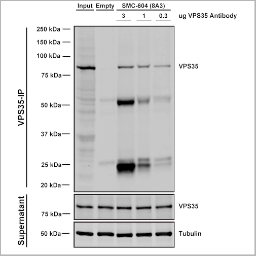
(Immunoprecipitation analysis using Mouse Anti-VPS35 Monoclonal Antibody, Clone 8A3. Tissue: embryonic fibroblast. Species: Mouse. Primary Antibody: Mouse Anti-VPS35 Monoclonal Antibody. Three amounts of (3, 1 and 0.3 ug) were non-covalently coupled to 10uL of A/G sepharose beads for 1 hour at 4 degree C and next incubated with 250ug of MEF lysate for 2 hours at 4 degree C.)
IP (Immunoprecipitation)
(Immunoprecipitation analysis using Mouse Anti-VPS35 Monoclonal Antibody, Clone 8A3. Tissue: embryonic fibroblast. Species: Mouse. Primary Antibody: Mouse Anti-VPS35 Monoclonal Antibody. Three amounts of (3, 1 and 0.3 ug) were non-covalently coupled to 10uL of A/G sepharose beads for 1 hour at 4 degree C and next incubated with 250ug of MEF lysate for 2 hours at 4 degree C.)
VPS35, Monoclonal Antibody (Cat# AAA27689)
Full Name
VPS35 Antibody, Clone 8A3: PerCP
Gene Names
VPS35; MEM3; PARK17
Reactivity
Human, Mouse, Rat
Applications
Western Blot, Immunocytochemistry, Immunofluorescence, Immunoprecipitation
Purity
Protein G Purified
Pricing
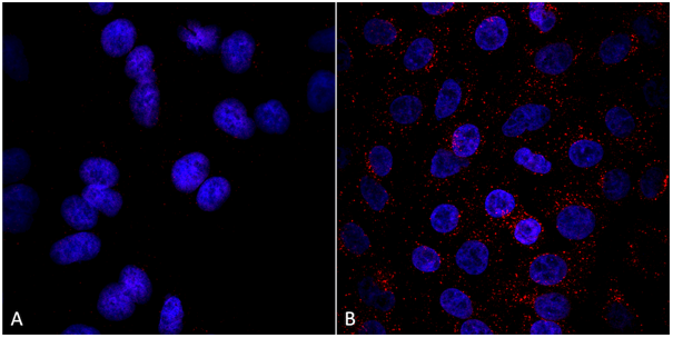
 IF (Immunofluorescence)
(Immunofluorescent analysis of HepG-2 cells using AAA26914 at a dilution of 1:100 and Alexa Fluor 488-congugated AffiniPure Goat Anti-Rabbit IgG(H+L))
IF (Immunofluorescence)
(Immunofluorescent analysis of HepG-2 cells using AAA26914 at a dilution of 1:100 and Alexa Fluor 488-congugated AffiniPure Goat Anti-Rabbit IgG(H+L))
ZNRF3, Polyclonal Antibody (Cat# AAA26914)
Full Name
ZNRF3 Antibody
Gene Names
ZNRF3; RNF203; BK747E2.3
Reactivity
Human
Applications
Immunofluorescence
Purity
>95%, Protein G purified
Pricing
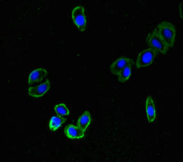
 IF (Immunofluorescence)
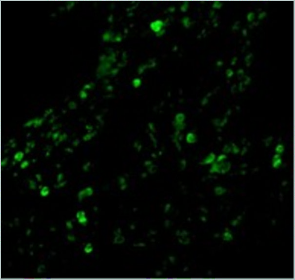
(Immunofluorescence Validation of cIAP in Human Lung Tissue Immunofluorescent analysis of 4% paraformaldehydefixed human lung tissue abeling cIAP with 3325 at 20 ug/mL, followed by goat anti-rabbit IgG secondary antibody at 1/500 dilution (green).)
IF (Immunofluorescence)
(Immunofluorescence Validation of cIAP in Human Lung Tissue Immunofluorescent analysis of 4% paraformaldehydefixed human lung tissue abeling cIAP with 3325 at 20 ug/mL, followed by goat anti-rabbit IgG secondary antibody at 1/500 dilution (green).)
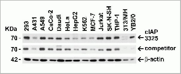
cIAP, Polyclonal Antibody (Cat# AAA10954)
Full Name
cIAP Antibody
Gene Names
BIRC2; API1; MIHB; HIAP2; RNF48; cIAP1; Hiap-2; c-IAP1
Reactivity
Human, Mouse
Applications
Western Blot, Immunohistochemistry, Immunofluorescence
Purity
cIAP Antibody is affinity chromatography purified via peptide column.
Pricing


 IP (Immunoprecipitation)
(Immunoprecipitation analysis using Mouse Anti-VPS35 Monoclonal Antibody, Clone 8A3. Tissue: embryonic fibroblast. Species: Mouse. Primary Antibody: Mouse Anti-VPS35 Monoclonal Antibody. Three amounts of (3, 1 and 0.3 ug) were non-covalently coupled to 10uL of A/G sepharose beads for 1 hour at 4 degree C and next incubated with 250ug of MEF lysate for 2 hours at 4 degree C.)
IP (Immunoprecipitation)
(Immunoprecipitation analysis using Mouse Anti-VPS35 Monoclonal Antibody, Clone 8A3. Tissue: embryonic fibroblast. Species: Mouse. Primary Antibody: Mouse Anti-VPS35 Monoclonal Antibody. Three amounts of (3, 1 and 0.3 ug) were non-covalently coupled to 10uL of A/G sepharose beads for 1 hour at 4 degree C and next incubated with 250ug of MEF lysate for 2 hours at 4 degree C.)
VPS35, Monoclonal Antibody (Cat# AAA27682)
Full Name
VPS35 Antibody, Clone 8A3
Gene Names
VPS35; MEM3; PARK17
Reactivity
Human, Mouse, Rat
Applications
Western Blot, Immunocytochemistry, Immunofluorescence, Immunoprecipitation
Purity
Protein G Purified
Pricing
 Application Data
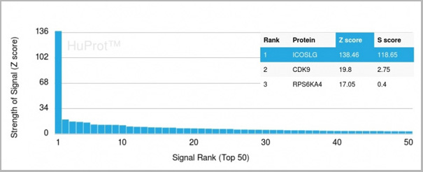
(Analysis of Protein Array containing more than 19,000 full-length human proteins using ICOS-L Mouse Monoclonal Antibody (ICOSL/3111). Z- and S- Score: The Z-score represents the strength of a signal that a monoclonal antibody (MAb) (in combination with a fluorescently-tagged anti-IgG secondary antibody) produces when binding to a particular protein on the HuProtTM array. Z-scores are described in units of standard deviations (SD’s) above the mean value of all signals generated on that array. If targets on HuProtTM are arranged in descending order of the Z-score, the S-score is the difference (also in units of SD’s) between the Z-score. S-score therefore represents the relative target specificity of a MAb to its intended target. A MAb is considered to specific to its intended target, if the MAb has an S-score of at least 2.5. For example, if a MAb binds to protein X with a Z-score of 43 and to protein Y with a Z-score of 14, then the S-score for the binding of that MAb to protein X is equal to 29.)
Application Data
(Analysis of Protein Array containing more than 19,000 full-length human proteins using ICOS-L Mouse Monoclonal Antibody (ICOSL/3111). Z- and S- Score: The Z-score represents the strength of a signal that a monoclonal antibody (MAb) (in combination with a fluorescently-tagged anti-IgG secondary antibody) produces when binding to a particular protein on the HuProtTM array. Z-scores are described in units of standard deviations (SD’s) above the mean value of all signals generated on that array. If targets on HuProtTM are arranged in descending order of the Z-score, the S-score is the difference (also in units of SD’s) between the Z-score. S-score therefore represents the relative target specificity of a MAb to its intended target. A MAb is considered to specific to its intended target, if the MAb has an S-score of at least 2.5. For example, if a MAb binds to protein X with a Z-score of 43 and to protein Y with a Z-score of 14, then the S-score for the binding of that MAb to protein X is equal to 29.)
ICOS-L/ICOS Ligand/B7RP-1 (Immuno-Oncology Target), Monoclonal Antibody (Cat# AAA23916)
Full Name
ICOS-L/ICOS Ligand/B7RP-1 (Immuno-Oncology Target)
Gene Names
ICOSLG; B7h; B7H2; GL50; B7-H2; B7RP1; CD275; ICOSL; LICOS; B7RP-1; ICOS-L
Reactivity
Human
Applications
Flow Cytometry, Immunofluorescence, Immunohistochemistry
Purity
Purified Ab with BSA and Azide at 200ug/ml
Pricing