Filters
Clonality
Type
Reactivity
Gene Name
Isotype
Host
Application
Clone
26 results for " Cullin" - showing 1-26


 FCM (Flow Cytometry)
(Flow cytometric analysis of Hela cells with Cullin 4a antibody at 1/100 dilution (green) compared with an unlabelled control (cells without incubation with primary antibody; red). Alexa Fluor 488-conjugated goat anti-rabbit IgG was used as the secondary antibody.)
FCM (Flow Cytometry)
(Flow cytometric analysis of Hela cells with Cullin 4a antibody at 1/100 dilution (green) compared with an unlabelled control (cells without incubation with primary antibody; red). Alexa Fluor 488-conjugated goat anti-rabbit IgG was used as the secondary antibody.)
Cullin 4a, Monoclonal Antibody (Cat# AAA30417)
Full Name
Cullin 4a Antibody
Reactivity
Human
Applications
Western Blot, Immunocytochemistry, Immunohistochemistry, Flow Cytometry
Purity
ProA affinity purified
Pricing
Cullin-1, Recombinant Protein (Cat# AAA10882)
Full Name
Recombinant Human Cullin-1
Purity
Greater than 95% as determined by SDS-PAGE.
Pricing


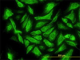
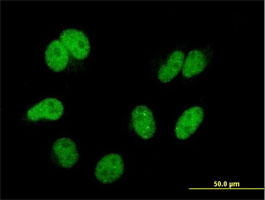
 IF (Immunofluorescence)
(Immunofluorescence of monoclonal antibody to CAND1 on HeLa cell. [antibody concentration 10 ug/ml])
IF (Immunofluorescence)
(Immunofluorescence of monoclonal antibody to CAND1 on HeLa cell. [antibody concentration 10 ug/ml])
CAND1, Monoclonal Antibody (Cat# AAA26649)
Full Name
CAND1 (Cullin-Associated and Neddylation-dissociated 1, DKFZp434M1414, FLJ10114, FLJ10929, FLJ38691, FLJ90441, KIAA0829, TIP120, TIP120A) (PE)
Gene Names
CAND1; TIP120; TIP120A
Applications
Immunofluorescence, Western Blot
Purity
Purified
Pricing


 IF (Immunofluorescence)
(Immunofluorescence of monoclonal antibody to CAND1 on HeLa cell. [antibody concentration 10 ug/ml])
IF (Immunofluorescence)
(Immunofluorescence of monoclonal antibody to CAND1 on HeLa cell. [antibody concentration 10 ug/ml])
CAND1, Monoclonal Antibody (Cat# AAA26384)
Full Name
CAND1 (Cullin-Associated and Neddylation-dissociated 1, DKFZp434M1414, FLJ10114, FLJ10929, FLJ38691, FLJ90441, KIAA0829, TIP120, TIP120A) (FITC)
Gene Names
CAND1; TIP120; TIP120A
Applications
Immunofluorescence, Western Blot
Purity
Purified
Pricing


 IF (Immunofluorescence)
(Immunofluorescence of monoclonal antibody to CAND1 on HeLa cell. [antibody concentration 10 ug/ml])
IF (Immunofluorescence)
(Immunofluorescence of monoclonal antibody to CAND1 on HeLa cell. [antibody concentration 10 ug/ml])
CAND1, Monoclonal Antibody (Cat# AAA25985)
Full Name
CAND1 (Cullin-Associated and Neddylation-dissociated 1, DKFZp434M1414, FLJ10114, FLJ10929, FLJ38691, FLJ90441, KIAA0829, TIP120, TIP120A) (AP)
Gene Names
CAND1; TIP120; TIP120A
Applications
Western Blot
Purity
Purified
Pricing


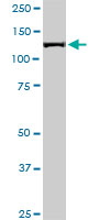
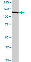
 WB (Western Blot)
(CAND1 monoclonal antibody (M04), clone 4D10. Western Blot analysis of CAND1 expression in Raw 264.7.)
WB (Western Blot)
(CAND1 monoclonal antibody (M04), clone 4D10. Western Blot analysis of CAND1 expression in Raw 264.7.)
CAND1, Monoclonal Antibody (Cat# AAA26233)
Full Name
CAND1 (Cullin-Associated and Neddylation-dissociated 1, DKFZp434M1414, FLJ10114, FLJ10929, FLJ38691, FLJ90441, KIAA0829, TIP120, TIP120A) (Biotin)
Gene Names
CAND1; TIP120; TIP120A
Applications
Immunofluorescence, Western Blot
Purity
Purified
Pricing


 IF (Immunofluorescence)
(Immunofluorescence of monoclonal antibody to CAND1 on HeLa cell. [antibody concentration 10 ug/ml])
IF (Immunofluorescence)
(Immunofluorescence of monoclonal antibody to CAND1 on HeLa cell. [antibody concentration 10 ug/ml])
CAND1, Monoclonal Antibody (Cat# AAA26517)
Full Name
CAND1 (Cullin-Associated and Neddylation-dissociated 1, DKFZp434M1414, FLJ10114, FLJ10929, FLJ38691, FLJ90441, KIAA0829, TIP120, TIP120A) (HRP)
Gene Names
CAND1; TIP120; TIP120A
Applications
Immunofluorescence, Western Blot
Purity
Purified
Pricing


 WB (Western Blot)
(CAND1 monoclonal antibody (M04), clone 4D10. Western Blot analysis of CAND1 expression in Raw 264.7.)
WB (Western Blot)
(CAND1 monoclonal antibody (M04), clone 4D10. Western Blot analysis of CAND1 expression in Raw 264.7.)
CAND1, Monoclonal Antibody (Cat# AAA26499)
Full Name
CAND1 (Cullin-Associated and Neddylation-dissociated 1, DKFZp434M1414, FLJ10114, FLJ10929, FLJ38691, FLJ90441, KIAA0829, TIP120, TIP120A) (HRP)
Gene Names
CAND1; TIP120; TIP120A
Applications
Immunofluorescence, Western Blot
Purity
Purified
Pricing


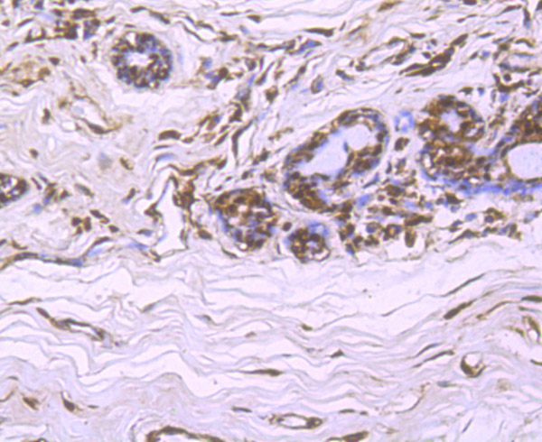
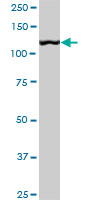
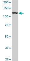
 WB (Western Blot)
(DCUN1D1 monoclonal antibody, Western Blot analysis of DCUN1D1 expression in HepG2.)
WB (Western Blot)
(DCUN1D1 monoclonal antibody, Western Blot analysis of DCUN1D1 expression in HepG2.)
DCUN1D1, Monoclonal Antibody (Cat# AAA25369)
Full Name
DCUN1D1 (DCN1-like Protein 1, DCUN1 Domain-containing Protein 1, Defective in Cullin Neddylation Protein 1-like Protein 1, Squamous Cell Carcinoma-related Oncogene, DCUN1L1, RP42, SCCRO) (HRP)
Gene Names
DCUN1D1; RP42; SCRO; Tes3; DCNL1; SCCRO; DCUN1L1
Reactivity
Human, Mouse, Rat
Applications
Western Blot
Purity
Purified by Protein A Affinity Chromatography.
Pricing


 WB (Western Blot)
(DCUN1D1 monoclonal antibody, Western Blot analysis of DCUN1D1 expression in HepG2.)
WB (Western Blot)
(DCUN1D1 monoclonal antibody, Western Blot analysis of DCUN1D1 expression in HepG2.)
DCUN1D1, Monoclonal Antibody (Cat# AAA25664)
Full Name
DCUN1D1 (DCN1-like Protein 1, DCUN1 Domain-containing Protein 1, Defective in Cullin Neddylation Protein 1-like Protein 1, Squamous Cell Carcinoma-related Oncogene, DCUN1L1, RP42, SCCRO) (PE)
Gene Names
DCUN1D1; RP42; SCRO; Tes3; DCNL1; SCCRO; DCUN1L1
Reactivity
Human, Mouse, Rat
Applications
Immunofluorescence, Western Blot
Purity
Purified by Protein A Affinity Chromatography.
Pricing


 WB (Western Blot)
(CAND1 monoclonal antibody (M04), clone 4D10. Western Blot analysis of CAND1 expression in Raw 264.7.)
WB (Western Blot)
(CAND1 monoclonal antibody (M04), clone 4D10. Western Blot analysis of CAND1 expression in Raw 264.7.)
CAND1, Monoclonal Antibody (Cat# AAA25967)
Full Name
CAND1 (Cullin-Associated and Neddylation-dissociated 1, DKFZp434M1414, FLJ10114, FLJ10929, FLJ38691, FLJ90441, KIAA0829, TIP120, TIP120A) (AP)
Gene Names
CAND1; TIP120; TIP120A
Applications
Western Blot
Purity
Purified
Pricing


 WB (Western Blot)
(CAND1 monoclonal antibody (M04), clone 4D10. Western Blot analysis of CAND1 expression in Raw 264.7.)
WB (Western Blot)
(CAND1 monoclonal antibody (M04), clone 4D10. Western Blot analysis of CAND1 expression in Raw 264.7.)
CAND1, Monoclonal Antibody (Cat# AAA26100)
Full Name
CAND1 (Cullin-Associated and Neddylation-dissociated 1, DKFZp434M1414, FLJ10114, FLJ10929, FLJ38691, FLJ90441, KIAA0829, TIP120, TIP120A) (APC)
Gene Names
CAND1; TIP120; TIP120A
Applications
EIA, IF, WB
Purity
Purified
Pricing


 IF (Immunofluorescence)
(Immunofluorescence of monoclonal antibody to CAND1 on HeLa cell. [antibody concentration 10 ug/ml])
IF (Immunofluorescence)
(Immunofluorescence of monoclonal antibody to CAND1 on HeLa cell. [antibody concentration 10 ug/ml])
CAND1, Monoclonal Antibody (Cat# AAA26251)
Full Name
CAND1 (Cullin-Associated and Neddylation-dissociated 1, DKFZp434M1414, FLJ10114, FLJ10929, FLJ38691, FLJ90441, KIAA0829, TIP120, TIP120A) (Biotin)
Gene Names
CAND1; TIP120; TIP120A
Applications
Immunofluorescence, Western Blot
Purity
Purified
Pricing


 WB (Western Blot)
(CAND1 monoclonal antibody (M04), clone 4D10. Western Blot analysis of CAND1 expression in Raw 264.7.)
WB (Western Blot)
(CAND1 monoclonal antibody (M04), clone 4D10. Western Blot analysis of CAND1 expression in Raw 264.7.)
CAND1, Monoclonal Antibody (Cat# AAA26631)
Full Name
CAND1 (Cullin-Associated and Neddylation-dissociated 1, DKFZp434M1414, FLJ10114, FLJ10929, FLJ38691, FLJ90441, KIAA0829, TIP120, TIP120A) (PE)
Gene Names
CAND1; TIP120; TIP120A
Applications
Immunofluorescence, Western Blot
Purity
Purified
Pricing


 IF (Immunofluorescence)
(Immunofluorescence of monoclonal antibody to CAND1 on HeLa cell. [antibody concentration 10 ug/ml])
IF (Immunofluorescence)
(Immunofluorescence of monoclonal antibody to CAND1 on HeLa cell. [antibody concentration 10 ug/ml])
CAND1, Monoclonal Antibody (Cat# AAA26118)
Full Name
CAND1 (Cullin-Associated and Neddylation-dissociated 1, DKFZp434M1414, FLJ10114, FLJ10929, FLJ38691, FLJ90441, KIAA0829, TIP120, TIP120A) (APC)
Gene Names
CAND1; TIP120; TIP120A
Applications
Immunofluorescence, Western Blot
Purity
Purified
Pricing


 WB (Western Blot)
(DCUN1D1 monoclonal antibody, Western Blot analysis of DCUN1D1 expression in HepG2.)
WB (Western Blot)
(DCUN1D1 monoclonal antibody, Western Blot analysis of DCUN1D1 expression in HepG2.)
DCUN1D1, Monoclonal Antibody (Cat# AAA24778)
Full Name
DCUN1D1 (DCN1-like Protein 1, DCUN1 Domain-containing Protein 1, Defective in Cullin Neddylation Protein 1-like Protein 1, Squamous Cell Carcinoma-related Oncogene, DCUN1L1, RP42, SCCRO) (Biotin)
Gene Names
DCUN1D1; RP42; SCRO; Tes3; DCNL1; SCCRO; DCUN1L1
Reactivity
Human, Mouse, Rat
Applications
EIA, IF, WB
Purity
Purified by Protein A Affinity Chromatography.
Pricing


 WB (Western Blot)
(DCUN1D1 monoclonal antibody, Western Blot analysis of DCUN1D1 expression in HepG2.)
WB (Western Blot)
(DCUN1D1 monoclonal antibody, Western Blot analysis of DCUN1D1 expression in HepG2.)
DCUN1D1, Monoclonal Antibody (Cat# AAA25075)
Full Name
DCUN1D1 (DCN1-like Protein 1, DCUN1 Domain-containing Protein 1, Defective in Cullin Neddylation Protein 1-like Protein 1, Squamous Cell Carcinoma-related Oncogene, DCUN1L1, RP42, SCCRO) (FITC)
Gene Names
DCUN1D1; RP42; SCRO; Tes3; DCNL1; SCCRO; DCUN1L1
Reactivity
Human, Mouse, Rat
Applications
EIA, IF, WB
Purity
Purified by Protein A Affinity Chromatography.
Pricing


 IF (Immunofluorescence)
(Immunofluorescence analysis of U2OS cells using CUL4B antibody.)
IF (Immunofluorescence)
(Immunofluorescence analysis of U2OS cells using CUL4B antibody.)
CUL4B, Polyclonal Antibody (Cat# AAA28142)
Full Name
CUL4B Polyclonal Antibody
Gene Names
CUL4B; SFM2; MRXSC; CUL-4B; MRXHF2; MRXS15
Reactivity
Human, Mouse, Rat
Applications
WB, IHC, IF
Purity
Affinity Purification
Pricing


 WB (Western Blot)
(DCUN1D1 monoclonal antibody, Western Blot analysis of DCUN1D1 expression in HepG2.)
WB (Western Blot)
(DCUN1D1 monoclonal antibody, Western Blot analysis of DCUN1D1 expression in HepG2.)
DCUN1D1, Monoclonal Antibody (Cat# AAA24483)
Full Name
DCUN1D1 (DCN1-like Protein 1, DCUN1 Domain-containing Protein 1, Defective in Cullin Neddylation Protein 1-like Protein 1, Squamous Cell Carcinoma-related Oncogene, DCUN1L1, RP42, SCCRO) APC
Gene Names
DCUN1D1; RP42; SCRO; Tes3; DCNL1; SCCRO; DCUN1L1
Reactivity
Human, Mouse, Rat
Applications
EIA, IF, WB
Purity
Purified by Protein A Affinity Chromatography.
Pricing


 WB (Western Blot)
(CAND1 monoclonal antibody (M04), clone 4D10. Western Blot analysis of CAND1 expression in Raw 264.7.)
WB (Western Blot)
(CAND1 monoclonal antibody (M04), clone 4D10. Western Blot analysis of CAND1 expression in Raw 264.7.)
CAND1, Monoclonal Antibody (Cat# AAA26366)
Full Name
CAND1 (Cullin-Associated and Neddylation-dissociated 1, DKFZp434M1414, FLJ10114, FLJ10929, FLJ38691, FLJ90441, KIAA0829, TIP120, TIP120A) (FITC)
Gene Names
CAND1; TIP120; TIP120A
Applications
EIA, IF, WB
Purity
Purified
Pricing


 WB (Western Blot)
(DCUN1D1 monoclonal antibody, Western Blot analysis of DCUN1D1 expression in HepG2.)
WB (Western Blot)
(DCUN1D1 monoclonal antibody, Western Blot analysis of DCUN1D1 expression in HepG2.)
DCUN1D1, Monoclonal Antibody (Cat# AAA24187)
Full Name
DCUN1D1 (DCN1-like Protein 1, DCUN1 Domain-containing Protein 1, Defective in Cullin Neddylation Protein 1-like Protein 1, Squamous Cell Carcinoma-related Oncogene, DCUN1L1, RP42, SCCRO) (AP)
Gene Names
DCUN1D1; RP42; SCRO; Tes3; DCNL1; SCCRO; DCUN1L1
Reactivity
Human, Mouse, Rat
Applications
EIA, WB
Purity
Purified by Protein A Affinity Chromatography.
Pricing


 Application Data
(Published customer image: Mouse anti V5 tag antibody, clone SV5-Pk1 used for the detection of V5 tagged WEEV_nsP3 protein by western blotting and immunofluorescenceImage caption: WEEV nsP3 interaction with host IKKbeta. A) U87MGs were transfected in a 6-well plate with 5 ug of pUC19 and WEEV_nsP3_HA for 24 hours. Cell lysates were resolved using SDS-PAGE and subsequently immunoblotted with V5 antibody and beta-actin served as a loading control. B) U87MGs were transfected with WEEV_nsP3_V5; cells were fixed after 24 hours and stained with antibodies against the endogenous IKKbeta and the V5 tag. Cells were incubated with appropriate secondary Alexa Fluor antibodies and the nuclei stained with DAPI. Co-localization of IKKbeta with WEEV_nsP3_V5 (yellow) was observed as shown by the arrows. B) Panels E -H serve as an example of transfected cells in a given field of view that show co-localization of IKKbeta and WEEV_nsP3_V5 24 hours post transfection. Panels I-L represent magnified images of other cells showing co-localization of IKKbeta and WEEV_nsP3_V5. Panel M is a magnified image of panel L. The co-localization was confirmed by Z-stack analysis. Co-localization was calculated to be approximately in 61% of cells (163 cells were counted of which 44% demonstrated expression of nsP3. Of those cells that expressed nsP3, 61% showed co-localization of both proteins). Images were taken using Nikon Eclipse TE2000-U at 60x magnification and are representative of 2 independent experiments.From: Amaya M, Voss K, Sampey G, Senina S, de la Fuente C, et al. (2014) The Role of IKKbeta in Venezuelan Equine Encephalitis Virus Infection. PLoS ONE 9(2): e86745.)
Application Data
(Published customer image: Mouse anti V5 tag antibody, clone SV5-Pk1 used for the detection of V5 tagged WEEV_nsP3 protein by western blotting and immunofluorescenceImage caption: WEEV nsP3 interaction with host IKKbeta. A) U87MGs were transfected in a 6-well plate with 5 ug of pUC19 and WEEV_nsP3_HA for 24 hours. Cell lysates were resolved using SDS-PAGE and subsequently immunoblotted with V5 antibody and beta-actin served as a loading control. B) U87MGs were transfected with WEEV_nsP3_V5; cells were fixed after 24 hours and stained with antibodies against the endogenous IKKbeta and the V5 tag. Cells were incubated with appropriate secondary Alexa Fluor antibodies and the nuclei stained with DAPI. Co-localization of IKKbeta with WEEV_nsP3_V5 (yellow) was observed as shown by the arrows. B) Panels E -H serve as an example of transfected cells in a given field of view that show co-localization of IKKbeta and WEEV_nsP3_V5 24 hours post transfection. Panels I-L represent magnified images of other cells showing co-localization of IKKbeta and WEEV_nsP3_V5. Panel M is a magnified image of panel L. The co-localization was confirmed by Z-stack analysis. Co-localization was calculated to be approximately in 61% of cells (163 cells were counted of which 44% demonstrated expression of nsP3. Of those cells that expressed nsP3, 61% showed co-localization of both proteins). Images were taken using Nikon Eclipse TE2000-U at 60x magnification and are representative of 2 independent experiments.From: Amaya M, Voss K, Sampey G, Senina S, de la Fuente C, et al. (2014) The Role of IKKbeta in Venezuelan Equine Encephalitis Virus Infection. PLoS ONE 9(2): e86745.)
V5-TAG, Monoclonal Antibody (Cat# AAA12081)
Full Name
MOUSE ANTI V5-TAG:HRP
Applications
Western Blot
Pricing


 Application Data
(Published customer image: Mouse anti V5 tag antibody, clone SV5-Pk1 used for the detection of V5 tagged WEEV_nsP3 protein by western blotting and immunofluorescenceImage caption: WEEV nsP3 interaction with host IKKbeta. A) U87MGs were transfected in a 6-well plate with 5 ug of pUC19 and WEEV_nsP3_HA for 24 hours. Cell lysates were resolved using SDS-PAGE and subsequently immunoblotted with V5 antibody and beta-actin served as a loading control. B) U87MGs were transfected with WEEV_nsP3_V5; cells were fixed after 24 hours and stained with antibodies against the endogenous IKKbeta and the V5 tag. Cells were incubated with appropriate secondary Alexa Fluor antibodies and the nuclei stained with DAPI. Co-localization of IKKbeta with WEEV_nsP3_V5 (yellow) was observed as shown by the arrows. B) Panels E -H serve as an example of transfected cells in a given field of view that show co-localization of IKKbeta and WEEV_nsP3_V5 24 hours post transfection. Panels I-L represent magnified images of other cells showing co-localization of IKKbeta and WEEV_nsP3_V5. Panel M is a magnified image of panel L. The co-localization was confirmed by Z-stack analysis. Co-localization was calculated to be approximately in 61% of cells (163 cells were counted of which 44% demonstrated expression of nsP3. Of those cells that expressed nsP3, 61% showed co-localization of both proteins). Images were taken using Nikon Eclipse TE2000-U at 60x magnification and are representative of 2 independent experiments.From: Amaya M, Voss K, Sampey G, Senina S, de la Fuente C, et al. (2014) The Role of IKKbeta in Venezuelan Equine Encephalitis Virus Infection. PLoS ONE 9(2): e86745.)
Application Data
(Published customer image: Mouse anti V5 tag antibody, clone SV5-Pk1 used for the detection of V5 tagged WEEV_nsP3 protein by western blotting and immunofluorescenceImage caption: WEEV nsP3 interaction with host IKKbeta. A) U87MGs were transfected in a 6-well plate with 5 ug of pUC19 and WEEV_nsP3_HA for 24 hours. Cell lysates were resolved using SDS-PAGE and subsequently immunoblotted with V5 antibody and beta-actin served as a loading control. B) U87MGs were transfected with WEEV_nsP3_V5; cells were fixed after 24 hours and stained with antibodies against the endogenous IKKbeta and the V5 tag. Cells were incubated with appropriate secondary Alexa Fluor antibodies and the nuclei stained with DAPI. Co-localization of IKKbeta with WEEV_nsP3_V5 (yellow) was observed as shown by the arrows. B) Panels E -H serve as an example of transfected cells in a given field of view that show co-localization of IKKbeta and WEEV_nsP3_V5 24 hours post transfection. Panels I-L represent magnified images of other cells showing co-localization of IKKbeta and WEEV_nsP3_V5. Panel M is a magnified image of panel L. The co-localization was confirmed by Z-stack analysis. Co-localization was calculated to be approximately in 61% of cells (163 cells were counted of which 44% demonstrated expression of nsP3. Of those cells that expressed nsP3, 61% showed co-localization of both proteins). Images were taken using Nikon Eclipse TE2000-U at 60x magnification and are representative of 2 independent experiments.From: Amaya M, Voss K, Sampey G, Senina S, de la Fuente C, et al. (2014) The Role of IKKbeta in Venezuelan Equine Encephalitis Virus Infection. PLoS ONE 9(2): e86745.)
V5-TAG, Monoclonal Antibody (Cat# AAA11864)
Full Name
MOUSE ANTI V5-TAG:FITC
Applications
Immunofluorescence
Pricing


 Application Data
(Published customer image: Mouse anti V5 tag antibody, clone SV5-Pk1 used for the detection of V5 tagged WEEV_nsP3 protein by western blotting and immunofluorescenceImage caption: WEEV nsP3 interaction with host IKKbeta. A) U87MGs were transfected in a 6-well plate with 5 ug of pUC19 and WEEV_nsP3_HA for 24 hours. Cell lysates were resolved using SDS-PAGE and subsequently immunoblotted with V5 antibody and beta-actin served as a loading control. B) U87MGs were transfected with WEEV_nsP3_V5; cells were fixed after 24 hours and stained with antibodies against the endogenous IKKbeta and the V5 tag. Cells were incubated with appropriate secondary Alexa Fluor antibodies and the nuclei stained with DAPI. Co-localization of IKKbeta with WEEV_nsP3_V5 (yellow) was observed as shown by the arrows. B) Panels E -H serve as an example of transfected cells in a given field of view that show co-localization of IKKbeta and WEEV_nsP3_V5 24 hours post transfection. Panels I-L represent magnified images of other cells showing co-localization of IKKbeta and WEEV_nsP3_V5. Panel M is a magnified image of panel L. The co-localization was confirmed by Z-stack analysis. Co-localization was calculated to be approximately in 61% of cells (163 cells were counted of which 44% demonstrated expression of nsP3. Of those cells that expressed nsP3, 61% showed co-localization of both proteins). Images were taken using Nikon Eclipse TE2000-U at 60x magnification and are representative of 2 independent experiments.From: Amaya M, Voss K, Sampey G, Senina S, de la Fuente C, et al. (2014) The Role of IKKbeta in Venezuelan Equine Encephalitis Virus Infection. PLoS ONE 9(2): e86745.)
Application Data
(Published customer image: Mouse anti V5 tag antibody, clone SV5-Pk1 used for the detection of V5 tagged WEEV_nsP3 protein by western blotting and immunofluorescenceImage caption: WEEV nsP3 interaction with host IKKbeta. A) U87MGs were transfected in a 6-well plate with 5 ug of pUC19 and WEEV_nsP3_HA for 24 hours. Cell lysates were resolved using SDS-PAGE and subsequently immunoblotted with V5 antibody and beta-actin served as a loading control. B) U87MGs were transfected with WEEV_nsP3_V5; cells were fixed after 24 hours and stained with antibodies against the endogenous IKKbeta and the V5 tag. Cells were incubated with appropriate secondary Alexa Fluor antibodies and the nuclei stained with DAPI. Co-localization of IKKbeta with WEEV_nsP3_V5 (yellow) was observed as shown by the arrows. B) Panels E -H serve as an example of transfected cells in a given field of view that show co-localization of IKKbeta and WEEV_nsP3_V5 24 hours post transfection. Panels I-L represent magnified images of other cells showing co-localization of IKKbeta and WEEV_nsP3_V5. Panel M is a magnified image of panel L. The co-localization was confirmed by Z-stack analysis. Co-localization was calculated to be approximately in 61% of cells (163 cells were counted of which 44% demonstrated expression of nsP3. Of those cells that expressed nsP3, 61% showed co-localization of both proteins). Images were taken using Nikon Eclipse TE2000-U at 60x magnification and are representative of 2 independent experiments.From: Amaya M, Voss K, Sampey G, Senina S, de la Fuente C, et al. (2014) The Role of IKKbeta in Venezuelan Equine Encephalitis Virus Infection. PLoS ONE 9(2): e86745.)
V5-TAG, Monoclonal Antibody (Cat# AAA11930)
Full Name
MOUSE ANTI V5-TAG
Applications
Immunohistochemistry, Flow Cytometry, Immunofluorescence, Immunoprecipitation, Western Blot, Radioimmunoassay
Pricing


 Application Data
(Published customer image: Mouse anti V5 tag antibody, clone SV5-Pk1 used for the detection of V5 tagged WEEV_nsP3 protein by western blotting and immunofluorescenceImage caption: WEEV nsP3 interaction with host IKKbeta. A) U87MGs were transfected in a 6-well plate with 5 ug of pUC19 and WEEV_nsP3_HA for 24 hours. Cell lysates were resolved using SDS-PAGE and subsequently immunoblotted with V5 antibody and beta-actin served as a loading control. B) U87MGs were transfected with WEEV_nsP3_V5; cells were fixed after 24 hours and stained with antibodies against the endogenous IKKbeta and the V5 tag. Cells were incubated with appropriate secondary Alexa Fluor antibodies and the nuclei stained with DAPI. Co-localization of IKKbeta with WEEV_nsP3_V5 (yellow) was observed as shown by the arrows. B) Panels E -H serve as an example of transfected cells in a given field of view that show co-localization of IKKbeta and WEEV_nsP3_V5 24 hours post transfection. Panels I-L represent magnified images of other cells showing co-localization of IKKbeta and WEEV_nsP3_V5. Panel M is a magnified image of panel L. The co-localization was confirmed by Z-stack analysis. Co-localization was calculated to be approximately in 61% of cells (163 cells were counted of which 44% demonstrated expression of nsP3. Of those cells that expressed nsP3, 61% showed co-localization of both proteins). Images were taken using Nikon Eclipse TE2000-U at 60x magnification and are representative of 2 independent experiments.From: Amaya M, Voss K, Sampey G, Senina S, de la Fuente C, et al. (2014) The Role of IKKbeta in Venezuelan Equine Encephalitis Virus Infection. PLoS ONE 9(2): e86745.)
Application Data
(Published customer image: Mouse anti V5 tag antibody, clone SV5-Pk1 used for the detection of V5 tagged WEEV_nsP3 protein by western blotting and immunofluorescenceImage caption: WEEV nsP3 interaction with host IKKbeta. A) U87MGs were transfected in a 6-well plate with 5 ug of pUC19 and WEEV_nsP3_HA for 24 hours. Cell lysates were resolved using SDS-PAGE and subsequently immunoblotted with V5 antibody and beta-actin served as a loading control. B) U87MGs were transfected with WEEV_nsP3_V5; cells were fixed after 24 hours and stained with antibodies against the endogenous IKKbeta and the V5 tag. Cells were incubated with appropriate secondary Alexa Fluor antibodies and the nuclei stained with DAPI. Co-localization of IKKbeta with WEEV_nsP3_V5 (yellow) was observed as shown by the arrows. B) Panels E -H serve as an example of transfected cells in a given field of view that show co-localization of IKKbeta and WEEV_nsP3_V5 24 hours post transfection. Panels I-L represent magnified images of other cells showing co-localization of IKKbeta and WEEV_nsP3_V5. Panel M is a magnified image of panel L. The co-localization was confirmed by Z-stack analysis. Co-localization was calculated to be approximately in 61% of cells (163 cells were counted of which 44% demonstrated expression of nsP3. Of those cells that expressed nsP3, 61% showed co-localization of both proteins). Images were taken using Nikon Eclipse TE2000-U at 60x magnification and are representative of 2 independent experiments.From: Amaya M, Voss K, Sampey G, Senina S, de la Fuente C, et al. (2014) The Role of IKKbeta in Venezuelan Equine Encephalitis Virus Infection. PLoS ONE 9(2): e86745.)
V5-TAG, Monoclonal Antibody (Cat# AAA12211)
Full Name
MOUSE ANTI V5-TAG
Applications
Immunohistochemistry, Flow Cytometry, Immunofluorescence, Immunoprecipitation, Western Blot, Radioimmunoassay
Pricing


 Application Data
(Published customer image: Mouse anti V5 tag antibody, clone SV5-Pk1 used for the detection of V5 tagged WEEV_nsP3 protein by western blotting and immunofluorescenceImage caption: WEEV nsP3 interaction with host IKKbeta. A) U87MGs were transfected in a 6-well plate with 5 ug of pUC19 and WEEV_nsP3_HA for 24 hours. Cell lysates were resolved using SDS-PAGE and subsequently immunoblotted with V5 antibody and beta-actin served as a loading control. B) U87MGs were transfected with WEEV_nsP3_V5; cells were fixed after 24 hours and stained with antibodies against the endogenous IKKbeta and the V5 tag. Cells were incubated with appropriate secondary Alexa Fluor antibodies and the nuclei stained with DAPI. Co-localization of IKKbeta with WEEV_nsP3_V5 (yellow) was observed as shown by the arrows. B) Panels E -H serve as an example of transfected cells in a given field of view that show co-localization of IKKbeta and WEEV_nsP3_V5 24 hours post transfection. Panels I-L represent magnified images of other cells showing co-localization of IKKbeta and WEEV_nsP3_V5. Panel M is a magnified image of panel L. The co-localization was confirmed by Z-stack analysis. Co-localization was calculated to be approximately in 61% of cells (163 cells were counted of which 44% demonstrated expression of nsP3. Of those cells that expressed nsP3, 61% showed co-localization of both proteins). Images were taken using Nikon Eclipse TE2000-U at 60x magnification and are representative of 2 independent experiments.From: Amaya M, Voss K, Sampey G, Senina S, de la Fuente C, et al. (2014) The Role of IKKbeta in Venezuelan Equine Encephalitis Virus Infection. PLoS ONE 9(2): e86745.)
Application Data
(Published customer image: Mouse anti V5 tag antibody, clone SV5-Pk1 used for the detection of V5 tagged WEEV_nsP3 protein by western blotting and immunofluorescenceImage caption: WEEV nsP3 interaction with host IKKbeta. A) U87MGs were transfected in a 6-well plate with 5 ug of pUC19 and WEEV_nsP3_HA for 24 hours. Cell lysates were resolved using SDS-PAGE and subsequently immunoblotted with V5 antibody and beta-actin served as a loading control. B) U87MGs were transfected with WEEV_nsP3_V5; cells were fixed after 24 hours and stained with antibodies against the endogenous IKKbeta and the V5 tag. Cells were incubated with appropriate secondary Alexa Fluor antibodies and the nuclei stained with DAPI. Co-localization of IKKbeta with WEEV_nsP3_V5 (yellow) was observed as shown by the arrows. B) Panels E -H serve as an example of transfected cells in a given field of view that show co-localization of IKKbeta and WEEV_nsP3_V5 24 hours post transfection. Panels I-L represent magnified images of other cells showing co-localization of IKKbeta and WEEV_nsP3_V5. Panel M is a magnified image of panel L. The co-localization was confirmed by Z-stack analysis. Co-localization was calculated to be approximately in 61% of cells (163 cells were counted of which 44% demonstrated expression of nsP3. Of those cells that expressed nsP3, 61% showed co-localization of both proteins). Images were taken using Nikon Eclipse TE2000-U at 60x magnification and are representative of 2 independent experiments.From: Amaya M, Voss K, Sampey G, Senina S, de la Fuente C, et al. (2014) The Role of IKKbeta in Venezuelan Equine Encephalitis Virus Infection. PLoS ONE 9(2): e86745.)
V5-TAG, Monoclonal Antibody (Cat# AAA11850)
Full Name
MOUSE ANTI V5-TAG:Biotin
Applications
Immunohistochemistry, Western Blot
Pricing