Filters
Clonality
Type
Reactivity
Gene Name
Isotype
Host
Application
Clone
56 results for " Anti Mouse Cytokine" - showing 1-50


 Application Data
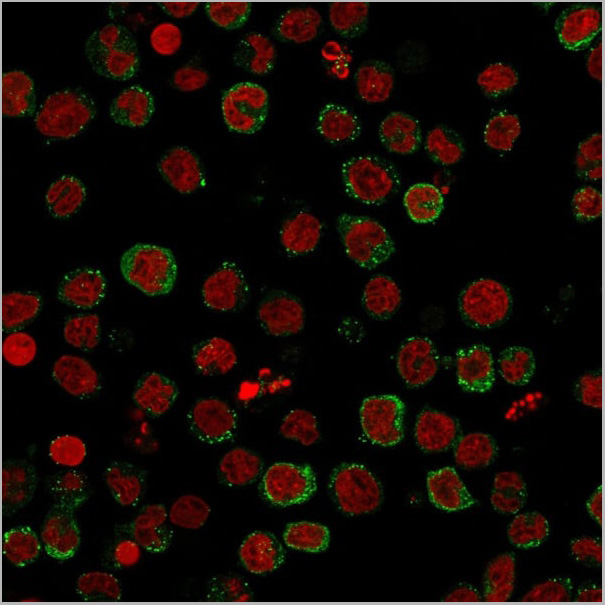
(Published customer image:Phycoerythrin conjugated Mouse anti Human CD83 antibody, clone HB15e used for the evaluation of CD83 expression on monocyte derived dendritic cells by flow cytometry.Phenotypic characterization of immunogenic and tolerogenic moDC populations by flow cytometry. Monocytes were negatively selected from PBMC using magnetic beads. Immature moDC were generated with IL-4 and GM-CSF for 6 days. 15d-PGJ2 (PGJ2 DC) and dexamethasone plus 1alpha,25-dihydroxyvitamin were added to generate tolerogenic moDC, respectively (PGJ2 DC and Dex/VD3 DC). To generate immunogenic moDC, immature moDC were stimulated for 24 h with LPS, polyI:C and a cytokine cocktail containing TNF-alpha, IL-1beta, IL-6 and PGE2, respectively. The phenotypes of the cells were analyzed by flow cytometry. Live cells were gated according to FSC/SSC. One representative experiment out of three is shown.From: Sprater F, Hovden A-O, Appel S (2012)Expression of ESE-3 Isoforms in Immunogenic and Tolerogenic Human Monocyte-Derived Dendritic Cells.PLoS ONE 7(11): e49577.)
Application Data
(Published customer image:Phycoerythrin conjugated Mouse anti Human CD83 antibody, clone HB15e used for the evaluation of CD83 expression on monocyte derived dendritic cells by flow cytometry.Phenotypic characterization of immunogenic and tolerogenic moDC populations by flow cytometry. Monocytes were negatively selected from PBMC using magnetic beads. Immature moDC were generated with IL-4 and GM-CSF for 6 days. 15d-PGJ2 (PGJ2 DC) and dexamethasone plus 1alpha,25-dihydroxyvitamin were added to generate tolerogenic moDC, respectively (PGJ2 DC and Dex/VD3 DC). To generate immunogenic moDC, immature moDC were stimulated for 24 h with LPS, polyI:C and a cytokine cocktail containing TNF-alpha, IL-1beta, IL-6 and PGE2, respectively. The phenotypes of the cells were analyzed by flow cytometry. Live cells were gated according to FSC/SSC. One representative experiment out of three is shown.From: Sprater F, Hovden A-O, Appel S (2012)Expression of ESE-3 Isoforms in Immunogenic and Tolerogenic Human Monocyte-Derived Dendritic Cells.PLoS ONE 7(11): e49577.)
CD83, Monoclonal Antibody (Cat# AAA12283)
Full Name
MOUSE ANTI HUMAN CD83
Reactivity
Human
Applications
Flow Cytometry, Immunohistochemistry, Immunohistochemistry, Immunofluorescence
Pricing


 FCM (Flow Cytometry)
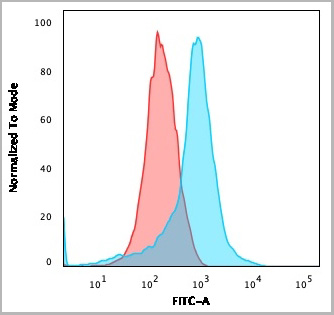
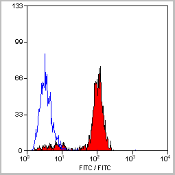
(SOCS1 Antibody (N-term) flow cytometric analysis of WiDr cells (right histogram) compared to a negative control cell (left histogram).FITC-conjugated goat-anti-rabbit secondary antibodies were used for the analysis.)
FCM (Flow Cytometry)
(SOCS1 Antibody (N-term) flow cytometric analysis of WiDr cells (right histogram) compared to a negative control cell (left histogram).FITC-conjugated goat-anti-rabbit secondary antibodies were used for the analysis.)
SOCS1, Polyclonal Antibody (Cat# AAA28752)
Full Name
SOCS1 Antibody (N-term)
Gene Names
SOCS1; JAB; CIS1; SSI1; TIP3; CISH1; SSI-1; SOCS-1
Reactivity
Mouse, rat
Applications
Western Blot, Immunohistochemistry, Immunofluorescence, Flow Cytometry
Purity
Purified Rabbit Polyclonal Antibody (Pab)
Pricing


 Application Data
(Published customer image:Phycoerythrin conjugated Mouse anti Human CD83 antibody, clone HB15e used for the evaluation of CD83 expression on monocyte derived dendritic cells by flow cytometry.Phenotypic characterization of immunogenic and tolerogenic moDC populations by flow cytometry. Monocytes were negatively selected from PBMC using magnetic beads. Immature moDC were generated with IL-4 and GM-CSF for 6 days. 15d-PGJ2 (PGJ2 DC) and dexamethasone plus 1alpha,25-dihydroxyvitamin were added to generate tolerogenic moDC, respectively (PGJ2 DC and Dex/VD3 DC). To generate immunogenic moDC, immature moDC were stimulated for 24 h with LPS, polyI:C and a cytokine cocktail containing TNF-alpha, IL-1beta, IL-6 and PGE2, respectively. The phenotypes of the cells were analyzed by flow cytometry. Live cells were gated according to FSC/SSC. One representative experiment out of three is shown.From: Sprater F, Hovden A-O, Appel S (2012)Expression of ESE-3 Isoforms in Immunogenic and Tolerogenic Human Monocyte-Derived Dendritic Cells.PLoS ONE 7(11): e49577.)
Application Data
(Published customer image:Phycoerythrin conjugated Mouse anti Human CD83 antibody, clone HB15e used for the evaluation of CD83 expression on monocyte derived dendritic cells by flow cytometry.Phenotypic characterization of immunogenic and tolerogenic moDC populations by flow cytometry. Monocytes were negatively selected from PBMC using magnetic beads. Immature moDC were generated with IL-4 and GM-CSF for 6 days. 15d-PGJ2 (PGJ2 DC) and dexamethasone plus 1alpha,25-dihydroxyvitamin were added to generate tolerogenic moDC, respectively (PGJ2 DC and Dex/VD3 DC). To generate immunogenic moDC, immature moDC were stimulated for 24 h with LPS, polyI:C and a cytokine cocktail containing TNF-alpha, IL-1beta, IL-6 and PGE2, respectively. The phenotypes of the cells were analyzed by flow cytometry. Live cells were gated according to FSC/SSC. One representative experiment out of three is shown.From: Sprater F, Hovden A-O, Appel S (2012)Expression of ESE-3 Isoforms in Immunogenic and Tolerogenic Human Monocyte-Derived Dendritic Cells.PLoS ONE 7(11): e49577.)
CD83, Monoclonal Antibody (Cat# AAA12282)
Full Name
MOUSE ANTI HUMAN CD83
Reactivity
Human
Applications
Flow Cytometry, Immunohistochemistry, Immunohistochemistry, Immunoprecipitation, Immunofluorescence
Pricing


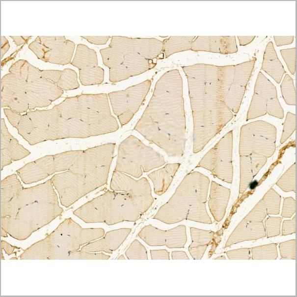
 IHC (Immunohistochemistry)
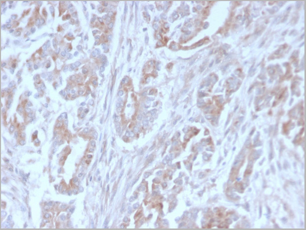
(At 1/100 staining Mouse kidney tissue by IHC-P. The sample was formaldehyde fixed and a heat mediated antigen retrieval step in citrate buffer was performed. The sample was then blocked and incubated with the primary antibody at 4 degree C overnight. An HRP conjugated anti-Rabbit antibody was used as the secondary antibody.)
IHC (Immunohistochemistry)
(At 1/100 staining Mouse kidney tissue by IHC-P. The sample was formaldehyde fixed and a heat mediated antigen retrieval step in citrate buffer was performed. The sample was then blocked and incubated with the primary antibody at 4 degree C overnight. An HRP conjugated anti-Rabbit antibody was used as the secondary antibody.)
p38 MAPK, Polyclonal Antibody (Cat# AAA31333)
Full Name
Phospho-p38 MAPK (Thr180/Tyr182) Antibody
Gene Names
MAPK14; RK; p38; CSBP; EXIP; Mxi2; CSBP1; CSBP2; CSPB1; PRKM14; PRKM15; SAPK2A; p38ALPHA
Reactivity
Human, Mouse, Rat
Predicted Reactivity: Pig (100%), Bovine (100%), Horse (100%), Sheep (100%), Rabbit (100%), Dog (100%), Xenopus (91%)
Predicted Reactivity: Pig (100%), Bovine (100%), Horse (100%), Sheep (100%), Rabbit (100%), Dog (100%), Xenopus (91%)
Applications
Western Blot, Immunohistochemistry, Immunofluorescence, Immunocytochemistry, Immunoprecipitation, Peptide ELISA
Purity
The antibody is from purified rabbit serum by affinity purification via sequential chromatography on phospho-peptide and non-phospho-peptide affinity columns.
Pricing


 FCM (Flow Cytometry)
(Flow cytometric analysis of THP-1 cells with MRP8/S100A8 antibody at 1/100 dilution (red) compared with an unlabelled control (cells without incubation with primary antibody; black).)
FCM (Flow Cytometry)
(Flow cytometric analysis of THP-1 cells with MRP8/S100A8 antibody at 1/100 dilution (red) compared with an unlabelled control (cells without incubation with primary antibody; black).)
MRP8/S100A8, Polyclonal Antibody (Cat# AAA29887)
Full Name
MRP8/S100A8 Antibody
Reactivity
Human, Mouse, Rat
Applications
Western Blot, Immunocytochemistry, Immunohistochemistry, Flow Cytometry
Purity
ProA affinity purified
Pricing


 Application Data
(Published customer image:Phycoerythrin conjugated Mouse anti Human CD83 antibody, clone HB15e used for the evaluation of CD83 expression on monocyte derived dendritic cells by flow cytometry.Phenotypic characterization of immunogenic and tolerogenic moDC populations by flow cytometry. Monocytes were negatively selected from PBMC using magnetic beads. Immature moDC were generated with IL-4 and GM-CSF for 6 days. 15d-PGJ2 (PGJ2 DC) and dexamethasone plus 1alpha,25-dihydroxyvitamin were added to generate tolerogenic moDC, respectively (PGJ2 DC and Dex/VD3 DC). To generate immunogenic moDC, immature moDC were stimulated for 24 h with LPS, polyI:C and a cytokine cocktail containing TNF-alpha, IL-1beta, IL-6 and PGE2, respectively. The phenotypes of the cells were analyzed by flow cytometry. Live cells were gated according to FSC/SSC. One representative experiment out of three is shown.From: Sprater F, Hovden A-O, Appel S (2012)Expression of ESE-3 Isoforms in Immunogenic and Tolerogenic Human Monocyte-Derived Dendritic Cells.PLoS ONE 7(11): e49577.)
Application Data
(Published customer image:Phycoerythrin conjugated Mouse anti Human CD83 antibody, clone HB15e used for the evaluation of CD83 expression on monocyte derived dendritic cells by flow cytometry.Phenotypic characterization of immunogenic and tolerogenic moDC populations by flow cytometry. Monocytes were negatively selected from PBMC using magnetic beads. Immature moDC were generated with IL-4 and GM-CSF for 6 days. 15d-PGJ2 (PGJ2 DC) and dexamethasone plus 1alpha,25-dihydroxyvitamin were added to generate tolerogenic moDC, respectively (PGJ2 DC and Dex/VD3 DC). To generate immunogenic moDC, immature moDC were stimulated for 24 h with LPS, polyI:C and a cytokine cocktail containing TNF-alpha, IL-1beta, IL-6 and PGE2, respectively. The phenotypes of the cells were analyzed by flow cytometry. Live cells were gated according to FSC/SSC. One representative experiment out of three is shown.From: Sprater F, Hovden A-O, Appel S (2012)Expression of ESE-3 Isoforms in Immunogenic and Tolerogenic Human Monocyte-Derived Dendritic Cells.PLoS ONE 7(11): e49577.)
CD83, Monoclonal Antibody (Cat# AAA12281)
Full Name
MOUSE ANTI HUMAN CD83: FITC
Reactivity
Human
Applications
Flow Cytometry
Pricing
 Application Data
Interferon-a Receptor, Type I, Subunit I (Ser535,539), Polyclonal Antibody (Cat# AAA14231)
Full Name
Anti-Phospho-Ser535,539 Interferon-alpha Receptor, Type I, Subunit I
Gene Names
IFNAR1; AVP; IFRC; IFNAR; IFNBR; IFN-alpha-REC
Reactivity
Rat
Applications
Western Blot, Immunohistochemistry
Purity
Affinity Purified (Prepared from rabbit serum by affinity purification via sequential chromatography on phospho- and dephosphopeptide affinity columns.)
Pricing


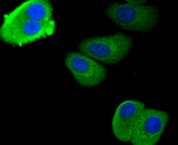
 IF (Immunofluorescence)
(Immunofluorescence analysis of HeLa cells using AIMP1 antibody. Blue: DAPI for nuclear staining.)
IF (Immunofluorescence)
(Immunofluorescence analysis of HeLa cells using AIMP1 antibody. Blue: DAPI for nuclear staining.)
AIMP1, Polyclonal Antibody (Cat# AAA28108)
Full Name
AIMP1 Polyclonal Antibody
Gene Names
AIMP1; p43; HLD3; EMAP2; SCYE1; EMAPII
Reactivity
Human, Mouse, Rat
Applications
Western Blot, Immunohistochemistry, Immunofluorescence
Purity
Affinity Purification
Pricing


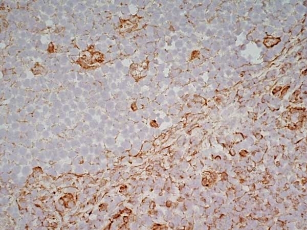
 Application Data
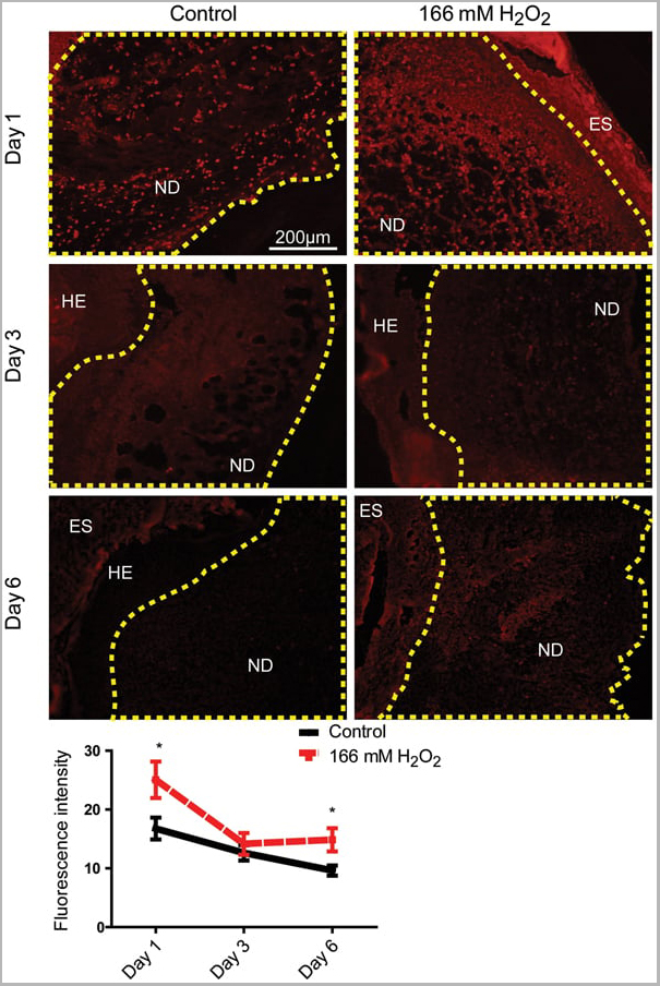
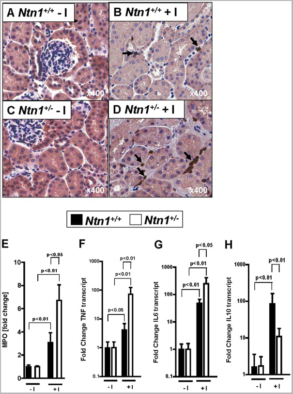
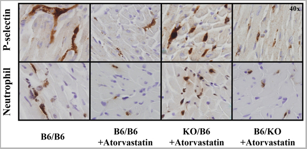
(Published customer image: Leukocyte infiltration in COX-2-M/-M and COX-2+/+ mice. MPO enzymatic activity (panel A) was statistically similar in COX-2-M/-M and COX-2+/+ livers at 6 h and 24 h post-IRI. Ly-6G+ neutrophil (panel B) and granulocyte (panel C) infiltration were also comparable in COX-2-M/-M and COX-2+/+ livers after IRI. Mac-1+ (panel D) and CD68 (panel E) infiltrating macrophages were significantly reduced in COX-2-M/-M livers at 24 h post-reperfusion, but were statistically indistinguishable in COX-2-M/-M and COX-2+/+ livers at 6 h after IRI. No statistical differences in MMP-9 expression (panel F) could be demonstrated in livers of COX-2-M/-M and COX-2+/+ mice post-IRI. Representative immunostaining (panel G) of infiltrating Ly-6G+ (a,b,e,f) and Mac-1+ (c,d,g,h) leukocytes in livers of COX-2+/+ (a,c,e,g) and COX-2-M/-M (b,d,f,h) mice at 6 h (a to d) and 24 h (e to h) post IRI; (n = 5 -6/group; * indicates p)
Application Data
(Published customer image: Leukocyte infiltration in COX-2-M/-M and COX-2+/+ mice. MPO enzymatic activity (panel A) was statistically similar in COX-2-M/-M and COX-2+/+ livers at 6 h and 24 h post-IRI. Ly-6G+ neutrophil (panel B) and granulocyte (panel C) infiltration were also comparable in COX-2-M/-M and COX-2+/+ livers after IRI. Mac-1+ (panel D) and CD68 (panel E) infiltrating macrophages were significantly reduced in COX-2-M/-M livers at 24 h post-reperfusion, but were statistically indistinguishable in COX-2-M/-M and COX-2+/+ livers at 6 h after IRI. No statistical differences in MMP-9 expression (panel F) could be demonstrated in livers of COX-2-M/-M and COX-2+/+ mice post-IRI. Representative immunostaining (panel G) of infiltrating Ly-6G+ (a,b,e,f) and Mac-1+ (c,d,g,h) leukocytes in livers of COX-2+/+ (a,c,e,g) and COX-2-M/-M (b,d,f,h) mice at 6 h (a to d) and 24 h (e to h) post IRI; (n = 5 -6/group; * indicates p)
CD68, Monoclonal Antibody (Cat# AAA12105)
Full Name
RAT ANTI MOUSE CD68:FITC
Gene Names
Cd68; Lamp4; gp110; Scard1
Applications
Flow Cytometry
Pricing


 Application Data
(Published customer image: Leukocyte infiltration in COX-2-M/-M and COX-2+/+ mice. MPO enzymatic activity (panel A) was statistically similar in COX-2-M/-M and COX-2+/+ livers at 6 h and 24 h post-IRI. Ly-6G+ neutrophil (panel B) and granulocyte (panel C) infiltration were also comparable in COX-2-M/-M and COX-2+/+ livers after IRI. Mac-1+ (panel D) and CD68 (panel E) infiltrating macrophages were significantly reduced in COX-2-M/-M livers at 24 h post-reperfusion, but were statistically indistinguishable in COX-2-M/-M and COX-2+/+ livers at 6 h after IRI. No statistical differences in MMP-9 expression (panel F) could be demonstrated in livers of COX-2-M/-M and COX-2+/+ mice post-IRI. Representative immunostaining (panel G) of infiltrating Ly-6G+ (a,b,e,f) and Mac-1+ (c,d,g,h) leukocytes in livers of COX-2+/+ (a,c,e,g) and COX-2-M/-M (b,d,f,h) mice at 6 h (a to d) and 24 h (e to h) post IRI; (n = 5 -6/group; * indicates p)
Application Data
(Published customer image: Leukocyte infiltration in COX-2-M/-M and COX-2+/+ mice. MPO enzymatic activity (panel A) was statistically similar in COX-2-M/-M and COX-2+/+ livers at 6 h and 24 h post-IRI. Ly-6G+ neutrophil (panel B) and granulocyte (panel C) infiltration were also comparable in COX-2-M/-M and COX-2+/+ livers after IRI. Mac-1+ (panel D) and CD68 (panel E) infiltrating macrophages were significantly reduced in COX-2-M/-M livers at 24 h post-reperfusion, but were statistically indistinguishable in COX-2-M/-M and COX-2+/+ livers at 6 h after IRI. No statistical differences in MMP-9 expression (panel F) could be demonstrated in livers of COX-2-M/-M and COX-2+/+ mice post-IRI. Representative immunostaining (panel G) of infiltrating Ly-6G+ (a,b,e,f) and Mac-1+ (c,d,g,h) leukocytes in livers of COX-2+/+ (a,c,e,g) and COX-2-M/-M (b,d,f,h) mice at 6 h (a to d) and 24 h (e to h) post IRI; (n = 5 -6/group; * indicates p)
CD68, Monoclonal Antibody (Cat# AAA12103)
Full Name
RAT ANTI MOUSE CD68:Biotin
Gene Names
Cd68; Lamp4; gp110; Scard1
Applications
Flow Cytometry
Pricing


 Application Data
(Published customer image: Leukocyte infiltration in COX-2-M/-M and COX-2+/+ mice. MPO enzymatic activity (panel A) was statistically similar in COX-2-M/-M and COX-2+/+ livers at 6 h and 24 h post-IRI. Ly-6G+ neutrophil (panel B) and granulocyte (panel C) infiltration were also comparable in COX-2-M/-M and COX-2+/+ livers after IRI. Mac-1+ (panel D) and CD68 (panel E) infiltrating macrophages were significantly reduced in COX-2-M/-M livers at 24 h post-reperfusion, but were statistically indistinguishable in COX-2-M/-M and COX-2+/+ livers at 6 h after IRI. No statistical differences in MMP-9 expression (panel F) could be demonstrated in livers of COX-2-M/-M and COX-2+/+ mice post-IRI. Representative immunostaining (panel G) of infiltrating Ly-6G+ (a,b,e,f) and Mac-1+ (c,d,g,h) leukocytes in livers of COX-2+/+ (a,c,e,g) and COX-2-M/-M (b,d,f,h) mice at 6 h (a to d) and 24 h (e to h) post IRI; (n = 5 -6/group; * indicates p)
Application Data
(Published customer image: Leukocyte infiltration in COX-2-M/-M and COX-2+/+ mice. MPO enzymatic activity (panel A) was statistically similar in COX-2-M/-M and COX-2+/+ livers at 6 h and 24 h post-IRI. Ly-6G+ neutrophil (panel B) and granulocyte (panel C) infiltration were also comparable in COX-2-M/-M and COX-2+/+ livers after IRI. Mac-1+ (panel D) and CD68 (panel E) infiltrating macrophages were significantly reduced in COX-2-M/-M livers at 24 h post-reperfusion, but were statistically indistinguishable in COX-2-M/-M and COX-2+/+ livers at 6 h after IRI. No statistical differences in MMP-9 expression (panel F) could be demonstrated in livers of COX-2-M/-M and COX-2+/+ mice post-IRI. Representative immunostaining (panel G) of infiltrating Ly-6G+ (a,b,e,f) and Mac-1+ (c,d,g,h) leukocytes in livers of COX-2+/+ (a,c,e,g) and COX-2-M/-M (b,d,f,h) mice at 6 h (a to d) and 24 h (e to h) post IRI; (n = 5 -6/group; * indicates p)
CD68, Monoclonal Antibody (Cat# AAA12110)
Full Name
RAT ANTI MOUSE CD68
Gene Names
Cd68; Lamp4; gp110; Scard1
Applications
Immunohistochemistry, Flow Cytometry, Immunofluorescence, Immunoprecipitation, Immunohistochemistry, Western Blot
Pricing


 Application Data
(Published customer image: Leukocyte infiltration in COX-2-M/-M and COX-2+/+ mice. MPO enzymatic activity (panel A) was statistically similar in COX-2-M/-M and COX-2+/+ livers at 6 h and 24 h post-IRI. Ly-6G+ neutrophil (panel B) and granulocyte (panel C) infiltration were also comparable in COX-2-M/-M and COX-2+/+ livers after IRI. Mac-1+ (panel D) and CD68 (panel E) infiltrating macrophages were significantly reduced in COX-2-M/-M livers at 24 h post-reperfusion, but were statistically indistinguishable in COX-2-M/-M and COX-2+/+ livers at 6 h after IRI. No statistical differences in MMP-9 expression (panel F) could be demonstrated in livers of COX-2-M/-M and COX-2+/+ mice post-IRI. Representative immunostaining (panel G) of infiltrating Ly-6G+ (a,b,e,f) and Mac-1+ (c,d,g,h) leukocytes in livers of COX-2+/+ (a,c,e,g) and COX-2-M/-M (b,d,f,h) mice at 6 h (a to d) and 24 h (e to h) post IRI; (n = 5 -6/group; * indicates p)
Application Data
(Published customer image: Leukocyte infiltration in COX-2-M/-M and COX-2+/+ mice. MPO enzymatic activity (panel A) was statistically similar in COX-2-M/-M and COX-2+/+ livers at 6 h and 24 h post-IRI. Ly-6G+ neutrophil (panel B) and granulocyte (panel C) infiltration were also comparable in COX-2-M/-M and COX-2+/+ livers after IRI. Mac-1+ (panel D) and CD68 (panel E) infiltrating macrophages were significantly reduced in COX-2-M/-M livers at 24 h post-reperfusion, but were statistically indistinguishable in COX-2-M/-M and COX-2+/+ livers at 6 h after IRI. No statistical differences in MMP-9 expression (panel F) could be demonstrated in livers of COX-2-M/-M and COX-2+/+ mice post-IRI. Representative immunostaining (panel G) of infiltrating Ly-6G+ (a,b,e,f) and Mac-1+ (c,d,g,h) leukocytes in livers of COX-2+/+ (a,c,e,g) and COX-2-M/-M (b,d,f,h) mice at 6 h (a to d) and 24 h (e to h) post IRI; (n = 5 -6/group; * indicates p)
CD68, Monoclonal Antibody (Cat# AAA12104)
Full Name
RAT ANTI MOUSE CD68:Biotin
Gene Names
Cd68; Lamp4; gp110; Scard1
Applications
Flow Cytometry
Pricing


 Application Data
(Published customer image: Leukocyte infiltration in COX-2-M/-M and COX-2+/+ mice. MPO enzymatic activity (panel A) was statistically similar in COX-2-M/-M and COX-2+/+ livers at 6 h and 24 h post-IRI. Ly-6G+ neutrophil (panel B) and granulocyte (panel C) infiltration were also comparable in COX-2-M/-M and COX-2+/+ livers after IRI. Mac-1+ (panel D) and CD68 (panel E) infiltrating macrophages were significantly reduced in COX-2-M/-M livers at 24 h post-reperfusion, but were statistically indistinguishable in COX-2-M/-M and COX-2+/+ livers at 6 h after IRI. No statistical differences in MMP-9 expression (panel F) could be demonstrated in livers of COX-2-M/-M and COX-2+/+ mice post-IRI. Representative immunostaining (panel G) of infiltrating Ly-6G+ (a,b,e,f) and Mac-1+ (c,d,g,h) leukocytes in livers of COX-2+/+ (a,c,e,g) and COX-2-M/-M (b,d,f,h) mice at 6 h (a to d) and 24 h (e to h) post IRI; (n = 5 -6/group; * indicates p)
Application Data
(Published customer image: Leukocyte infiltration in COX-2-M/-M and COX-2+/+ mice. MPO enzymatic activity (panel A) was statistically similar in COX-2-M/-M and COX-2+/+ livers at 6 h and 24 h post-IRI. Ly-6G+ neutrophil (panel B) and granulocyte (panel C) infiltration were also comparable in COX-2-M/-M and COX-2+/+ livers after IRI. Mac-1+ (panel D) and CD68 (panel E) infiltrating macrophages were significantly reduced in COX-2-M/-M livers at 24 h post-reperfusion, but were statistically indistinguishable in COX-2-M/-M and COX-2+/+ livers at 6 h after IRI. No statistical differences in MMP-9 expression (panel F) could be demonstrated in livers of COX-2-M/-M and COX-2+/+ mice post-IRI. Representative immunostaining (panel G) of infiltrating Ly-6G+ (a,b,e,f) and Mac-1+ (c,d,g,h) leukocytes in livers of COX-2+/+ (a,c,e,g) and COX-2-M/-M (b,d,f,h) mice at 6 h (a to d) and 24 h (e to h) post IRI; (n = 5 -6/group; * indicates p)
CD68, Monoclonal Antibody (Cat# AAA12107)
Full Name
RAT ANTI MOUSE CD68
Gene Names
Cd68; Lamp4; gp110; Scard1
Applications
Immunohistochemistry, Flow Cytometry, Immunofluorescence, Immunoprecipitation, Immunohistochemistry, Western Blot
Purity
Purified
Purified IgG - liquid
Purified IgG - liquid
Pricing


 Application Data
(Published customer image:Phycoerythrin conjugated Mouse anti Human CD83 antibody, clone HB15e used for the evaluation of CD83 expression on monocyte derived dendritic cells by flow cytometry.Phenotypic characterization of immunogenic and tolerogenic moDC populations by flow cytometry. Monocytes were negatively selected from PBMC using magnetic beads. Immature moDC were generated with IL-4 and GM-CSF for 6 days. 15d-PGJ2 (PGJ2 DC) and dexamethasone plus 1alpha,25-dihydroxyvitamin were added to generate tolerogenic moDC, respectively (PGJ2 DC and Dex/VD3 DC). To generate immunogenic moDC, immature moDC were stimulated for 24 h with LPS, polyI:C and a cytokine cocktail containing TNF-alpha, IL-1beta, IL-6 and PGE2, respectively. The phenotypes of the cells were analyzed by flow cytometry. Live cells were gated according to FSC/SSC. One representative experiment out of three is shown.From: Sprater F, Hovden A-O, Appel S (2012)Expression of ESE-3 Isoforms in Immunogenic and Tolerogenic Human Monocyte-Derived Dendritic Cells.PLoS ONE 7(11): e49577.)
Application Data
(Published customer image:Phycoerythrin conjugated Mouse anti Human CD83 antibody, clone HB15e used for the evaluation of CD83 expression on monocyte derived dendritic cells by flow cytometry.Phenotypic characterization of immunogenic and tolerogenic moDC populations by flow cytometry. Monocytes were negatively selected from PBMC using magnetic beads. Immature moDC were generated with IL-4 and GM-CSF for 6 days. 15d-PGJ2 (PGJ2 DC) and dexamethasone plus 1alpha,25-dihydroxyvitamin were added to generate tolerogenic moDC, respectively (PGJ2 DC and Dex/VD3 DC). To generate immunogenic moDC, immature moDC were stimulated for 24 h with LPS, polyI:C and a cytokine cocktail containing TNF-alpha, IL-1beta, IL-6 and PGE2, respectively. The phenotypes of the cells were analyzed by flow cytometry. Live cells were gated according to FSC/SSC. One representative experiment out of three is shown.From: Sprater F, Hovden A-O, Appel S (2012)Expression of ESE-3 Isoforms in Immunogenic and Tolerogenic Human Monocyte-Derived Dendritic Cells.PLoS ONE 7(11): e49577.)
CD83, Monoclonal Antibody (Cat# AAA12280)
Full Name
MOUSE ANTI HUMAN CD83
Reactivity
Human
Applications
Flow Cytometry, Immunohistochemistry, Immunohistochemistry, Immunoprecipitation, Immunofluorescence
Pricing


 Application Data
(At 25 degree C. The primary antibody was diluted at 1/200 and incubated with the sample for 1 hour at 37 degree C. An Alexa Fluor 594 conjugated goat anti-rabbit IgG (H+L) antibody(Red), diluted at 1/600, was used as secondary antibody.)
Application Data
(At 25 degree C. The primary antibody was diluted at 1/200 and incubated with the sample for 1 hour at 37 degree C. An Alexa Fluor 594 conjugated goat anti-rabbit IgG (H+L) antibody(Red), diluted at 1/600, was used as secondary antibody.)
JAK3, Polyclonal Antibody (Cat# AAA31404)
Full Name
Phospho-JAK3 (Tyr904) Antibody
Gene Names
JAK3; JAKL; LJAK; JAK-3; L-JAK; JAK3_HUMAN
Reactivity
Human, Mouse, Rat
Predicted Reactivity: Bovine (100%), Sheep (100%), Dog (100%)
Predicted Reactivity: Bovine (100%), Sheep (100%), Dog (100%)
Applications
Western Blot, Immunohistochemistry, Immunofluorescence, Immunocytochemistry, Peptide ELISA
Purity
The antibody is from purified rabbit serum by affinity purification via sequential chromatography on phospho-peptide and non-phospho-peptide affinity columns.
Pricing


 IP (Immunoprecipitation)
(Immunoprecipitation analysis of 200ug extracts of U-87MG cells using 3ug MAPK14 antibody. Western blot was performed from the immunoprecipitate using MAPK14 antibody at a dilition of 1:1000.)
IP (Immunoprecipitation)
(Immunoprecipitation analysis of 200ug extracts of U-87MG cells using 3ug MAPK14 antibody. Western blot was performed from the immunoprecipitate using MAPK14 antibody at a dilition of 1:1000.)
MAPK14, Polyclonal Antibody (Cat# AAA28274)
Full Name
MAPK14 Polyclonal Antibody
Gene Names
MAPK14; RK; p38; CSBP; EXIP; Mxi2; CSBP1; CSBP2; CSPB1; PRKM14; PRKM15; SAPK2A; p38ALPHA
Reactivity
Human, Mouse, Rat
Applications
Western Blot, Immunohistochemistry, Immunoprecipitation
Purity
Affinity Purification
Pricing


 Application Data
(Published customer image: Leukocyte infiltration in COX-2-M/-M and COX-2+/+ mice. MPO enzymatic activity (panel A) was statistically similar in COX-2-M/-M and COX-2+/+ livers at 6 h and 24 h post-IRI. Ly-6G+ neutrophil (panel B) and granulocyte (panel C) infiltration were also comparable in COX-2-M/-M and COX-2+/+ livers after IRI. Mac-1+ (panel D) and CD68 (panel E) infiltrating macrophages were significantly reduced in COX-2-M/-M livers at 24 h post-reperfusion, but were statistically indistinguishable in COX-2-M/-M and COX-2+/+ livers at 6 h after IRI. No statistical differences in MMP-9 expression (panel F) could be demonstrated in livers of COX-2-M/-M and COX-2+/+ mice post-IRI. Representative immunostaining (panel G) of infiltrating Ly-6G+ (a,b,e,f) and Mac-1+ (c,d,g,h) leukocytes in livers of COX-2+/+ (a,c,e,g) and COX-2-M/-M (b,d,f,h) mice at 6 h (a to d) and 24 h (e to h) post IRI; (n = 5 -6/group; * indicates p)
Application Data
(Published customer image: Leukocyte infiltration in COX-2-M/-M and COX-2+/+ mice. MPO enzymatic activity (panel A) was statistically similar in COX-2-M/-M and COX-2+/+ livers at 6 h and 24 h post-IRI. Ly-6G+ neutrophil (panel B) and granulocyte (panel C) infiltration were also comparable in COX-2-M/-M and COX-2+/+ livers after IRI. Mac-1+ (panel D) and CD68 (panel E) infiltrating macrophages were significantly reduced in COX-2-M/-M livers at 24 h post-reperfusion, but were statistically indistinguishable in COX-2-M/-M and COX-2+/+ livers at 6 h after IRI. No statistical differences in MMP-9 expression (panel F) could be demonstrated in livers of COX-2-M/-M and COX-2+/+ mice post-IRI. Representative immunostaining (panel G) of infiltrating Ly-6G+ (a,b,e,f) and Mac-1+ (c,d,g,h) leukocytes in livers of COX-2+/+ (a,c,e,g) and COX-2-M/-M (b,d,f,h) mice at 6 h (a to d) and 24 h (e to h) post IRI; (n = 5 -6/group; * indicates p)
CD68, Monoclonal Antibody (Cat# AAA12108)
Full Name
RAT ANTI MOUSE CD68:RPE
Gene Names
Cd68; Lamp4; gp110; Scard1
Applications
Flow Cytometry
Pricing


 Application Data
(Published customer image: Leukocyte infiltration in COX-2-M/-M and COX-2+/+ mice. MPO enzymatic activity (panel A) was statistically similar in COX-2-M/-M and COX-2+/+ livers at 6 h and 24 h post-IRI. Ly-6G+ neutrophil (panel B) and granulocyte (panel C) infiltration were also comparable in COX-2-M/-M and COX-2+/+ livers after IRI. Mac-1+ (panel D) and CD68 (panel E) infiltrating macrophages were significantly reduced in COX-2-M/-M livers at 24 h post-reperfusion, but were statistically indistinguishable in COX-2-M/-M and COX-2+/+ livers at 6 h after IRI. No statistical differences in MMP-9 expression (panel F) could be demonstrated in livers of COX-2-M/-M and COX-2+/+ mice post-IRI. Representative immunostaining (panel G) of infiltrating Ly-6G+ (a,b,e,f) and Mac-1+ (c,d,g,h) leukocytes in livers of COX-2+/+ (a,c,e,g) and COX-2-M/-M (b,d,f,h) mice at 6 h (a to d) and 24 h (e to h) post IRI; (n = 5 -6/group; * indicates p)
Application Data
(Published customer image: Leukocyte infiltration in COX-2-M/-M and COX-2+/+ mice. MPO enzymatic activity (panel A) was statistically similar in COX-2-M/-M and COX-2+/+ livers at 6 h and 24 h post-IRI. Ly-6G+ neutrophil (panel B) and granulocyte (panel C) infiltration were also comparable in COX-2-M/-M and COX-2+/+ livers after IRI. Mac-1+ (panel D) and CD68 (panel E) infiltrating macrophages were significantly reduced in COX-2-M/-M livers at 24 h post-reperfusion, but were statistically indistinguishable in COX-2-M/-M and COX-2+/+ livers at 6 h after IRI. No statistical differences in MMP-9 expression (panel F) could be demonstrated in livers of COX-2-M/-M and COX-2+/+ mice post-IRI. Representative immunostaining (panel G) of infiltrating Ly-6G+ (a,b,e,f) and Mac-1+ (c,d,g,h) leukocytes in livers of COX-2+/+ (a,c,e,g) and COX-2-M/-M (b,d,f,h) mice at 6 h (a to d) and 24 h (e to h) post IRI; (n = 5 -6/group; * indicates p)
CD68, Monoclonal Antibody (Cat# AAA12102)
Full Name
RAT ANTI MOUSE CD68
Gene Names
Cd68; Lamp4; gp110; Scard1
Applications
Immunohistochemistry, Flow Cytometry, Immunofluorescence, Immunoprecipitation, Immunohistochemistry, Western Blot
Pricing


 FCM (Flow Cytometry)
(Dual staining of pig peripheral blood lymphocytes with Mouse anti Pig CD335 detected with Goat anti Mouse IgG (H/L):FITC (STAR117F), and Mouse anti Pig wCD8a:RPE)
FCM (Flow Cytometry)
(Dual staining of pig peripheral blood lymphocytes with Mouse anti Pig CD335 detected with Goat anti Mouse IgG (H/L):FITC (STAR117F), and Mouse anti Pig wCD8a:RPE)
CD335, Monoclonal Antibody (Cat# AAA12253)
Full Name
MOUSE ANTI PIG CD335
Reactivity
Pig
Applications
Flow Cytometry, Immunofluorescence
Pricing


 Application Data
(At 25 degree C. The primary antibody was diluted at 1/200 and incubated with the sample for 1 hour at 37 degree C. An Alexa Fluor 594 conjugated goat anti-rabbit IgG (H+L) antibody(Red), diluted at 1/600, was used as secondary antibody.)
Application Data
(At 25 degree C. The primary antibody was diluted at 1/200 and incubated with the sample for 1 hour at 37 degree C. An Alexa Fluor 594 conjugated goat anti-rabbit IgG (H+L) antibody(Red), diluted at 1/600, was used as secondary antibody.)
JAK3, Polyclonal Antibody (Cat# AAA31405)
Full Name
Phospho-JAK3 (Tyr981) Antibody
Gene Names
JAK3; JAKL; LJAK; JAK-3; L-JAK; JAK3_HUMAN
Reactivity
Human, Mouse, Rat, Monkey
Predicted Reactivity: Pig (100%), Bovine (91%), Horse (100%), Sheep (100%), Dog (100%)
Predicted Reactivity: Pig (100%), Bovine (91%), Horse (100%), Sheep (100%), Dog (100%)
Applications
Western Blot, Immunohistochemistry, Immunofluorescence, Immunocytochemistry, Peptide ELISA
Purity
The antibody is from purified rabbit serum by affinity purification via sequential chromatography on phospho-peptide and non-phospho-peptide affinity columns.
Pricing


 FCM (Flow Cytometry)
(Dual staining of pig peripheral blood lymphocytes with Mouse anti Pig CD335 detected with Goat anti Mouse IgG (H/L):FITC (STAR117F), and Mouse anti Pig wCD8a:RPE)
FCM (Flow Cytometry)
(Dual staining of pig peripheral blood lymphocytes with Mouse anti Pig CD335 detected with Goat anti Mouse IgG (H/L):FITC (STAR117F), and Mouse anti Pig wCD8a:RPE)
CD335, Monoclonal Antibody (Cat# AAA12251)
Full Name
MOUSE ANTI PIG CD335: APC
Reactivity
Pig
Applications
Flow Cytometry
Pricing


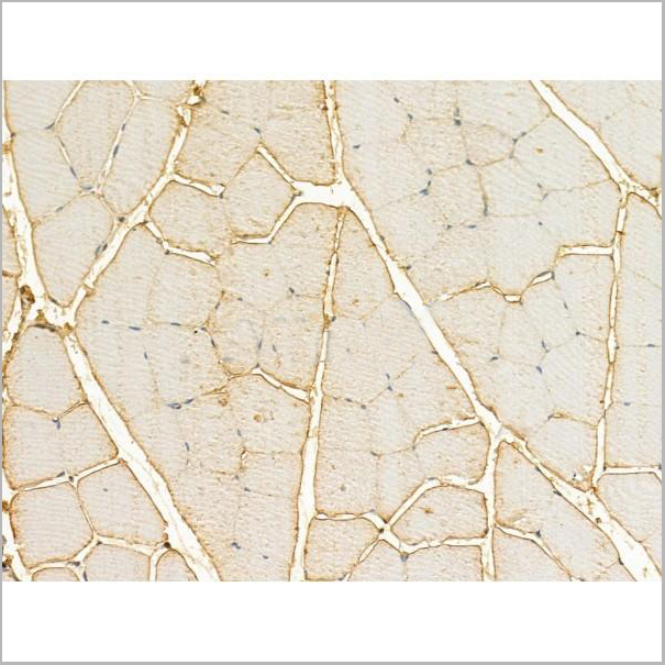
 IHC (Immunohistchemistry)
(Immunohistochemical analysis of paraffin-embedded liver cancer tissues using CCL2 mouse mAb with DAB staining.)
IHC (Immunohistchemistry)
(Immunohistochemical analysis of paraffin-embedded liver cancer tissues using CCL2 mouse mAb with DAB staining.)
CCL2, Monoclonal Antibody (Cat# AAA14116)
Full Name
Anti-CCL2 Mouse mAb
Gene Names
CCL2; MCP-1
Reactivity
Human, Mouse, Monkey
Applications
Western Blot, Immunohistochemistry, Immunocytochemistry, Flow Cytometry
Pricing


 IF (Immunofluorescence)
(Immunofluorescence analysis of NIH-3T3 cells using IFNAR1 Rabbit pAb at dilution of 1:100 (40x lens). Blue: DAPI for nuclear staining.)
IF (Immunofluorescence)
(Immunofluorescence analysis of NIH-3T3 cells using IFNAR1 Rabbit pAb at dilution of 1:100 (40x lens). Blue: DAPI for nuclear staining.)
IFNAR1, Polyclonal Antibody (Cat# AAA28371)
Full Name
IFNAR1 Rabbit pAb
Gene Names
IFNAR1; AVP; IFRC; IFNAR; IFNBR; IFN-alpha-REC
Reactivity
Human, Mouse, Rat
Applications
WB, IHC, IF
Purity
Affinity purification
Pricing


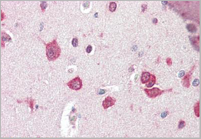
 IHC (Immunohistchemistry)
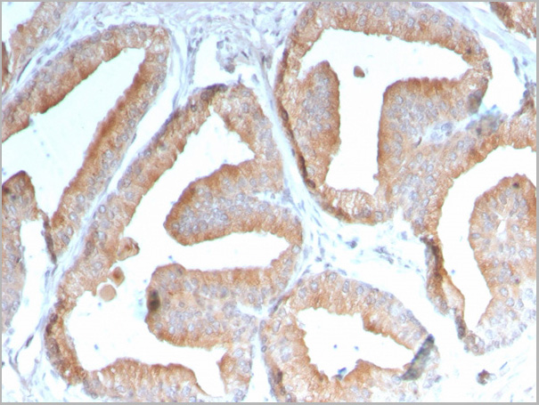
(Figure 6. IHC analysis of Flt3 / CD135 using anti-Flt3 / CD135 antibody (AAA19130).Flt3 / CD135 was detected in paraffin-embedded section of mouse kidney tissue. Heat mediated antigen retrieval was performed in citrate buffer (pH6, epitope retrieval solution) for 20 mins. The tissue section was blocked with 10% goat serum. The tissue section was then incubated with 1ug/ml rabbit anti-Flt3 / CD135 Antibody (AAA19130) overnight at 4 degree C. Biotinylated goat anti-rabbit IgG was used as secondary antibody and incubated for 30 minutes at 37 degree C. The tissue section was developed using Strepavidin-Biotin-Complex (SABC) with DAB as the chromogen.)
IHC (Immunohistchemistry)
(Figure 6. IHC analysis of Flt3 / CD135 using anti-Flt3 / CD135 antibody (AAA19130).Flt3 / CD135 was detected in paraffin-embedded section of mouse kidney tissue. Heat mediated antigen retrieval was performed in citrate buffer (pH6, epitope retrieval solution) for 20 mins. The tissue section was blocked with 10% goat serum. The tissue section was then incubated with 1ug/ml rabbit anti-Flt3 / CD135 Antibody (AAA19130) overnight at 4 degree C. Biotinylated goat anti-rabbit IgG was used as secondary antibody and incubated for 30 minutes at 37 degree C. The tissue section was developed using Strepavidin-Biotin-Complex (SABC) with DAB as the chromogen.)
Flt3/CD135, Polyclonal Antibody (Cat# AAA19130)
Full Name
Anti-Flt3/CD135 Picoband Antibody
Reactivity
Mouse, Rat
No cross reactivity with other proteins.
No cross reactivity with other proteins.
Applications
EIA, IHC, WB
Purity
Immunogen affinity purified
Pricing


 ICC (Immunocytochemistry)
(ICC staining RANTES in HUVEC cells (green). The nuclear counter stain is DAPI (blue). Cells were fixed in paraformaldehyde, permeabilised with 0.25% Triton X100/PBS.)
ICC (Immunocytochemistry)
(ICC staining RANTES in HUVEC cells (green). The nuclear counter stain is DAPI (blue). Cells were fixed in paraformaldehyde, permeabilised with 0.25% Triton X100/PBS.)
RANTES, Monoclonal Antibody (Cat# AAA30380)
Full Name
RANTES Antibody
Gene Names
CCL5; SISd; eoCP; SCYA5; RANTES; TCP228; D17S136E; SIS-delta
Reactivity
Human, Mouse
Applications
Western Blot, Immunocytochemistry, Immunofluorescence, Immunohistochemistry
Purity
ProA affinity purified
Pricing
 Standard Curve (Sample)
growth differentiation factor 15, ELISA Kit (Cat# AAA18194)
Full Name
Mouse Growth/differentiation factor 15, GDF15 ELISA Kit
Gene Names
Gdf15; SBF; MIC-1; NAG-1
Reactivity
Mouse
Pricing


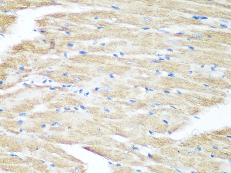
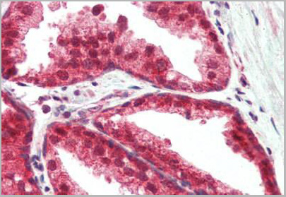
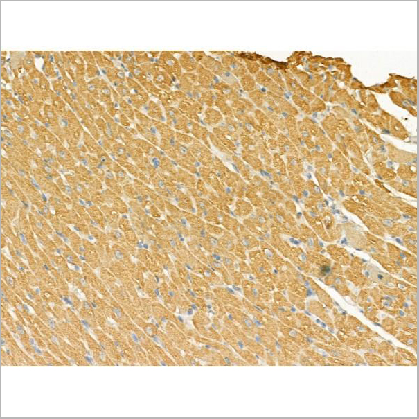
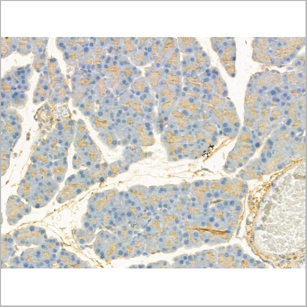
 IHC (Immunohistchemistry)
(DAB staining on IHC-P;Samples: Mouse Testis Tissue.)
IHC (Immunohistchemistry)
(DAB staining on IHC-P;Samples: Mouse Testis Tissue.)
Monocyte Chemotactic Protein 3 (MCP3), Antibody (Cat# AAA20140)
Full Name
Polyclonal Antibody to Monocyte Chemotactic Protein 3 (MCP3)
Gene Names
Ccl7; fic; marc; mcp3; MCP-3; Scya7
Reactivity
Mouse
Applications
WB, IHC, ICC, IP
Purity
Antigen-specific affinity chromatography followed by Protein A affinity chromatography
Pricing
 Standard Curve (Sample)
Growth Differentiation Factor 15, ELISA Kit (Cat# AAA15970)
Full Name
Mouse Growth Differentiation Factor 15 ELISA Kit
Gene Names
GDF15; PDF; MIC1; PLAB; MIC-1; NAG-1; PTGFB; GDF-15
Reactivity
Mouse
Pricing


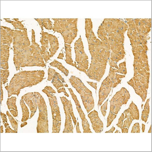
 WB (Western Blot)
(Western Blot detection against Immunogen (51.96kD).)
WB (Western Blot)
(Western Blot detection against Immunogen (51.96kD).)
FAM3B, Monoclonal Antibody (Cat# AAA14793)
Full Name
FAM3B (Protein FAM3B, Cytokine-like Protein 2-21, Pancreatic-derived Factor, PANDER, UNQ320/PRO365, PRED44, C21orf76, C21orf11, D21M16SJHU19e)
Reactivity
Human
Applications
EL/EIA, WB, IP, IHC
Purity
Affinity Purified
Purified by Protein A affinity chromatography.
Purified by Protein A affinity chromatography.
Pricing


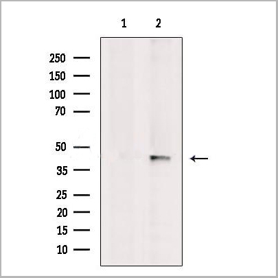
 WB (Western Blot)
(ASB9 monoclonal antibody, Western Blot analysis of ASB9 expression in HepG2.)
WB (Western Blot)
(ASB9 monoclonal antibody, Western Blot analysis of ASB9 expression in HepG2.)
ASB9, Monoclonal Antibody (Cat# AAA14792)
Full Name
ASB9 (Ankyrin Repeat and SOCS Box Protein 9, ASB-9, DKFZp564L0862, FLJ20636, MGC4954)
Reactivity
Human
Applications
EL/EIA, WB, IP, IHC
Purity
Affinity Purified
Purified by Protein A affinity chromatography.
Purified by Protein A affinity chromatography.
Pricing
 Application Data
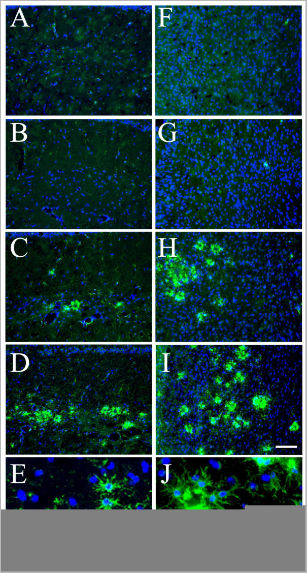
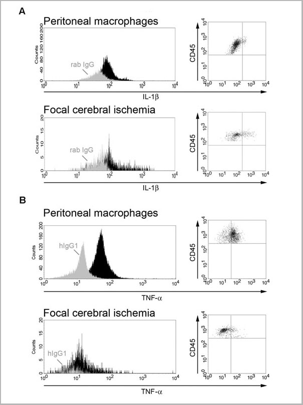
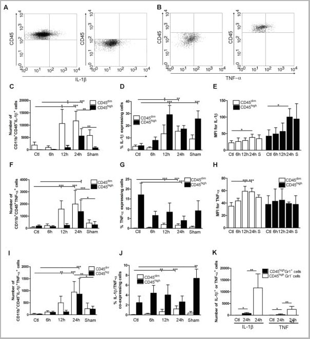
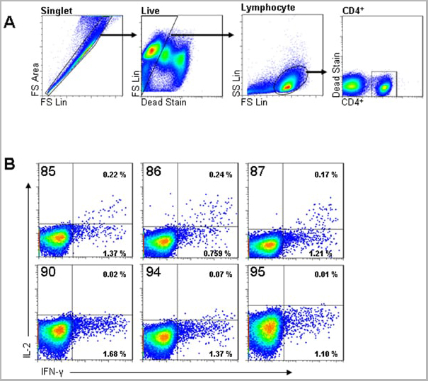
(Published customer image: Cytokine expression in segregated populations of cells following stroke. (A, B) Dot plots showing CD11b+CD45high macrophages/granulocytes (upper right quadrants) and CD11b+CD45dim microglia (bottom right quadrants) expressing IL-1beta (A) or TNF-a (B). (C-J) Bar graphs showing numbers and proportions of IL-1beta (C, D), TNF-a (F, G) and IL-1beta/TNF-a co-expressing (I, J) CD11b+CD45dim microglia and CD11b+CD45high macrophages/granulocytes in unmanipulated control mice (n = 10), in mice 6 (n = 7), 12 (n = 7), or 24 hours after pMCAO (n = 10), and in sham-operated mice 24 hours after pMCAO (n = 7). (E, H) Comparison of the MFI values for IL-1beta (E) and TNF-a (H) in viable CD11b+CD45dim microglia and CD11b+CD45high macrophages/granulocytes in unmanipulated mice, in mice 6, 12, or 24 hours after pMCAO, and in sham-operated mice 24 hours after pMCAO. Macrophages/granulocytes express significantly more IL-1beta than do microglial in unmanipulated mice, in mice 6, 12, or 24 hours after pMCAO, and in sham-operated mice 24 hours after pMCAO (E), whereas microglial cells express significantly higher levels of TNF-a than do macrophages/granulocytes at 12 h and 24 hours, and in sham-operated mice 24 hours after pMCAO (H). (K) CD11b+CD45highGr1- macrophages and not CD11b+CD45highGr1+ granulocytes are the main producers of IL-1beta and TNF-a 24 hours after pMCAO. *P < 0.05, **P < 0.01, and ***P < 0.001.From: http://www.jneuroinflammation.com/content/5/1/46.)
Application Data
(Published customer image: Cytokine expression in segregated populations of cells following stroke. (A, B) Dot plots showing CD11b+CD45high macrophages/granulocytes (upper right quadrants) and CD11b+CD45dim microglia (bottom right quadrants) expressing IL-1beta (A) or TNF-a (B). (C-J) Bar graphs showing numbers and proportions of IL-1beta (C, D), TNF-a (F, G) and IL-1beta/TNF-a co-expressing (I, J) CD11b+CD45dim microglia and CD11b+CD45high macrophages/granulocytes in unmanipulated control mice (n = 10), in mice 6 (n = 7), 12 (n = 7), or 24 hours after pMCAO (n = 10), and in sham-operated mice 24 hours after pMCAO (n = 7). (E, H) Comparison of the MFI values for IL-1beta (E) and TNF-a (H) in viable CD11b+CD45dim microglia and CD11b+CD45high macrophages/granulocytes in unmanipulated mice, in mice 6, 12, or 24 hours after pMCAO, and in sham-operated mice 24 hours after pMCAO. Macrophages/granulocytes express significantly more IL-1beta than do microglial in unmanipulated mice, in mice 6, 12, or 24 hours after pMCAO, and in sham-operated mice 24 hours after pMCAO (E), whereas microglial cells express significantly higher levels of TNF-a than do macrophages/granulocytes at 12 h and 24 hours, and in sham-operated mice 24 hours after pMCAO (H). (K) CD11b+CD45highGr1- macrophages and not CD11b+CD45highGr1+ granulocytes are the main producers of IL-1beta and TNF-a 24 hours after pMCAO. *P < 0.05, **P < 0.01, and ***P < 0.001.From: http://www.jneuroinflammation.com/content/5/1/46.)
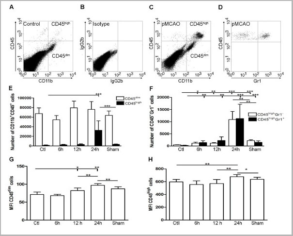
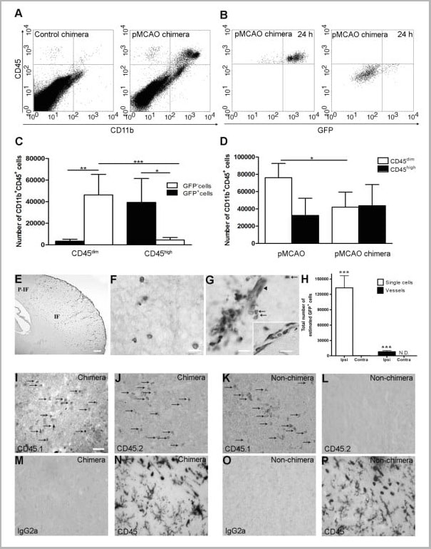
CD45, Monoclonal Antibody (Cat# AAA11896)
Full Name
RAT ANTI MOUSE CD45
Gene Names
Ptprc; loc; B220; Cd45; L-CA; Ly-5; T200; CD45R; Lyt-4
Applications
Immunohistochemistry, Flow Cytometry, Immunofluorescence, Immunoprecipitation
Pricing
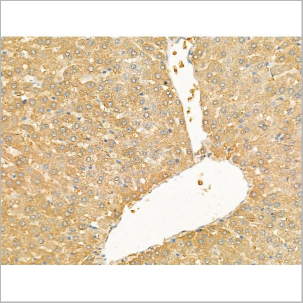
 IHC (Immunohistochemistry)
(Anti-GDF15 antibody IHC of human placenta. Immunohistochemistry of formalin-fixed, paraffin-embedded tissue after heat-induced antigen retrieval. Antibody dilution 10 ug/ml.)
IHC (Immunohistochemistry)
(Anti-GDF15 antibody IHC of human placenta. Immunohistochemistry of formalin-fixed, paraffin-embedded tissue after heat-induced antigen retrieval. Antibody dilution 10 ug/ml.)
GDF15, Monoclonal Antibody (Cat# AAA12357)
Full Name
Rat Monoclonal [clone 6D12.H10.E4] (IgG) to Human GDF15
Gene Names
GDF15; PDF; MIC1; PLAB; MIC-1; NAG-1; PTGFB; GDF-15
Reactivity
Chimpanzee, Macaque
Applications
Immunohistochemistry, Immunohistochemistry, Western Blot
Purity
Protein G Purified chromatography
Pricing
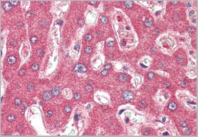 IHC (Immunohistochemistry)
(Anti-TSLP antibody IHC of human liver. Immunohistochemistry of formalin-fixed, paraffin-embedded tissue after heat-induced antigen retrieval. Antibody concentration 5 ug/ml.)
IHC (Immunohistochemistry)
(Anti-TSLP antibody IHC of human liver. Immunohistochemistry of formalin-fixed, paraffin-embedded tissue after heat-induced antigen retrieval. Antibody concentration 5 ug/ml.)
TSLP, Polyclonal Antibody (Cat# AAA12321)
Full Name
Rabbit Polyclonal to Human TSLP
Reactivity
Human, Mouse
Applications
Immunohistochemistry, Western Blot
Purity
Immunoaffinity Purified
Pricing
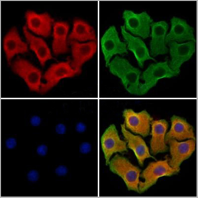 IF (Immunofluorescence)
(AAA31197 staining Hela cells by IF/ICC. The samples were fixed with PFA and permeabilized in 0.1% Triton X-100,then blocked in 10% serum for 45 minutes at 25°C. Samples were then incubated with primary Ab(AAA31197 1:200) and mouse anti-beta tubulin Ab(T0023 1:200) for 1 hour at 37°C. An AlexaFluor594 conjugated goat anti-rabbit IgG(H+L) Ab(Red) and an AlexaFluor488 conjugated goat anti-mouse IgG(H+L) Ab(Green) were used as the secondary antibody.The nuclear counter stain is DAPI(blue).)
IF (Immunofluorescence)
(AAA31197 staining Hela cells by IF/ICC. The samples were fixed with PFA and permeabilized in 0.1% Triton X-100,then blocked in 10% serum for 45 minutes at 25°C. Samples were then incubated with primary Ab(AAA31197 1:200) and mouse anti-beta tubulin Ab(T0023 1:200) for 1 hour at 37°C. An AlexaFluor594 conjugated goat anti-rabbit IgG(H+L) Ab(Red) and an AlexaFluor488 conjugated goat anti-mouse IgG(H+L) Ab(Green) were used as the secondary antibody.The nuclear counter stain is DAPI(blue).)
CXCL16, Polyclonal Antibody (Cat# AAA31197)
Full Name
CXCL16 Antibody
Gene Names
CXCL16; SRPSOX; CXCLG16; SR-PSOX
Reactivity
Human, Mouse, Rat
Applications
Western Blot, Immunofluorescence, Immunocytochemistry
Purity
The antiserum was purified by peptide affinity chromatography using SulfoLink Coupling Resin (Thermo Fisher Scientific).
Pricing
 Application Data
(Analysis of Protein Array containing more than 19,000 full-length human proteins using ICOS-L Mouse Monoclonal Antibody (ICOSL/3111). Z- and S- Score: The Z-score represents the strength of a signal that a monoclonal antibody (MAb) (in combination with a fluorescently-tagged anti-IgG secondary antibody) produces when binding to a particular protein on the HuProtTM array. Z-scores are described in units of standard deviations (SD’s) above the mean value of all signals generated on that array. If targets on HuProtTM are arranged in descending order of the Z-score, the S-score is the difference (also in units of SD’s) between the Z-score. S-score therefore represents the relative target specificity of a MAb to its intended target. A MAb is considered to specific to its intended target, if the MAb has an S-score of at least 2.5. For example, if a MAb binds to protein X with a Z-score of 43 and to protein Y with a Z-score of 14, then the S-score for the binding of that MAb to protein X is equal to 29.)
Application Data
(Analysis of Protein Array containing more than 19,000 full-length human proteins using ICOS-L Mouse Monoclonal Antibody (ICOSL/3111). Z- and S- Score: The Z-score represents the strength of a signal that a monoclonal antibody (MAb) (in combination with a fluorescently-tagged anti-IgG secondary antibody) produces when binding to a particular protein on the HuProtTM array. Z-scores are described in units of standard deviations (SD’s) above the mean value of all signals generated on that array. If targets on HuProtTM are arranged in descending order of the Z-score, the S-score is the difference (also in units of SD’s) between the Z-score. S-score therefore represents the relative target specificity of a MAb to its intended target. A MAb is considered to specific to its intended target, if the MAb has an S-score of at least 2.5. For example, if a MAb binds to protein X with a Z-score of 43 and to protein Y with a Z-score of 14, then the S-score for the binding of that MAb to protein X is equal to 29.)
ICOS-L/ICOS Ligand/B7RP-1 (Immuno-Oncology Target), Monoclonal Antibody (Cat# AAA23916)
Full Name
ICOS-L/ICOS Ligand/B7RP-1 (Immuno-Oncology Target)
Gene Names
ICOSLG; B7h; B7H2; GL50; B7-H2; B7RP1; CD275; ICOSL; LICOS; B7RP-1; ICOS-L
Reactivity
Human
Applications
Flow Cytometry, Immunofluorescence, Immunohistochemistry
Purity
Purified Ab with BSA and Azide at 200ug/ml
Pricing


 Application Data
(Staining of New Zealand Black mouse peripheral blood granulocytes with Rat anti Mouse Ly-6B.2 conjugated to FITC Data)
Application Data
(Staining of New Zealand Black mouse peripheral blood granulocytes with Rat anti Mouse Ly-6B.2 conjugated to FITC Data)
Ly-6B.2 ALLOANTIGEN, Monoclonal Antibody (Cat# AAA12197)
Full Name
RAT ANTI MOUSE Ly-6B.2 ALLOANTIGEN:RPE
Applications
Flow Cytometry
Pricing


 Application Data
(Staining of New Zealand Black mouse peripheral blood granulocytes with Rat anti Mouse Ly-6B.2 conjugated to FITC Data)
Application Data
(Staining of New Zealand Black mouse peripheral blood granulocytes with Rat anti Mouse Ly-6B.2 conjugated to FITC Data)
Ly-6B.2 ALLOANTIGEN, Monoclonal Antibody (Cat# AAA12193)
Full Name
RAT ANTI MOUSE Ly-6B.2 ALLOANTIGEN:FITC
Applications
Flow Cytometry
Pricing


 Application Data
(Staining of New Zealand Black mouse peripheral blood granulocytes with Rat anti Mouse Ly-6B.2 conjugated to FITC Data)
Application Data
(Staining of New Zealand Black mouse peripheral blood granulocytes with Rat anti Mouse Ly-6B.2 conjugated to FITC Data)
Ly-6B.2 ALLOANTIGEN, Monoclonal Antibody (Cat# AAA12192)
Full Name
RAT ANTI MOUSE Ly-6B.2 ALLOANTIGEN:FITC
Applications
Flow Cytometry
Pricing


 Application Data
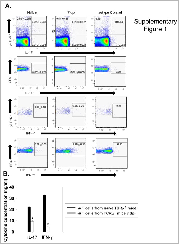
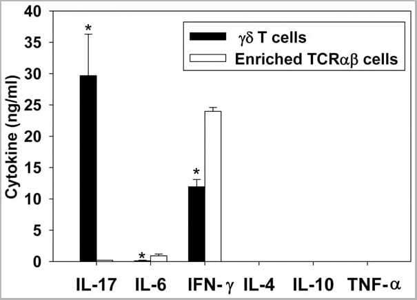
(Published customer image: gamma delta T cells are the primary source of IL-17 during B. abortus infection. C57BL/6 mice were infected i.p. with 5x10⁴ CFUs of B. abortus 2308, and two weeks later gamma delta T cells (>95% purity) and an enriched TCRalphabeta (~55% CD4+, 25% CD8+) cell fraction were isolated from the spleens of infected mice. Cells were stimulated with 500 ng/ml ionomycin and 50 ng/ml PMA for three days, and cell-free supernatants from triplicate wells were assayed for cytokine production via ELISA. The mean +/- SD is shown; * P)
Application Data
(Published customer image: gamma delta T cells are the primary source of IL-17 during B. abortus infection. C57BL/6 mice were infected i.p. with 5x10⁴ CFUs of B. abortus 2308, and two weeks later gamma delta T cells (>95% purity) and an enriched TCRalphabeta (~55% CD4+, 25% CD8+) cell fraction were isolated from the spleens of infected mice. Cells were stimulated with 500 ng/ml ionomycin and 50 ng/ml PMA for three days, and cell-free supernatants from triplicate wells were assayed for cytokine production via ELISA. The mean +/- SD is shown; * P)
IFN GAMMA, Monoclonal Antibody (Cat# AAA12093)
Full Name
MOUSE ANTI BOVINE INTERFERON GAMMA
Reactivity
Dog, Dolphin, Ferret, Fin Whale, Goat, Horse, Human, Mink, Rabbit, Pig, Sheep.
Based on sequence similarity, is expected to react with: Mustelid
N.B. Antibody reactivity and working conditions may vary between species.
Based on sequence similarity, is expected to react with: Mustelid
N.B. Antibody reactivity and working conditions may vary between species.
Applications
Flow Cytometry
Pricing


 Application Data
(Staining of New Zealand Black mouse peripheral blood granulocytes with Rat anti Mouse Ly-6B.2 conjugated to FITC Data)
Application Data
(Staining of New Zealand Black mouse peripheral blood granulocytes with Rat anti Mouse Ly-6B.2 conjugated to FITC Data)
Ly-6B.2 ALLOANTIGEN, Monoclonal Antibody (Cat# AAA12194)
Full Name
RAT ANTI MOUSE Ly-6B.2 ALLOANTIGEN
Applications
Immunohistochemistry, Flow Cytometry, Immunofluorescence, Immunohistochemistry, Western Blot
Pricing


 Application Data
(Staining of New Zealand Black mouse peripheral blood granulocytes with Rat anti Mouse Ly-6B.2 conjugated to FITC Data)
Application Data
(Staining of New Zealand Black mouse peripheral blood granulocytes with Rat anti Mouse Ly-6B.2 conjugated to FITC Data)
Ly-6B.2 ALLOANTIGEN, Monoclonal Antibody (Cat# AAA12198)
Full Name
RAT ANTI MOUSE Ly-6B.2 ALLOANTIGEN:RPE
Applications
Flow Cytometry
Pricing


 Application Data
(Staining of New Zealand Black mouse peripheral blood granulocytes with Rat anti Mouse Ly-6B.2 conjugated to FITC Data)
Application Data
(Staining of New Zealand Black mouse peripheral blood granulocytes with Rat anti Mouse Ly-6B.2 conjugated to FITC Data)
Ly-6B.2 ALLOANTIGEN, Monoclonal Antibody (Cat# AAA12195)
Full Name
RAT ANTI MOUSE Ly-6B.2 ALLOANTIGEN
Applications
Immunohistochemistry, Flow Cytometry, Immunofluorescence, Immunohistochemistry, Western Blot
Pricing


 Application Data
(Published customer image: gamma delta T cells are the primary source of IL-17 during B. abortus infection. C57BL/6 mice were infected i.p. with 5x10⁴ CFUs of B. abortus 2308, and two weeks later gamma delta T cells (>95% purity) and an enriched TCRalphabeta (~55% CD4+, 25% CD8+) cell fraction were isolated from the spleens of infected mice. Cells were stimulated with 500 ng/ml ionomycin and 50 ng/ml PMA for three days, and cell-free supernatants from triplicate wells were assayed for cytokine production via ELISA. The mean +/- SD is shown; * P)
Application Data
(Published customer image: gamma delta T cells are the primary source of IL-17 during B. abortus infection. C57BL/6 mice were infected i.p. with 5x10⁴ CFUs of B. abortus 2308, and two weeks later gamma delta T cells (>95% purity) and an enriched TCRalphabeta (~55% CD4+, 25% CD8+) cell fraction were isolated from the spleens of infected mice. Cells were stimulated with 500 ng/ml ionomycin and 50 ng/ml PMA for three days, and cell-free supernatants from triplicate wells were assayed for cytokine production via ELISA. The mean +/- SD is shown; * P)
IFN GAMMA, Monoclonal Antibody (Cat# AAA12095)
Full Name
MOUSE ANTI BOVINE INTERFERON GAMMA:FITC
Applications
Flow Cytometry
Pricing


 Application Data
(Published customer image: gamma delta T cells are the primary source of IL-17 during B. abortus infection. C57BL/6 mice were infected i.p. with 5x10⁴ CFUs of B. abortus 2308, and two weeks later gamma delta T cells (>95% purity) and an enriched TCRalphabeta (~55% CD4+, 25% CD8+) cell fraction were isolated from the spleens of infected mice. Cells were stimulated with 500 ng/ml ionomycin and 50 ng/ml PMA for three days, and cell-free supernatants from triplicate wells were assayed for cytokine production via ELISA. The mean +/- SD is shown; * P)
Application Data
(Published customer image: gamma delta T cells are the primary source of IL-17 during B. abortus infection. C57BL/6 mice were infected i.p. with 5x10⁴ CFUs of B. abortus 2308, and two weeks later gamma delta T cells (>95% purity) and an enriched TCRalphabeta (~55% CD4+, 25% CD8+) cell fraction were isolated from the spleens of infected mice. Cells were stimulated with 500 ng/ml ionomycin and 50 ng/ml PMA for three days, and cell-free supernatants from triplicate wells were assayed for cytokine production via ELISA. The mean +/- SD is shown; * P)
IFN GAMMA, Monoclonal Antibody (Cat# AAA12096)
Full Name
MOUSE ANTI BOVINE INTERFERON GAMMA:RPE
Applications
Flow Cytometry
Pricing


 Application Data
(Staining of New Zealand Black mouse peripheral blood granulocytes with Rat anti Mouse Ly-6B.2 conjugated to FITC Data)
Application Data
(Staining of New Zealand Black mouse peripheral blood granulocytes with Rat anti Mouse Ly-6B.2 conjugated to FITC Data)
Ly-6B.2 ALLOANTIGEN, Monoclonal Antibody (Cat# AAA12190)
Full Name
RAT ANTI MOUSE Ly-6B.2 ALLOANTIGEN:FITC
Applications
Flow Cytometry
Pricing


 Application Data
(Staining of New Zealand Black mouse peripheral blood granulocytes with Rat anti Mouse Ly-6B.2 conjugated to FITC Data)
Application Data
(Staining of New Zealand Black mouse peripheral blood granulocytes with Rat anti Mouse Ly-6B.2 conjugated to FITC Data)
Ly-6B.2 ALLOANTIGEN, Monoclonal Antibody (Cat# AAA12189)
Full Name
RAT ANTI MOUSE Ly-6B.2 ALLOANTIGEN:Low Endotoxin
Applications
Immunohistochemistry, Flow Cytometry, Immunofluorescence, Immunohistochemistry, Western Blot
Pricing


 Application Data
(Staining of New Zealand Black mouse peripheral blood granulocytes with Rat anti Mouse Ly-6B.2 conjugated to FITC AAA12191)
Application Data
(Staining of New Zealand Black mouse peripheral blood granulocytes with Rat anti Mouse Ly-6B.2 conjugated to FITC AAA12191)
Ly-6B.2 ALLOANTIGEN, Monoclonal Antibody (Cat# AAA12191)
Full Name
RAT ANTI MOUSE Ly-6B.2 ALLOANTIGEN:FITC
Applications
Flow Cytometry
Pricing


 Application Data
(Staining of New Zealand Black mouse peripheral blood granulocytes with Rat anti Mouse Ly-6B.2 conjugated to FITC Data)
Application Data
(Staining of New Zealand Black mouse peripheral blood granulocytes with Rat anti Mouse Ly-6B.2 conjugated to FITC Data)
Ly-6B.2 ALLOANTIGEN, Monoclonal Antibody (Cat# AAA12188)
Full Name
RAT ANTI MOUSE Ly-6B.2 ALLOANTIGEN:Biotin
Applications
Flow Cytometry
Pricing


 Application Data
(Published customer image: gamma delta T cells are the primary source of IL-17 during B. abortus infection. C57BL/6 mice were infected i.p. with 5x10⁴ CFUs of B. abortus 2308, and two weeks later gamma delta T cells (>95% purity) and an enriched TCRalphabeta (~55% CD4+, 25% CD8+) cell fraction were isolated from the spleens of infected mice. Cells were stimulated with 500 ng/ml ionomycin and 50 ng/ml PMA for three days, and cell-free supernatants from triplicate wells were assayed for cytokine production via ELISA. The mean +/- SD is shown; * P)
Application Data
(Published customer image: gamma delta T cells are the primary source of IL-17 during B. abortus infection. C57BL/6 mice were infected i.p. with 5x10⁴ CFUs of B. abortus 2308, and two weeks later gamma delta T cells (>95% purity) and an enriched TCRalphabeta (~55% CD4+, 25% CD8+) cell fraction were isolated from the spleens of infected mice. Cells were stimulated with 500 ng/ml ionomycin and 50 ng/ml PMA for three days, and cell-free supernatants from triplicate wells were assayed for cytokine production via ELISA. The mean +/- SD is shown; * P)
IFN GAMMA, Monoclonal Antibody (Cat# AAA12094)
Full Name
MOUSE ANTI BOVINE INTERFERON GAMMA:Biotin
Applications
Flow Cytometry
Pricing


 Application Data
(Staining of New Zealand Black mouse peripheral blood granulocytes with Rat anti Mouse Ly-6B.2 conjugated to FITC Data)
Application Data
(Staining of New Zealand Black mouse peripheral blood granulocytes with Rat anti Mouse Ly-6B.2 conjugated to FITC Data)
Ly-6B.2 ALLOANTIGEN, Monoclonal Antibody (Cat# AAA12196)
Full Name
RAT ANTI MOUSE Ly-6B.2 ALLOANTIGEN
Applications
Immunohistochemistry, Flow Cytometry, Immunofluorescence, Immunohistochemistry, Western Blot
Pricing