Filters
Clonality
Type
Reactivity
Gene Name
Isotype
Host
Application
Clone
45 results for "Mouse IgG2a Isotype Control" - showing 1-45


 Application Data
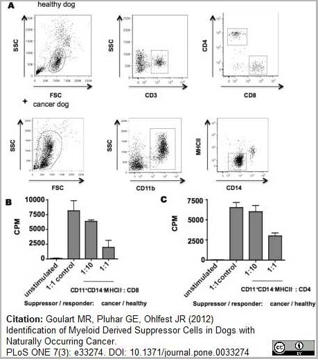
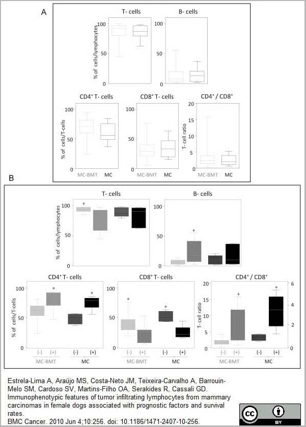
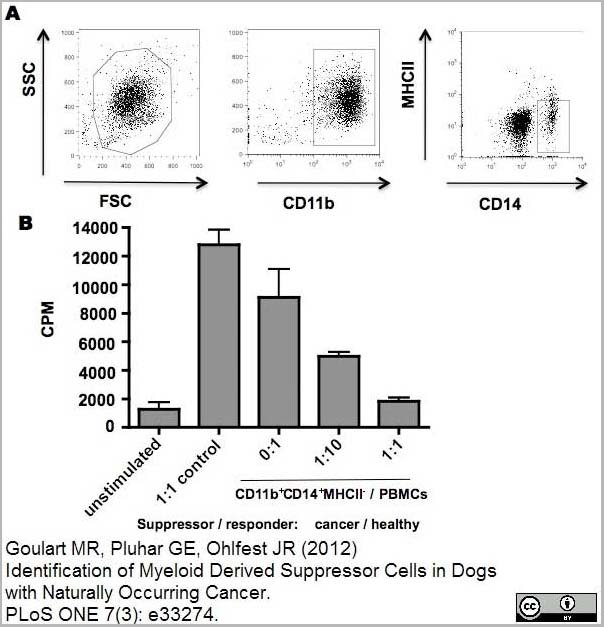
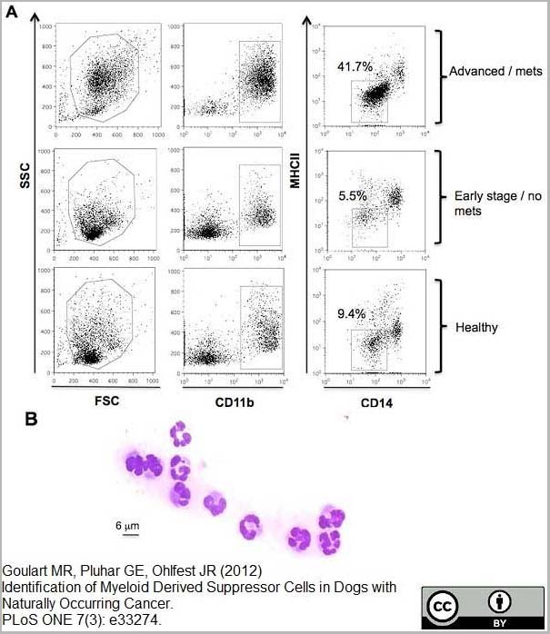
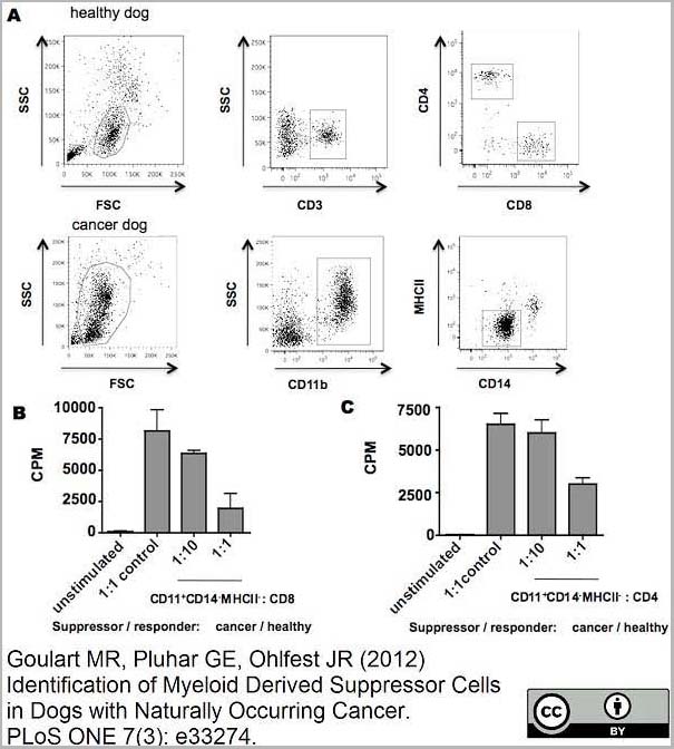
(Published customer image:RPE conjugated Mouse anti Canine CD4 antibody, clone YKIC302.9used for the assessment of CD4 levels on canine cells by flow cytometry.Image caption:Immunophenotypic profile of tumor infiltrating lymphocyte in canine mammary carcinomas. Analysis of tumor infiltrating T-cells, B-lymphocytes and T-cell subsets from MC-BMT or MC (A), further subcategorized according to the absence (-) or presence (+) of lymph node metastasis (-) (B). Lymphocyte populations and subsets were identified by flow cytometric immunostaining as described in Material and Methods. Data were expressed as percentage of positive cells within gated lymphocytes and CD4+/CD8+ T-cell ratio. Significant differences at p < 0.05 are highlighted by asterisk.)
Application Data
(Published customer image:RPE conjugated Mouse anti Canine CD4 antibody, clone YKIC302.9used for the assessment of CD4 levels on canine cells by flow cytometry.Image caption:Immunophenotypic profile of tumor infiltrating lymphocyte in canine mammary carcinomas. Analysis of tumor infiltrating T-cells, B-lymphocytes and T-cell subsets from MC-BMT or MC (A), further subcategorized according to the absence (-) or presence (+) of lymph node metastasis (-) (B). Lymphocyte populations and subsets were identified by flow cytometric immunostaining as described in Material and Methods. Data were expressed as percentage of positive cells within gated lymphocytes and CD4+/CD8+ T-cell ratio. Significant differences at p < 0.05 are highlighted by asterisk.)
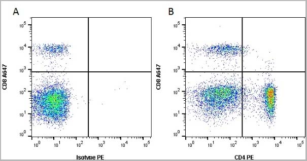
CD4, Monoclonal Antibody (Cat# AAA12278)
Full Name
RAT ANTI DOG CD4: APC
Reactivity
Dog
Applications
Flow Cytometry
Pricing


 FCM (Flow Cytometry)
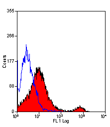
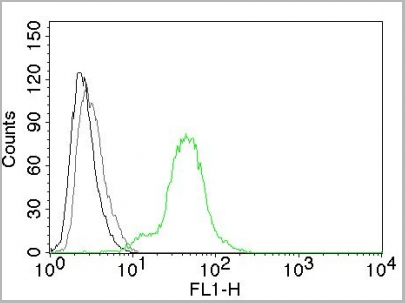
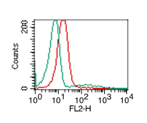
(Overlay histogram showing HepG2 cells stained with AAA28061 (red line) at 1:100. The cells were fixed in 4% formaldehyde and permeated by 0.2% TritonX-100. Then 10% normal goat serum was Incubated to block non-specific protein-protein interactions followed by the antibody (1ug/1*106cells) for 1 h at 4 degree C. The secondary antibody used was FITC-conjugated Goat Anti-Mouse IgG(H+L) at 1/100 dilution for 30min at 4 degree C. Isotype control antibody (green line) was mouse IgG2b (1ug/1*106cells) used under the same conditions. Acquisition of >10,000 events was performed.)
FCM (Flow Cytometry)
(Overlay histogram showing HepG2 cells stained with AAA28061 (red line) at 1:100. The cells were fixed in 4% formaldehyde and permeated by 0.2% TritonX-100. Then 10% normal goat serum was Incubated to block non-specific protein-protein interactions followed by the antibody (1ug/1*106cells) for 1 h at 4 degree C. The secondary antibody used was FITC-conjugated Goat Anti-Mouse IgG(H+L) at 1/100 dilution for 30min at 4 degree C. Isotype control antibody (green line) was mouse IgG2b (1ug/1*106cells) used under the same conditions. Acquisition of >10,000 events was performed.)
CD63, Monoclonal Antibody (Cat# AAA28061)
Full Name
CD63 Monoclonal Antibody
Gene Names
Cd63; ME491; C75951; Tspan30
Reactivity
Human
Applications
Western Blot, Immunohistochemistry, Immunofluorescence, Flow Cytometry
Purity
>95%, Protein A purified
Pricing


 Application Data
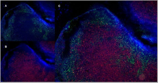
(Published Customer Image:Mouse CD31 antibody, clone ER-MP12 used for the demonstration of vasculature in mouse brain by immunofluorescence.Image caption:Inhibition of 2-AG hydrolysis reduces LPS-induced BBB permeability. a, b Fibrinogen levels in b plasma and the a ratio of brain to plasma fibrinogen were assessed by ELISA. n?=?5/7 mice per group. c, d Fluorescent immunostaining in the striatum for fibrinogen (red) and vascular marker (CD31; green) demonstrated leakage of fibrinogen into the brain with vehicle treatment, whereas vascular integrity was preserved when (e, f) MAGL was inhibited. g Extravascular fibrinogen was semi-quantitated in fluorescently labeled sections of the striatum. Bar graphs were plotted with mean?+/-SEM and data analyzed using one-way analysis of variance (ANOVA) with Tukey post-hoc comparisons. n = 5/7 mice per group. Significance is shown as *p?)
Application Data
(Published Customer Image:Mouse CD31 antibody, clone ER-MP12 used for the demonstration of vasculature in mouse brain by immunofluorescence.Image caption:Inhibition of 2-AG hydrolysis reduces LPS-induced BBB permeability. a, b Fibrinogen levels in b plasma and the a ratio of brain to plasma fibrinogen were assessed by ELISA. n?=?5/7 mice per group. c, d Fluorescent immunostaining in the striatum for fibrinogen (red) and vascular marker (CD31; green) demonstrated leakage of fibrinogen into the brain with vehicle treatment, whereas vascular integrity was preserved when (e, f) MAGL was inhibited. g Extravascular fibrinogen was semi-quantitated in fluorescently labeled sections of the striatum. Bar graphs were plotted with mean?+/-SEM and data analyzed using one-way analysis of variance (ANOVA) with Tukey post-hoc comparisons. n = 5/7 mice per group. Significance is shown as *p?)
CD31, Monoclonal Antibody (Cat# AAA12258)
Full Name
Rat Anti Mouse CD31: FITC
Gene Names
Pecam1; Cd31; Pecam; C85791; PECAM-1
Reactivity
Mouse
Applications
Flow Cytometry
Purity
Purified IgG prepared by affinity chromatography on Protein G from tissue culture supernatant
Pricing


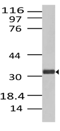
 SDS-PAGE
(SDS Page analysis of purified S100B Mouse Monoclonal Antibody (S100B/1012).)
SDS-PAGE
(SDS Page analysis of purified S100B Mouse Monoclonal Antibody (S100B/1012).)
S100B, Monoclonal Antibody (Cat# AAA23893)
Full Name
S100B (Astrocyte and Melanoma Marker)
Gene Names
S100B; NEF; S100; S100-B; S100beta
Reactivity
Human, Mouse, Rat, Cow. Others not known.
Applications
Flow Cytometry, Immunofluorescence, Western Blot, Immunohistochemistry
Pricing


 SDS-PAGE
(Purified ER-beta Mouse Monoclonal Antibody (ESR2/686). Confirmation of Integrity and Purity of Antibody.)
SDS-PAGE
(Purified ER-beta Mouse Monoclonal Antibody (ESR2/686). Confirmation of Integrity and Purity of Antibody.)
ER-beta1 (Estrogen Receptor beta-1), Monoclonal Antibody (Cat# AAA13846)
Full Name
ER-beta1 (Estrogen Receptor beta-1) Mouse Monoclonal Antibody
Gene Names
ESR2; Erb; ESRB; ESTRB; NR3A2; ER-BETA; ESR-BETA
Reactivity
Human
Applications
Flow Cytometry, Immunofluorescence, Western Blot, Immunohistochemistry
Pricing


 FCM (Flow Cytometry)
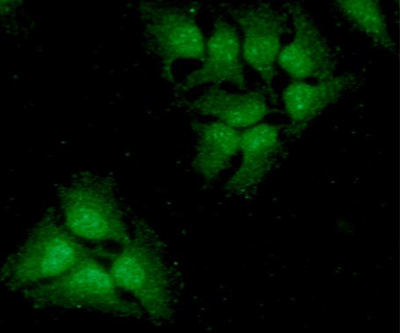
(Overlay histogram showing MCF-7 cells stained with CSB-MA013563A0m (red line) at 1:400. The cells were incubated in 1x PBS /10% normal goat serum to block non-specific protein-protein interactions followed by primary antibody for 1 h at 4 degree C. The secondary antibody used was FITC goat anti-mouse IgG(H+L) at 1/200 dilution for 1 h at 4 degree C. Isotype control antibody (green line) was used under the same conditions. Acquisition of >10,000 events was performed.)
FCM (Flow Cytometry)
(Overlay histogram showing MCF-7 cells stained with CSB-MA013563A0m (red line) at 1:400. The cells were incubated in 1x PBS /10% normal goat serum to block non-specific protein-protein interactions followed by primary antibody for 1 h at 4 degree C. The secondary antibody used was FITC goat anti-mouse IgG(H+L) at 1/200 dilution for 1 h at 4 degree C. Isotype control antibody (green line) was used under the same conditions. Acquisition of >10,000 events was performed.)
CD146, Monoclonal Antibody (Cat# AAA27014)
Full Name
CD146 Monoclonal Antibody
Gene Names
MCAM; CD146; MUC18; HEMCAM; METCAM; MelCAM
Reactivity
Human
Applications
Western Blot, Immunofluorescence, Flow Cytometry
Purity
>95%
Protein G Purified
Protein G Purified
Pricing


 Application Data
(Overlay histogram showing HL-60 cells stained with AAA27009 (red line) at 1:500. The cells were incubated in 1x PBS /10% normal goat serum to block non-specific protein-protein interactions followed by primary antibody for 1 h at 4 degree C.The secondary antibody used was FITC goat anti-mouse IgG (H+L) at 1/200 dilution for 1 h at 4 degree C. Isotype control antibody (green line) was used under the same conditions. Acquisition of >10, 000 events was performed.)
Application Data
(Overlay histogram showing HL-60 cells stained with AAA27009 (red line) at 1:500. The cells were incubated in 1x PBS /10% normal goat serum to block non-specific protein-protein interactions followed by primary antibody for 1 h at 4 degree C.The secondary antibody used was FITC goat anti-mouse IgG (H+L) at 1/200 dilution for 1 h at 4 degree C. Isotype control antibody (green line) was used under the same conditions. Acquisition of >10, 000 events was performed.)
CD31, Monoclonal Antibody (Cat# AAA27009)
Full Name
CD31 Monoclonal Antibody
Gene Names
PECAM1; CD31; PECA1; GPIIA'; PECAM-1; endoCAM; CD31/EndoCAM
Reactivity
Human
Applications
Western Blot, Immunohistochemistry, Immunofluorescence, Flow Cytometry
Purity
>95%, Protein G purified
Pricing


 FCM (Flow Cytometry)
(Figure 12. Flow Cytometry analysis of A549 cells using anti-Transketolase/TKT antibody (AAA19367).Overlay histogram showing A549 cells stained with AAA19367 (Blue line). The cells were blocked with 10% normal goat serum. And then incubated with mouse anti- Transketolase/TKT Antibody (AAA19367, 1μg/1x106 cells) for 30 min at 20 degree C. DyLight®488 conjugated goat anti-mouse IgG (BA1126, 5-10μg/1x106 cells) was used as secondary antibody for 30 minutes at 20 degree C. Isotype control antibody (Green line) was mouse IgG (1μg/1x106) used under the same conditions. Unlabelled sample (Red line) was also used as a control.)
FCM (Flow Cytometry)
(Figure 12. Flow Cytometry analysis of A549 cells using anti-Transketolase/TKT antibody (AAA19367).Overlay histogram showing A549 cells stained with AAA19367 (Blue line). The cells were blocked with 10% normal goat serum. And then incubated with mouse anti- Transketolase/TKT Antibody (AAA19367, 1μg/1x106 cells) for 30 min at 20 degree C. DyLight®488 conjugated goat anti-mouse IgG (BA1126, 5-10μg/1x106 cells) was used as secondary antibody for 30 minutes at 20 degree C. Isotype control antibody (Green line) was mouse IgG (1μg/1x106) used under the same conditions. Unlabelled sample (Red line) was also used as a control.)
Transketolase/TKT, Monoclonal Antibody (Cat# AAA19367)
Full Name
Anti-Transketolase/TKT Antibody (monoclonal, 3E5)
Gene Names
TKT; TK; TKT1; HEL107
Reactivity
Human, Mouse, Rat
Applications
WB, IHC-P, ICC, IF, FC/FACS/FCM
Purity
Immunogen affinity purified.
Pricing


 Application Data
(Figure A. FITC conjugated Rat anti Mouse CD19 . Figure B. FITC conjugated Rat anti Mouse CD19 and SBV440 conjugated Rat anti Mouse CD45R . All experiments performed on mouse splenocytes in the presence of 10% mouse seru)
Application Data
(Figure A. FITC conjugated Rat anti Mouse CD19 . Figure B. FITC conjugated Rat anti Mouse CD19 and SBV440 conjugated Rat anti Mouse CD45R . All experiments performed on mouse splenocytes in the presence of 10% mouse seru)
CD19, Monoclonal Antibody (Cat# AAA12286)
Full Name
Rat anti Mouse CD19:Amethyst Orange
Gene Names
Cd19; AW495831
Reactivity
Mouse
Applications
Flow Cytometry
Purity
Purified IgG prepared by affinity chromatography on Protein G from tissue culture supernatant
Pricing


 Application Data
(Figure A. FITC conjugated mouse anti bovine CD2 and RPE conjugated mouse IgG1 isotype control . Figure B. FITC conjugated mouse anti bovine CD2 and RPE conjugated mouse anti bovine CD335 . All experiments performed on red cell lysed bovine blood gated on mononuclear cells.)
Application Data
(Figure A. FITC conjugated mouse anti bovine CD2 and RPE conjugated mouse IgG1 isotype control . Figure B. FITC conjugated mouse anti bovine CD2 and RPE conjugated mouse anti bovine CD335 . All experiments performed on red cell lysed bovine blood gated on mononuclear cells.)
CD2, Monoclonal Antibody (Cat# AAA12261)
Full Name
Mouse Anti Bovine CD2
Reactivity
Bovine, Goat
Applications
Flow Cytometry, Immunofluorescence, Immunoprecipitation, Immunohistochemistry
Purity
Purified IgG prepared by affinity chromatography on Protein G from tissue culture supernatant
Pricing


 IHC (Immunohistchemistry-Paraffin)
(Immunohistochemical analysis of paraffin-embedded R. kidney section using VEGFR3(Cat#NA). NA was diluted at 1:25 dilution. A peroxidase-conjugated goat anti-mouse IgG at 1:400 dilution was used as the secondary antibody, followed by DAB staining.)
IHC (Immunohistchemistry-Paraffin)
(Immunohistochemical analysis of paraffin-embedded R. kidney section using VEGFR3(Cat#NA). NA was diluted at 1:25 dilution. A peroxidase-conjugated goat anti-mouse IgG at 1:400 dilution was used as the secondary antibody, followed by DAB staining.)
VEGFR3, Monoclonal Antibody (Cat# AAA28806)
Full Name
VEGFR3 Antibody
Gene Names
FLT4; PCL; FLT-4; FLT41; LMPH1A; LMPHM1; VEGFR3; VEGFR-3
Reactivity
Human
Applications
Western Blot, Immunohistochemistry, Flow Cytometry
Pricing


 Application Data
(Figure A. Alexa Fluor 488 conjugated Rat anti Mouse CD4 . Figure B. Alexa Fluor 488 conjugated Rat anti Mouse CD4 and StarBright Violet 610 conjugated Rat anti Mouse CD3 . All experiments performed on red blood lys)
Application Data
(Figure A. Alexa Fluor 488 conjugated Rat anti Mouse CD4 . Figure B. Alexa Fluor 488 conjugated Rat anti Mouse CD4 and StarBright Violet 610 conjugated Rat anti Mouse CD3 . All experiments performed on red blood lys)
CD4, Monoclonal Antibody (Cat# AAA12287)
Full Name
Rat anti Mouse CD4:Amethyst Orange
Gene Names
Cd4; L3T4; Ly-4
Reactivity
Mouse
Applications
ELISA
Purity
Purified IgG prepared by affinity chromatography on Protein G from tissue culture supernatant
Pricing


 Application Data
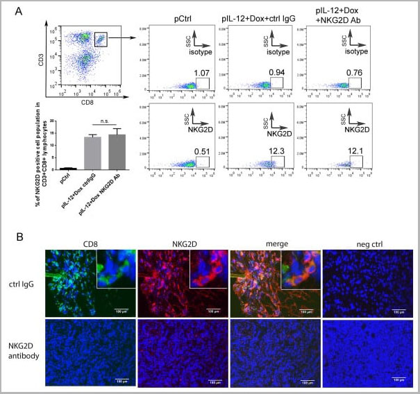
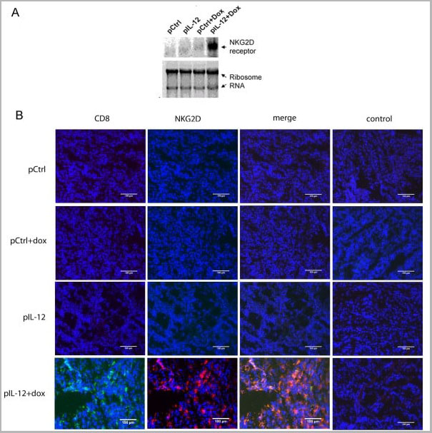
(Published clone specific image Alloimmunity-associated cytotoxicity is mediated through the NKG2D receptor. (A) Liver expression of nkg2d on day ten after liver transplantation. (B) Representative NKG2D expression levels in blood NK cells (left) and monocytes (right) of allogeneic (black) and syngeneic (grey) recipients. Isotype was used as control (dashed lines). (C) Sorted blood NK cell cytotoxicity inhibition with anti-NKG2D antibody or with anti-NKp30 antibody. (D) Levels of NKG2D ligand (rae1l, rrlt and irp94) expression in the liver on day ten after transplantation. (E) Levels of NKG2D ligand (rae1l, rrlt and irp94) expression in rat HCC cell lines. (F) Representative level of recombinant NKG2D-Fc binding to rat HCC cells lines. *p)
Application Data
(Published clone specific image Alloimmunity-associated cytotoxicity is mediated through the NKG2D receptor. (A) Liver expression of nkg2d on day ten after liver transplantation. (B) Representative NKG2D expression levels in blood NK cells (left) and monocytes (right) of allogeneic (black) and syngeneic (grey) recipients. Isotype was used as control (dashed lines). (C) Sorted blood NK cell cytotoxicity inhibition with anti-NKG2D antibody or with anti-NKp30 antibody. (D) Levels of NKG2D ligand (rae1l, rrlt and irp94) expression in the liver on day ten after transplantation. (E) Levels of NKG2D ligand (rae1l, rrlt and irp94) expression in rat HCC cell lines. (F) Representative level of recombinant NKG2D-Fc binding to rat HCC cells lines. *p)
CD172a, Monoclonal Antibody (Cat# AAA11875)
Full Name
MOUSE ANTI RAT CD172a:FITC
Gene Names
Sirpa; Bit; Ptpns1; SHPS-1
Applications
Flow Cytometry
Pricing


 FCM (Flow Cytometry)
(Figure 8. Flow Cytometry analysis of SiHa cells using anti- ASS1 antibody (AAA19369).Overlay histogram showing SiHa cells stained with AAA19369 (Blue line). The cells were blocked with 10% normal goat serum. And then incubated with mouse anti-ASS1 Antibody (AAA19369, 1μg/1x106 cells) for 30 min at 20 degree C. DyLight®488 conjugated goat anti-mouse IgG (BA1126, 5-10μg/1x106 cells) was used as secondary antibody for 30 minutes at 20 degree C. Isotype control antibody (Green line) was mouse IgG (1μg/1x106) used under the same conditions. Unlabelled sample (Red line) was also used as a control.)
FCM (Flow Cytometry)
(Figure 8. Flow Cytometry analysis of SiHa cells using anti- ASS1 antibody (AAA19369).Overlay histogram showing SiHa cells stained with AAA19369 (Blue line). The cells were blocked with 10% normal goat serum. And then incubated with mouse anti-ASS1 Antibody (AAA19369, 1μg/1x106 cells) for 30 min at 20 degree C. DyLight®488 conjugated goat anti-mouse IgG (BA1126, 5-10μg/1x106 cells) was used as secondary antibody for 30 minutes at 20 degree C. Isotype control antibody (Green line) was mouse IgG (1μg/1x106) used under the same conditions. Unlabelled sample (Red line) was also used as a control.)
ASS1, Monoclonal Antibody (Cat# AAA19369)
Full Name
Anti-ASS1 Antibody (monoclonal, 5I5)
Gene Names
ASS1; ASS; CTLN1
Reactivity
Human, Mouse, Rat, Monkey
Applications
WB, IHC-P, ICC, IF, FC/FACS/FCM
Purity
Immunogen affinity purified.
Pricing


 FCM (Flow Cytometry)
(Figure 6. Flow Cytometry analysis of A549 cells using anti-MCM5 antibody (AAA19377).Overlay histogram showing A549 cells stained with AAA19377 (Blue line). The cells were blocked with 10% normal goat serum. And then incubated with mouse anti- MCM5 Antibody (AAA19377, 1μg/1x106 cells) for 30 min at 20 degree C. DyLight®488 conjugated goat anti-mouse IgG (BA1126, 5-10μg/1x106 cells) was used as secondary antibody for 30 minutes at 20 degree C. Isotype control antibody (Green line) was mouse IgG (1μg/1x106) used under the same conditions. Unlabelled sample (Red line) was also used as a control.)
FCM (Flow Cytometry)
(Figure 6. Flow Cytometry analysis of A549 cells using anti-MCM5 antibody (AAA19377).Overlay histogram showing A549 cells stained with AAA19377 (Blue line). The cells were blocked with 10% normal goat serum. And then incubated with mouse anti- MCM5 Antibody (AAA19377, 1μg/1x106 cells) for 30 min at 20 degree C. DyLight®488 conjugated goat anti-mouse IgG (BA1126, 5-10μg/1x106 cells) was used as secondary antibody for 30 minutes at 20 degree C. Isotype control antibody (Green line) was mouse IgG (1μg/1x106) used under the same conditions. Unlabelled sample (Red line) was also used as a control.)
MCM5, Monoclonal Antibody (Cat# AAA19377)
Full Name
Anti-MCM5 Antibody (monoclonal, 4G10)
Gene Names
MCM5; CDC46; P1-CDC46
Reactivity
Human, Mouse, Rat
Applications
WB, IHC-P, ICC, IF, FC/FACS/FCM
Purity
Immunogen affinity purified.
Pricing


 Application Data
(Published clone specific image Alloimmunity-associated cytotoxicity is mediated through the NKG2D receptor. (A) Liver expression of nkg2d on day ten after liver transplantation. (B) Representative NKG2D expression levels in blood NK cells (left) and monocytes (right) of allogeneic (black) and syngeneic (grey) recipients. Isotype was used as control (dashed lines). (C) Sorted blood NK cell cytotoxicity inhibition with anti-NKG2D antibody or with anti-NKp30 antibody. (D) Levels of NKG2D ligand (rae1l, rrlt and irp94) expression in the liver on day ten after transplantation. (E) Levels of NKG2D ligand (rae1l, rrlt and irp94) expression in rat HCC cell lines. (F) Representative level of recombinant NKG2D-Fc binding to rat HCC cells lines. *p)
Application Data
(Published clone specific image Alloimmunity-associated cytotoxicity is mediated through the NKG2D receptor. (A) Liver expression of nkg2d on day ten after liver transplantation. (B) Representative NKG2D expression levels in blood NK cells (left) and monocytes (right) of allogeneic (black) and syngeneic (grey) recipients. Isotype was used as control (dashed lines). (C) Sorted blood NK cell cytotoxicity inhibition with anti-NKG2D antibody or with anti-NKp30 antibody. (D) Levels of NKG2D ligand (rae1l, rrlt and irp94) expression in the liver on day ten after transplantation. (E) Levels of NKG2D ligand (rae1l, rrlt and irp94) expression in rat HCC cell lines. (F) Representative level of recombinant NKG2D-Fc binding to rat HCC cells lines. *p)
CD172a, Monoclonal Antibody (Cat# AAA11967)
Full Name
MOUSE ANTI RAT CD172a
Gene Names
Sirpa; Bit; Ptpns1; SHPS-1
Applications
Immunohistochemistry, Flow Cytometry, Immunohistochemistry, Western Blot
Pricing


 Application Data
(Published clone specific image Alloimmunity-associated cytotoxicity is mediated through the NKG2D receptor. (A) Liver expression of nkg2d on day ten after liver transplantation. (B) Representative NKG2D expression levels in blood NK cells (left) and monocytes (right) of allogeneic (black) and syngeneic (grey) recipients. Isotype was used as control (dashed lines). (C) Sorted blood NK cell cytotoxicity inhibition with anti-NKG2D antibody or with anti-NKp30 antibody. (D) Levels of NKG2D ligand (rae1l, rrlt and irp94) expression in the liver on day ten after transplantation. (E) Levels of NKG2D ligand (rae1l, rrlt and irp94) expression in rat HCC cell lines. (F) Representative level of recombinant NKG2D-Fc binding to rat HCC cells lines. *p)
Application Data
(Published clone specific image Alloimmunity-associated cytotoxicity is mediated through the NKG2D receptor. (A) Liver expression of nkg2d on day ten after liver transplantation. (B) Representative NKG2D expression levels in blood NK cells (left) and monocytes (right) of allogeneic (black) and syngeneic (grey) recipients. Isotype was used as control (dashed lines). (C) Sorted blood NK cell cytotoxicity inhibition with anti-NKG2D antibody or with anti-NKp30 antibody. (D) Levels of NKG2D ligand (rae1l, rrlt and irp94) expression in the liver on day ten after transplantation. (E) Levels of NKG2D ligand (rae1l, rrlt and irp94) expression in rat HCC cell lines. (F) Representative level of recombinant NKG2D-Fc binding to rat HCC cells lines. *p)
CD172a, Monoclonal Antibody (Cat# AAA11968)
Full Name
MOUSE ANTI RAT CD172a
Gene Names
Sirpa; Bit; Ptpns1; SHPS-1
Applications
Immunohistochemistry, Flow Cytometry, Immunohistochemistry, Western Blot
Pricing


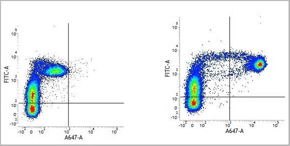
 FCM (Flow Cytometry)
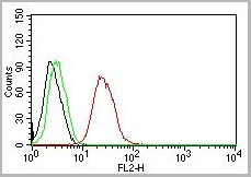
(Flow cytometry analysis of GOT1 in HeLa cell line, staining at 2-5ug for 1x10⁶cells (red line). The secondary antibody used goat anti-mouse IgG Alexa fluor 488 conjugate. Isotype control antibody was mouse IgG (black line).)
FCM (Flow Cytometry)
(Flow cytometry analysis of GOT1 in HeLa cell line, staining at 2-5ug for 1x10⁶cells (red line). The secondary antibody used goat anti-mouse IgG Alexa fluor 488 conjugate. Isotype control antibody was mouse IgG (black line).)
GOT1, Monoclonal Antibody (Cat# AAA11733)
Full Name
GOT1 antibody
Gene Names
GOT1; AST1; cCAT; GIG18; cAspAT; ASTQTL1
Reactivity
Human
Applications
Western Blot, Flow Cytometry, Immunocytochemistry, Immunofluorescence
Purity
By protein-A affinity chromatography
Pricing


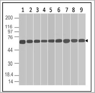
 WB (Western Blot)
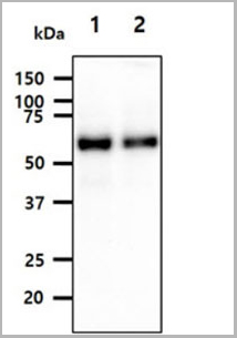
(Western Blot Analysis of Raji cell lysate using CD20 Mouse Monoclonal Antibody (L26).)
WB (Western Blot)
(Western Blot Analysis of Raji cell lysate using CD20 Mouse Monoclonal Antibody (L26).)
CD20/MS4A1, Monoclonal Antibody (Cat# AAA23946)
Full Name
CD20/MS4A1 (B-Cell Marker)
Gene Names
MS4A1; B1; S7; Bp35; CD20; CVID5; MS4A2; LEU-16
Reactivity
Human
Applications
FC/FACS, IF, WB, IHC
Purity
Purified Ab with BSA and Azide at 200ug/ml OR Purified Ab WITHOUT BSA and Azide at 1.0mg/ml
Pricing


 FCM (Flow Cytometry)
(Figure 7. Flow Cytometry analysis of U87 cells using anti-MPI antibody (AAA19349).Overlay histogram showing U87 cells stained with AAA19349 (Blue line). The cells were blocked with 10% normal goat serum. And then incubated with mouse anti- MPI Antibody (AAA19349, 1μg/1x106 cells) for 30 min at 20 degree C. DyLight®488 conjugated goat anti-mouse IgG (BA1126, 5-10μg/1x106 cells) was used as secondary antibody for 30 minutes at 20 degree C. Isotype control antibody (Green line) was mouse IgG (1μg/1x106) used under the same conditions. Unlabelled sample (Red line) was also used as a control.)
FCM (Flow Cytometry)
(Figure 7. Flow Cytometry analysis of U87 cells using anti-MPI antibody (AAA19349).Overlay histogram showing U87 cells stained with AAA19349 (Blue line). The cells were blocked with 10% normal goat serum. And then incubated with mouse anti- MPI Antibody (AAA19349, 1μg/1x106 cells) for 30 min at 20 degree C. DyLight®488 conjugated goat anti-mouse IgG (BA1126, 5-10μg/1x106 cells) was used as secondary antibody for 30 minutes at 20 degree C. Isotype control antibody (Green line) was mouse IgG (1μg/1x106) used under the same conditions. Unlabelled sample (Red line) was also used as a control.)
MPI, Monoclonal Antibody (Cat# AAA19349)
Full Name
Anti-MPI Antibody (monoclonal, 5G5)
Gene Names
MPI; PMI; PMI1; CDG1B
Reactivity
Human, Rat
Applications
WB, IHC-P, ICC, IF, FC/FACS/FCM
Purity
Immunogen affinity purified.
Pricing


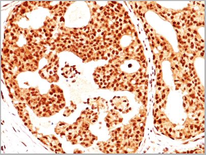
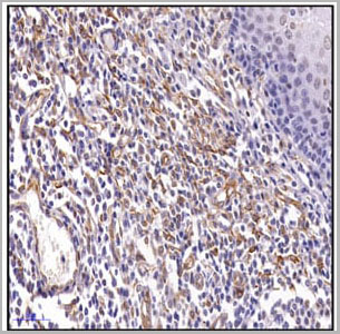
 IHC (Immunohistchemistry)
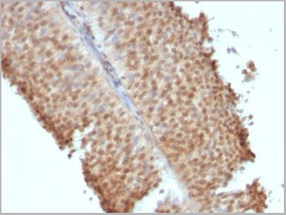
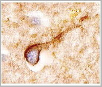
(Figure 6. IHC analysis of PP2A-alpha/PPP2CA using anti-PP2A-alpha/PPP2CA antibody (AAA19366).PP2A-alpha/PPP2CA was detected in paraffin-embedded section of rat brain tissue. Heat mediated antigen retrieval was performed in EDTA buffer (pH8. 0, epitope retrieval solution). The tissue section was blocked with 10% goat serum. The tissue section was then incubated with 1μg/ml mouse anti-PP2A-alpha/PPP2CA Antibody (AAA19366) overnight at 4 degree C. Biotinylated goat anti-mouse IgG was used as secondary antibody and incubated for 30 minutes at 37 degree C. The tissue section was developed using Strepavidin-Biotin-Complex (SABC) (Catalog # with DAB as the chromogen.)
IHC (Immunohistchemistry)
(Figure 6. IHC analysis of PP2A-alpha/PPP2CA using anti-PP2A-alpha/PPP2CA antibody (AAA19366).PP2A-alpha/PPP2CA was detected in paraffin-embedded section of rat brain tissue. Heat mediated antigen retrieval was performed in EDTA buffer (pH8. 0, epitope retrieval solution). The tissue section was blocked with 10% goat serum. The tissue section was then incubated with 1μg/ml mouse anti-PP2A-alpha/PPP2CA Antibody (AAA19366) overnight at 4 degree C. Biotinylated goat anti-mouse IgG was used as secondary antibody and incubated for 30 minutes at 37 degree C. The tissue section was developed using Strepavidin-Biotin-Complex (SABC) (Catalog # with DAB as the chromogen.)
PP2A-alpha/PPP2CA, Monoclonal Antibody (Cat# AAA19366)
Full Name
Anti-PP2A-alpha/PPP2CA Antibody (monoclonal, 3B6)
Gene Names
PPP2CA; RP-C; PP2Ac; PP2CA; PP2Calpha
Reactivity
Human, Monkey, Mouse, Rat
Applications
WB, IHC-P, ICC, IF, FC/FACS/FCM
Purity
Immunogen affinity purified.
Pricing


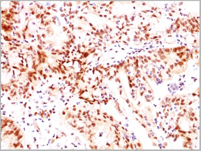
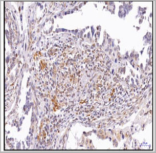
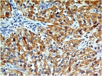
 IHC (Immunohistochemistry)
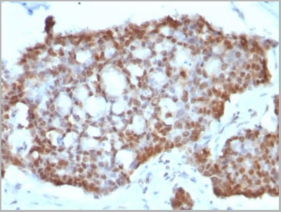
(Figure 11. IHC analysis of U2AF65/U2AF2 using anti U2AF65/U2AF2 antibody (AAA19376).U2AF65/U2AF2 was detected in paraffin-embedded section of human gallbladder adenocarcinoma tissue. Heat mediated antigen retrieval was performed in EDTA buffer (pH8. 0, epitope retrieval solution). The tissue section was blocked with 10% goat serum. The tissue section was then incubated with 2μg/ml mouse anti-U2AF65/U2AF2 Antibody (AAA19376) overnight at 4 degree C. Biotinylated goat anti-mouse IgG was used as secondary antibody and incubated for 30 minutes at 37 degree C. The tissue section was developed using Strepavidin-Biotin-Complex (SABC) (Catalog # with DAB as the chromogen.)
IHC (Immunohistochemistry)
(Figure 11. IHC analysis of U2AF65/U2AF2 using anti U2AF65/U2AF2 antibody (AAA19376).U2AF65/U2AF2 was detected in paraffin-embedded section of human gallbladder adenocarcinoma tissue. Heat mediated antigen retrieval was performed in EDTA buffer (pH8. 0, epitope retrieval solution). The tissue section was blocked with 10% goat serum. The tissue section was then incubated with 2μg/ml mouse anti-U2AF65/U2AF2 Antibody (AAA19376) overnight at 4 degree C. Biotinylated goat anti-mouse IgG was used as secondary antibody and incubated for 30 minutes at 37 degree C. The tissue section was developed using Strepavidin-Biotin-Complex (SABC) (Catalog # with DAB as the chromogen.)
U2AF65/U2AF2, Monoclonal Antibody (Cat# AAA19376)
Full Name
Anti-U2AF65/U2AF2 Antibody (monoclonal, 3G9)
Gene Names
U2AF2; U2AF65
Reactivity
Human, Mouse, Rat
Applications
WB, IHC-P, ICC, IF, FC/FACS/FCM
Purity
Immunogen affinity purified.
Pricing


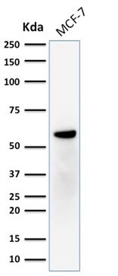
 FCM (Flow Cytometry)
(Figure 6. Flow Cytometry analysis of MCF-7 cells using anti-DDX1 antibody (AAA19379).Overlay histogram showing MCF-7 cells stained with AAA19379 (Blue line). The cells were blocked with 10% normal goat serum. And then incubated with mouse anti- DDX1 Antibody (AAA19379, 1μg/1x106 cells) for 30 min at 20 degree C. DyLight®488 conjugated goat anti-mouse IgG (BA1126, 5-10μg/1x106 cells) was used as secondary antibody for 30 minutes at 20 degree C. Isotype control antibody (Green line) was mouse IgG (1μg/1x106) used under the same conditions. Unlabelled sample (Red line) was also used as a control.)
FCM (Flow Cytometry)
(Figure 6. Flow Cytometry analysis of MCF-7 cells using anti-DDX1 antibody (AAA19379).Overlay histogram showing MCF-7 cells stained with AAA19379 (Blue line). The cells were blocked with 10% normal goat serum. And then incubated with mouse anti- DDX1 Antibody (AAA19379, 1μg/1x106 cells) for 30 min at 20 degree C. DyLight®488 conjugated goat anti-mouse IgG (BA1126, 5-10μg/1x106 cells) was used as secondary antibody for 30 minutes at 20 degree C. Isotype control antibody (Green line) was mouse IgG (1μg/1x106) used under the same conditions. Unlabelled sample (Red line) was also used as a control.)
DDX1, Monoclonal Antibody (Cat# AAA19379)
Full Name
Anti-DDX1 Antibody (monoclonal, 2D4)
Gene Names
DDX1; DBP-RB; UKVH5d
Reactivity
Human, Mouse, Rat
Applications
WB, IHC-P, FC/FACS/FCM
Purity
Immunogen affinity purified.
Pricing
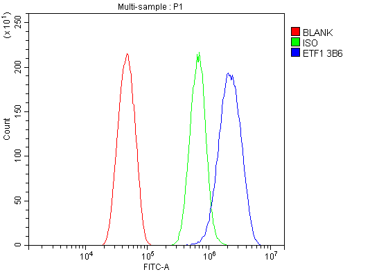


 FCM (Flow Cytometry)
(Figure 11. Flow Cytometry analysis of RH35 cells using anti- eRF1/ETF1 antibody (AAA19382).Overlay histogram showing RH35 cells stained with AAA19382 (Blue line). The cells were blocked with 10% normal goat serum. And then incubated with mouse anti-eRF1/ETF1 Antibody (AAA19382, 1μg/1x106 cells) for 30 min at 20 degree C. DyLight®488 conjugated goat anti-mouse IgG (BA1126, 5-10μg/1x106 cells) was used as secondary antibody for 30 minutes at 20 degree C. Isotype control antibody (Green line) was mouse IgG (1μg/1x106) used under the same conditions. Unlabelled sample (Red line) was also used as a control.)
FCM (Flow Cytometry)
(Figure 11. Flow Cytometry analysis of RH35 cells using anti- eRF1/ETF1 antibody (AAA19382).Overlay histogram showing RH35 cells stained with AAA19382 (Blue line). The cells were blocked with 10% normal goat serum. And then incubated with mouse anti-eRF1/ETF1 Antibody (AAA19382, 1μg/1x106 cells) for 30 min at 20 degree C. DyLight®488 conjugated goat anti-mouse IgG (BA1126, 5-10μg/1x106 cells) was used as secondary antibody for 30 minutes at 20 degree C. Isotype control antibody (Green line) was mouse IgG (1μg/1x106) used under the same conditions. Unlabelled sample (Red line) was also used as a control.)
eRF1/ETF1, Monoclonal Antibody (Cat# AAA19382)
Full Name
Anti-eRF1/ETF1 Antibody (monoclonal, 3B6)
Gene Names
ETF1; ERF; RF1; ERF1; TB3-1; D5S1995; SUP45L1
Reactivity
Human, Mouse, Rat
Applications
WB, IHC-P, ICC, IF, FC/FACS/FCM
Purity
Immunogen affinity purified.
Pricing


 FCM (Flow Cytometry)
(Figure 8. Flow Cytometry analysis of THP-1 cells using anti-INPPL1 antibody (AAA19364).Overlay histogram showing THP-1 cells stained with AAA19364 (Blue line). The cells were blocked with 10% normal goat serum. And then incubated with mouse anti- INPPL1 Antibody (AAA19364, 1μg/1x106 cells) for 30 min at 20 degree C. DyLight®488 conjugated goat anti-mouse IgG (BA1126, 5-10μg/1x106 cells) was used as secondary antibody for 30 minutes at 20 degree C. Isotype control antibody (Green line) was mouse IgG (1μg/1x106) used under the same conditions. Unlabelled sample (Red line) was also used as a control.)
FCM (Flow Cytometry)
(Figure 8. Flow Cytometry analysis of THP-1 cells using anti-INPPL1 antibody (AAA19364).Overlay histogram showing THP-1 cells stained with AAA19364 (Blue line). The cells were blocked with 10% normal goat serum. And then incubated with mouse anti- INPPL1 Antibody (AAA19364, 1μg/1x106 cells) for 30 min at 20 degree C. DyLight®488 conjugated goat anti-mouse IgG (BA1126, 5-10μg/1x106 cells) was used as secondary antibody for 30 minutes at 20 degree C. Isotype control antibody (Green line) was mouse IgG (1μg/1x106) used under the same conditions. Unlabelled sample (Red line) was also used as a control.)
INPPL1, Monoclonal Antibody (Cat# AAA19364)
Full Name
Anti-INPPL1 Antibody (monoclonal, 8C13)
Gene Names
INPPL1; OPSMD; SHIP2
Reactivity
Human, Mouse, Rat
Applications
WB, IHC-P, ICC, IF, FC/FACS/FCM
Purity
Immunogen affinity purified.
Pricing


 Application Data
(Analysis of Protein Array containing more than 19,000 full-length human proteins using ROR-gamma / RORC Mouse Monoclonal Antibody (RORC/2941). Z- and S- Score: The Z-score represents the strength of a signal that a monoclonal antibody (MAb) (in combination with a fluorescently-tagged anti-IgG secondary antibody) produces when binding to a particular protein on the HuProtTM array. Z-scores are described in units of standard deviations (SD's) above the mean value of all signals generated on that array. If targets on HuProtTM are arranged in descending order of the Z-score, the S-score is the difference (also in units of SD's) between the Z-score. S-score therefore represents the relative target specificity of a MAb to its intended target. A MAb is considered to specific to its intended target, if the MAb has an S-score of at least 2.5. For example, if a MAb binds to protein X with a Z-score of 43 and to protein Y with a Z-score of 14, then the S-score for the binding of that MAb to protein X is equal to 29.)
Application Data
(Analysis of Protein Array containing more than 19,000 full-length human proteins using ROR-gamma / RORC Mouse Monoclonal Antibody (RORC/2941). Z- and S- Score: The Z-score represents the strength of a signal that a monoclonal antibody (MAb) (in combination with a fluorescently-tagged anti-IgG secondary antibody) produces when binding to a particular protein on the HuProtTM array. Z-scores are described in units of standard deviations (SD's) above the mean value of all signals generated on that array. If targets on HuProtTM are arranged in descending order of the Z-score, the S-score is the difference (also in units of SD's) between the Z-score. S-score therefore represents the relative target specificity of a MAb to its intended target. A MAb is considered to specific to its intended target, if the MAb has an S-score of at least 2.5. For example, if a MAb binds to protein X with a Z-score of 43 and to protein Y with a Z-score of 14, then the S-score for the binding of that MAb to protein X is equal to 29.)
ROR-gamma/RORC, Monoclonal Antibody (Cat# AAA23951)
Full Name
ROR-gamma/RORC (RAR-related Orphan Receptor C)
Gene Names
RORC; TOR; RORG; RZRG; IMD42; NR1F3; RZR-GAMMA
Reactivity
Human
Applications
FC, IHC
Purity
Purified Ab with BSA and Azide at 200ug/ml or Purified Ab with BSA and Azide at 200ug/ml or Purified Ab WITHOUT BSA and Azide at 1.0mg/ml
Pricing
 Application Data
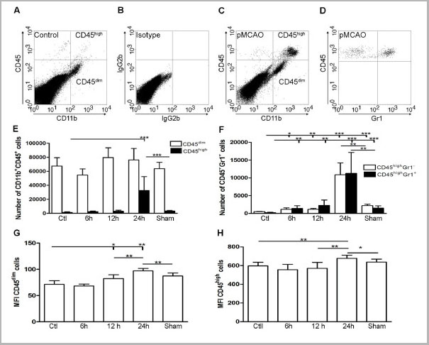
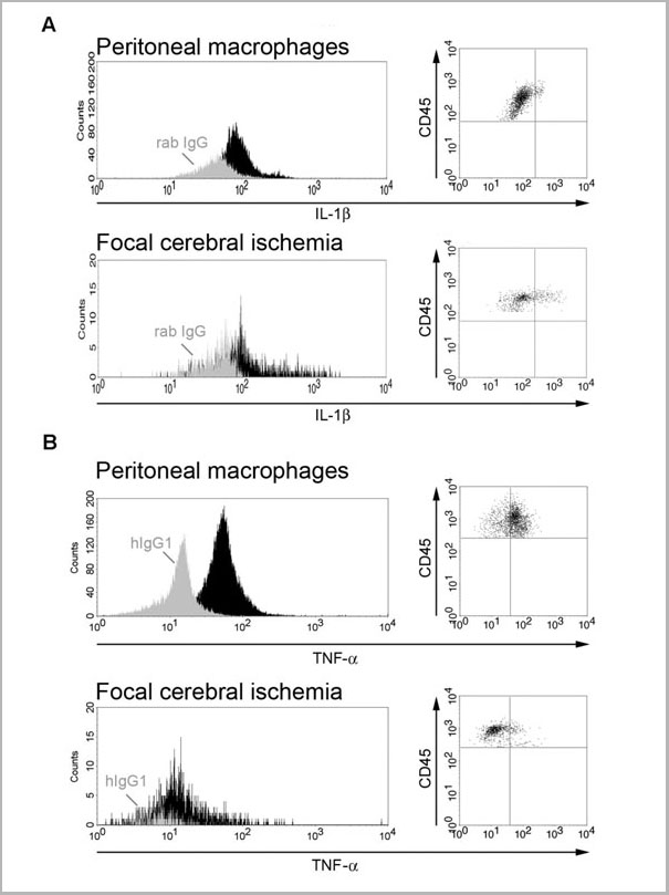
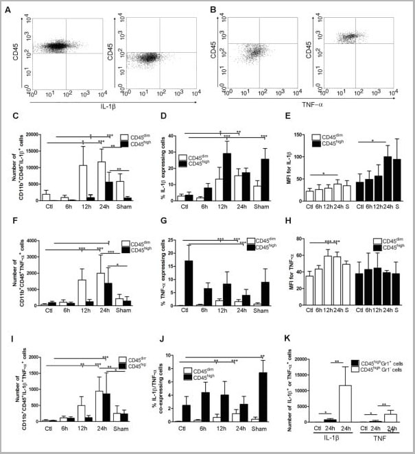
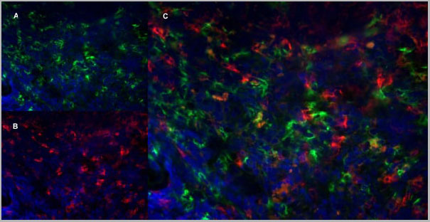
(Published customer image: Cytokine expression in segregated populations of cells following stroke. (A, B) Dot plots showing CD11b+CD45high macrophages/granulocytes (upper right quadrants) and CD11b+CD45dim microglia (bottom right quadrants) expressing IL-1beta (A) or TNF-a (B). (C-J) Bar graphs showing numbers and proportions of IL-1beta (C, D), TNF-a (F, G) and IL-1beta/TNF-a co-expressing (I, J) CD11b+CD45dim microglia and CD11b+CD45high macrophages/granulocytes in unmanipulated control mice (n = 10), in mice 6 (n = 7), 12 (n = 7), or 24 hours after pMCAO (n = 10), and in sham-operated mice 24 hours after pMCAO (n = 7). (E, H) Comparison of the MFI values for IL-1beta (E) and TNF-a (H) in viable CD11b+CD45dim microglia and CD11b+CD45high macrophages/granulocytes in unmanipulated mice, in mice 6, 12, or 24 hours after pMCAO, and in sham-operated mice 24 hours after pMCAO. Macrophages/granulocytes express significantly more IL-1beta than do microglial in unmanipulated mice, in mice 6, 12, or 24 hours after pMCAO, and in sham-operated mice 24 hours after pMCAO (E), whereas microglial cells express significantly higher levels of TNF-a than do macrophages/granulocytes at 12 h and 24 hours, and in sham-operated mice 24 hours after pMCAO (H). (K) CD11b+CD45highGr1- macrophages and not CD11b+CD45highGr1+ granulocytes are the main producers of IL-1beta and TNF-a 24 hours after pMCAO. *P < 0.05, **P < 0.01, and ***P < 0.001.From: http://www.jneuroinflammation.com/content/5/1/46.)
Application Data
(Published customer image: Cytokine expression in segregated populations of cells following stroke. (A, B) Dot plots showing CD11b+CD45high macrophages/granulocytes (upper right quadrants) and CD11b+CD45dim microglia (bottom right quadrants) expressing IL-1beta (A) or TNF-a (B). (C-J) Bar graphs showing numbers and proportions of IL-1beta (C, D), TNF-a (F, G) and IL-1beta/TNF-a co-expressing (I, J) CD11b+CD45dim microglia and CD11b+CD45high macrophages/granulocytes in unmanipulated control mice (n = 10), in mice 6 (n = 7), 12 (n = 7), or 24 hours after pMCAO (n = 10), and in sham-operated mice 24 hours after pMCAO (n = 7). (E, H) Comparison of the MFI values for IL-1beta (E) and TNF-a (H) in viable CD11b+CD45dim microglia and CD11b+CD45high macrophages/granulocytes in unmanipulated mice, in mice 6, 12, or 24 hours after pMCAO, and in sham-operated mice 24 hours after pMCAO. Macrophages/granulocytes express significantly more IL-1beta than do microglial in unmanipulated mice, in mice 6, 12, or 24 hours after pMCAO, and in sham-operated mice 24 hours after pMCAO (E), whereas microglial cells express significantly higher levels of TNF-a than do macrophages/granulocytes at 12 h and 24 hours, and in sham-operated mice 24 hours after pMCAO (H). (K) CD11b+CD45highGr1- macrophages and not CD11b+CD45highGr1+ granulocytes are the main producers of IL-1beta and TNF-a 24 hours after pMCAO. *P < 0.05, **P < 0.01, and ***P < 0.001.From: http://www.jneuroinflammation.com/content/5/1/46.)
CD45, Monoclonal Antibody (Cat# AAA11896)
Full Name
RAT ANTI MOUSE CD45
Gene Names
Ptprc; loc; B220; Cd45; L-CA; Ly-5; T200; CD45R; Lyt-4
Applications
Immunohistochemistry, Flow Cytometry, Immunofluorescence, Immunoprecipitation
Pricing
 Application Data
(Published customer image:RPE conjugated Mouse anti Canine CD4 antibody, clone YKIC302.9used for the assessment of CD4 levels on canine cells by flow cytometry.Image caption:Immunophenotypic profile of tumor infiltrating lymphocyte in canine mammary carcinomas. Analysis of tumor infiltrating T-cells, B-lymphocytes and T-cell subsets from MC-BMT or MC (A), further subcategorized according to the absence (-) or presence (+) of lymph node metastasis (-) (B). Lymphocyte populations and subsets were identified by flow cytometric immunostaining as described in Material and Methods. Data were expressed as percentage of positive cells within gated lymphocytes and CD4+/CD8+ T-cell ratio. Significant differences at p < 0.05 are highlighted by asterisk.From: From: Estrela-Lima et al.BMC Cancer 2010 10:256.)
Application Data
(Published customer image:RPE conjugated Mouse anti Canine CD4 antibody, clone YKIC302.9used for the assessment of CD4 levels on canine cells by flow cytometry.Image caption:Immunophenotypic profile of tumor infiltrating lymphocyte in canine mammary carcinomas. Analysis of tumor infiltrating T-cells, B-lymphocytes and T-cell subsets from MC-BMT or MC (A), further subcategorized according to the absence (-) or presence (+) of lymph node metastasis (-) (B). Lymphocyte populations and subsets were identified by flow cytometric immunostaining as described in Material and Methods. Data were expressed as percentage of positive cells within gated lymphocytes and CD4+/CD8+ T-cell ratio. Significant differences at p < 0.05 are highlighted by asterisk.From: From: Estrela-Lima et al.BMC Cancer 2010 10:256.)
CD4, Monoclonal Antibody (Cat# AAA12279)
Full Name
RAT ANTI DOG CD4: RPE-Cy7
Applications
Flow Cytometry
Purity
Purified IgG prepared by affinity chromatography on Protein G from tissue culture supernatant
Pricing
 Application Data
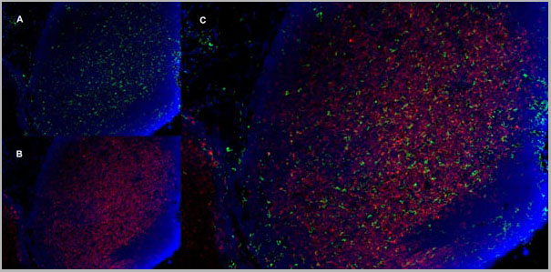
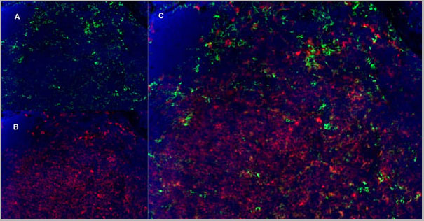
(Immunofluorescence staining of mouse lymph node cryosection with Rat anti Mouse CD11b, clone 5C6 , green in A and Rat anti Mouse CD8, clone YTS105.18 , red in B. C is the merged image with nuclei counterstained blue using DAPI. High power)
Application Data
(Immunofluorescence staining of mouse lymph node cryosection with Rat anti Mouse CD11b, clone 5C6 , green in A and Rat anti Mouse CD8, clone YTS105.18 , red in B. C is the merged image with nuclei counterstained blue using DAPI. High power)
CD8 ALPHA, Monoclonal Antibody (Cat# AAA11918)
Full Name
RAT ANTI MOUSE CD8
Gene Names
Cd8a; Ly-2; Ly-B; Ly-35; Lyt-2; BB154331
Applications
Immunohistochemistry, Flow Cytometry, Immunofluorescence
Pricing
 Application Data
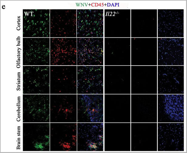
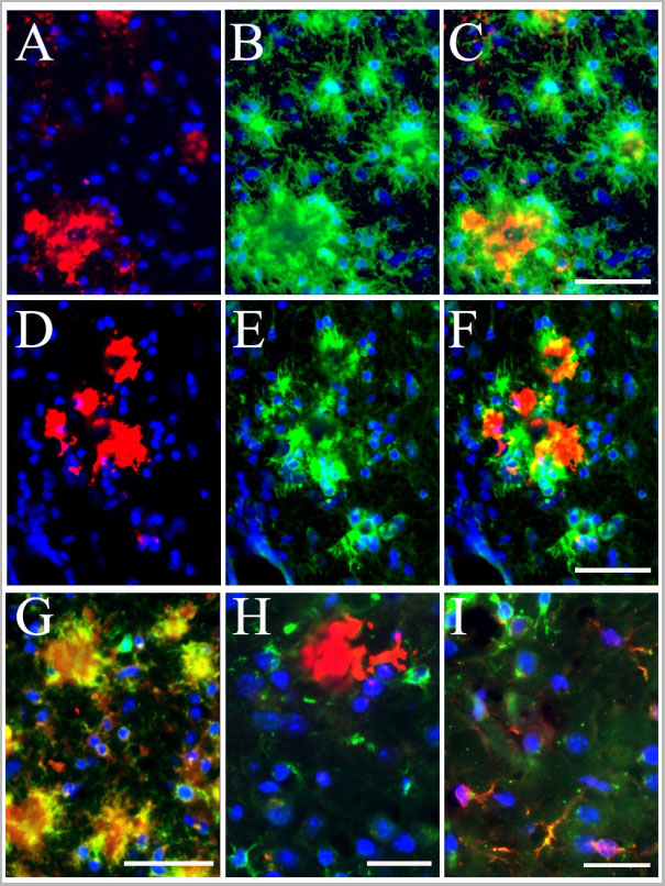
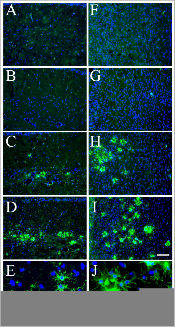
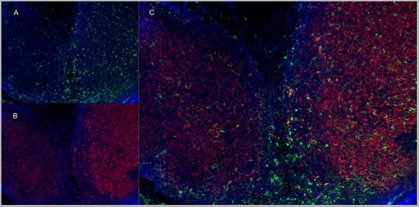
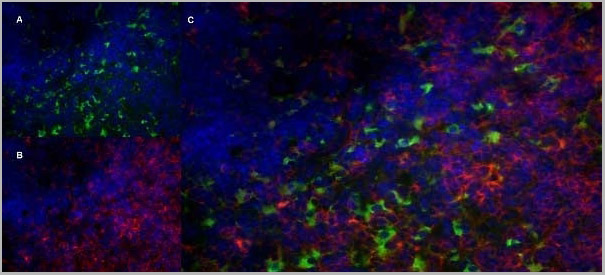
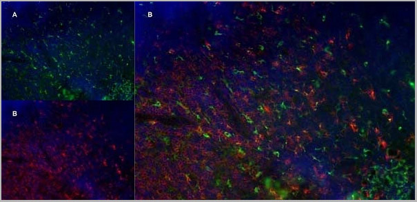
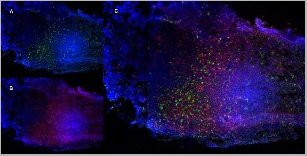
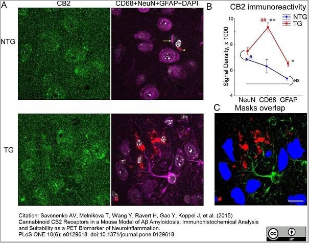
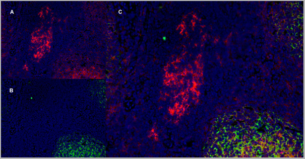
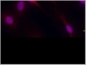
(Published customer image:Rat anti Mouse CD68 antibody, clone FA-11 used for the identification of microglia in mouse brain by immunofluorescence.Image caption:Comparison of CB2 immunoreactivity in neurons, activated microglia and astrocytes. ()
Application Data
(Published customer image:Rat anti Mouse CD68 antibody, clone FA-11 used for the identification of microglia in mouse brain by immunofluorescence.Image caption:Comparison of CB2 immunoreactivity in neurons, activated microglia and astrocytes. ()
CD68, Monoclonal Antibody (Cat# AAA12289)
Full Name
Rat anti Mouse CD68:Amethyst Orange
Gene Names
Cd68; Lamp4; gp110; Scard1
Reactivity
Mouse
Applications
Flow Cytometry
Purity
Purified IgG prepared by affinity chromatography on Protein G from tissue culture supernatant
Pricing


 Application Data
(Immunofluorescence staining of mouse lymph node cryosection with Rat anti Mouse CD11b, clone 5C6 , green in A and Rat anti Mouse CD8, clone YTS105.18 , red in B. C is the merged image with nuclei counterstained blue using DAPI. High power)
Application Data
(Immunofluorescence staining of mouse lymph node cryosection with Rat anti Mouse CD11b, clone 5C6 , green in A and Rat anti Mouse CD8, clone YTS105.18 , red in B. C is the merged image with nuclei counterstained blue using DAPI. High power)
CD8 ALPHA, Monoclonal Antibody (Cat# AAA12069)
Full Name
RAT ANTI MOUSE CD8
Gene Names
Cd8a; Ly-2; Ly-B; Ly-35; Lyt-2; BB154331
Reactivity
Mouse
Applications
Immunohistochemistry, Flow Cytometry, Immunofluorescence
Pricing


 Application Data
(Staining of human peripheral blood lymphocytes with Mouse anti Human CD40:RPE)
Application Data
(Staining of human peripheral blood lymphocytes with Mouse anti Human CD40:RPE)
CD40, Monoclonal Antibody (Cat# AAA12090)
Full Name
MOUSE ANTI HUMAN CD40
Gene Names
CD40; p50; Bp50; CDW40; TNFRSF5
Applications
Immunohistochemistry, Flow Cytometry, Immunoprecipitation, Immunohistochemistry
Pricing


 Application Data
(Staining of human peripheral blood lymphocytes with Mouse anti Human CD40:RPE)
Application Data
(Staining of human peripheral blood lymphocytes with Mouse anti Human CD40:RPE)
CD40, Monoclonal Antibody (Cat# AAA11867)
Full Name
MOUSE ANTI HUMAN CD40:FITC
Gene Names
CD40; p50; Bp50; CDW40; TNFRSF5
Applications
Flow Cytometry
Pricing


 Application Data
(Staining of human peripheral blood lymphocytes with Mouse anti Human CD40:RPE)
Application Data
(Staining of human peripheral blood lymphocytes with Mouse anti Human CD40:RPE)
CD40, Monoclonal Antibody (Cat# AAA11936)
Full Name
MOUSE ANTI HUMAN CD40
Gene Names
CD40; p50; Bp50; CDW40; TNFRSF5
Reactivity
Dog
Applications
Immunohistochemistry, Flow Cytometry, Immunoprecipitation, Immunohistochemistry
Pricing


 Application Data
(Staining of human peripheral blood lymphocytes with Mouse anti Human CD40:RPE)
Application Data
(Staining of human peripheral blood lymphocytes with Mouse anti Human CD40:RPE)
CD40, Monoclonal Antibody (Cat# AAA12089)
Full Name
MOUSE ANTI HUMAN CD40:RPE
Gene Names
CD40; p50; Bp50; CDW40; TNFRSF5
Applications
Flow Cytometry
Pricing


 Application Data
(Staining of human peripheral blood lymphocytes with Mouse anti Human CD40:RPE)
Application Data
(Staining of human peripheral blood lymphocytes with Mouse anti Human CD40:RPE)
CD40, Monoclonal Antibody (Cat# AAA12031)
Full Name
MOUSE ANTI HUMAN CD40:RPE
Gene Names
CD40; p50; Bp50; CDW40; TNFRSF5
Applications
Flow Cytometry
Pricing
 WB (Western Blot)
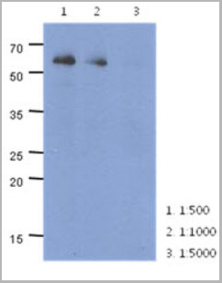
(Western Blot Analysis of human MCF-7 cell lysate using ER-beta1 Mouse Monoclonal Antibody (ERb455).)
WB (Western Blot)
(Western Blot Analysis of human MCF-7 cell lysate using ER-beta1 Mouse Monoclonal Antibody (ERb455).)
ER-beta1 (Estrogen Receptor beta-1), Monoclonal Antibody (Cat# AAA13814)
Full Name
ER-beta1 (Estrogen Receptor beta-1) Mouse Monoclonal Antibody
Gene Names
ESR2; Erb; ESRB; ESTRB; NR3A2; ER-BETA; ESR-BETA
Reactivity
Human
Applications
Flow Cytometry, Immunofluorescence, Western Blot, Immunohistochemistry
Pricing
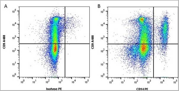
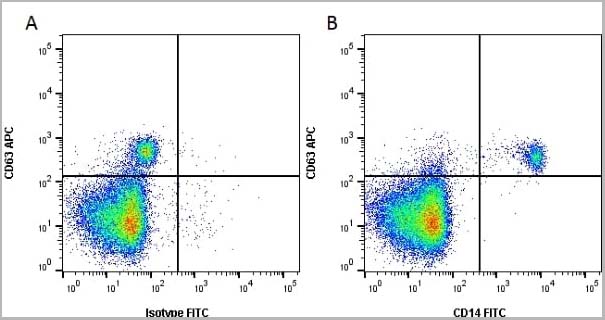
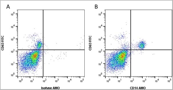
 Application Data
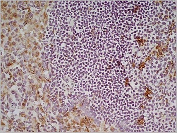
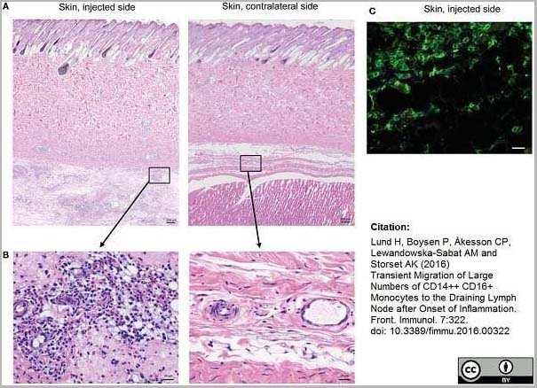
(Mouse anti Human CD14 antibody, clone Tük4 used to identify bovine monocytes in the skin and subcutaneous tissue of adjuvant injected calves by immunofluorescence.Image caption:Cellular recruitment to skin and subcutaneous tissues. (A) HE stained sections of skin with subcutaneous tissue from the side injected with adjuvant and the contralateral side, at 24 h post-injection. Scale bars: 200 ?m. (B) Enlargement of outlined areas in A, as indicated. Scale bars: 20 ?m. (C) Immunofluorescent labeling of subcutaneous tissue on the injected side with antibody against CD14 (green). Scale bar: 20 ?m.From: Lund H, Boysen P, Åkesson CP, Lewandowska-Sabat AM and Storset AK (2016)Transient Migration of Large Numbers of CD14++ CD16+ Monocytes to the Draining Lymph Node after Onset of Inflammation.Front. Immunol. 7:322.This is from an open access article distributed under the terms of the Creative Commons Attribution License.)
Application Data
(Mouse anti Human CD14 antibody, clone Tük4 used to identify bovine monocytes in the skin and subcutaneous tissue of adjuvant injected calves by immunofluorescence.Image caption:Cellular recruitment to skin and subcutaneous tissues. (A) HE stained sections of skin with subcutaneous tissue from the side injected with adjuvant and the contralateral side, at 24 h post-injection. Scale bars: 200 ?m. (B) Enlargement of outlined areas in A, as indicated. Scale bars: 20 ?m. (C) Immunofluorescent labeling of subcutaneous tissue on the injected side with antibody against CD14 (green). Scale bar: 20 ?m.From: Lund H, Boysen P, Åkesson CP, Lewandowska-Sabat AM and Storset AK (2016)Transient Migration of Large Numbers of CD14++ CD16+ Monocytes to the Draining Lymph Node after Onset of Inflammation.Front. Immunol. 7:322.This is from an open access article distributed under the terms of the Creative Commons Attribution License.)
CD14, Monoclonal Antibody (Cat# AAA12266)
Full Name
Mouse Anti Human CD14: Amethyst Orange
Reactivity
Human
Applications
Flow Cytometry
Purity
Purified IgG prepared by affinity chromatography on Protein A from tissue culture supernatant.
Pricing
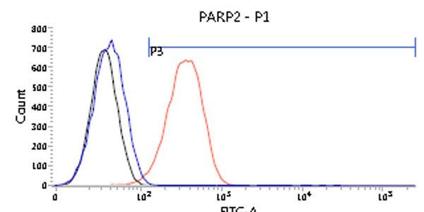 FCM (Flow Cytometry)
(Flow cytometry analysis of PARP2 in U87MG cells. The cell was stained with AAA11732 at 2-5ug for 1x10^6cells (red). A Goat anti mouse IgG (Alexa fluor 488) was used as the secondary antibody. Mouse monoclonal IgG was used as the isotype control (blue), cells without incubation with primary and secondary antibody was used as the negative control (black))
FCM (Flow Cytometry)
(Flow cytometry analysis of PARP2 in U87MG cells. The cell was stained with AAA11732 at 2-5ug for 1x10^6cells (red). A Goat anti mouse IgG (Alexa fluor 488) was used as the secondary antibody. Mouse monoclonal IgG was used as the isotype control (blue), cells without incubation with primary and secondary antibody was used as the negative control (black))
PARP2, Monoclonal Antibody (Cat# AAA11732)
Full Name
PARP2 antibody
Gene Names
PARP2; ARTD2; ADPRT2; PARP-2; ADPRTL2; ADPRTL3; pADPRT-2
Reactivity
Human
Applications
Western Blot, Flow Cytometry, Immunocytochemistry, Immunofluorescence
Purity
By protein-A affinity chromatography
Pricing
 WB (Western Blot)
(Western blot analysis of PD-L1. Anti-PD-L1 antibody (Clone: ABM4E54) was tested at 2 ug/ml on h Spleen lysate.)
WB (Western Blot)
(Western blot analysis of PD-L1. Anti-PD-L1 antibody (Clone: ABM4E54) was tested at 2 ug/ml on h Spleen lysate.)
PD-L1, Monoclonal Antibody (Cat# AAA14873)
Full Name
Monoclonal Antibody to PD-L1 (Clone: ABM4E54)
Gene Names
CD274; B7-H; B7H1; PDL1; PD-L1; PDCD1L1; PDCD1LG1
Reactivity
Human
Applications
Immunohistochemistry, Flow Cytometry, Western Blot
Purity
Purified
Protein G Chromatography
Protein G Chromatography
Pricing
 IHC (Immunohistochemistry)
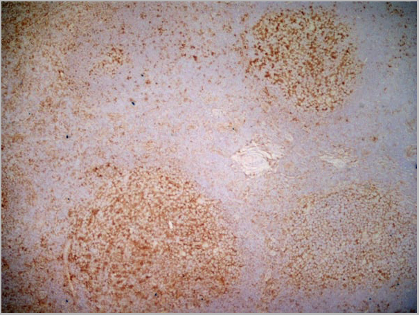
(Immunohistochemical analysis of Dectin-2 in Renal Cell Carcinoma using Dectin-2 antibody (Clone: ABM2H28) at 5 ug/ml.)
IHC (Immunohistochemistry)
(Immunohistochemical analysis of Dectin-2 in Renal Cell Carcinoma using Dectin-2 antibody (Clone: ABM2H28) at 5 ug/ml.)
Dectin-2, Monoclonal Antibody (Cat# AAA14862)
Full Name
Dectin-2 Monoclonal Antibody
Gene Names
CLEC6A; CLEC4N; CLECSF10
Reactivity
Human, Mouse
Applications
Western Blot, Flow Cytometry, Immunohistochemistry
Purity
Protein G Chromatography
Pricing

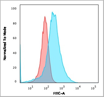
 FCM (Flow Cytometry)
(Flow Cytometry analyis of SSTR2 in human cell line. Human breast cancer cell line was stained with AAA14651 (filled histogram) or isotype control antibody (open histogram), followed by Allophycocyanin-conjugated Anti-Mouse IgG F(ab')2 secondary antibody. To facilitate intracellular staining, cells were fixed with paraformaldehyde and permeabilized with saponin.)
FCM (Flow Cytometry)
(Flow Cytometry analyis of SSTR2 in human cell line. Human breast cancer cell line was stained with AAA14651 (filled histogram) or isotype control antibody (open histogram), followed by Allophycocyanin-conjugated Anti-Mouse IgG F(ab')2 secondary antibody. To facilitate intracellular staining, cells were fixed with paraformaldehyde and permeabilized with saponin.)
Somatostatin Receptor Type 2, Monoclonal Antibody (Cat# AAA14651)
Full Name
Somatostatin Receptor Type 2 (SSTr2)
Gene Names
SSTR2; SSTR2
Reactivity
Human
Applications
Immunohistochemistry, Immunocytochemistry, Flow Cytometry
Purity
Purified by Protein A affinity chromatography from hybridoma culture supernatant.
Pricing
 Application Data
(Analysis of Protein Array containing more than 19,000 full-length human proteins using Annexin A1 Mouse Monoclonal Antibody (ANXA1/3566). Z- and S- Score: The Z-score represents the strength of a signal that a monoclonal antibody (MAb) (in combination with a fluorescently-tagged anti-IgG secondary antibody) produces when binding to a particular protein on the HuProtTM array. Z-scores are described in units of standard deviations (SD’s) above the mean value of all signals generated on that array. If targets on HuProtTM are arranged in descending order of the Z-score, the S-score is the difference (also in units of SD’s) between the Z-score. S-score therefore represents the relative target specificity of a MAb to its intended target. A MAb is considered to specific to its intended target, if the MAb has an S-score of at least 2.5. For example, if a MAb binds to protein X with a Z-score of 43 and to protein Y with a Z-score of 14, then the S-score for the binding of that MAb to protein X is equal to 29.)
Application Data
(Analysis of Protein Array containing more than 19,000 full-length human proteins using Annexin A1 Mouse Monoclonal Antibody (ANXA1/3566). Z- and S- Score: The Z-score represents the strength of a signal that a monoclonal antibody (MAb) (in combination with a fluorescently-tagged anti-IgG secondary antibody) produces when binding to a particular protein on the HuProtTM array. Z-scores are described in units of standard deviations (SD’s) above the mean value of all signals generated on that array. If targets on HuProtTM are arranged in descending order of the Z-score, the S-score is the difference (also in units of SD’s) between the Z-score. S-score therefore represents the relative target specificity of a MAb to its intended target. A MAb is considered to specific to its intended target, if the MAb has an S-score of at least 2.5. For example, if a MAb binds to protein X with a Z-score of 43 and to protein Y with a Z-score of 14, then the S-score for the binding of that MAb to protein X is equal to 29.)
Annexin A1, Monoclonal Antibody (Cat# AAA23918)
Full Name
Annexin A1/(Hairy Cell Leukemia Marker)
Gene Names
ANXA1; ANX1; LPC1
Reactivity
Human
Applications
Flow Cytometry, Immunofluorescence, Immunohistochemistry
Purity
Purified Ab with BSA and Azide at 200ug/ml OR Purified Ab WITHOUT BSA and Azide at 1.0mg/ml
Pricing
 FCM (Flow Cytometry)
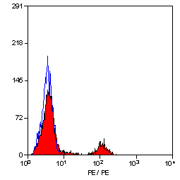
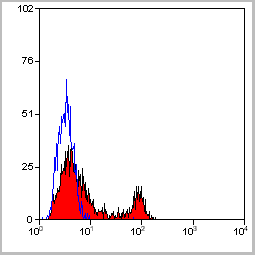
(Staining of C57BL/6 splenocytes with 0.25 ug of Mouse IgG2a kappa Isotype Control (FITC) (open histogram) or 0.25 ug of Qa-2 antibody (FITC) (filled histogram). Total viable cells were used for analysis.)
FCM (Flow Cytometry)
(Staining of C57BL/6 splenocytes with 0.25 ug of Mouse IgG2a kappa Isotype Control (FITC) (open histogram) or 0.25 ug of Qa-2 antibody (FITC) (filled histogram). Total viable cells were used for analysis.)
Qa2, Monoclonal Antibody (Cat# AAA17929)
Full Name
Qa2 antibody (FITC)
Gene Names
H2-Q7; Q9; Ped; Qa7; Qa-2; Qa-7; H-2Q7
Applications
Flow Cytometry
Pricing
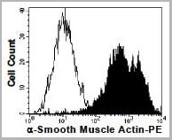 Application Data
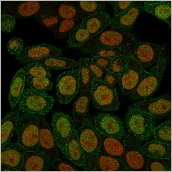
(Flow Cytometry analysis of intracellular staining in mouse fibroblast cell line L929 using AAA14733 (filled histogram) or with isotype control (open histogram).)
Application Data
(Flow Cytometry analysis of intracellular staining in mouse fibroblast cell line L929 using AAA14733 (filled histogram) or with isotype control (open histogram).)
Actin, alpha, Smooth Muscle, Monoclonal Antibody (Cat# AAA14733)
Full Name
Actin, alpha, Smooth Muscle (PE)
Gene Names
ACTA2; AAT6; ACTSA; MYMY5
Reactivity
Human
Applications
Flow Cytometry
Purity
Purified by Protein A affinity chromatography from hybridoma culture supernatant.
Pricing