Filters
Clonality
Type
Reactivity
Gene Name
Isotype
Host
Application
Clone
27 results for " Virus Antigen Detection" - showing 1-27


 ELISA
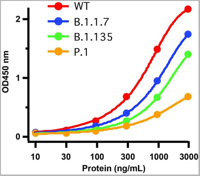
(Figure 6 ELISA Validation of Delta Variant Spike S1 Protein with SARS-CoV-2 Spike S1 Antibodies Coating Antigen: SARS-CoV-2 spike S1 proteins, including WT and Delta variant (B.1.671.2), 5 μg/mL, incubated at 4 ˚C overnight.Detection Antibodies: SARS-CoV-2 Spike S1 antibody, dilution: 0.3-1000 ng/mL, incubate at RT for 1 hr.Secondary Antibodies: Goat anti-rabbit HRP at 1:20,000, incubate at RT for 1 hr.Antibody 9083 does not distinguish between WT and Delta.)
ELISA
(Figure 6 ELISA Validation of Delta Variant Spike S1 Protein with SARS-CoV-2 Spike S1 Antibodies Coating Antigen: SARS-CoV-2 spike S1 proteins, including WT and Delta variant (B.1.671.2), 5 μg/mL, incubated at 4 ˚C overnight.Detection Antibodies: SARS-CoV-2 Spike S1 antibody, dilution: 0.3-1000 ng/mL, incubate at RT for 1 hr.Secondary Antibodies: Goat anti-rabbit HRP at 1:20,000, incubate at RT for 1 hr.Antibody 9083 does not distinguish between WT and Delta.)
COVID 19 Delta Variant Spike S1 (His-Avi Tag) Coronavirus, Recombinant Protein (Cat# AAA11040)
Full Name
SARS-CoV-2 (COVID-19) Delta Variant Spike S1 (His-Avi Tag) Recombinant Protein
Applications
Western Blot
Purity
> 90% as determined by Bis-Tris PAGE
Pricing


 WB (Western Blot)
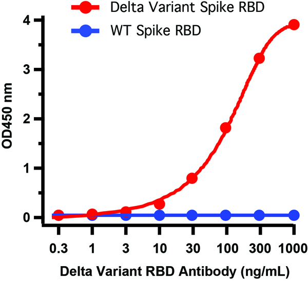
(Figure 6 WB Validation of Delta Variant Spike RBD Protein with SARS-CoV-2 Spike RBD Antibodies Loading: 20 ng of SARS-CoV-2 spike RBD proteins, including WT and Delta variant (B.1.671.2).Detection Antibodies: SARS-CoV-2 Spike RBD antibody, 1 μg/mL, incubate at RT for 1 hr.Secondary Antibodies: Goat anti-rabbit HRP at 1:20,000, incubate at RT for 1 hr.)
WB (Western Blot)
(Figure 6 WB Validation of Delta Variant Spike RBD Protein with SARS-CoV-2 Spike RBD Antibodies Loading: 20 ng of SARS-CoV-2 spike RBD proteins, including WT and Delta variant (B.1.671.2).Detection Antibodies: SARS-CoV-2 Spike RBD antibody, 1 μg/mL, incubate at RT for 1 hr.Secondary Antibodies: Goat anti-rabbit HRP at 1:20,000, incubate at RT for 1 hr.)
COVID 19 Delta Variant Spike RBD (His-Avi Tag) Coronavirus, Recombinant Protein (Cat# AAA11039)
Full Name
SARS-CoV-2 (COVID-19) Delta Variant Spike RBD (His-Avi Tag) Recombinant Protein
Applications
Western Blot
Purity
> 95% as determined by Bis-Tris PAGE
Pricing
 Standard Curve (Sample)
CD46 molecule, complement regulatory protein, ELISA Kit (Cat# AAA15581)
Full Name
Human membrane cofactor protein, MCP ELISA Kit
Gene Names
CD46; MCP; TLX; AHUS2; MIC10; TRA2.10
Reactivity
Human
Pricing


 IHC (Immunohistchemistry)
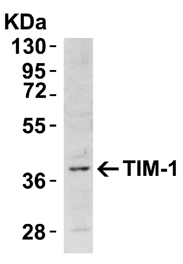
(Figure 5 Immunohistochemistry Validation of TIM-1Immunohistochemical analysis of paraffin-embedded human uterus tissue using anti-TIM-1 antibody (3809) at 10 μg/ml. Tissue was fixed with formaldehyde and blocked with 10% serum for 1 h at RT; antigen retrieval was by heat mediation with a citrate buffer (pH6). Samples were incubated with primary antibody overnight at 4˚ C. A goat anti-rabbit IgG H&L (HRP) at 1/250 was used as secondary. Counter stained with Hematoxylin.)
IHC (Immunohistchemistry)
(Figure 5 Immunohistochemistry Validation of TIM-1Immunohistochemical analysis of paraffin-embedded human uterus tissue using anti-TIM-1 antibody (3809) at 10 μg/ml. Tissue was fixed with formaldehyde and blocked with 10% serum for 1 h at RT; antigen retrieval was by heat mediation with a citrate buffer (pH6). Samples were incubated with primary antibody overnight at 4˚ C. A goat anti-rabbit IgG H&L (HRP) at 1/250 was used as secondary. Counter stained with Hematoxylin.)
TIM-1, Polyclonal Antibody (Cat# AAA10935)
Full Name
TIM-1 Antibody
Gene Names
HAVCR1; TIM; KIM1; TIM1; HAVCR; KIM-1; TIM-1; TIMD1; TIMD-1; HAVCR-1
Reactivity
Human, Mouse
Applications
Western Blot, Immunohistochemistry, Immunofluorescence
Purity
TIM-1 Antibody is affinity chromatography purified via peptide column.
Pricing
 Standard Curve (Sample)
hepatitis D virus (HDV) antigen, ELISA Kit (Cat# AAA18262)
Full Name
Human hepatitis D virus, HDV antigen ELISA Kit
Reactivity
Human
Pricing


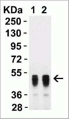
 WB (Western Blot)
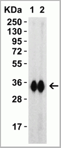
(Western blot analysis of SARS-CoV-2 spike S1 recombinant protein probed with Rabbit anti SARS-CoV-2 spike protein RBD antibody (Lane 1: 2.5ug/ml, Lane 2: 5ug/ml, AAA12292). Arrow points to SARS-CoV-2 spike protein.)
WB (Western Blot)
(Western blot analysis of SARS-CoV-2 spike S1 recombinant protein probed with Rabbit anti SARS-CoV-2 spike protein RBD antibody (Lane 1: 2.5ug/ml, Lane 2: 5ug/ml, AAA12292). Arrow points to SARS-CoV-2 spike protein.)
COVID 19 Spike Protein Rbd Coronavirus, Polyclonal Antibody (Cat# AAA12292)
Full Name
Rabbit anti SARS-CoV-2 Spike Protein Rbd
Reactivity
Viral
Applications
Immunofluorescence, Western Blot
Purity
Rabbit polyclonal antibody purified by affinity chromatography
Pricing


 Application Data
Quantitative, For Hepatitis B Virus Specific T Cells, Detection Kit (Cat# AAA27976)
Full Name
Quantitative Detection Kit, For Hepatitis B Virus Specific T Cells
Pricing


 IHC (Immunohistchemistry)
(Figure 6. IHC analysis of DYNLT1 using anti-DYNLT1 antibody (AAA19175).DYNLT1 was detected in paraffin-embedded section of rat lung tissue. Heat mediated antigen retrieval was performed in citrate buffer (pH6, epitope retrieval solution) for 20 mins. The tissue section was blocked with 10% goat serum. The tissue section was then incubated with 1ug/ml rabbit anti-DYNLT1 Antibody (AAA19175) overnight at 4 degree C. Biotinylated goat anti-rabbit IgG was used as secondary antibody and incubated for 30 minutes at 37 degree C. The tissue section was developed using Strepavidin-Biotin-Complex (SABC) with DAB as the chromogen.)
IHC (Immunohistchemistry)
(Figure 6. IHC analysis of DYNLT1 using anti-DYNLT1 antibody (AAA19175).DYNLT1 was detected in paraffin-embedded section of rat lung tissue. Heat mediated antigen retrieval was performed in citrate buffer (pH6, epitope retrieval solution) for 20 mins. The tissue section was blocked with 10% goat serum. The tissue section was then incubated with 1ug/ml rabbit anti-DYNLT1 Antibody (AAA19175) overnight at 4 degree C. Biotinylated goat anti-rabbit IgG was used as secondary antibody and incubated for 30 minutes at 37 degree C. The tissue section was developed using Strepavidin-Biotin-Complex (SABC) with DAB as the chromogen.)
DYNLT1, Polyclonal Antibody (Cat# AAA19175)
Full Name
Anti-DYNLT1 Picoband antibody
Gene Names
DYNLT1; CW-1; TCTEL1; tctex-1
Reactivity
Human, Mouse, Rat
No cross reactivity with other proteins.
No cross reactivity with other proteins.
Applications
EIA, IHC, WB
Pricing


 FCM (Flow Cytometry)
(Figure 7. Flow Cytometry analysis of PC-3 cells using anti-PVRL1/NECTIN1 antibody (AAA19303).Overlay histogram showing PC-3 cells stained with AAA19303 (Blue line). The cells were blocked with 10% normal goat serum. And then incubated with rabbit anti-PVRL1/NECTIN1 Antibody (AAA19303, 1μg/1x106 cells) for 30 min at 20 degree C. DyLight®488 conjugated goat anti-rabbit IgG (5-10μg/1x106 cells) was used as secondary antibody for 30 minutes at 20 degree C. Isotype control antibody (Green line) was rabbit IgG (1μg/1x106) used under the same conditions. Unlabelled sample (Red line) was also used as a control.)
FCM (Flow Cytometry)
(Figure 7. Flow Cytometry analysis of PC-3 cells using anti-PVRL1/NECTIN1 antibody (AAA19303).Overlay histogram showing PC-3 cells stained with AAA19303 (Blue line). The cells were blocked with 10% normal goat serum. And then incubated with rabbit anti-PVRL1/NECTIN1 Antibody (AAA19303, 1μg/1x106 cells) for 30 min at 20 degree C. DyLight®488 conjugated goat anti-rabbit IgG (5-10μg/1x106 cells) was used as secondary antibody for 30 minutes at 20 degree C. Isotype control antibody (Green line) was rabbit IgG (1μg/1x106) used under the same conditions. Unlabelled sample (Red line) was also used as a control.)
PVRL1/NECTIN1, Polyclonal Antibody (Cat# AAA19303)
Full Name
Anti-PVRL1/NECTIN1 Antibody
Gene Names
PVRL1; ED4; PRR; HIgR; HVEC; OFC7; PRR1; PVRR; CD111; PVRR1; SK-12; CLPED1; nectin-1
Reactivity
Human, Mouse, Rat
Applications
WB, IHC-P, FC/FACS/FCM, EIA
Purity
Immunogen affinity purified.
Pricing


 FCM (Flow Cytometry)
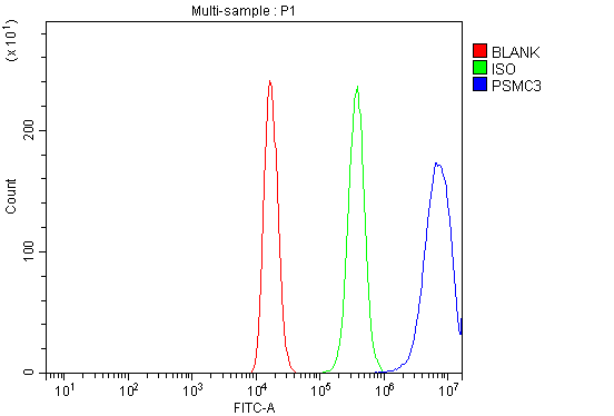
(Figure 7. Flow Cytometry analysis of 293T cells using anti-TBP-1/PSMC3 antibody (AAA19314).Overlay histogram showing 293T cells stained with AAA19314 (Blue line). The cells were blocked with 10% normal goat serum. And then incubated with rabbit anti-TBP-1/PSMC3 Antibody (AAA19314, 1μg/1x106 cells) for 30 min at 20 degree C. DyLight®488 conjugated goat anti-rabbit IgG (5-10μg/1x106 cells) was used as secondary antibody for 30 minutes at 20 degree C. Isotype control antibody (Green line) was rabbit IgG (1μg/1x106) used under the same conditions. Unlabelled sample (Red line) was also used as a control.)
FCM (Flow Cytometry)
(Figure 7. Flow Cytometry analysis of 293T cells using anti-TBP-1/PSMC3 antibody (AAA19314).Overlay histogram showing 293T cells stained with AAA19314 (Blue line). The cells were blocked with 10% normal goat serum. And then incubated with rabbit anti-TBP-1/PSMC3 Antibody (AAA19314, 1μg/1x106 cells) for 30 min at 20 degree C. DyLight®488 conjugated goat anti-rabbit IgG (5-10μg/1x106 cells) was used as secondary antibody for 30 minutes at 20 degree C. Isotype control antibody (Green line) was rabbit IgG (1μg/1x106) used under the same conditions. Unlabelled sample (Red line) was also used as a control.)
TBP-1/PSMC3, Polyclonal Antibody (Cat# AAA19314)
Full Name
Anti-TBP-1/PSMC3 Antibody
Gene Names
PSMC3; TBP1
Reactivity
Human, Mouse, Rat
Applications
WB, IHC-P, FC/FACS/FCM, EIA
Purity
Immunogen affinity purified.
Pricing

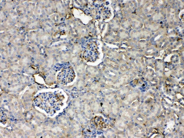


 IHC (Immunohistchemistry)
(Figure 6. IHC analysis of Thrombopoietin using anti- Thrombopoietin antibody (AAA19166).Thrombopoietin was detected in paraffin-embedded section of rat kidney tissues. Heat mediated antigen retrieval was performed in citrate buffer (pH6, epitope retrieval solution) for 20 mins. The tissue section was blocked with 10% goat serum. The tissue section was then incubated with 1ug/ml rabbit anti- Thrombopoietin Antibody (AAA19166) overnight at 4 degree C. Biotinylated goat anti-rabbit IgG was used as secondary antibody and incubated for 30 minutes at 37 degree C. The tissue section was developed using Strepavidin-Biotin-Complex (SABC) with DAB as the chromogen.)
IHC (Immunohistchemistry)
(Figure 6. IHC analysis of Thrombopoietin using anti- Thrombopoietin antibody (AAA19166).Thrombopoietin was detected in paraffin-embedded section of rat kidney tissues. Heat mediated antigen retrieval was performed in citrate buffer (pH6, epitope retrieval solution) for 20 mins. The tissue section was blocked with 10% goat serum. The tissue section was then incubated with 1ug/ml rabbit anti- Thrombopoietin Antibody (AAA19166) overnight at 4 degree C. Biotinylated goat anti-rabbit IgG was used as secondary antibody and incubated for 30 minutes at 37 degree C. The tissue section was developed using Strepavidin-Biotin-Complex (SABC) with DAB as the chromogen.)
Thrombopoietin, Polyclonal Antibody (Cat# AAA19166)
Full Name
Anti-Thrombopoietin Picoband Antibody
Gene Names
Thpo; Ml; Tpo; Mgdf; Mpllg
Reactivity
Mouse, Rat
No cross reactivity with other proteins
No cross reactivity with other proteins
Applications
EIA, IHC, WB
Purity
Immunogen affinity purified
Pricing
West Nile Envelope Virus, Recombinant Protein (Cat# AAA10847)
Full Name
Recombinant West Nile Envelope WNV Envelope
Applications
Westen Blot, Lateral Flow
Purity
Protein is >95% pure as determined by SDS-PAGE
Purification Method: Purified by proprietary chromatographic technique.
Purification Method: Purified by proprietary chromatographic technique.
Pricing


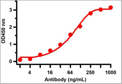
 WB (Western Blot)
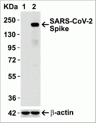
(Figure 12 Overexpression Validation in Spike Transfected 293 Cells Loading: 15 ug per lane of 293 cell lysate. Antibodies: SARS-CoV-2 (COVID-19) Spike, AAA10931 (1 ug/mL), 1h incubation at RT in 5% NFDM/TBST. Secondary: Goat anti rabbit IgG HRP conjugate at 1:10000 dilution. Lane 1: WT293 cells and Lane 2: SARS-CoV-2 Spike overexpressed 293 cells.)
WB (Western Blot)
(Figure 12 Overexpression Validation in Spike Transfected 293 Cells Loading: 15 ug per lane of 293 cell lysate. Antibodies: SARS-CoV-2 (COVID-19) Spike, AAA10931 (1 ug/mL), 1h incubation at RT in 5% NFDM/TBST. Secondary: Goat anti rabbit IgG HRP conjugate at 1:10000 dilution. Lane 1: WT293 cells and Lane 2: SARS-CoV-2 Spike overexpressed 293 cells.)
COVID 19 Spike Protein Coronavirus, Polyclonal Antibody (Cat# AAA10931)
Full Name
SARS-CoV-2 (COVID-19, 2019-nCoV) Spike Antibody
Reactivity
Virus
Applications
Immunofluorescence, Immunohistochemistry, Western Blot
Purity
SARS-CoV-2 (COVID-19, 2019-nCoV) Spike Antibody is affinity chromatography purified via peptide column.
Pricing


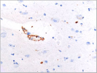
 IHC (Immunohistochemistry)
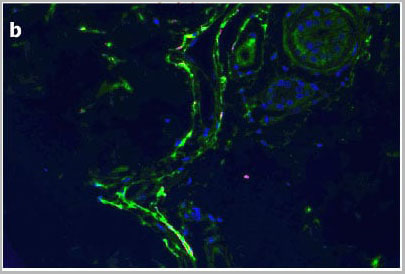
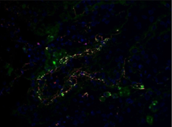
(IHC Validation of Envelope in COVID-19 Patient Skin (Magro et al., 2020) Detection of SARS-CoV-2 Envelope protein in the blood vessels of COVID-19 patients that were confirmed by PCR. The staining shows Envelope protein expression (green) detected by envelope antibodies (AAA10932, 3 ug/mL) in the endothelial cytoplasms in thrombosed and normal appearing blood vessels with hematoxylin counterstain. The staining was negative in control normal skin/lung (not shown).)
IHC (Immunohistochemistry)
(IHC Validation of Envelope in COVID-19 Patient Skin (Magro et al., 2020) Detection of SARS-CoV-2 Envelope protein in the blood vessels of COVID-19 patients that were confirmed by PCR. The staining shows Envelope protein expression (green) detected by envelope antibodies (AAA10932, 3 ug/mL) in the endothelial cytoplasms in thrombosed and normal appearing blood vessels with hematoxylin counterstain. The staining was negative in control normal skin/lung (not shown).)
COVID 19 Envelope Coronavirus, Polyclonal Antibody (Cat# AAA10932)
Full Name
SARS-CoV-2 (COVID-19, 2019-nCoV) Envelope antibody
Reactivity
Virus
Applications
Immunofluorescence, Immunohistochemistry
Purity
SARS-CoV-2 (COVID-19, 2019-nCoV) Envelope Antibody is affinity chromatography purified via peptide column.
Pricing
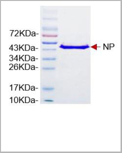 SDS-PAGE
(Nucleoprotein (NP) of SARS-CoV-2)
SDS-PAGE
(Nucleoprotein (NP) of SARS-CoV-2)
COVID 19 Nucleocapsid (NP) Coronavirus, Recombinant Protein (Cat# AAA13610)
Full Name
COVID-19 Nucleocapsid protein (NP)
Applications
Functional Assay, Immunoassay
Purity
>95% by SDS-PAGE
Pricing
 ELISA
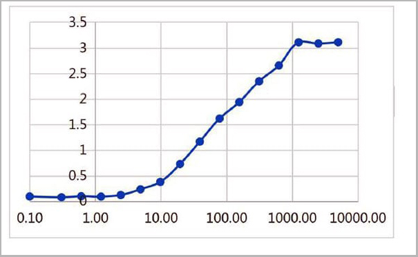
(Figure 2 ELISA TestAntibodies: SARS-CoV-2 Nucleocapsid antibody, AAA11029. An ELISA was performed using human SARS-CoV-2 Nucleocapsid recombinant protein as coating antigen and the SARS-CoV-2 Nucleocapsid antibody, as the capture antibody. Secondary: Goat anti-mouse IgG HRP conjugate at 1:20000 dilution. Detection range is from 2 ng/mL to 1000 ng/mL.)
ELISA
(Figure 2 ELISA TestAntibodies: SARS-CoV-2 Nucleocapsid antibody, AAA11029. An ELISA was performed using human SARS-CoV-2 Nucleocapsid recombinant protein as coating antigen and the SARS-CoV-2 Nucleocapsid antibody, as the capture antibody. Secondary: Goat anti-mouse IgG HRP conjugate at 1:20000 dilution. Detection range is from 2 ng/mL to 1000 ng/mL.)
COVID 19 Nucleocapsid (NP) Coronavirus, Monoclonal Antibody (Cat# AAA11029)
Full Name
SARS-CoV-2 (COVID-19) Nucleoprotein
Reactivity
Virus
Applications
Immunofluorescence
Purity
SARS-CoV-2 (COVID-19, 2019-nCoV) Nucleoprotein antibody is purified from ascites fluid or culture medium by protein A chromoatography or sequential differential precipitations.
Pricing
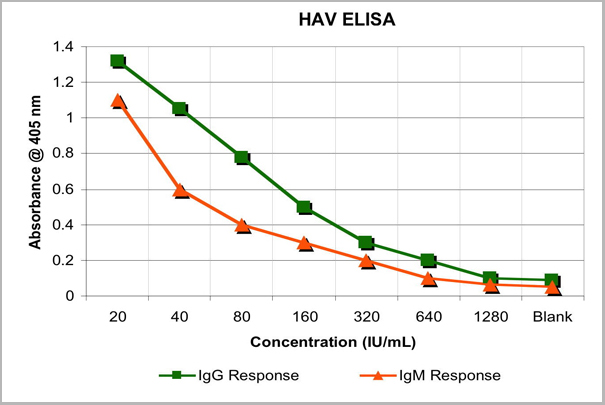 Application Data
(The HAV Antigen can be used in a direct ELISA for the detection of either IgG or IgM antibodies. The HAV antigen has demonstrated reactivity with antibodies of individuals vaccinated with the HAVRIX vaccine.)
Application Data
(The HAV Antigen can be used in a direct ELISA for the detection of either IgG or IgM antibodies. The HAV antigen has demonstrated reactivity with antibodies of individuals vaccinated with the HAVRIX vaccine.)
Hepatitis A Virus, pHM175, Infectious Disease (Cat# AAA14849)
Full Name
Hepatitis A Virus, pHM175 (HAV)
Applications
Western Blot
Purity
Purified
Pricing
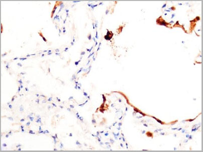
 IHC (Immunohistochemistry)
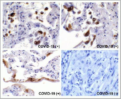
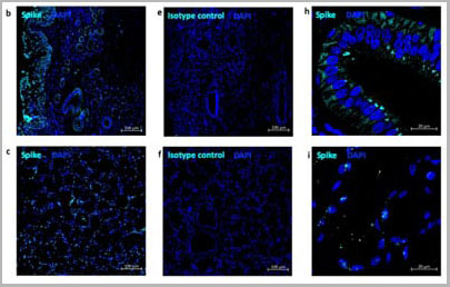
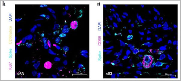
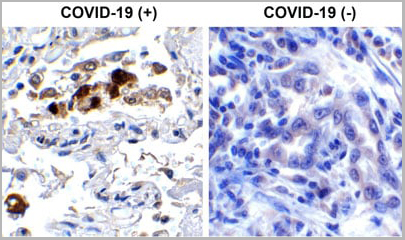
(Immunohistochemistry Validation of SARSCoV-2 (COVID-19) Spike RBD in COVID-19 Patient Lung Immunohistochemical analysis of paraffin-embedded COVID-19 patient lung tissue using anti- SARS-CoV-2 (COVID-19) Spike RBD antibody (AAA11030, 0.5 µg/mL). Tissue was fixed with formaldehyde and blocked with 10% serum for 1 h at RT; antigen retrieval was by heat mediation with a citrate buffer (pH6). Samples were incubated with primary antibody overnight at 4°C. A goat anti-rabbit IgG H&L (HRP) at 1/250 was used as secondary. Counter stained with Hematoxylin. Strong signal of SARS-COV-2 Spike RBD protein was observed in macrophage of COVID-19 patient lung, but not in non-COVID-19 patient lung.)
IHC (Immunohistochemistry)
(Immunohistochemistry Validation of SARSCoV-2 (COVID-19) Spike RBD in COVID-19 Patient Lung Immunohistochemical analysis of paraffin-embedded COVID-19 patient lung tissue using anti- SARS-CoV-2 (COVID-19) Spike RBD antibody (AAA11030, 0.5 µg/mL). Tissue was fixed with formaldehyde and blocked with 10% serum for 1 h at RT; antigen retrieval was by heat mediation with a citrate buffer (pH6). Samples were incubated with primary antibody overnight at 4°C. A goat anti-rabbit IgG H&L (HRP) at 1/250 was used as secondary. Counter stained with Hematoxylin. Strong signal of SARS-COV-2 Spike RBD protein was observed in macrophage of COVID-19 patient lung, but not in non-COVID-19 patient lung.)
COVID 19 Spike RBD Coronavirus, Polyclonal Antibody (Cat# AAA11030)
Full Name
SARS-CoV-2 (COVID-19) Spike RBD Antibody
Gene Names
S; spike glycoprotein
Reactivity
Virus
Applications
Immunofluorescence, Immunohistochemistry, Western Blot
Purity
SARS-CoV-2 (COVID-19) Spike RBD antibody is affinity chromatography purified via peptide column.
Pricing


 Application Data
(Published customer image: Mouse anti V5 tag antibody, clone SV5-Pk1 used for the detection of V5 tagged WEEV_nsP3 protein by western blotting and immunofluorescenceImage caption: WEEV nsP3 interaction with host IKKbeta. A) U87MGs were transfected in a 6-well plate with 5 ug of pUC19 and WEEV_nsP3_HA for 24 hours. Cell lysates were resolved using SDS-PAGE and subsequently immunoblotted with V5 antibody and beta-actin served as a loading control. B) U87MGs were transfected with WEEV_nsP3_V5; cells were fixed after 24 hours and stained with antibodies against the endogenous IKKbeta and the V5 tag. Cells were incubated with appropriate secondary Alexa Fluor antibodies and the nuclei stained with DAPI. Co-localization of IKKbeta with WEEV_nsP3_V5 (yellow) was observed as shown by the arrows. B) Panels E -H serve as an example of transfected cells in a given field of view that show co-localization of IKKbeta and WEEV_nsP3_V5 24 hours post transfection. Panels I-L represent magnified images of other cells showing co-localization of IKKbeta and WEEV_nsP3_V5. Panel M is a magnified image of panel L. The co-localization was confirmed by Z-stack analysis. Co-localization was calculated to be approximately in 61% of cells (163 cells were counted of which 44% demonstrated expression of nsP3. Of those cells that expressed nsP3, 61% showed co-localization of both proteins). Images were taken using Nikon Eclipse TE2000-U at 60x magnification and are representative of 2 independent experiments.From: Amaya M, Voss K, Sampey G, Senina S, de la Fuente C, et al. (2014) The Role of IKKbeta in Venezuelan Equine Encephalitis Virus Infection. PLoS ONE 9(2): e86745.)
Application Data
(Published customer image: Mouse anti V5 tag antibody, clone SV5-Pk1 used for the detection of V5 tagged WEEV_nsP3 protein by western blotting and immunofluorescenceImage caption: WEEV nsP3 interaction with host IKKbeta. A) U87MGs were transfected in a 6-well plate with 5 ug of pUC19 and WEEV_nsP3_HA for 24 hours. Cell lysates were resolved using SDS-PAGE and subsequently immunoblotted with V5 antibody and beta-actin served as a loading control. B) U87MGs were transfected with WEEV_nsP3_V5; cells were fixed after 24 hours and stained with antibodies against the endogenous IKKbeta and the V5 tag. Cells were incubated with appropriate secondary Alexa Fluor antibodies and the nuclei stained with DAPI. Co-localization of IKKbeta with WEEV_nsP3_V5 (yellow) was observed as shown by the arrows. B) Panels E -H serve as an example of transfected cells in a given field of view that show co-localization of IKKbeta and WEEV_nsP3_V5 24 hours post transfection. Panels I-L represent magnified images of other cells showing co-localization of IKKbeta and WEEV_nsP3_V5. Panel M is a magnified image of panel L. The co-localization was confirmed by Z-stack analysis. Co-localization was calculated to be approximately in 61% of cells (163 cells were counted of which 44% demonstrated expression of nsP3. Of those cells that expressed nsP3, 61% showed co-localization of both proteins). Images were taken using Nikon Eclipse TE2000-U at 60x magnification and are representative of 2 independent experiments.From: Amaya M, Voss K, Sampey G, Senina S, de la Fuente C, et al. (2014) The Role of IKKbeta in Venezuelan Equine Encephalitis Virus Infection. PLoS ONE 9(2): e86745.)
V5-TAG, Monoclonal Antibody (Cat# AAA12081)
Full Name
MOUSE ANTI V5-TAG:HRP
Applications
Western Blot
Pricing


 Application Data
(Published customer image: Mouse anti V5 tag antibody, clone SV5-Pk1 used for the detection of V5 tagged WEEV_nsP3 protein by western blotting and immunofluorescenceImage caption: WEEV nsP3 interaction with host IKKbeta. A) U87MGs were transfected in a 6-well plate with 5 ug of pUC19 and WEEV_nsP3_HA for 24 hours. Cell lysates were resolved using SDS-PAGE and subsequently immunoblotted with V5 antibody and beta-actin served as a loading control. B) U87MGs were transfected with WEEV_nsP3_V5; cells were fixed after 24 hours and stained with antibodies against the endogenous IKKbeta and the V5 tag. Cells were incubated with appropriate secondary Alexa Fluor antibodies and the nuclei stained with DAPI. Co-localization of IKKbeta with WEEV_nsP3_V5 (yellow) was observed as shown by the arrows. B) Panels E -H serve as an example of transfected cells in a given field of view that show co-localization of IKKbeta and WEEV_nsP3_V5 24 hours post transfection. Panels I-L represent magnified images of other cells showing co-localization of IKKbeta and WEEV_nsP3_V5. Panel M is a magnified image of panel L. The co-localization was confirmed by Z-stack analysis. Co-localization was calculated to be approximately in 61% of cells (163 cells were counted of which 44% demonstrated expression of nsP3. Of those cells that expressed nsP3, 61% showed co-localization of both proteins). Images were taken using Nikon Eclipse TE2000-U at 60x magnification and are representative of 2 independent experiments.From: Amaya M, Voss K, Sampey G, Senina S, de la Fuente C, et al. (2014) The Role of IKKbeta in Venezuelan Equine Encephalitis Virus Infection. PLoS ONE 9(2): e86745.)
Application Data
(Published customer image: Mouse anti V5 tag antibody, clone SV5-Pk1 used for the detection of V5 tagged WEEV_nsP3 protein by western blotting and immunofluorescenceImage caption: WEEV nsP3 interaction with host IKKbeta. A) U87MGs were transfected in a 6-well plate with 5 ug of pUC19 and WEEV_nsP3_HA for 24 hours. Cell lysates were resolved using SDS-PAGE and subsequently immunoblotted with V5 antibody and beta-actin served as a loading control. B) U87MGs were transfected with WEEV_nsP3_V5; cells were fixed after 24 hours and stained with antibodies against the endogenous IKKbeta and the V5 tag. Cells were incubated with appropriate secondary Alexa Fluor antibodies and the nuclei stained with DAPI. Co-localization of IKKbeta with WEEV_nsP3_V5 (yellow) was observed as shown by the arrows. B) Panels E -H serve as an example of transfected cells in a given field of view that show co-localization of IKKbeta and WEEV_nsP3_V5 24 hours post transfection. Panels I-L represent magnified images of other cells showing co-localization of IKKbeta and WEEV_nsP3_V5. Panel M is a magnified image of panel L. The co-localization was confirmed by Z-stack analysis. Co-localization was calculated to be approximately in 61% of cells (163 cells were counted of which 44% demonstrated expression of nsP3. Of those cells that expressed nsP3, 61% showed co-localization of both proteins). Images were taken using Nikon Eclipse TE2000-U at 60x magnification and are representative of 2 independent experiments.From: Amaya M, Voss K, Sampey G, Senina S, de la Fuente C, et al. (2014) The Role of IKKbeta in Venezuelan Equine Encephalitis Virus Infection. PLoS ONE 9(2): e86745.)
V5-TAG, Monoclonal Antibody (Cat# AAA11864)
Full Name
MOUSE ANTI V5-TAG:FITC
Applications
Immunofluorescence
Pricing


 Application Data
(Published customer image: Mouse anti V5 tag antibody, clone SV5-Pk1 used for the detection of V5 tagged WEEV_nsP3 protein by western blotting and immunofluorescenceImage caption: WEEV nsP3 interaction with host IKKbeta. A) U87MGs were transfected in a 6-well plate with 5 ug of pUC19 and WEEV_nsP3_HA for 24 hours. Cell lysates were resolved using SDS-PAGE and subsequently immunoblotted with V5 antibody and beta-actin served as a loading control. B) U87MGs were transfected with WEEV_nsP3_V5; cells were fixed after 24 hours and stained with antibodies against the endogenous IKKbeta and the V5 tag. Cells were incubated with appropriate secondary Alexa Fluor antibodies and the nuclei stained with DAPI. Co-localization of IKKbeta with WEEV_nsP3_V5 (yellow) was observed as shown by the arrows. B) Panels E -H serve as an example of transfected cells in a given field of view that show co-localization of IKKbeta and WEEV_nsP3_V5 24 hours post transfection. Panels I-L represent magnified images of other cells showing co-localization of IKKbeta and WEEV_nsP3_V5. Panel M is a magnified image of panel L. The co-localization was confirmed by Z-stack analysis. Co-localization was calculated to be approximately in 61% of cells (163 cells were counted of which 44% demonstrated expression of nsP3. Of those cells that expressed nsP3, 61% showed co-localization of both proteins). Images were taken using Nikon Eclipse TE2000-U at 60x magnification and are representative of 2 independent experiments.From: Amaya M, Voss K, Sampey G, Senina S, de la Fuente C, et al. (2014) The Role of IKKbeta in Venezuelan Equine Encephalitis Virus Infection. PLoS ONE 9(2): e86745.)
Application Data
(Published customer image: Mouse anti V5 tag antibody, clone SV5-Pk1 used for the detection of V5 tagged WEEV_nsP3 protein by western blotting and immunofluorescenceImage caption: WEEV nsP3 interaction with host IKKbeta. A) U87MGs were transfected in a 6-well plate with 5 ug of pUC19 and WEEV_nsP3_HA for 24 hours. Cell lysates were resolved using SDS-PAGE and subsequently immunoblotted with V5 antibody and beta-actin served as a loading control. B) U87MGs were transfected with WEEV_nsP3_V5; cells were fixed after 24 hours and stained with antibodies against the endogenous IKKbeta and the V5 tag. Cells were incubated with appropriate secondary Alexa Fluor antibodies and the nuclei stained with DAPI. Co-localization of IKKbeta with WEEV_nsP3_V5 (yellow) was observed as shown by the arrows. B) Panels E -H serve as an example of transfected cells in a given field of view that show co-localization of IKKbeta and WEEV_nsP3_V5 24 hours post transfection. Panels I-L represent magnified images of other cells showing co-localization of IKKbeta and WEEV_nsP3_V5. Panel M is a magnified image of panel L. The co-localization was confirmed by Z-stack analysis. Co-localization was calculated to be approximately in 61% of cells (163 cells were counted of which 44% demonstrated expression of nsP3. Of those cells that expressed nsP3, 61% showed co-localization of both proteins). Images were taken using Nikon Eclipse TE2000-U at 60x magnification and are representative of 2 independent experiments.From: Amaya M, Voss K, Sampey G, Senina S, de la Fuente C, et al. (2014) The Role of IKKbeta in Venezuelan Equine Encephalitis Virus Infection. PLoS ONE 9(2): e86745.)
V5-TAG, Monoclonal Antibody (Cat# AAA11930)
Full Name
MOUSE ANTI V5-TAG
Applications
Immunohistochemistry, Flow Cytometry, Immunofluorescence, Immunoprecipitation, Western Blot, Radioimmunoassay
Pricing


 Application Data
(Published customer image: Mouse anti V5 tag antibody, clone SV5-Pk1 used for the detection of V5 tagged WEEV_nsP3 protein by western blotting and immunofluorescenceImage caption: WEEV nsP3 interaction with host IKKbeta. A) U87MGs were transfected in a 6-well plate with 5 ug of pUC19 and WEEV_nsP3_HA for 24 hours. Cell lysates were resolved using SDS-PAGE and subsequently immunoblotted with V5 antibody and beta-actin served as a loading control. B) U87MGs were transfected with WEEV_nsP3_V5; cells were fixed after 24 hours and stained with antibodies against the endogenous IKKbeta and the V5 tag. Cells were incubated with appropriate secondary Alexa Fluor antibodies and the nuclei stained with DAPI. Co-localization of IKKbeta with WEEV_nsP3_V5 (yellow) was observed as shown by the arrows. B) Panels E -H serve as an example of transfected cells in a given field of view that show co-localization of IKKbeta and WEEV_nsP3_V5 24 hours post transfection. Panels I-L represent magnified images of other cells showing co-localization of IKKbeta and WEEV_nsP3_V5. Panel M is a magnified image of panel L. The co-localization was confirmed by Z-stack analysis. Co-localization was calculated to be approximately in 61% of cells (163 cells were counted of which 44% demonstrated expression of nsP3. Of those cells that expressed nsP3, 61% showed co-localization of both proteins). Images were taken using Nikon Eclipse TE2000-U at 60x magnification and are representative of 2 independent experiments.From: Amaya M, Voss K, Sampey G, Senina S, de la Fuente C, et al. (2014) The Role of IKKbeta in Venezuelan Equine Encephalitis Virus Infection. PLoS ONE 9(2): e86745.)
Application Data
(Published customer image: Mouse anti V5 tag antibody, clone SV5-Pk1 used for the detection of V5 tagged WEEV_nsP3 protein by western blotting and immunofluorescenceImage caption: WEEV nsP3 interaction with host IKKbeta. A) U87MGs were transfected in a 6-well plate with 5 ug of pUC19 and WEEV_nsP3_HA for 24 hours. Cell lysates were resolved using SDS-PAGE and subsequently immunoblotted with V5 antibody and beta-actin served as a loading control. B) U87MGs were transfected with WEEV_nsP3_V5; cells were fixed after 24 hours and stained with antibodies against the endogenous IKKbeta and the V5 tag. Cells were incubated with appropriate secondary Alexa Fluor antibodies and the nuclei stained with DAPI. Co-localization of IKKbeta with WEEV_nsP3_V5 (yellow) was observed as shown by the arrows. B) Panels E -H serve as an example of transfected cells in a given field of view that show co-localization of IKKbeta and WEEV_nsP3_V5 24 hours post transfection. Panels I-L represent magnified images of other cells showing co-localization of IKKbeta and WEEV_nsP3_V5. Panel M is a magnified image of panel L. The co-localization was confirmed by Z-stack analysis. Co-localization was calculated to be approximately in 61% of cells (163 cells were counted of which 44% demonstrated expression of nsP3. Of those cells that expressed nsP3, 61% showed co-localization of both proteins). Images were taken using Nikon Eclipse TE2000-U at 60x magnification and are representative of 2 independent experiments.From: Amaya M, Voss K, Sampey G, Senina S, de la Fuente C, et al. (2014) The Role of IKKbeta in Venezuelan Equine Encephalitis Virus Infection. PLoS ONE 9(2): e86745.)
V5-TAG, Monoclonal Antibody (Cat# AAA12211)
Full Name
MOUSE ANTI V5-TAG
Applications
Immunohistochemistry, Flow Cytometry, Immunofluorescence, Immunoprecipitation, Western Blot, Radioimmunoassay
Pricing
 ELISA
(ELISA on Denge Virus-Like Particles using AAA14182 Dengue Virus Serotype 2 VLPs were coated onto a plate at 5µg/ml. Anti-flavivirus antibody (AAA14182) added to plate in a 3-fold serial dilution starting at 1000 ng/ml. Detection performed using HRP labelled goat anti-mouse IgG.)
ELISA
(ELISA on Denge Virus-Like Particles using AAA14182 Dengue Virus Serotype 2 VLPs were coated onto a plate at 5µg/ml. Anti-flavivirus antibody (AAA14182) added to plate in a 3-fold serial dilution starting at 1000 ng/ml. Detection performed using HRP labelled goat anti-mouse IgG.)
Flavivirus group antigen, Recombinant Antibody (Cat# AAA14182)
Full Name
Anti-Flavivirus group antigen [D1-4G2-4-15 (4G2)]
Reactivity
Dengue Virus, Zika Virus, West Nile Virus, Flaviviridae, Yellow Fever Virus
Applications
Immunofluorescence, Western Blot, Neutralization Assay, Flow Cytometry, Immunohistochemistry
Purity
Protein A Affinity Purified
Pricing
 ELISA
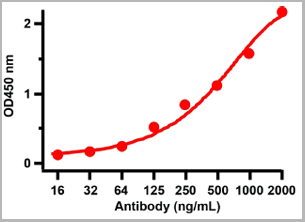
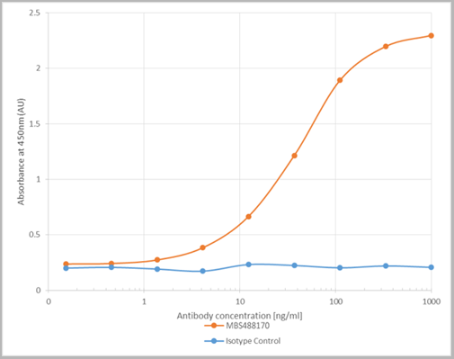
(ELISA plate was coated by the 2019-nCoV S1 protein, 100 uL/cell at 5ug/ml. The indirect ELISA analysis was performed by loading 100 uL per well of the anti-2019-nCoV S1 mAb (S1) at various concentrations. The plate was incubated for 1 hours at 37 degree C, then washed 5 times. An anti hFc HRP conjugated mAb at a concentration of 1:2000, 100uL/well was used as the secondary antibody. Again, the plate was incubated for 1 hours at 37 degree C, then washed 5 times. Detection was performed using TMB substrate for 10 minutes at room temperature in the dark. The plate was stopped with 2M sulfuric acid. Signal was read on a spectrophotometer at 450 nm.)
ELISA
(ELISA plate was coated by the 2019-nCoV S1 protein, 100 uL/cell at 5ug/ml. The indirect ELISA analysis was performed by loading 100 uL per well of the anti-2019-nCoV S1 mAb (S1) at various concentrations. The plate was incubated for 1 hours at 37 degree C, then washed 5 times. An anti hFc HRP conjugated mAb at a concentration of 1:2000, 100uL/well was used as the secondary antibody. Again, the plate was incubated for 1 hours at 37 degree C, then washed 5 times. Detection was performed using TMB substrate for 10 minutes at room temperature in the dark. The plate was stopped with 2M sulfuric acid. Signal was read on a spectrophotometer at 450 nm.)
COVID 19 Spike S1 Protein Coronavirus, Monoclonal Antibody (Cat# AAA27959)
Full Name
human anti-2019-nCoV Spike S1 mAb (S1)
Applications
ELISA
Purity
Purity: ≥ 95% as determined by SDS-PAGE
Purification: Protein A
Purification: Protein A
Pricing


 Application Data
(Published customer image: Mouse anti V5 tag antibody, clone SV5-Pk1 used for the detection of V5 tagged WEEV_nsP3 protein by western blotting and immunofluorescenceImage caption: WEEV nsP3 interaction with host IKKbeta. A) U87MGs were transfected in a 6-well plate with 5 ug of pUC19 and WEEV_nsP3_HA for 24 hours. Cell lysates were resolved using SDS-PAGE and subsequently immunoblotted with V5 antibody and beta-actin served as a loading control. B) U87MGs were transfected with WEEV_nsP3_V5; cells were fixed after 24 hours and stained with antibodies against the endogenous IKKbeta and the V5 tag. Cells were incubated with appropriate secondary Alexa Fluor antibodies and the nuclei stained with DAPI. Co-localization of IKKbeta with WEEV_nsP3_V5 (yellow) was observed as shown by the arrows. B) Panels E -H serve as an example of transfected cells in a given field of view that show co-localization of IKKbeta and WEEV_nsP3_V5 24 hours post transfection. Panels I-L represent magnified images of other cells showing co-localization of IKKbeta and WEEV_nsP3_V5. Panel M is a magnified image of panel L. The co-localization was confirmed by Z-stack analysis. Co-localization was calculated to be approximately in 61% of cells (163 cells were counted of which 44% demonstrated expression of nsP3. Of those cells that expressed nsP3, 61% showed co-localization of both proteins). Images were taken using Nikon Eclipse TE2000-U at 60x magnification and are representative of 2 independent experiments.From: Amaya M, Voss K, Sampey G, Senina S, de la Fuente C, et al. (2014) The Role of IKKbeta in Venezuelan Equine Encephalitis Virus Infection. PLoS ONE 9(2): e86745.)
Application Data
(Published customer image: Mouse anti V5 tag antibody, clone SV5-Pk1 used for the detection of V5 tagged WEEV_nsP3 protein by western blotting and immunofluorescenceImage caption: WEEV nsP3 interaction with host IKKbeta. A) U87MGs were transfected in a 6-well plate with 5 ug of pUC19 and WEEV_nsP3_HA for 24 hours. Cell lysates were resolved using SDS-PAGE and subsequently immunoblotted with V5 antibody and beta-actin served as a loading control. B) U87MGs were transfected with WEEV_nsP3_V5; cells were fixed after 24 hours and stained with antibodies against the endogenous IKKbeta and the V5 tag. Cells were incubated with appropriate secondary Alexa Fluor antibodies and the nuclei stained with DAPI. Co-localization of IKKbeta with WEEV_nsP3_V5 (yellow) was observed as shown by the arrows. B) Panels E -H serve as an example of transfected cells in a given field of view that show co-localization of IKKbeta and WEEV_nsP3_V5 24 hours post transfection. Panels I-L represent magnified images of other cells showing co-localization of IKKbeta and WEEV_nsP3_V5. Panel M is a magnified image of panel L. The co-localization was confirmed by Z-stack analysis. Co-localization was calculated to be approximately in 61% of cells (163 cells were counted of which 44% demonstrated expression of nsP3. Of those cells that expressed nsP3, 61% showed co-localization of both proteins). Images were taken using Nikon Eclipse TE2000-U at 60x magnification and are representative of 2 independent experiments.From: Amaya M, Voss K, Sampey G, Senina S, de la Fuente C, et al. (2014) The Role of IKKbeta in Venezuelan Equine Encephalitis Virus Infection. PLoS ONE 9(2): e86745.)
V5-TAG, Monoclonal Antibody (Cat# AAA11850)
Full Name
MOUSE ANTI V5-TAG:Biotin
Applications
Immunohistochemistry, Western Blot
Pricing
 Application Data
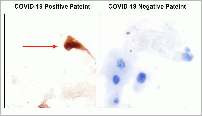
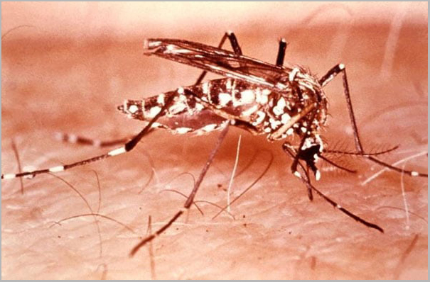
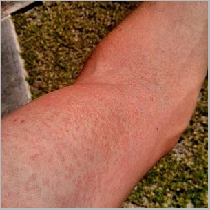
(Zika rash: arm.)
Application Data
(Zika rash: arm.)
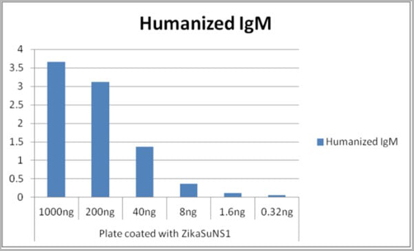
Zika Virus NS1, Monoclonal Antibody (Cat# AAA24113)
Full Name
Human Anti-Zika Virus NS1 (Zika Virus Nonstructural Protein)
Applications
ELISA
Purity
>98% (SDS-PAGE). Purified from cell culture supernatant by lectin affinity chromatography.
Pricing
Hepatitis B Virus e Antigen, HBeAg, ELISA Kit (Cat# AAA15174)
Full Name
Human Hepatitis B Virus e Antigen, HBeAg ELISA Kit
Reactivity
Human
Pricing