Filters
Clonality
Type
Reactivity
Gene Name
Isotype
Host
Application
Clone
162 results for " Tumor Necrosis Factor" - showing 1-50
 Standard Curve (Sample)
Osteoprotegerin, OPG, ELISA Kit (Cat# AAA11197)
Full Name
Human Osteoprotegerin, OPG ELISA Kit
Gene Names
TNFRSF11B; OPG; TR1; OCIF; PDB5
Reactivity
Human
Pricing


 FCM (Flow Cytometry)
(Flow cytometric analysis of HL-60 cells with TNF Receptor II antibody at 1/50 dilution (red) compared with an unlabelled control (cells without incubation with primary antibody; black). Alexa Fluor 488-conjugated goat anti rabbit IgG was used as the secondary antibody.)
FCM (Flow Cytometry)
(Flow cytometric analysis of HL-60 cells with TNF Receptor II antibody at 1/50 dilution (red) compared with an unlabelled control (cells without incubation with primary antibody; black). Alexa Fluor 488-conjugated goat anti rabbit IgG was used as the secondary antibody.)
TNF II, Monoclonal Antibody (Cat# AAA30327)
Full Name
TNF Receptor II Antibody
Gene Names
TNFRSF1B; p75; TBPII; TNFBR; TNFR2; CD120b; TNFR1B; TNFR80; TNF-R75; p75TNFR; TNF-R-II
Reactivity
Human, Mouse, Rat
Applications
Western Blot, Immunocytochemistry, Immunofluorescence, Immunohistochemistry, Flow Cytometry, Immunoprecipitation
Purity
ProA affinity purified
Pricing


 Application Data
(Figure 10 KD Validation in HeLa cells (Horinaka et al., 2005)HeLa cells were transfected with DR4siRNA or LacZ control siRNA. At 24 h after transfection, the cells were treated with or without 20 ?M luteolin for 24 h. Western blot analysis was carried out with anti-DR4 antibodies (1139). DR4 expression was markedly reduced after DR4 knockdown.)
Application Data
(Figure 10 KD Validation in HeLa cells (Horinaka et al., 2005)HeLa cells were transfected with DR4siRNA or LacZ control siRNA. At 24 h after transfection, the cells were treated with or without 20 ?M luteolin for 24 h. Western blot analysis was carried out with anti-DR4 antibodies (1139). DR4 expression was markedly reduced after DR4 knockdown.)
DR4, Polyclonal Antibody (Cat# AAA10952)
Full Name
DR4 Antibody
Gene Names
TNFRSF10A; DR4; APO2; CD261; TRAILR1; TRAILR-1
Reactivity
Human
Applications
Western Blot, Immunocytochemistry
Purity
DR4 Antibody is Antibody is affinity chromatography purified via peptide column.
Pricing


 Application Data
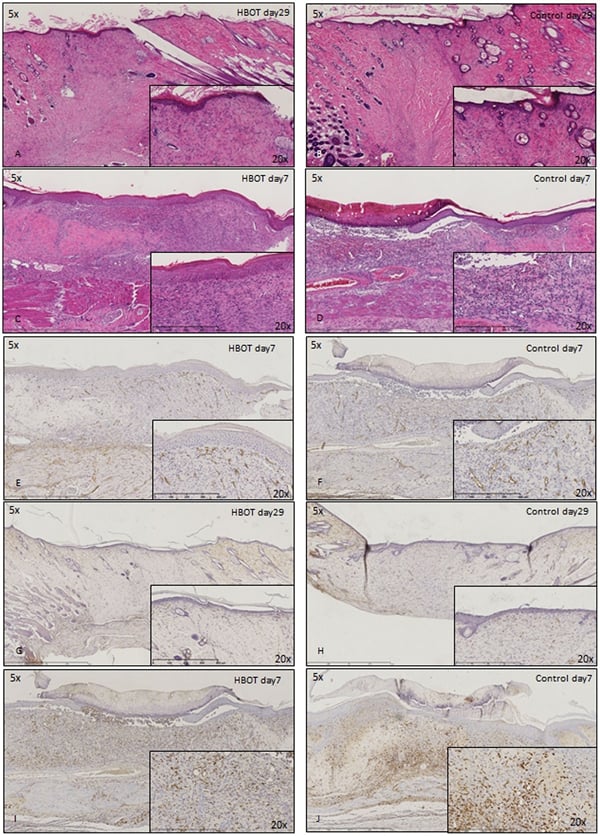
(Published customer image: Histological staining of control and HBOT wounds at post-wounding days 7 and 29. A -D) H&E staining. E -H) CD34 immunohistochemistry. I+J) CD68 immunohistochemistry.From: uk B, Tong M, Fijneman EMG, van Neck JW (2014) Hyperbaric Oxygen Therapy to Treat Diabetes Impaired Wound Healing in Rats. PLoS ONE 9(10): e108533.)
Application Data
(Published customer image: Histological staining of control and HBOT wounds at post-wounding days 7 and 29. A -D) H&E staining. E -H) CD34 immunohistochemistry. I+J) CD68 immunohistochemistry.From: uk B, Tong M, Fijneman EMG, van Neck JW (2014) Hyperbaric Oxygen Therapy to Treat Diabetes Impaired Wound Healing in Rats. PLoS ONE 9(10): e108533.)
CD68, Monoclonal Antibody (Cat# AAA12150)
Full Name
MOUSE ANTI RAT CD68:RPE
Applications
Flow Cytometry
Pricing


 FCM (Flow Cytometry)
(Flow cytometric analysis of THP-1 cells with CD40L antibody at 1/50 dilution (red) compared with an unlabelled control (cells without incubation with primary antibody; black). Alexa Fluor 488-conjugated goat anti rabbit IgG was used as the secondary antibody.)
FCM (Flow Cytometry)
(Flow cytometric analysis of THP-1 cells with CD40L antibody at 1/50 dilution (red) compared with an unlabelled control (cells without incubation with primary antibody; black). Alexa Fluor 488-conjugated goat anti rabbit IgG was used as the secondary antibody.)
CD40L, Monoclonal Antibody (Cat# AAA30328)
Full Name
CD40L Antibody
Gene Names
CD40LG; IGM; IMD3; TRAP; gp39; CD154; CD40L; HIGM1; T-BAM; TNFSF5; hCD40L
Reactivity
Human, Mouse
Applications
Western Blot, Immunocytochemistry, Immunofluorescence, Immunohistochemistry, Flow Cytometry
Purity
ProA affinity purified
Pricing
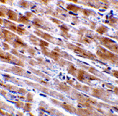


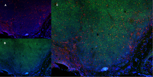
 IF (Immunofluorescence)
(Immunofluorescence of CTRP3 in Mouse Heart tissue with CTRP3 antibody at 10 μg/mL.)
IF (Immunofluorescence)
(Immunofluorescence of CTRP3 in Mouse Heart tissue with CTRP3 antibody at 10 μg/mL.)
CTRP3, Polyclonal Antibody (Cat# AAA10961)
Full Name
CTRP3 Antibody
Gene Names
C1QTNF3; CORS; CORCS; CTRP3; CORS26; C1ATNF3; CORS-26
Reactivity
Human, Mouse, Rat
Applications
Western Blot, Immunohistochemistry, Immunofluorescence
Purity
CTRP3 Antibody is affinity chromatography purified via peptide column.
Pricing


 Application Data
(Published customer image: Histological staining of control and HBOT wounds at post-wounding days 7 and 29. A -D) H&E staining. E -H) CD34 immunohistochemistry. I+J) CD68 immunohistochemistry.From: uk B, Tong M, Fijneman EMG, van Neck JW (2014) Hyperbaric Oxygen Therapy to Treat Diabetes Impaired Wound Healing in Rats. PLoS ONE 9(10): e108533.)
Application Data
(Published customer image: Histological staining of control and HBOT wounds at post-wounding days 7 and 29. A -D) H&E staining. E -H) CD34 immunohistochemistry. I+J) CD68 immunohistochemistry.From: uk B, Tong M, Fijneman EMG, van Neck JW (2014) Hyperbaric Oxygen Therapy to Treat Diabetes Impaired Wound Healing in Rats. PLoS ONE 9(10): e108533.)
CD68, Monoclonal Antibody (Cat# AAA12149)
Full Name
MOUSE ANTI RAT CD68
Applications
Immunohistochemistry, Flow Cytometry, Immunofluorescence, Immunoprecipitation, Immunohistochemistry
Pricing


 Application Data
(Staining of mouse splenocytes with Rat anti Mouse CD40: RPE)
Application Data
(Staining of mouse splenocytes with Rat anti Mouse CD40: RPE)
CD40, Monoclonal Antibody (Cat# AAA12023)
Full Name
RAT ANTI MOUSE CD40:RPE
Gene Names
Cd40; IGM; p50; Bp50; GP39; IMD3; TRAP; HIGM1; T-BAM; Tnfrsf5; AI326936
Applications
Flow Cytometry
Pricing


 FCM (Flow Cytometry)
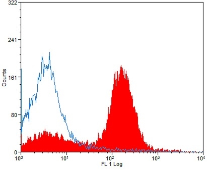
(Flow cytometric analysis of Jurkat cells with CD137 antibody at 1/100 dilution (red) compared with an unlabelled control (cells without incubation with primary antibody; black).)
FCM (Flow Cytometry)
(Flow cytometric analysis of Jurkat cells with CD137 antibody at 1/100 dilution (red) compared with an unlabelled control (cells without incubation with primary antibody; black).)
CD137, Monoclonal Antibody (Cat# AAA29865)
Full Name
CD137 Antibody
Gene Names
TNFRSF9; ILA; 4-1BB; CD137; CDw137
Reactivity
Human, Mouse, Rat
Applications
Western Blot, Immunohistochemistry, Immunocytochemistry, Flow Cytometry
Purity
ProA affinity purified
Pricing


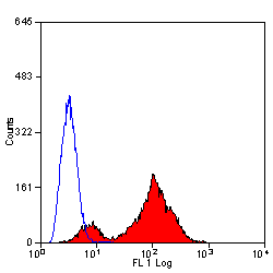
 FCM (Flow Cytometry)
(Flow cytometry analysis of LIGHT overexpressing HEK293 cells using LIGHT antibody and control mouse IgG antibody at 10 μg/ml. Blue: Untransfected HEK293 cells. Yellow: LIGHT overexpressing HEK293 cells.)
FCM (Flow Cytometry)
(Flow cytometry analysis of LIGHT overexpressing HEK293 cells using LIGHT antibody and control mouse IgG antibody at 10 μg/ml. Blue: Untransfected HEK293 cells. Yellow: LIGHT overexpressing HEK293 cells.)
LIGHT, Monoclonal Antibody (Cat# AAA11011)
Full Name
LIGHT Antibody [8D4]
Gene Names
TNFSF14; LTg; CD258; HVEML; LIGHT
Reactivity
Human
Applications
Western Blot, Immunohistochemistry, Immunocytochemistry, Immunofluorescence, Flow Cytometry
Purity
Protein A purified
Pricing


 Application Data
(Published customer image Infiltration of GFP+ BM-cells in infarct and peri-infarct regions. (A-B) Dot plots of viable macrophages/granulocytes (CD11b+CD45high, top right quadrants) and microglia (CD11b+CD45dim, bottom right quadrants) in cortex from BM-chimeric unmanipulated mice and mice exposed to pMCAO. (C) Bar graph showing mean numbers of CD11b+CD45dim microglia and CD11b+CD45high macrophages/granulocytes in BM-chimeric mice 24 hours after pMCAO, subdivided based on expression of GFP (n = 5). Approximately 92% of of the CD45high population were GFP+. (D) Estimation and comparison of mean numbers of CD11b+CD45dim microglia in non-chimeric (n = 10) versus BM-chimeric mice (n = 5) 24 hours after of pMCAO shows significantly fewer CD11b+CD45dim microglial cells in irradiated mice. (E) Overview, showing distribution of infiltrating GFP+ BM-derived cells into infarct (IF) and peri-infarct (P-IF) regions 24 hours after pMCAO. (E-G) By 24 hours, GFP+ single cells (F) and vessel-associated aggregates of GFP+ cells (arrows in G) were observed in infarct and peri-infarct regions. Some of the vessel-associated cells were round, leukocyte-like cells (arrows) while others were elongated cells lining the vasculature (arrow heads in G and in insert). (H) Bar graph showing mean numbers of single GFP+ cells and vessel-associated aggregates of GFP+ cells in ipsi- and contralateral cortex 24 hours after surgery (n = 10). (I-P) Immunohistochemical staining of CD45.1 (I, K), CD45.2 (J, L), IgG2a (M, O) and CD45 (N, P) in ischemic tissue in BM-chimeric (I, J, M, N) and non-chimeric mice (K, L, O, P) 24 hours after pMCAO. N.D, none detected. Scale bars: 200 um (A), 10 um (B, C). 50 um (I-P) *P < 0.05, **P < 0.01, and ***P < 0.001.From: Clausen BH, Lambertsen KL, Babcock AA, Holm TH, Dagnaes-Hansen F, Finsen B. Interleukin-1beta and tumor necrosis factor-alpha are expressed by different subsets of microglia and macrophages after ischemic stroke in mice. J Neuroinflammation. 2008 Oct 23;5:46.)
Application Data
(Published customer image Infiltration of GFP+ BM-cells in infarct and peri-infarct regions. (A-B) Dot plots of viable macrophages/granulocytes (CD11b+CD45high, top right quadrants) and microglia (CD11b+CD45dim, bottom right quadrants) in cortex from BM-chimeric unmanipulated mice and mice exposed to pMCAO. (C) Bar graph showing mean numbers of CD11b+CD45dim microglia and CD11b+CD45high macrophages/granulocytes in BM-chimeric mice 24 hours after pMCAO, subdivided based on expression of GFP (n = 5). Approximately 92% of of the CD45high population were GFP+. (D) Estimation and comparison of mean numbers of CD11b+CD45dim microglia in non-chimeric (n = 10) versus BM-chimeric mice (n = 5) 24 hours after of pMCAO shows significantly fewer CD11b+CD45dim microglial cells in irradiated mice. (E) Overview, showing distribution of infiltrating GFP+ BM-derived cells into infarct (IF) and peri-infarct (P-IF) regions 24 hours after pMCAO. (E-G) By 24 hours, GFP+ single cells (F) and vessel-associated aggregates of GFP+ cells (arrows in G) were observed in infarct and peri-infarct regions. Some of the vessel-associated cells were round, leukocyte-like cells (arrows) while others were elongated cells lining the vasculature (arrow heads in G and in insert). (H) Bar graph showing mean numbers of single GFP+ cells and vessel-associated aggregates of GFP+ cells in ipsi- and contralateral cortex 24 hours after surgery (n = 10). (I-P) Immunohistochemical staining of CD45.1 (I, K), CD45.2 (J, L), IgG2a (M, O) and CD45 (N, P) in ischemic tissue in BM-chimeric (I, J, M, N) and non-chimeric mice (K, L, O, P) 24 hours after pMCAO. N.D, none detected. Scale bars: 200 um (A), 10 um (B, C). 50 um (I-P) *P < 0.05, **P < 0.01, and ***P < 0.001.From: Clausen BH, Lambertsen KL, Babcock AA, Holm TH, Dagnaes-Hansen F, Finsen B. Interleukin-1beta and tumor necrosis factor-alpha are expressed by different subsets of microglia and macrophages after ischemic stroke in mice. J Neuroinflammation. 2008 Oct 23;5:46.)
CD11b, Monoclonal Antibody (Cat# AAA12182)
Full Name
RAT ANTI MOUSE CD11b:FITC
Gene Names
Itgam; CR3; CR3A; MAC1; Cd11b; Ly-40; Mac-1; Mac-1a; CD11b/CD18; F730045J24Rik
Applications
Flow Cytometry
Pricing


 Application Data
(Published customer image Infiltration of GFP+ BM-cells in infarct and peri-infarct regions. (A-B) Dot plots of viable macrophages/granulocytes (CD11b+CD45high, top right quadrants) and microglia (CD11b+CD45dim, bottom right quadrants) in cortex from BM-chimeric unmanipulated mice and mice exposed to pMCAO. (C) Bar graph showing mean numbers of CD11b+CD45dim microglia and CD11b+CD45high macrophages/granulocytes in BM-chimeric mice 24 hours after pMCAO, subdivided based on expression of GFP (n = 5). Approximately 92% of of the CD45high population were GFP+. (D) Estimation and comparison of mean numbers of CD11b+CD45dim microglia in non-chimeric (n = 10) versus BM-chimeric mice (n = 5) 24 hours after of pMCAO shows significantly fewer CD11b+CD45dim microglial cells in irradiated mice. (E) Overview, showing distribution of infiltrating GFP+ BM-derived cells into infarct (IF) and peri-infarct (P-IF) regions 24 hours after pMCAO. (E-G) By 24 hours, GFP+ single cells (F) and vessel-associated aggregates of GFP+ cells (arrows in G) were observed in infarct and peri-infarct regions. Some of the vessel-associated cells were round, leukocyte-like cells (arrows) while others were elongated cells lining the vasculature (arrow heads in G and in insert). (H) Bar graph showing mean numbers of single GFP+ cells and vessel-associated aggregates of GFP+ cells in ipsi- and contralateral cortex 24 hours after surgery (n = 10). (I-P) Immunohistochemical staining of CD45.1 (I, K), CD45.2 (J, L), IgG2a (M, O) and CD45 (N, P) in ischemic tissue in BM-chimeric (I, J, M, N) and non-chimeric mice (K, L, O, P) 24 hours after pMCAO. N.D, none detected. Scale bars: 200 um (A), 10 um (B, C). 50 um (I-P) *P < 0.05, **P < 0.01, and ***P < 0.001.From: Clausen BH, Lambertsen KL, Babcock AA, Holm TH, Dagnaes-Hansen F, Finsen B. Interleukin-1beta and tumor necrosis factor-alpha are expressed by different subsets of microglia and macrophages after ischemic stroke in mice. J Neuroinflammation. 2008 Oct 23;5:46.)
Application Data
(Published customer image Infiltration of GFP+ BM-cells in infarct and peri-infarct regions. (A-B) Dot plots of viable macrophages/granulocytes (CD11b+CD45high, top right quadrants) and microglia (CD11b+CD45dim, bottom right quadrants) in cortex from BM-chimeric unmanipulated mice and mice exposed to pMCAO. (C) Bar graph showing mean numbers of CD11b+CD45dim microglia and CD11b+CD45high macrophages/granulocytes in BM-chimeric mice 24 hours after pMCAO, subdivided based on expression of GFP (n = 5). Approximately 92% of of the CD45high population were GFP+. (D) Estimation and comparison of mean numbers of CD11b+CD45dim microglia in non-chimeric (n = 10) versus BM-chimeric mice (n = 5) 24 hours after of pMCAO shows significantly fewer CD11b+CD45dim microglial cells in irradiated mice. (E) Overview, showing distribution of infiltrating GFP+ BM-derived cells into infarct (IF) and peri-infarct (P-IF) regions 24 hours after pMCAO. (E-G) By 24 hours, GFP+ single cells (F) and vessel-associated aggregates of GFP+ cells (arrows in G) were observed in infarct and peri-infarct regions. Some of the vessel-associated cells were round, leukocyte-like cells (arrows) while others were elongated cells lining the vasculature (arrow heads in G and in insert). (H) Bar graph showing mean numbers of single GFP+ cells and vessel-associated aggregates of GFP+ cells in ipsi- and contralateral cortex 24 hours after surgery (n = 10). (I-P) Immunohistochemical staining of CD45.1 (I, K), CD45.2 (J, L), IgG2a (M, O) and CD45 (N, P) in ischemic tissue in BM-chimeric (I, J, M, N) and non-chimeric mice (K, L, O, P) 24 hours after pMCAO. N.D, none detected. Scale bars: 200 um (A), 10 um (B, C). 50 um (I-P) *P < 0.05, **P < 0.01, and ***P < 0.001.From: Clausen BH, Lambertsen KL, Babcock AA, Holm TH, Dagnaes-Hansen F, Finsen B. Interleukin-1beta and tumor necrosis factor-alpha are expressed by different subsets of microglia and macrophages after ischemic stroke in mice. J Neuroinflammation. 2008 Oct 23;5:46.)
CD11b, Monoclonal Antibody (Cat# AAA12184)
Full Name
RAT ANTI MOUSE CD11b
Gene Names
Itgam; CR3; CR3A; MAC1; Cd11b; Ly-40; Mac-1; Mac-1a; CD11b/CD18; F730045J24Rik
Applications
Immunohistochemistry, Flow Cytometry, Immunofluorescence, Immunoprecipitation
Pricing


 WB (Western Blot)
(Figure 9 KD Validation in HeLa Cells (Horinaka et al., 2005)HeLa cells were transfected with DR4siRNA or LacZ control siRNA. At 24 h after transfection, the cells were treated with or without 20 μM luteolin for 24 h. Western blot analysis was carried out with anti-DR4 antibodies. DR4 expression was markedly reduced after DR4 knockdown.)
WB (Western Blot)
(Figure 9 KD Validation in HeLa Cells (Horinaka et al., 2005)HeLa cells were transfected with DR4siRNA or LacZ control siRNA. At 24 h after transfection, the cells were treated with or without 20 μM luteolin for 24 h. Western blot analysis was carried out with anti-DR4 antibodies. DR4 expression was markedly reduced after DR4 knockdown.)
DR4, Polyclonal Antibody (Cat# AAA10950)
Full Name
DR4 Antibody
Gene Names
TNFRSF10A; DR4; APO2; CD261; TRAILR1; TRAILR-1
Reactivity
Human
Applications
Western Blot
Purity
DR4 Antibody is affinity chromatography purified via peptide column.
Pricing


 Application Data
(Published customer image: Histological staining of control and HBOT wounds at post-wounding days 7 and 29. A -D) H&E staining. E -H) CD34 immunohistochemistry. I+J) CD68 immunohistochemistry.From: uk B, Tong M, Fijneman EMG, van Neck JW (2014) Hyperbaric Oxygen Therapy to Treat Diabetes Impaired Wound Healing in Rats. PLoS ONE 9(10): e108533.)
Application Data
(Published customer image: Histological staining of control and HBOT wounds at post-wounding days 7 and 29. A -D) H&E staining. E -H) CD34 immunohistochemistry. I+J) CD68 immunohistochemistry.From: uk B, Tong M, Fijneman EMG, van Neck JW (2014) Hyperbaric Oxygen Therapy to Treat Diabetes Impaired Wound Healing in Rats. PLoS ONE 9(10): e108533.)
CD68, Monoclonal Antibody (Cat# AAA12147)
Full Name
MOUSE ANTI RAT CD68:Biotin
Applications
Flow Cytometry
Pricing


 Application Data
(Staining of mouse splenocytes with Rat anti Mouse CD40: RPE)
Application Data
(Staining of mouse splenocytes with Rat anti Mouse CD40: RPE)
CD40, Monoclonal Antibody (Cat# AAA11919)
Full Name
RAT ANTI MOUSE CD40
Gene Names
Cd40; IGM; p50; Bp50; GP39; IMD3; TRAP; HIGM1; T-BAM; Tnfrsf5; AI326936
Applications
Immunohistochemistry, Flow Cytometry
Pricing
 Standard Curve (Sample)
Tumor Necrosis Factor Alpha, ELISA Kit (Cat# AAA17469)
Full Name
Guinea pig Tumor Necrosis Factor Alpha ELISA Kit
Reactivity
Guinea pig
Pricing


 Application Data
(Published customer image Infiltration of GFP+ BM-cells in infarct and peri-infarct regions. (A-B) Dot plots of viable macrophages/granulocytes (CD11b+CD45high, top right quadrants) and microglia (CD11b+CD45dim, bottom right quadrants) in cortex from BM-chimeric unmanipulated mice and mice exposed to pMCAO. (C) Bar graph showing mean numbers of CD11b+CD45dim microglia and CD11b+CD45high macrophages/granulocytes in BM-chimeric mice 24 hours after pMCAO, subdivided based on expression of GFP (n = 5). Approximately 92% of of the CD45high population were GFP+. (D) Estimation and comparison of mean numbers of CD11b+CD45dim microglia in non-chimeric (n = 10) versus BM-chimeric mice (n = 5) 24 hours after of pMCAO shows significantly fewer CD11b+CD45dim microglial cells in irradiated mice. (E) Overview, showing distribution of infiltrating GFP+ BM-derived cells into infarct (IF) and peri-infarct (P-IF) regions 24 hours after pMCAO. (E-G) By 24 hours, GFP+ single cells (F) and vessel-associated aggregates of GFP+ cells (arrows in G) were observed in infarct and peri-infarct regions. Some of the vessel-associated cells were round, leukocyte-like cells (arrows) while others were elongated cells lining the vasculature (arrow heads in G and in insert). (H) Bar graph showing mean numbers of single GFP+ cells and vessel-associated aggregates of GFP+ cells in ipsi- and contralateral cortex 24 hours after surgery (n = 10). (I-P) Immunohistochemical staining of CD45.1 (I, K), CD45.2 (J, L), IgG2a (M, O) and CD45 (N, P) in ischemic tissue in BM-chimeric (I, J, M, N) and non-chimeric mice (K, L, O, P) 24 hours after pMCAO. N.D, none detected. Scale bars: 200 um (A), 10 um (B, C). 50 um (I-P) *P < 0.05, **P < 0.01, and ***P < 0.001.From: Clausen BH, Lambertsen KL, Babcock AA, Holm TH, Dagnaes-Hansen F, Finsen B. Interleukin-1beta and tumor necrosis factor-alpha are expressed by different subsets of microglia and macrophages after ischemic stroke in mice. J Neuroinflammation. 2008 Oct 23;5:46.)
Application Data
(Published customer image Infiltration of GFP+ BM-cells in infarct and peri-infarct regions. (A-B) Dot plots of viable macrophages/granulocytes (CD11b+CD45high, top right quadrants) and microglia (CD11b+CD45dim, bottom right quadrants) in cortex from BM-chimeric unmanipulated mice and mice exposed to pMCAO. (C) Bar graph showing mean numbers of CD11b+CD45dim microglia and CD11b+CD45high macrophages/granulocytes in BM-chimeric mice 24 hours after pMCAO, subdivided based on expression of GFP (n = 5). Approximately 92% of of the CD45high population were GFP+. (D) Estimation and comparison of mean numbers of CD11b+CD45dim microglia in non-chimeric (n = 10) versus BM-chimeric mice (n = 5) 24 hours after of pMCAO shows significantly fewer CD11b+CD45dim microglial cells in irradiated mice. (E) Overview, showing distribution of infiltrating GFP+ BM-derived cells into infarct (IF) and peri-infarct (P-IF) regions 24 hours after pMCAO. (E-G) By 24 hours, GFP+ single cells (F) and vessel-associated aggregates of GFP+ cells (arrows in G) were observed in infarct and peri-infarct regions. Some of the vessel-associated cells were round, leukocyte-like cells (arrows) while others were elongated cells lining the vasculature (arrow heads in G and in insert). (H) Bar graph showing mean numbers of single GFP+ cells and vessel-associated aggregates of GFP+ cells in ipsi- and contralateral cortex 24 hours after surgery (n = 10). (I-P) Immunohistochemical staining of CD45.1 (I, K), CD45.2 (J, L), IgG2a (M, O) and CD45 (N, P) in ischemic tissue in BM-chimeric (I, J, M, N) and non-chimeric mice (K, L, O, P) 24 hours after pMCAO. N.D, none detected. Scale bars: 200 um (A), 10 um (B, C). 50 um (I-P) *P < 0.05, **P < 0.01, and ***P < 0.001.From: Clausen BH, Lambertsen KL, Babcock AA, Holm TH, Dagnaes-Hansen F, Finsen B. Interleukin-1beta and tumor necrosis factor-alpha are expressed by different subsets of microglia and macrophages after ischemic stroke in mice. J Neuroinflammation. 2008 Oct 23;5:46.)
CD11b, Monoclonal Antibody (Cat# AAA12185)
Full Name
RAT ANTI MOUSE CD11b
Gene Names
Itgam; CR3; CR3A; MAC1; Cd11b; Ly-40; Mac-1; Mac-1a; CD11b/CD18; F730045J24Rik
Applications
Immunohistochemistry, Flow Cytometry, Immunofluorescence, Immunoprecipitation
Pricing


 Application Data
(Published customer image Infiltration of GFP+ BM-cells in infarct and peri-infarct regions. (A-B) Dot plots of viable macrophages/granulocytes (CD11b+CD45high, top right quadrants) and microglia (CD11b+CD45dim, bottom right quadrants) in cortex from BM-chimeric unmanipulated mice and mice exposed to pMCAO. (C) Bar graph showing mean numbers of CD11b+CD45dim microglia and CD11b+CD45high macrophages/granulocytes in BM-chimeric mice 24 hours after pMCAO, subdivided based on expression of GFP (n = 5). Approximately 92% of of the CD45high population were GFP+. (D) Estimation and comparison of mean numbers of CD11b+CD45dim microglia in non-chimeric (n = 10) versus BM-chimeric mice (n = 5) 24 hours after of pMCAO shows significantly fewer CD11b+CD45dim microglial cells in irradiated mice. (E) Overview, showing distribution of infiltrating GFP+ BM-derived cells into infarct (IF) and peri-infarct (P-IF) regions 24 hours after pMCAO. (E-G) By 24 hours, GFP+ single cells (F) and vessel-associated aggregates of GFP+ cells (arrows in G) were observed in infarct and peri-infarct regions. Some of the vessel-associated cells were round, leukocyte-like cells (arrows) while others were elongated cells lining the vasculature (arrow heads in G and in insert). (H) Bar graph showing mean numbers of single GFP+ cells and vessel-associated aggregates of GFP+ cells in ipsi- and contralateral cortex 24 hours after surgery (n = 10). (I-P) Immunohistochemical staining of CD45.1 (I, K), CD45.2 (J, L), IgG2a (M, O) and CD45 (N, P) in ischemic tissue in BM-chimeric (I, J, M, N) and non-chimeric mice (K, L, O, P) 24 hours after pMCAO. N.D, none detected. Scale bars: 200 um (A), 10 um (B, C). 50 um (I-P) *P < 0.05, **P < 0.01, and ***P < 0.001.From: Clausen BH, Lambertsen KL, Babcock AA, Holm TH, Dagnaes-Hansen F, Finsen B. Interleukin-1beta and tumor necrosis factor-alpha are expressed by different subsets of microglia and macrophages after ischemic stroke in mice. J Neuroinflammation. 2008 Oct 23;5:46.)
Application Data
(Published customer image Infiltration of GFP+ BM-cells in infarct and peri-infarct regions. (A-B) Dot plots of viable macrophages/granulocytes (CD11b+CD45high, top right quadrants) and microglia (CD11b+CD45dim, bottom right quadrants) in cortex from BM-chimeric unmanipulated mice and mice exposed to pMCAO. (C) Bar graph showing mean numbers of CD11b+CD45dim microglia and CD11b+CD45high macrophages/granulocytes in BM-chimeric mice 24 hours after pMCAO, subdivided based on expression of GFP (n = 5). Approximately 92% of of the CD45high population were GFP+. (D) Estimation and comparison of mean numbers of CD11b+CD45dim microglia in non-chimeric (n = 10) versus BM-chimeric mice (n = 5) 24 hours after of pMCAO shows significantly fewer CD11b+CD45dim microglial cells in irradiated mice. (E) Overview, showing distribution of infiltrating GFP+ BM-derived cells into infarct (IF) and peri-infarct (P-IF) regions 24 hours after pMCAO. (E-G) By 24 hours, GFP+ single cells (F) and vessel-associated aggregates of GFP+ cells (arrows in G) were observed in infarct and peri-infarct regions. Some of the vessel-associated cells were round, leukocyte-like cells (arrows) while others were elongated cells lining the vasculature (arrow heads in G and in insert). (H) Bar graph showing mean numbers of single GFP+ cells and vessel-associated aggregates of GFP+ cells in ipsi- and contralateral cortex 24 hours after surgery (n = 10). (I-P) Immunohistochemical staining of CD45.1 (I, K), CD45.2 (J, L), IgG2a (M, O) and CD45 (N, P) in ischemic tissue in BM-chimeric (I, J, M, N) and non-chimeric mice (K, L, O, P) 24 hours after pMCAO. N.D, none detected. Scale bars: 200 um (A), 10 um (B, C). 50 um (I-P) *P < 0.05, **P < 0.01, and ***P < 0.001.From: Clausen BH, Lambertsen KL, Babcock AA, Holm TH, Dagnaes-Hansen F, Finsen B. Interleukin-1beta and tumor necrosis factor-alpha are expressed by different subsets of microglia and macrophages after ischemic stroke in mice. J Neuroinflammation. 2008 Oct 23;5:46.)
CD11b, Monoclonal Antibody (Cat# AAA12183)
Full Name
RAT ANTI MOUSE CD11b:FITC
Gene Names
Itgam; CR3; CR3A; MAC1; Cd11b; Ly-40; Mac-1; Mac-1a; CD11b/CD18; F730045J24Rik
Applications
Flow Cytometry
Pricing


 WB (Western Blot)
(WB Suggested Anti-PTX3 Antibody Titration: 0.2-1 ug/mlELISA Titer: 1:312500Positive Control: MCF7 cell lysate)
WB (Western Blot)
(WB Suggested Anti-PTX3 Antibody Titration: 0.2-1 ug/mlELISA Titer: 1:312500Positive Control: MCF7 cell lysate)
PTX3, Polyclonal Antibody (Cat# AAA23569)
Full Name
PTX3 antibody - N-terminal region
Gene Names
PTX3; TSG-14; TNFAIP5
Reactivity
Cow, Guinea Pig, Human, Mouse, Rabbit, Rat
Applications
Immunohistochemistry, Western Blot
Purity
Affinity Purified
Pricing


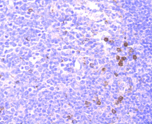
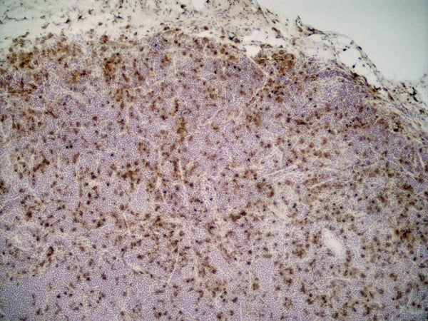
 IHC (Immunohistochemistry)
(Immunohistochemistry of paraffin-embedded mouse spleen using TNFRSF11B Antibody at dilution of 1:200 (40x lens).)
IHC (Immunohistochemistry)
(Immunohistochemistry of paraffin-embedded mouse spleen using TNFRSF11B Antibody at dilution of 1:200 (40x lens).)
TNFRSF11B, Polyclonal Antibody (Cat# AAA10748)
Full Name
TNFRSF11B Polyclonal Antibody
Gene Names
TNFRSF11B; OPG; TR1; OCIF
Reactivity
Human, Mouse, Rat
Applications
Western Blot, Immunohistochemistry, Immunofluorescence
Purity
Affinity Purification
Pricing
 FCM (Flow Cytometry)
(Test Results:Tissue Distribution by Flow Cytometry Analysis:Mouse Strain: BALB/cCell Concentration: 1 x 10^6 cells per test.Antibody Concentration Used: 1.0ug/10^6 cells)
FCM (Flow Cytometry)
(Test Results:Tissue Distribution by Flow Cytometry Analysis:Mouse Strain: BALB/cCell Concentration: 1 x 10^6 cells per test.Antibody Concentration Used: 1.0ug/10^6 cells)
CD134L/OX40L, Monoclonal Antibody (Cat# AAA14267)
Full Name
Anti-Mouse CD134L/OX40L, Purified (Clone OX89) (rat IgG1)
Reactivity
Mouse
Applications
Immunohistochemistry, Functional Assay
Purity
Purified from ascitic fluid via Protein G Chromatography
Pricing
 Standard Curve (Sample)
tumor necrosis factor (TNF superfamily, member 2), ELISA Kit (Cat# AAA15045)
Full Name
Rat TNF-alpha ELISA Kit
Gene Names
TNF; TNFA; TNFSF2
Reactivity
Rat
Pricing
 Standard Curve (Sample)
tumor necrosis factor, alpha-induced protein 6, ELISA Kit (Cat# AAA18167)
Full Name
Mouse Tumor necrosis factor-inducible gene 6 protein, TNFAIP6 ELISA Kit
Gene Names
Tnfaip6; Tsg6; TSG-6; Tnfip6
Reactivity
Mouse
Pricing


 Application Data
(Published customer image: Histological staining of control and HBOT wounds at post-wounding days 7 and 29. A -D) H&E staining. E -H) CD34 immunohistochemistry. I+J) CD68 immunohistochemistry.From: uk B, Tong M, Fijneman EMG, van Neck JW (2014) Hyperbaric Oxygen Therapy to Treat Diabetes Impaired Wound Healing in Rats. PLoS ONE 9(10): e108533.)
Application Data
(Published customer image: Histological staining of control and HBOT wounds at post-wounding days 7 and 29. A -D) H&E staining. E -H) CD34 immunohistochemistry. I+J) CD68 immunohistochemistry.From: uk B, Tong M, Fijneman EMG, van Neck JW (2014) Hyperbaric Oxygen Therapy to Treat Diabetes Impaired Wound Healing in Rats. PLoS ONE 9(10): e108533.)
CD68, Monoclonal Antibody (Cat# AAA12151)
Full Name
MOUSE ANTI RAT CD68
Applications
Immunohistochemistry, Flow Cytometry, Immunofluorescence, Immunoprecipitation, Immunohistochemistry, Radioimmunoassay, Western Blot
Pricing


 FCM (Flow Cytometry)
(Flow cytometric analysis of SW480 cells with DR5 antibody at 1/100 dilution (red) compared with an unlabelled control (cells without incubation with primary antibody; black).)
FCM (Flow Cytometry)
(Flow cytometric analysis of SW480 cells with DR5 antibody at 1/100 dilution (red) compared with an unlabelled control (cells without incubation with primary antibody; black).)
DR5, Monoclonal Antibody (Cat# AAA30353)
Full Name
DR5 Antibody
Gene Names
TNFRSF10B; DR5; CD262; KILLER; TRICK2; TRICKB; ZTNFR9; TRAILR2; TRICK2A; TRICK2B; TRAIL-R2; KILLER/DR5
Reactivity
Human
Applications
Western Blot, Immunoprecipitation, Immunohistochemistry, Flow Cytometry
Purity
ProA affinity purified
Pricing

 WB (Western Blot)
(Peptide-+ Western blot analysis of extracts from RAW264.7 cells, HT-29 cells and LOVO cells, using TNF Receptor II antibody. Immunofluorescence analysis of HeLa cells, using TNF Receptor II antibody.)
WB (Western Blot)
(Peptide-+ Western blot analysis of extracts from RAW264.7 cells, HT-29 cells and LOVO cells, using TNF Receptor II antibody. Immunofluorescence analysis of HeLa cells, using TNF Receptor II antibody.)
TNF Receptor II, Antibody (Cat# AAA17967)
Full Name
TNF Receptor II Antibody
Gene Names
TNFRSF1B; p75; TBPII; TNFBR; TNFR2; CD120b; TNFR1B; TNFR80; TNF-R75; p75TNFR; TNF-R-II
Reactivity
Human, Mouse, Rat
Applications
Western Blot, Immunofluorescence
Purity
Affinity-purified from rabbit antiserum by affinity-chromatography using epitope-specific immunogen.
Pricing


 WB (Western Blot)
(Sample: Recombinant TRAIL, Human;Antibody: Rabbit Anti-Human TRAIL Ab)
WB (Western Blot)
(Sample: Recombinant TRAIL, Human;Antibody: Rabbit Anti-Human TRAIL Ab)
Tumor Necrosis Factor Related Apoptosis Inducing Ligand (TRAIL), Active Protein (Cat# AAA21106)
Full Name
Active Tumor Necrosis Factor Related Apoptosis Inducing Ligand (TRAIL)
Gene Names
TNFSF10; TL2; APO2L; CD253; TRAIL; Apo-2L; TNLG6A
Reactivity
Homo sapiens (Human)
Applications
Cell culture; Activity Assays; In vivo assays
Purity
>95%
Pricing


 Application Data
(Published customer image Infiltration of GFP+ BM-cells in infarct and peri-infarct regions. (A-B) Dot plots of viable macrophages/granulocytes (CD11b+CD45high, top right quadrants) and microglia (CD11b+CD45dim, bottom right quadrants) in cortex from BM-chimeric unmanipulated mice and mice exposed to pMCAO. (C) Bar graph showing mean numbers of CD11b+CD45dim microglia and CD11b+CD45high macrophages/granulocytes in BM-chimeric mice 24 hours after pMCAO, subdivided based on expression of GFP (n = 5). Approximately 92% of of the CD45high population were GFP+. (D) Estimation and comparison of mean numbers of CD11b+CD45dim microglia in non-chimeric (n = 10) versus BM-chimeric mice (n = 5) 24 hours after of pMCAO shows significantly fewer CD11b+CD45dim microglial cells in irradiated mice. (E) Overview, showing distribution of infiltrating GFP+ BM-derived cells into infarct (IF) and peri-infarct (P-IF) regions 24 hours after pMCAO. (E-G) By 24 hours, GFP+ single cells (F) and vessel-associated aggregates of GFP+ cells (arrows in G) were observed in infarct and peri-infarct regions. Some of the vessel-associated cells were round, leukocyte-like cells (arrows) while others were elongated cells lining the vasculature (arrow heads in G and in insert). (H) Bar graph showing mean numbers of single GFP+ cells and vessel-associated aggregates of GFP+ cells in ipsi- and contralateral cortex 24 hours after surgery (n = 10). (I-P) Immunohistochemical staining of CD45.1 (I, K), CD45.2 (J, L), IgG2a (M, O) and CD45 (N, P) in ischemic tissue in BM-chimeric (I, J, M, N) and non-chimeric mice (K, L, O, P) 24 hours after pMCAO. N.D, none detected. Scale bars: 200 um (A), 10 um (B, C). 50 um (I-P) *P < 0.05, **P < 0.01, and ***P < 0.001.From: Clausen BH, Lambertsen KL, Babcock AA, Holm TH, Dagnaes-Hansen F, Finsen B. Interleukin-1beta and tumor necrosis factor-alpha are expressed by different subsets of microglia and macrophages after ischemic stroke in mice. J Neuroinflammation. 2008 Oct 23;5:46.)
Application Data
(Published customer image Infiltration of GFP+ BM-cells in infarct and peri-infarct regions. (A-B) Dot plots of viable macrophages/granulocytes (CD11b+CD45high, top right quadrants) and microglia (CD11b+CD45dim, bottom right quadrants) in cortex from BM-chimeric unmanipulated mice and mice exposed to pMCAO. (C) Bar graph showing mean numbers of CD11b+CD45dim microglia and CD11b+CD45high macrophages/granulocytes in BM-chimeric mice 24 hours after pMCAO, subdivided based on expression of GFP (n = 5). Approximately 92% of of the CD45high population were GFP+. (D) Estimation and comparison of mean numbers of CD11b+CD45dim microglia in non-chimeric (n = 10) versus BM-chimeric mice (n = 5) 24 hours after of pMCAO shows significantly fewer CD11b+CD45dim microglial cells in irradiated mice. (E) Overview, showing distribution of infiltrating GFP+ BM-derived cells into infarct (IF) and peri-infarct (P-IF) regions 24 hours after pMCAO. (E-G) By 24 hours, GFP+ single cells (F) and vessel-associated aggregates of GFP+ cells (arrows in G) were observed in infarct and peri-infarct regions. Some of the vessel-associated cells were round, leukocyte-like cells (arrows) while others were elongated cells lining the vasculature (arrow heads in G and in insert). (H) Bar graph showing mean numbers of single GFP+ cells and vessel-associated aggregates of GFP+ cells in ipsi- and contralateral cortex 24 hours after surgery (n = 10). (I-P) Immunohistochemical staining of CD45.1 (I, K), CD45.2 (J, L), IgG2a (M, O) and CD45 (N, P) in ischemic tissue in BM-chimeric (I, J, M, N) and non-chimeric mice (K, L, O, P) 24 hours after pMCAO. N.D, none detected. Scale bars: 200 um (A), 10 um (B, C). 50 um (I-P) *P < 0.05, **P < 0.01, and ***P < 0.001.From: Clausen BH, Lambertsen KL, Babcock AA, Holm TH, Dagnaes-Hansen F, Finsen B. Interleukin-1beta and tumor necrosis factor-alpha are expressed by different subsets of microglia and macrophages after ischemic stroke in mice. J Neuroinflammation. 2008 Oct 23;5:46.)
CD11b, Monoclonal Antibody (Cat# AAA12186)
Full Name
RAT ANTI MOUSE CD11b:RPE
Gene Names
Itgam; CR3; CR3A; MAC1; Cd11b; Ly-40; Mac-1; Mac-1a; CD11b/CD18; F730045J24Rik
Applications
Flow Cytometry
Pricing


 Application Data
(Published customer image Infiltration of GFP+ BM-cells in infarct and peri-infarct regions. (A-B) Dot plots of viable macrophages/granulocytes (CD11b+CD45high, top right quadrants) and microglia (CD11b+CD45dim, bottom right quadrants) in cortex from BM-chimeric unmanipulated mice and mice exposed to pMCAO. (C) Bar graph showing mean numbers of CD11b+CD45dim microglia and CD11b+CD45high macrophages/granulocytes in BM-chimeric mice 24 hours after pMCAO, subdivided based on expression of GFP (n = 5). Approximately 92% of of the CD45high population were GFP+. (D) Estimation and comparison of mean numbers of CD11b+CD45dim microglia in non-chimeric (n = 10) versus BM-chimeric mice (n = 5) 24 hours after of pMCAO shows significantly fewer CD11b+CD45dim microglial cells in irradiated mice. (E) Overview, showing distribution of infiltrating GFP+ BM-derived cells into infarct (IF) and peri-infarct (P-IF) regions 24 hours after pMCAO. (E-G) By 24 hours, GFP+ single cells (F) and vessel-associated aggregates of GFP+ cells (arrows in G) were observed in infarct and peri-infarct regions. Some of the vessel-associated cells were round, leukocyte-like cells (arrows) while others were elongated cells lining the vasculature (arrow heads in G and in insert). (H) Bar graph showing mean numbers of single GFP+ cells and vessel-associated aggregates of GFP+ cells in ipsi- and contralateral cortex 24 hours after surgery (n = 10). (I-P) Immunohistochemical staining of CD45.1 (I, K), CD45.2 (J, L), IgG2a (M, O) and CD45 (N, P) in ischemic tissue in BM-chimeric (I, J, M, N) and non-chimeric mice (K, L, O, P) 24 hours after pMCAO. N.D, none detected. Scale bars: 200 um (A), 10 um (B, C). 50 um (I-P) *P < 0.05, **P < 0.01, and ***P < 0.001.From: Clausen BH, Lambertsen KL, Babcock AA, Holm TH, Dagnaes-Hansen F, Finsen B. Interleukin-1beta and tumor necrosis factor-alpha are expressed by different subsets of microglia and macrophages after ischemic stroke in mice. J Neuroinflammation. 2008 Oct 23;5:46.)
Application Data
(Published customer image Infiltration of GFP+ BM-cells in infarct and peri-infarct regions. (A-B) Dot plots of viable macrophages/granulocytes (CD11b+CD45high, top right quadrants) and microglia (CD11b+CD45dim, bottom right quadrants) in cortex from BM-chimeric unmanipulated mice and mice exposed to pMCAO. (C) Bar graph showing mean numbers of CD11b+CD45dim microglia and CD11b+CD45high macrophages/granulocytes in BM-chimeric mice 24 hours after pMCAO, subdivided based on expression of GFP (n = 5). Approximately 92% of of the CD45high population were GFP+. (D) Estimation and comparison of mean numbers of CD11b+CD45dim microglia in non-chimeric (n = 10) versus BM-chimeric mice (n = 5) 24 hours after of pMCAO shows significantly fewer CD11b+CD45dim microglial cells in irradiated mice. (E) Overview, showing distribution of infiltrating GFP+ BM-derived cells into infarct (IF) and peri-infarct (P-IF) regions 24 hours after pMCAO. (E-G) By 24 hours, GFP+ single cells (F) and vessel-associated aggregates of GFP+ cells (arrows in G) were observed in infarct and peri-infarct regions. Some of the vessel-associated cells were round, leukocyte-like cells (arrows) while others were elongated cells lining the vasculature (arrow heads in G and in insert). (H) Bar graph showing mean numbers of single GFP+ cells and vessel-associated aggregates of GFP+ cells in ipsi- and contralateral cortex 24 hours after surgery (n = 10). (I-P) Immunohistochemical staining of CD45.1 (I, K), CD45.2 (J, L), IgG2a (M, O) and CD45 (N, P) in ischemic tissue in BM-chimeric (I, J, M, N) and non-chimeric mice (K, L, O, P) 24 hours after pMCAO. N.D, none detected. Scale bars: 200 um (A), 10 um (B, C). 50 um (I-P) *P < 0.05, **P < 0.01, and ***P < 0.001.From: Clausen BH, Lambertsen KL, Babcock AA, Holm TH, Dagnaes-Hansen F, Finsen B. Interleukin-1beta and tumor necrosis factor-alpha are expressed by different subsets of microglia and macrophages after ischemic stroke in mice. J Neuroinflammation. 2008 Oct 23;5:46.)
CD11b, Monoclonal Antibody (Cat# AAA12231)
Full Name
RAT ANTI MOUSE CD11b:Low Endotoxin
Gene Names
Itgam; CR3; CR3A; MAC1; Cd11b; Ly-40; Mac-1; Mac-1a; CD11b/CD18; F730045J24Rik
Applications
Immunohistochemistry, Flow Cytometry, Functional Assay, Immunofluorescence, Immunoprecipitation
Pricing
 Standard Curve (Sample)
TNF-alpha (Tumor Necrosis Factor-alpha), ELISA Kit (Cat# AAA27544)
Full Name
Hamster TNF-alpha (Tumor Necrosis Factor-alpha) ELISA Kit
Reactivity
Hamster
Pricing


 Application Data
(Published customer image: Histological staining of control and HBOT wounds at post-wounding days 7 and 29. A -D) H&E staining. E -H) CD34 immunohistochemistry. I+J) CD68 immunohistochemistry.From: uk B, Tong M, Fijneman EMG, van Neck JW (2014) Hyperbaric Oxygen Therapy to Treat Diabetes Impaired Wound Healing in Rats. PLoS ONE 9(10): e108533.)
Application Data
(Published customer image: Histological staining of control and HBOT wounds at post-wounding days 7 and 29. A -D) H&E staining. E -H) CD34 immunohistochemistry. I+J) CD68 immunohistochemistry.From: uk B, Tong M, Fijneman EMG, van Neck JW (2014) Hyperbaric Oxygen Therapy to Treat Diabetes Impaired Wound Healing in Rats. PLoS ONE 9(10): e108533.)
CD68, Monoclonal Antibody (Cat# AAA12148)
Full Name
MOUSE ANTI RAT CD68:FITC
Applications
Flow Cytometry
Pricing


 Application Data
(Published customer image Infiltration of GFP+ BM-cells in infarct and peri-infarct regions. (A-B) Dot plots of viable macrophages/granulocytes (CD11b+CD45high, top right quadrants) and microglia (CD11b+CD45dim, bottom right quadrants) in cortex from BM-chimeric unmanipulated mice and mice exposed to pMCAO. (C) Bar graph showing mean numbers of CD11b+CD45dim microglia and CD11b+CD45high macrophages/granulocytes in BM-chimeric mice 24 hours after pMCAO, subdivided based on expression of GFP (n = 5). Approximately 92% of of the CD45high population were GFP+. (D) Estimation and comparison of mean numbers of CD11b+CD45dim microglia in non-chimeric (n = 10) versus BM-chimeric mice (n = 5) 24 hours after of pMCAO shows significantly fewer CD11b+CD45dim microglial cells in irradiated mice. (E) Overview, showing distribution of infiltrating GFP+ BM-derived cells into infarct (IF) and peri-infarct (P-IF) regions 24 hours after pMCAO. (E-G) By 24 hours, GFP+ single cells (F) and vessel-associated aggregates of GFP+ cells (arrows in G) were observed in infarct and peri-infarct regions. Some of the vessel-associated cells were round, leukocyte-like cells (arrows) while others were elongated cells lining the vasculature (arrow heads in G and in insert). (H) Bar graph showing mean numbers of single GFP+ cells and vessel-associated aggregates of GFP+ cells in ipsi- and contralateral cortex 24 hours after surgery (n = 10). (I-P) Immunohistochemical staining of CD45.1 (I, K), CD45.2 (J, L), IgG2a (M, O) and CD45 (N, P) in ischemic tissue in BM-chimeric (I, J, M, N) and non-chimeric mice (K, L, O, P) 24 hours after pMCAO. N.D, none detected. Scale bars: 200 um (A), 10 um (B, C). 50 um (I-P) *P < 0.05, **P < 0.01, and ***P < 0.001.From: Clausen BH, Lambertsen KL, Babcock AA, Holm TH, Dagnaes-Hansen F, Finsen B. Interleukin-1beta and tumor necrosis factor-alpha are expressed by different subsets of microglia and macrophages after ischemic stroke in mice. J Neuroinflammation. 2008 Oct 23;5:46.)
Application Data
(Published customer image Infiltration of GFP+ BM-cells in infarct and peri-infarct regions. (A-B) Dot plots of viable macrophages/granulocytes (CD11b+CD45high, top right quadrants) and microglia (CD11b+CD45dim, bottom right quadrants) in cortex from BM-chimeric unmanipulated mice and mice exposed to pMCAO. (C) Bar graph showing mean numbers of CD11b+CD45dim microglia and CD11b+CD45high macrophages/granulocytes in BM-chimeric mice 24 hours after pMCAO, subdivided based on expression of GFP (n = 5). Approximately 92% of of the CD45high population were GFP+. (D) Estimation and comparison of mean numbers of CD11b+CD45dim microglia in non-chimeric (n = 10) versus BM-chimeric mice (n = 5) 24 hours after of pMCAO shows significantly fewer CD11b+CD45dim microglial cells in irradiated mice. (E) Overview, showing distribution of infiltrating GFP+ BM-derived cells into infarct (IF) and peri-infarct (P-IF) regions 24 hours after pMCAO. (E-G) By 24 hours, GFP+ single cells (F) and vessel-associated aggregates of GFP+ cells (arrows in G) were observed in infarct and peri-infarct regions. Some of the vessel-associated cells were round, leukocyte-like cells (arrows) while others were elongated cells lining the vasculature (arrow heads in G and in insert). (H) Bar graph showing mean numbers of single GFP+ cells and vessel-associated aggregates of GFP+ cells in ipsi- and contralateral cortex 24 hours after surgery (n = 10). (I-P) Immunohistochemical staining of CD45.1 (I, K), CD45.2 (J, L), IgG2a (M, O) and CD45 (N, P) in ischemic tissue in BM-chimeric (I, J, M, N) and non-chimeric mice (K, L, O, P) 24 hours after pMCAO. N.D, none detected. Scale bars: 200 um (A), 10 um (B, C). 50 um (I-P) *P < 0.05, **P < 0.01, and ***P < 0.001.From: Clausen BH, Lambertsen KL, Babcock AA, Holm TH, Dagnaes-Hansen F, Finsen B. Interleukin-1beta and tumor necrosis factor-alpha are expressed by different subsets of microglia and macrophages after ischemic stroke in mice. J Neuroinflammation. 2008 Oct 23;5:46.)
CD11b, Monoclonal Antibody (Cat# AAA12181)
Full Name
RAT ANTI MOUSE CD11b
Gene Names
Itgam; CR3; CR3A; MAC1; Cd11b; Ly-40; Mac-1; Mac-1a; CD11b/CD18; F730045J24Rik
Reactivity
Human
Applications
Immunohistochemistry, Flow Cytometry, Immunofluorescence, Immunoprecipitation
Pricing
 Standard Curve (Sample)
TNF alpha, ELISA Kit (Cat# AAA17896)
Full Name
Human TNF alpha ELISA Kit
Gene Names
TNF; DIF; TNFA; TNFSF2; TNF-alpha
Reactivity
Human
Applications
SE
Pricing
 Standard Curve (Sample)
Tumor necrosis factor alpha, TNF-alpha, ELISA Kit (Cat# AAA15193)
Full Name
Chicken Tumor necrosis factor alpha, TNF-alpha ELISA Kit
Reactivity
Chicken
Pricing


 FCM (Flow Cytometry)
(Flow cytometric analysis of HepG2 cells with TNFAIP3 antibody at 1/50 dilution (red) compared with an unlabelled control (cells without incubation with primary antibody; black). Alexa Fluor 488-conjugated goat anti rabbit IgG was used as the secondary antibody)
FCM (Flow Cytometry)
(Flow cytometric analysis of HepG2 cells with TNFAIP3 antibody at 1/50 dilution (red) compared with an unlabelled control (cells without incubation with primary antibody; black). Alexa Fluor 488-conjugated goat anti rabbit IgG was used as the secondary antibody)
TNFAIP3, Monoclonal Antibody (Cat# AAA30161)
Full Name
TNFAIP3 Antibody
Gene Names
TNFAIP3; A20; OTUD7C; TNFA1P2
Reactivity
Human
Applications
Western Blot, Immunocytochemistry, Immunofluorescence, Immunohistochemistry, Flow Cytometry
Purity
ProA affinity purified
Pricing
 Standard Curve (Sample)
Tumor Necrosis Factor Alpha, ELISA Kit (Cat# AAA17378)
Full Name
Canine Tumor Necrosis Factor Alpha ELISA Kit
Reactivity
Canine
Pricing
 Standard Curve (Sample)
tumor necrosis factor (TNF superfamily, member 2), ELISA Kit (Cat# AAA15072)
Full Name
Monkey Tumor necrosis factor alpha, TNF-alpha ELISA Kit
Gene Names
TNF; TNF-ALPHA
Reactivity
Monkey
Pricing


 IF (Immunofluorescence)
(Immunofluorescence analysis of HeLa cells using TRAP1 antibody. Blue: DAPI for nuclear staining.)
IF (Immunofluorescence)
(Immunofluorescence analysis of HeLa cells using TRAP1 antibody. Blue: DAPI for nuclear staining.)
TRAP1, Polyclonal Antibody (Cat# AAA10761)
Full Name
TRAP1 Polyclonal Antibody
Gene Names
TRAP1; HSP75; HSP 75; HSP90L; TRAP-1
Reactivity
Human, Mouse, Rat
Applications
Western Blot, Immunohistochemistry, Immunofluorescence, Flow Cytometry
Purity
Affinity Purification
Pricing

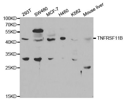
 WB (Western Blot)
((0.5ug/ml) staining of A549 (A), HepG2 (B) and K562 (C) lysates (35ug protein in RIPA buffer). Primary incubation was 1 hour. Detected by chemiluminescence.)
WB (Western Blot)
((0.5ug/ml) staining of A549 (A), HepG2 (B) and K562 (C) lysates (35ug protein in RIPA buffer). Primary incubation was 1 hour. Detected by chemiluminescence.)
TNFRSF1A/TNFR1, Polyclonal Antibody (Cat# AAA13672)
Full Name
Goat anti-TNFRSF1A/TNFR1 Antibody
Gene Names
TNFRSF1A; FPF; MS5; p55; p60; TBP1; TNF-R; TNFAR; TNFR1; p55-R; CD120a; TNFR55; TNFR60; TNF-R-I; TNF-R55; TNFR1-d2
Reactivity
Tested: Human; Expected from sequence similarity: Human, Rat, Pig
Applications
Peptide ELISA, Western Blot
Purity
Purified from goat serum by ammonium sulphate precipitation followed by antigen affinity chromatography using the immunizing peptide.
Pricing
 Standard Curve (Sample)
Tumor Necrosis Factor Alpha, ELISA Kit (Cat# AAA17624)
Full Name
Human Tumor Necrosis Factor Alpha ELISA Kit
Reactivity
Human
Pricing
 Standard Curve (Sample)
Receptor Activator Of Nuclear Factor Kappa B Ligand (RANkL), ELISA Kit (Cat# AAA22441)
Full Name
Rabbit Receptor Activator Of Nuclear Factor Kappa B Ligand (RANkL) ELISA Kit
Gene Names
TNFSF11; ODF; OPGL; sOdf; CD254; OPTB2; RANKL; TRANCE; hRANKL2
Reactivity
Rabbit
Pricing
 Standard Curve (Sample)
tumor necrosis factor receptor superfamily, member 11b, ELISA Kit (Cat# AAA15033)
Full Name
Rat Osteoprotegerin, OPG ELISA Kit
Gene Names
Tnfrsf11b; Opg
Reactivity
Rat
Pricing
 Standard Curve (Sample)
tumor necrosis factor (TNF superfamily, member 2), ELISA Kit (Cat# AAA15213)
Full Name
Sheep Tumor Necrosis Factor alpha, TNF-alpha ELISA Kit
Gene Names
TNF; TNF-alpha
Reactivity
Sheep
Pricing
 Standard Curve (Sample)
Complement 1q, ELISA Kit (Cat# AAA15988)
Full Name
Rat Complement 1q ELISA Kit
Gene Names
C1QTNF5; CTRP5
Reactivity
Rat
Pricing
 Standard Curve (Sample)
Tumor Necrosis Factor Alpha, ELISA Kit (Cat# AAA17368)
Full Name
Porcine Tumor Necrosis Factor Alpha ELISA Kit
Reactivity
Porcine
Pricing

 Application Data
TNFRSF1B, Active Protein (Cat# AAA23888)
Full Name
Recombinant Human TNFRSF1B
Gene Names
TNFRSF1B; p75; TBPII; TNFBR; TNFR2; CD120b; TNFR1B; TNFR80; TNF-R75; p75TNFR; TNF-R-II
Purity
>95% as determined by SDS-PAGE
Pricing
 Standard Curve (Sample)
Erythroferrone (ERFE), ELISA Kit (Cat# AAA18916)
Full Name
Human Erythroferrone (ERFE) ELISA Kit
Gene Names
ERFE; CTRP15; FAM132B; C1QTNF15
Reactivity
Human
Pricing
 Standard Curve (Sample)
Tumor Necrosis Factor Beta (TNFb), ELISA Kit (Cat# AAA22910)
Full Name
Human Tumor Necrosis Factor Beta (TNFb) ELISA Kit
Gene Names
LTA; LT; TNFB; TNFSF1; TNLG1E
Reactivity
Human
Pricing
 Standard Curve (Sample)
Complement C1q tumor necrosis factor-related protein 1, ELISA Kit (Cat# AAA17390)
Full Name
Mouse Complement C1q tumor necrosis factor-related protein 1 ELISA Kit
Gene Names
C1QTNF1; GIP; CTRP1; ZSIG37
Reactivity
Mouse
Pricing
 Standard Curve (Sample)
Tumor Necrosis Factor alpha, ELISA Kit (Cat# AAA16584)
Full Name
Guinea pig Tumor Necrosis Factor alpha ELISA Kit
Gene Names
TNF; DIF; TNFA; TNFSF2; TNF-alpha
Reactivity
Guinea Pig
Pricing