Filters
Clonality
Type
Reactivity
Gene Name
Isotype
Host
Application
Clone
20 results for " Structures" - showing 1-20

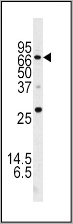
 SDS-PAGE
(SDS-PAGE of ~14 kDa Active Human Recombinant Alpha Synuclein Protein Monomer (SPR-321). Lane 1: Molecular Weight Ladder (MW). Lane 2: BSA (5 ug). Lane 3: BSA (2.5 ug). Lane 4: Active Alpha Synuclein Protein Monomer (5 ug) (SPR-321). Lane 5: Active Alpha Synuclein Protein Monomer (2.5 ug) (SPR-321).)
SDS-PAGE
(SDS-PAGE of ~14 kDa Active Human Recombinant Alpha Synuclein Protein Monomer (SPR-321). Lane 1: Molecular Weight Ladder (MW). Lane 2: BSA (5 ug). Lane 3: BSA (2.5 ug). Lane 4: Active Alpha Synuclein Protein Monomer (5 ug) (SPR-321). Lane 5: Active Alpha Synuclein Protein Monomer (2.5 ug) (SPR-321).)
Alpha Synuclein, Active Protein (Cat# AAA27658)
Full Name
Alpha Synuclein Protein
Gene Names
SNCA; PD1; NACP; PARK1; PARK4
Applications
Western Blot
Purity
Purity: >95%
Purification: Ion-exchange Purified
Purification: Ion-exchange Purified
Pricing


 Application Data
(At 25 degree C. Samples were then incubated with primary Ab(At 37 degree C. An AlexaFluor594 conjugated goat anti-rabbit IgG(H+L) Ab(Red) and an AlexaFluor488 conjugated goat anti-mouse IgG(H+L) Ab(Green) were used as the secondary antibody.The nuclear counter stain is DAPI (blue).)
Application Data
(At 25 degree C. Samples were then incubated with primary Ab(At 37 degree C. An AlexaFluor594 conjugated goat anti-rabbit IgG(H+L) Ab(Red) and an AlexaFluor488 conjugated goat anti-mouse IgG(H+L) Ab(Green) were used as the secondary antibody.The nuclear counter stain is DAPI (blue).)
LRRK2, Polyclonal Antibody (Cat# AAA31287)
Full Name
Phospho-LRRK2 (Ser935) Antibody
Gene Names
LRRK2; PARK8; RIPK7; ROCO2; AURA17; DARDARIN
Reactivity
Human, Mouse, Rat
Applications
Western Blot, Immunohistochemistry, Immunofluorescence, Immunocytochemistry, Peptide ELISA
Purity
The antibody is from purified rabbit serum by affinity purification via sequential chromatography on phospho-peptide and non-phospho-peptide affinity columns.
Pricing


 Application Data
(At 25 degree C. Samples were then incubated with primary Ab(At 37 degree C. An AlexaFluor594 conjugated goat anti-rabbit IgG(H+L) Ab(Red) and an AlexaFluor488 conjugated goat anti-mouse IgG(H+L) Ab(Green) were used as the secondary antibody.The nuclear counter stain is DAPI (blue).)
Application Data
(At 25 degree C. Samples were then incubated with primary Ab(At 37 degree C. An AlexaFluor594 conjugated goat anti-rabbit IgG(H+L) Ab(Red) and an AlexaFluor488 conjugated goat anti-mouse IgG(H+L) Ab(Green) were used as the secondary antibody.The nuclear counter stain is DAPI (blue).)
TOP2A, Polyclonal Antibody (Cat# AAA31306)
Full Name
Phospho-TOP2A (Thr1343) Antibody
Gene Names
TOP2A; TOP2; TP2A
Reactivity
Human, Mouse
Applications
Immunohistochemistry, Immunofluorescence, Immunocytochemistry, Peptide ELISA
Purity
The antibody is from purified rabbit serum by affinity purification via sequential chromatography on phospho-peptide and non-phospho-peptide affinity columns.
Pricing
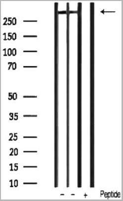
 SDS-PAGE
(Lane 1: 5 uL MW Marker. Lane 2: Reduced Mouse IgG Whole Molecule. Lane 3: Reduced Mouse F(c) Fragment. Lane 4: Reduced Mouse F(ab) Fragment. Lane 5: Mouse IgM Kappa Myeloma Protein. Load: 1 ug per lane. Predicted/Observed size: IgG at 50 and 25 kDa; F(c) at 25 kDa; F(ab) at 25 kDa; IgM K at 70 and 23 kDa. Observed F(c) Fragment migrates slightly higher.)
SDS-PAGE
(Lane 1: 5 uL MW Marker. Lane 2: Reduced Mouse IgG Whole Molecule. Lane 3: Reduced Mouse F(c) Fragment. Lane 4: Reduced Mouse F(ab) Fragment. Lane 5: Mouse IgM Kappa Myeloma Protein. Load: 1 ug per lane. Predicted/Observed size: IgG at 50 and 25 kDa; F(c) at 25 kDa; F(ab) at 25 kDa; IgM K at 70 and 23 kDa. Observed F(c) Fragment migrates slightly higher.)
Mouse IgG (Fc), Immunoglobulin (Cat# AAA14352)
Full Name
Mouse IgG (Fc)
Purity
Single arc observed by IEP.
Pricing


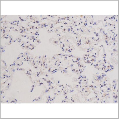
 IHC (Immunohistochemistry)
(At 1/100 staining Rat heart tissue by IHC-P. The sample was formaldehyde fixed and a heat mediated antigen retrieval step in citrate buffer was performed. The sample was then blocked and incubated with the primary antibody at 4 degree C overnight. An HRP conjugated anti-Rabbit antibody was used as the secondary antibody.)
IHC (Immunohistochemistry)
(At 1/100 staining Rat heart tissue by IHC-P. The sample was formaldehyde fixed and a heat mediated antigen retrieval step in citrate buffer was performed. The sample was then blocked and incubated with the primary antibody at 4 degree C overnight. An HRP conjugated anti-Rabbit antibody was used as the secondary antibody.)
ULK1, Polyclonal Antibody (Cat# AAA31359)
Full Name
Phospho-ULK1 (Ser757)[Ser758] Antibody
Gene Names
ULK1; ATG1; ATG1A; UNC51; hATG1; Unc51.1
Reactivity
Human, Mouse, Rat
Predicted Reactivity: Pig (100%), Bovine (100%), Horse (100%), Sheep (100%), Rabbit (100%), Dog (100%), Xenopus (88%)
Predicted Reactivity: Pig (100%), Bovine (100%), Horse (100%), Sheep (100%), Rabbit (100%), Dog (100%), Xenopus (88%)
Applications
Western Blot, Immunohistochemistry, Peptide ELISA
Purity
The antibody is from purified rabbit serum by affinity purification via sequential chromatography on phospho-peptide and non-phospho-peptide affinity columns.
Pricing


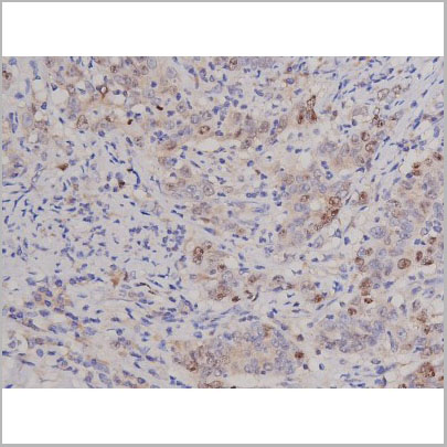
 IHC (Immunohistochemistry)
(At 1/100 staining Human gastric cancer and adjacent normal tissues by IHC-P. The sample was formaldehyde fixed and a heat mediated antigen retrieval step in citrate buffer was performed. The sample was then blocked and incubated with the primary antibody at 4 degree C overnight. An HRP conjugated anti-Rabbit antibody was used as the secondary antibody.)
IHC (Immunohistochemistry)
(At 1/100 staining Human gastric cancer and adjacent normal tissues by IHC-P. The sample was formaldehyde fixed and a heat mediated antigen retrieval step in citrate buffer was performed. The sample was then blocked and incubated with the primary antibody at 4 degree C overnight. An HRP conjugated anti-Rabbit antibody was used as the secondary antibody.)
AIRE, Polyclonal Antibody (Cat# AAA31324)
Full Name
Phospho-AIRE (Ser156) Antibody
Gene Names
AIRE; APS1; APSI; PGA1; AIRE1; APECED
Reactivity
Human, Mouse, Rat
Applications
Immunohistochemistry, Peptide ELISA
Purity
The antibody is from purified rabbit serum by affinity purification via sequential chromatography on phospho-peptide and non-phospho-peptide affinity columns.
Pricing


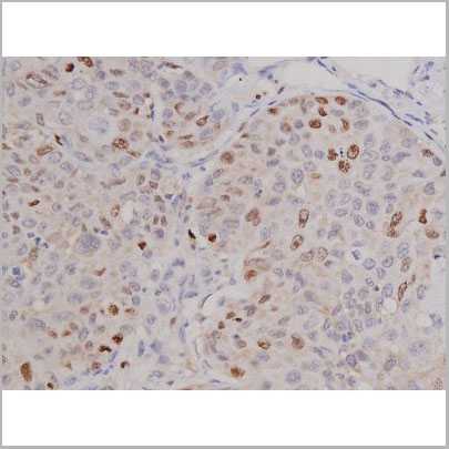
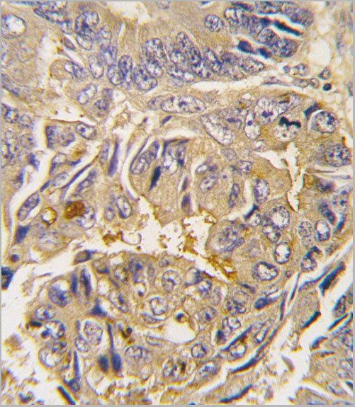
 IHC (Immunohistochemistry)
(At 1/100 staining Human colorectal cancer by IHC-P. The sample was formaldehyde fixed and a heat mediated antigen retrieval step in citrate buffer was performed. The sample was then blocked and incubated with the primary antibody at 4 degree C overnight. An HRP conjugated anti-Rabbit antibody was used as the secondary antibody.)
IHC (Immunohistochemistry)
(At 1/100 staining Human colorectal cancer by IHC-P. The sample was formaldehyde fixed and a heat mediated antigen retrieval step in citrate buffer was performed. The sample was then blocked and incubated with the primary antibody at 4 degree C overnight. An HRP conjugated anti-Rabbit antibody was used as the secondary antibody.)
CD227/MUC1, Polyclonal Antibody (Cat# AAA31426)
Full Name
Phospho-CD227/MUC1 (Ser1227) Antibody
Gene Names
MUC1; EMA; PEM; PUM; KL-6; MAM6; PEMT; CD227; H23AG; MUC-1; CA 15-3; MUC-1/X; MUC1/ZD; MUC-1/SEC
Reactivity
Human, Mouse, Rat
Applications
Western Blot, Immunohistochemistry, Peptide ELISA
Purity
The antibody is from purified rabbit serum by affinity purification via sequential chromatography on phospho-peptide and non-phospho-peptide affinity columns.
Pricing


 Application Data
(Ref. 3: Isolation, culture and immunocytochemical characterization of HUCPC. The dissection under sterile condition of foetal and full-term cords was performed to expose the WJ, the vein and arteries (A). After the in vitro expansion of foetal HUCPC (B) the cells ..)
Application Data
(Ref. 3: Isolation, culture and immunocytochemical characterization of HUCPC. The dissection under sterile condition of foetal and full-term cords was performed to expose the WJ, the vein and arteries (A). After the in vitro expansion of foetal HUCPC (B) the cells ..)
Chick Embryo Extract, Ultrafiltrate, Reagent (Cat# AAA14749)
Full Name
Chick Embryo Extract, Ultrafiltrate (CEE)
Purity
Molecular Biology Grade
Pricing
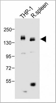
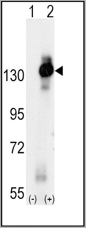
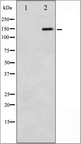
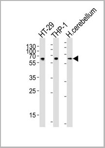
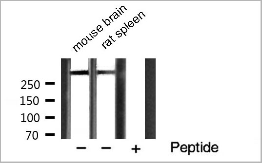
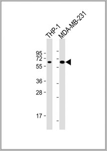 WB (Western Blot)
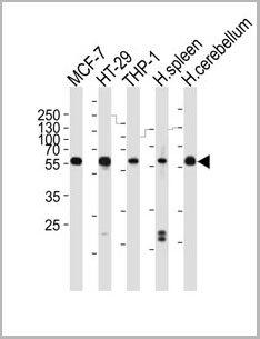
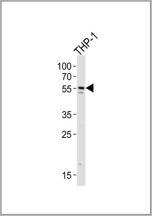
(All lanes : Anti-MB21D1 Antibody at 1:4000 dilutionLane 1: THP-1 whole cell lysateLane 2: MDA-MB-231 whole cell lysateLysates/proteins at 20 ug per lane. SecondaryGoat Anti-mouse IgG, (H+L), Peroxidase conjugated at 1/10000 dilution. Predicted band size : 59kDaBlocking/Dilution buffer: 5% NFDM/TBST.)
WB (Western Blot)
(All lanes : Anti-MB21D1 Antibody at 1:4000 dilutionLane 1: THP-1 whole cell lysateLane 2: MDA-MB-231 whole cell lysateLysates/proteins at 20 ug per lane. SecondaryGoat Anti-mouse IgG, (H+L), Peroxidase conjugated at 1/10000 dilution. Predicted band size : 59kDaBlocking/Dilution buffer: 5% NFDM/TBST.)
MB21D1, Monoclonal Antibody (Cat# AAA28800)
Full Name
MB21D1 Antibody
Gene Names
MB21D1; cGAS; h-cGAS; C6orf150
Reactivity
Human
Applications
Western Blot
Pricing
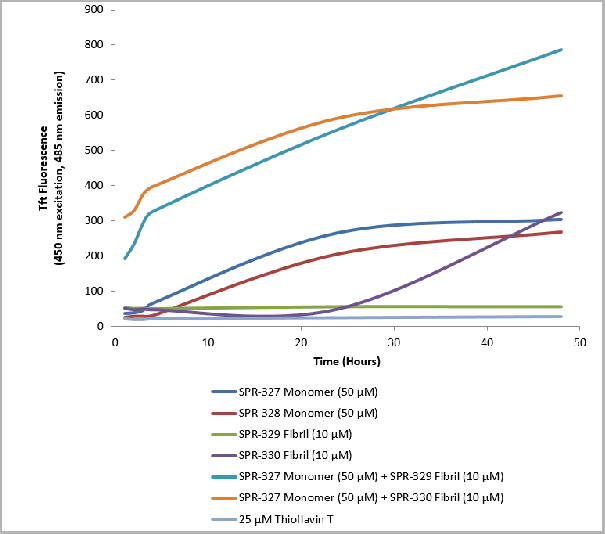
 Application Data
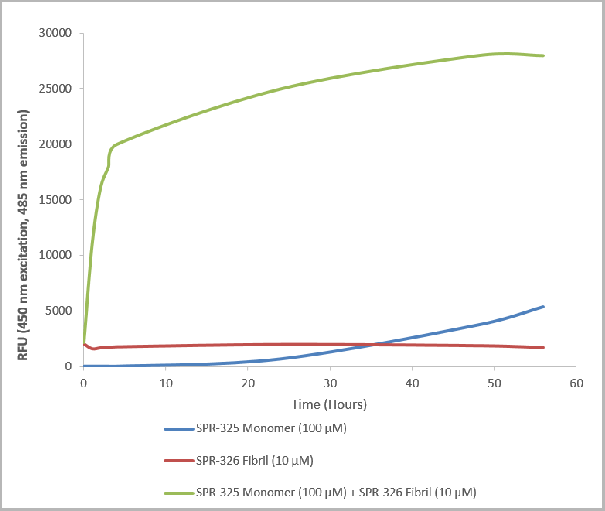
(Thioflavin T is a fluorescent dye that binds to beta sheet-rich structures such as those in alpha synuclein fibrils. Upon binding, the emission spectrum of the dye experiences a red-shift and increased fluorescence intensity. Thioflavin T emission curves show a limited increase in fluorescence (correlated to alpha synuclein aggregation) over time in A53T alpha synuclein monomers . A much greater increase in fluorescence is seen when 100 uM monomer is combined with 10 nM of fibrils (AAA27661) as the fibrils seed the formation of new fibrils from the pool of active monomers. Thioflavin T ex = 450 nm, em = 485 nm.)
Application Data
(Thioflavin T is a fluorescent dye that binds to beta sheet-rich structures such as those in alpha synuclein fibrils. Upon binding, the emission spectrum of the dye experiences a red-shift and increased fluorescence intensity. Thioflavin T emission curves show a limited increase in fluorescence (correlated to alpha synuclein aggregation) over time in A53T alpha synuclein monomers . A much greater increase in fluorescence is seen when 100 uM monomer is combined with 10 nM of fibrils (AAA27661) as the fibrils seed the formation of new fibrils from the pool of active monomers. Thioflavin T ex = 450 nm, em = 485 nm.)
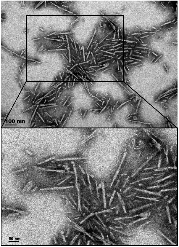
A53T Mutant Alpha Synuclein, Active Protein (Cat# AAA27661)
Full Name
Active Human Recombinant A53T Mutant Alpha Synuclein Protein Preformed Fibrils (Type 1)
Gene Names
SNCA; PD1; NACP; PARK1; PARK4
Applications
Western Blot
Purity
>95% pure using SDS-PAGE analysis.
Ion-Exchange Purified
Ion-Exchange Purified
Pricing

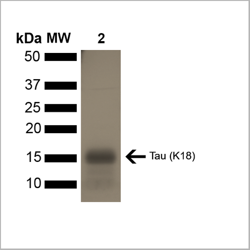
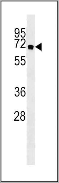
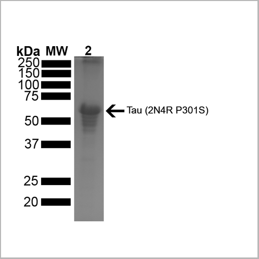 SDS-PAGE
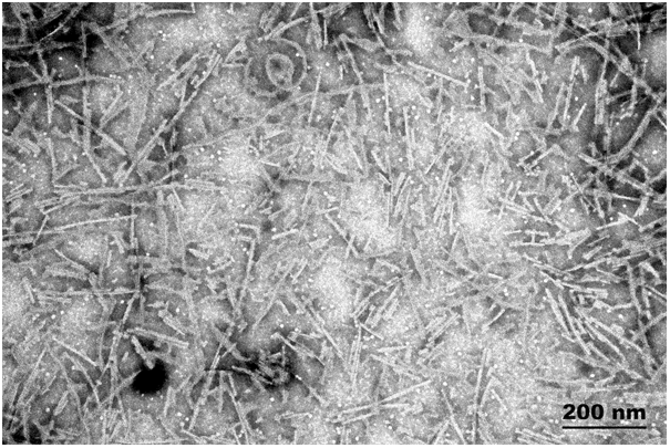
(SDS-PAGE of ~67 kDa Human Tau Protein 2N4R P301S Preformed Fibrils. Lane 1: MW Ladder. Lane 2: Tau Protein Preformed Fibrils)
SDS-PAGE
(SDS-PAGE of ~67 kDa Human Tau Protein 2N4R P301S Preformed Fibrils. Lane 1: MW Ladder. Lane 2: Tau Protein Preformed Fibrils)
Tau441, Active Protein (Cat# AAA27663)
Full Name
Active Human Recombinant Tau441 (2N4R), P301S Mutant Protein Preformed Fibrils
Gene Names
MAPT; TAU; MSTD; PPND; DDPAC; MAPTL; MTBT1; MTBT2; FTDP-17; PPP1R103
Applications
Western Blot
Purity
>95%
Ion-Exchange Purified
Ion-Exchange Purified
Pricing
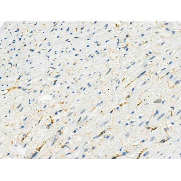
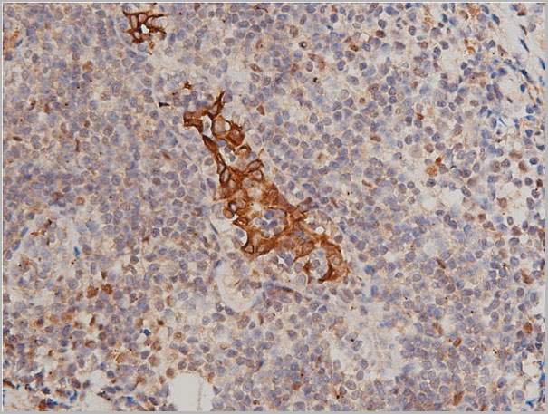
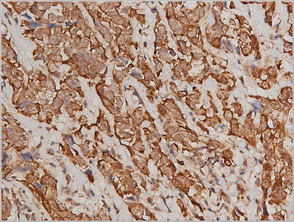
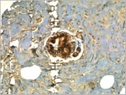
 IHC (Immunohistochemistry)
(Formalin-fixed, paraffin-embedded human Tonsil stained with NuMA Monoclonal Antibody (A73-B/D12))
IHC (Immunohistochemistry)
(Formalin-fixed, paraffin-embedded human Tonsil stained with NuMA Monoclonal Antibody (A73-B/D12))
Nuclear Mitotic Apparatus Protein (NuMA), Monoclonal Antibody (Cat# AAA13819)
Full Name
Nuclear Mitotic Apparatus Protein (NuMA) Mouse Monoclonal Antibody
Gene Names
NUMA1; NUMA; NMP-22
Reactivity
Human (Others not known)
Applications
Flow Cytometry, Immunofluorescence, Immunohistochemistry
Pricing
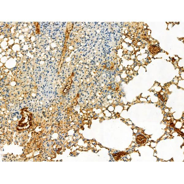
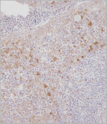
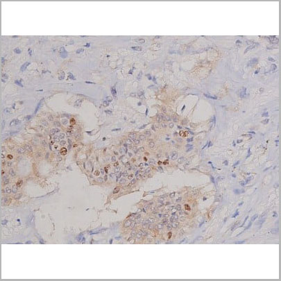
 IHC (Immunohistochemistry)
(AAA30985 at 1/50 staining human breast cancer tissue sections by IHC-P. The tissue was formaldehyde fixed and a heat mediated antigen retrieval step in citrate buffer was performed. The tissue was then blocked and incubated with the antibody for 1.5 hours at 22 degree C. An HRP conjugated goat anti-rabbit antibody was used as the secondary.)
IHC (Immunohistochemistry)
(AAA30985 at 1/50 staining human breast cancer tissue sections by IHC-P. The tissue was formaldehyde fixed and a heat mediated antigen retrieval step in citrate buffer was performed. The tissue was then blocked and incubated with the antibody for 1.5 hours at 22 degree C. An HRP conjugated goat anti-rabbit antibody was used as the secondary.)
Keratin 8, Polyclonal Antibody (Cat# AAA30985)
Full Name
Phospho-Keratin 8 (Ser432) Antibody
Gene Names
KRT8; K8; KO; CK8; CK-8; CYK8; K2C8; CARD2
Reactivity
Human, Mouse, Rat
Applications
Western Blot, Immunohistochemistry, Immunofluorescence, Immunocytochemistry
Purity
From purified rabbit serum by affinity purification via sequential chromatography on phospho-and non-phospho-peptide affinity columns.
Pricing
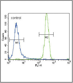
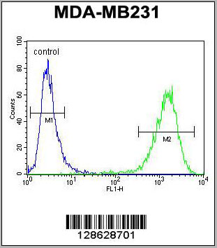
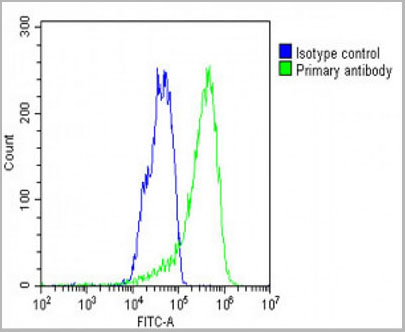 FCM (Flow Cytometry)
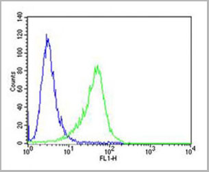
(WB - MCSF Receptor (CSF1R) Antibody (C-term) AAA28757 detail IHC-P - MCSF Receptor (CSF1R) Antibody (C-term) AAA28757 detail IHC-P - MCSF Receptor (CSF1R) Antibody (C-term) AAA28757 detail FC - MCSF Receptor (CSF1R) Antibody (C-term) AAA28757 detail Overlay histogram showing HepG2 cells stained with AAA28757(green line). The cells were fixed with 2% paraformaldehyde (10 min) and then permeabilized with 90% methanol for 10 min. The cells were then icubated in 2% bovine serum albumin to block non-specific protein-protein interactions followed by the antibody (AAA28757, 1:25 dilution) for 60 min at 37ºC. The secondary antibody used was Goat-Anti-Rabbit IgG, DyLight® 488 Conjugated Highly Cross-Adsorbed(1583138) at 1/200 dilution for 40 min at 37ºC. Isotype control antibody (blue line) was rabbit IgG1 (1ug/1x10^6 cells) used under the same conditions. Acquisition of >10, 000 events was performed.)
FCM (Flow Cytometry)
(WB - MCSF Receptor (CSF1R) Antibody (C-term) AAA28757 detail IHC-P - MCSF Receptor (CSF1R) Antibody (C-term) AAA28757 detail IHC-P - MCSF Receptor (CSF1R) Antibody (C-term) AAA28757 detail FC - MCSF Receptor (CSF1R) Antibody (C-term) AAA28757 detail Overlay histogram showing HepG2 cells stained with AAA28757(green line). The cells were fixed with 2% paraformaldehyde (10 min) and then permeabilized with 90% methanol for 10 min. The cells were then icubated in 2% bovine serum albumin to block non-specific protein-protein interactions followed by the antibody (AAA28757, 1:25 dilution) for 60 min at 37ºC. The secondary antibody used was Goat-Anti-Rabbit IgG, DyLight® 488 Conjugated Highly Cross-Adsorbed(1583138) at 1/200 dilution for 40 min at 37ºC. Isotype control antibody (blue line) was rabbit IgG1 (1ug/1x10^6 cells) used under the same conditions. Acquisition of >10, 000 events was performed.)
MCSF Receptor (CSF1R), Polyclonal Antibody (Cat# AAA28757)
Full Name
MCSF Receptor (CSF1R) Antibody (C-term)
Gene Names
CSF1R; FMS; CSFR; FIM2; HDLS; C-FMS; CD115; CSF-1R; M-CSF-R
Reactivity
Human
Applications
Western Blot, Flow Cytometry, Immunohistochemistry
Purity
Purified Rabbit Polyclonal Antibody (Pab)
Pricing
 Application Data
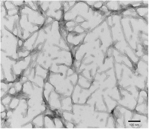
(Thioflavin T is a fluorescent dye that binds to beta sheet-rich structures such as those in tau fibrils. Upon binding, the emission spectrum of the dye experiences a red-shift, and increased fluorescence intensity. Thioflavin T emission curves show increased fluorescence (correlated to tau aggregation) in tau K18 P301L monomers over time. Thioflavin T ex = 450 nm, em = 485 nm.)
Application Data
(Thioflavin T is a fluorescent dye that binds to beta sheet-rich structures such as those in tau fibrils. Upon binding, the emission spectrum of the dye experiences a red-shift, and increased fluorescence intensity. Thioflavin T emission curves show increased fluorescence (correlated to tau aggregation) in tau K18 P301L monomers over time. Thioflavin T ex = 450 nm, em = 485 nm.)
Tau, Active Protein (Cat# AAA27662)
Full Name
Active Human Recombinant Tau (K18), P301L Mutant Protein Monomer
Gene Names
MAPT; TAU; MSTD; PPND; DDPAC; MAPTL; MTBT1; MTBT2; FTDP-17; PPP1R103
Applications
Western Blot
Purity
Purity: >95%
Purification: Ion-exchange Purified
Purification: Ion-exchange Purified
Pricing
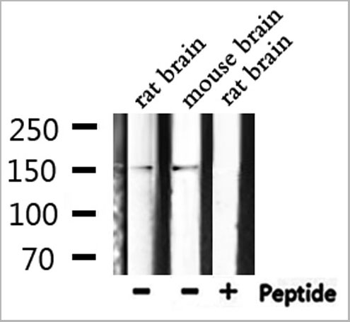
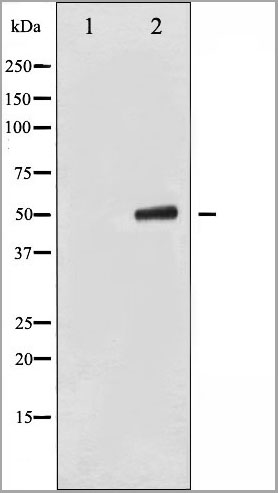
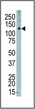
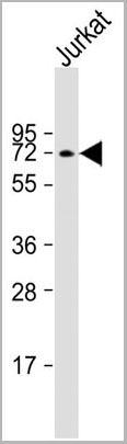
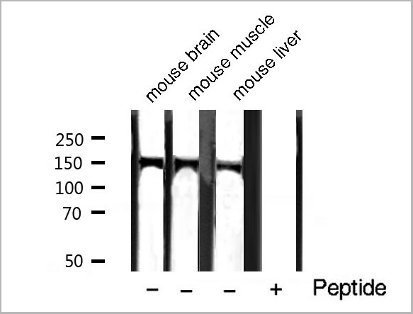 WB (Western Blot)
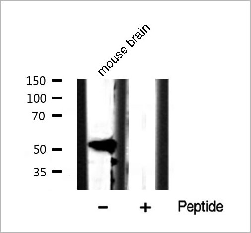
(Western blot analysis of extracts of various tissue sample, using Phospho-PLCG1 (Tyr771) Antibody.)
WB (Western Blot)
(Western blot analysis of extracts of various tissue sample, using Phospho-PLCG1 (Tyr771) Antibody.)
PLCG1, Polyclonal Antibody (Cat# AAA31021)
Full Name
Phospho-PLCG1 (Tyr771) Antibody
Gene Names
PLCG1; PLC1; NCKAP3; PLC-II; PLC148; PLCgamma1
Reactivity
Human, Mouse, Rat, Monkey
Applications
Western Blot, Immunohistochemistry, Immunofluorescence, Immunocytochemistry
Purity
From purified rabbit serum by affinity purification via sequential chromatography on phospho-and non-phospho-peptide affinity columns.
Pricing
 Application Data
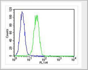
(Overlay histogram showing U-2OS cells stained with AP10510c (green line). The cells were fixed with 2% paraformaldehyde (10 min) and then permeabilized with 90% methanol for 10 min. The cells were then icubated n 2% bovine serum albumin to block non-specificprotein-protein interactions followed by the antibody (AP10510c, 1:25 dilution) for 60 min at 37ºC. The secondary antibody used wasGoat-Anti-Rabbit IgG, DyLight® 488 Conjugated Highly Cross-Adsorbed(NA168821) at 1/400 dilution for 40 min at 37ºC. Isotype control antibody (blue line) was rabbit IgG (1?g/1x10^6 cells) used under the same conditions. Acquisition of >10, 000 events wasperformed)
Application Data
(Overlay histogram showing U-2OS cells stained with AP10510c (green line). The cells were fixed with 2% paraformaldehyde (10 min) and then permeabilized with 90% methanol for 10 min. The cells were then icubated n 2% bovine serum albumin to block non-specificprotein-protein interactions followed by the antibody (AP10510c, 1:25 dilution) for 60 min at 37ºC. The secondary antibody used wasGoat-Anti-Rabbit IgG, DyLight® 488 Conjugated Highly Cross-Adsorbed(NA168821) at 1/400 dilution for 40 min at 37ºC. Isotype control antibody (blue line) was rabbit IgG (1?g/1x10^6 cells) used under the same conditions. Acquisition of >10, 000 events wasperformed)
CF150, Polyclonal Antibody (Cat# AAA28789)
Full Name
CF150 Antibody (Center)
Gene Names
MB21D1; cGAS; h-cGAS; C6orf150
Reactivity
Human
Applications
Flow Cytometry, Western Blot
Purity
Peptide Affinity Purified Rabbit Polyclonal Antibody (Pab)
Pricing
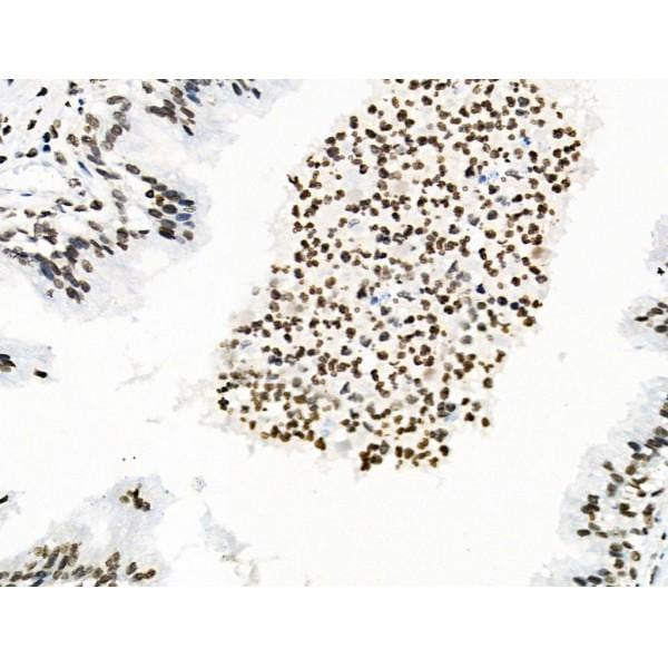
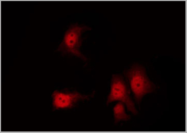
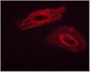
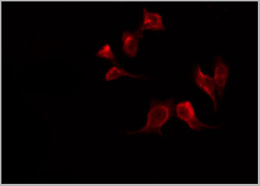
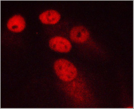 IF (Immunofluorescence)
(AAA30924 staining MCF-7 cells by ICC/IF. Cells were fixed with PFA and permeabilized in 0.1% saponin prior to blocking in 10% serum for 45 minutes at 37 degree C. The primary antibody was diluted 1/400 and incubated with the sample for 1 hour at 37 degree C. A Alexa Fluor 594 conjugated goat polyclonal to rabbit IgG (H+L), diluted 1/600 was used as secondary antibody.)
IF (Immunofluorescence)
(AAA30924 staining MCF-7 cells by ICC/IF. Cells were fixed with PFA and permeabilized in 0.1% saponin prior to blocking in 10% serum for 45 minutes at 37 degree C. The primary antibody was diluted 1/400 and incubated with the sample for 1 hour at 37 degree C. A Alexa Fluor 594 conjugated goat polyclonal to rabbit IgG (H+L), diluted 1/600 was used as secondary antibody.)
Ki67, Polyclonal Antibody (Cat# AAA30924)
Full Name
Ki67 Antibody
Gene Names
MKI67; KIA; MIB-; MIB-1; PPP1R105
Reactivity
Human, Mouse, Rat
Applications
Western Blot, Immunohistochemistry, Immunofluorescence, Immunocytochemistry
Purity
The antiserum was purified by peptide affinity chromatography using SulfoLink Coupling Resin (Thermo Fisher Scientifiv).
Pricing
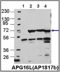
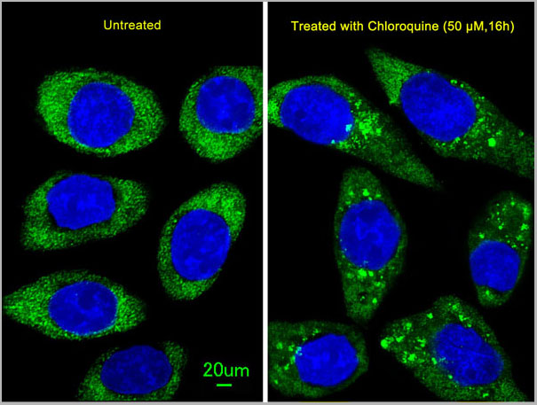
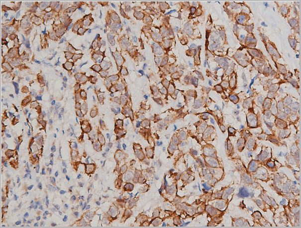
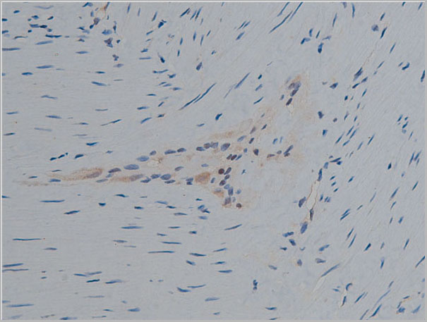
 IHC (Immunohistchemistry)
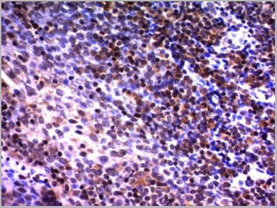
(Formalin-fixed and paraffin-embedded human colon carcinoma tissue reacted with Autophagy APG16L antibody (L176), which was peroxidase-conjugated to the secondary antibody, followed by DAB staining.This data demonstrates the use of this antibody for immunohistochemistry;clinical relevance has not been evaluated.)
IHC (Immunohistchemistry)
(Formalin-fixed and paraffin-embedded human colon carcinoma tissue reacted with Autophagy APG16L antibody (L176), which was peroxidase-conjugated to the secondary antibody, followed by DAB staining.This data demonstrates the use of this antibody for immunohistochemistry;clinical relevance has not been evaluated.)
ATG16L, Polyclonal Antibody (Cat# AAA28729)
Full Name
ATG16L Antibody
Gene Names
ATG16L1; IBD10; WDR30; APG16L; ATG16A; ATG16L
Reactivity
Human, mouse
Applications
Immunofluorescence, Western Blot, Immunohistochemistry
Purity
Purified Rabbit Polyclonal Antibody (Pab)
Pricing


 IF (Immunofluorescence)
(MILIN1 is expressed in association with lymphatic vessels. Cryostat sections of normal mouse tissues stained with anti-EMILIN1 (green) antibodies. In all mouse tissues and organs examined, EMILIN1 was uniformly distributed in the stroma. In the skin, EMILIN1 staining colocalizes with LYVE-1-positive lymphatic vessels surrounding hair follicles. In the small intestine, EMILIN1 colocalizes with LYVE-1-positive lacteals and submucosal lymphatic vessels. At higher magnification, in the lung and lymph nodes, it is more evident that EMILIN1 is distributed at the abluminal surfaces of LECs. In the lymph node, EMILIN1-positive fibers connecting LECs to the surrounding ECM are evident.)
IF (Immunofluorescence)
(MILIN1 is expressed in association with lymphatic vessels. Cryostat sections of normal mouse tissues stained with anti-EMILIN1 (green) antibodies. In all mouse tissues and organs examined, EMILIN1 was uniformly distributed in the stroma. In the skin, EMILIN1 staining colocalizes with LYVE-1-positive lymphatic vessels surrounding hair follicles. In the small intestine, EMILIN1 colocalizes with LYVE-1-positive lacteals and submucosal lymphatic vessels. At higher magnification, in the lung and lymph nodes, it is more evident that EMILIN1 is distributed at the abluminal surfaces of LECs. In the lymph node, EMILIN1-positive fibers connecting LECs to the surrounding ECM are evident.)
Emilin-1, Monoclonal Antibody (Cat# AAA14943)
Full Name
Rat Anti-Mouse Emilin-1
Gene Names
Emilin1; gp115; AW229038; BB105748; EMILIN-1; 5830419M17Rik
Reactivity
Mouse
Applications
WB, IHC
Purity
Protein G purified
Pricing