Filters
Clonality
Type
Reactivity
Gene Name
Isotype
Host
Application
Clone
7 results for " Metabolic Signaling" - showing 1-7


 Application Data
(All lanes use the Antibody at 1:5K dilution for 1 hour at room temperature.)
Application Data
(All lanes use the Antibody at 1:5K dilution for 1 hour at room temperature.)
GBA/Glucosylceramidase, Monoclonal Antibody (Cat# AAA11697)
Full Name
Anti-GBA/Glucosylceramidase Rabbit Monoclonal Antibody
Gene Names
GBA; GCB; GBA1; GLUC
Reactivity
Human, Rat
Applications
Immunohistochemistry, Western Blot
Purity
Affinity-chromatography
Pricing


 WB (Western Blot)
(Western Blot (WB) Sample: Recombinant ADIPOR1, Human; Antibody: Rabbit Anti-Human ADIPOR1 Ab)
WB (Western Blot)
(Western Blot (WB) Sample: Recombinant ADIPOR1, Human; Antibody: Rabbit Anti-Human ADIPOR1 Ab)
Adiponectin Receptor 1 (ADIPOR1), Active Protein (Cat# AAA21107)
Full Name
Active Adiponectin Receptor 1 (ADIPOR1)
Gene Names
ADIPOR1; CGI45; PAQR1; ACDCR1; CGI-45; TESBP1A
Reactivity
Homo sapiens (Human)
Applications
Cell culture; Activity Assays.
Purity
>98%
Pricing


 IHC (Immunohistchemistry)
(Figure 6. IHC analysis of GALE using anti-GALE antibody (AAA19135).GALE was detected in paraffin-embedded section of rat kidney tissue. Heat mediated antigen retrieval was performed in citrate buffer (pH6, epitope retrieval solution) for 20 mins. The tissue section was blocked with 10% goat serum. The tissue section was then incubated with 1ug/ml rabbit anti-GALE Antibody (AAA19135) overnight at 4 degree C. Biotinylated goat anti-rabbit IgG was used as secondary antibody and incubated for 30 minutes at 37 degree C. The tissue section was developed using Strepavidin-Biotin-Complex (SABC) with DAB as the chromogen.)
IHC (Immunohistchemistry)
(Figure 6. IHC analysis of GALE using anti-GALE antibody (AAA19135).GALE was detected in paraffin-embedded section of rat kidney tissue. Heat mediated antigen retrieval was performed in citrate buffer (pH6, epitope retrieval solution) for 20 mins. The tissue section was blocked with 10% goat serum. The tissue section was then incubated with 1ug/ml rabbit anti-GALE Antibody (AAA19135) overnight at 4 degree C. Biotinylated goat anti-rabbit IgG was used as secondary antibody and incubated for 30 minutes at 37 degree C. The tissue section was developed using Strepavidin-Biotin-Complex (SABC) with DAB as the chromogen.)
GALE, Polyclonal Antibody (Cat# AAA19135)
Full Name
Anti-GALE Picoband antibody
Gene Names
GALE; SDR1E1
Reactivity
Human, Mouse, Rat
No cross reactivity with other proteins.
No cross reactivity with other proteins.
Applications
EIA, IHC, WB
Pricing


 IHC (Immunohistchemistry)
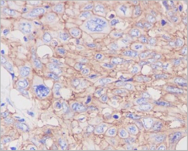
(Figure 6. IHC analysis of AMD1 using anti-AMD1 antibody (AAA19177).AMD1 was detected in paraffin-embedded section of human mammary cancer tissue. Heat mediated antigen retrieval was performed in citrate buffer (pH6, epitope retrieval solution) for 20 mins. The tissue section was blocked with 10% goat serum. The tissue section was then incubated with 1ug/ml rabbit anti-AMD1 Antibody (AAA19177) overnight at 4 degree C. Biotinylated goat anti-rabbit IgG was used as secondary antibody and incubated for 30 minutes at 37 degree C. The tissue section was developed using Strepavidin-Biotin-Complex (SABC) with DAB as the chromogen.)
IHC (Immunohistchemistry)
(Figure 6. IHC analysis of AMD1 using anti-AMD1 antibody (AAA19177).AMD1 was detected in paraffin-embedded section of human mammary cancer tissue. Heat mediated antigen retrieval was performed in citrate buffer (pH6, epitope retrieval solution) for 20 mins. The tissue section was blocked with 10% goat serum. The tissue section was then incubated with 1ug/ml rabbit anti-AMD1 Antibody (AAA19177) overnight at 4 degree C. Biotinylated goat anti-rabbit IgG was used as secondary antibody and incubated for 30 minutes at 37 degree C. The tissue section was developed using Strepavidin-Biotin-Complex (SABC) with DAB as the chromogen.)
AMD1/Adometdc, Polyclonal Antibody (Cat# AAA19177)
Full Name
Anti-AMD1/Adometdc Antibody
Gene Names
AMD1; AMD; SAMDC; ADOMETDC
Reactivity
Human, Mouse, Rat
No cross reactivity with other proteins.
No cross reactivity with other proteins.
Applications
IHC, WB
Purity
Immunogen affinity purified
Pricing
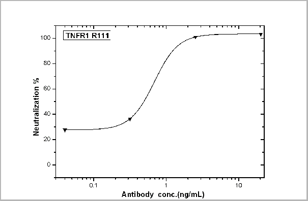 Application Data
(TNFR1/TNFRSF1A-mediated inhibition of cytotoxicity was Neutralized by Human TNFR1 Antibody. Recombinant Human TNFR1/TNFRSF1A inhibits Recombinant Human TNFa induced cytotoxicity in the L-929 mouse fibroblast cell line. Inhibition of Recombinant Human TNFa (0.2 ng/mL) activity elicited by Recombinant Human TNFR1/TNFRSF1A (0.3 ug/mL) is neutralized by increasing concentrations of Human TNFR1/TNFRSF1A Monoclonal Antibody. The IC50 is typically 0.5-1.5 ug/mL in the presence of the metabolic inhibitor actinomycin D (1 ug/mL).)
Application Data
(TNFR1/TNFRSF1A-mediated inhibition of cytotoxicity was Neutralized by Human TNFR1 Antibody. Recombinant Human TNFR1/TNFRSF1A inhibits Recombinant Human TNFa induced cytotoxicity in the L-929 mouse fibroblast cell line. Inhibition of Recombinant Human TNFa (0.2 ng/mL) activity elicited by Recombinant Human TNFR1/TNFRSF1A (0.3 ug/mL) is neutralized by increasing concentrations of Human TNFR1/TNFRSF1A Monoclonal Antibody. The IC50 is typically 0.5-1.5 ug/mL in the presence of the metabolic inhibitor actinomycin D (1 ug/mL).)
TNFR1/CD120a/TNFRSF1A, Monoclonal Antibody (Cat# AAA27740)
Full Name
TNFR1/CD120a/TNFRSF1A Neutralizing Antibody
Gene Names
TNFRSF1A; FPF; MS5; p55; p60; TBP1; TNF-R; TNFAR; TNFR1; p55-R; CD120a; TNFR55; TNFR60; TNF-R-I; TNF-R55; TNFR1-d2
Applications
Neutralization
Pricing
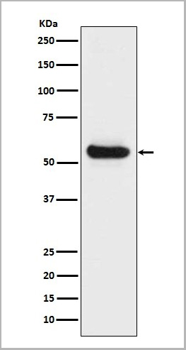 WB (Western Blot)
(Western blot analysis of Lipoprotein lipase expression in Human fetal liver lysate (AAA11694).Electrophoresis was performed on a 5-20% SDS-PAGE gel at 70V (Stacking gel) / 90V (Resolving gel) for 2-3 hours. The sample well of each lane was loaded with 50ug of sample under reducing conditions. After Electrophoresis, proteins were transferred to a Nitrocellulose membrane at 150mA for 50-90 minutes. Blocked the membrane with 5% Non-fat Milk/ TBS for 1.5 hour at RT. The membrane was incubated with rabbit anti-LPL monoclonal antibody overnight at 4 degree C, then washed with TBS-0.1%Tween 3 times with 5 minutes each and probed with a goat anti-rabbit IgG-HRP secondary antibody at a dilution of 1:10000 for 1.5 hour at RT. The signal is developed using an Enhanced Chemiluminescent detection (ECL) kit with Tanon 5200 system. A specific band was detected for LPL)
WB (Western Blot)
(Western blot analysis of Lipoprotein lipase expression in Human fetal liver lysate (AAA11694).Electrophoresis was performed on a 5-20% SDS-PAGE gel at 70V (Stacking gel) / 90V (Resolving gel) for 2-3 hours. The sample well of each lane was loaded with 50ug of sample under reducing conditions. After Electrophoresis, proteins were transferred to a Nitrocellulose membrane at 150mA for 50-90 minutes. Blocked the membrane with 5% Non-fat Milk/ TBS for 1.5 hour at RT. The membrane was incubated with rabbit anti-LPL monoclonal antibody overnight at 4 degree C, then washed with TBS-0.1%Tween 3 times with 5 minutes each and probed with a goat anti-rabbit IgG-HRP secondary antibody at a dilution of 1:10000 for 1.5 hour at RT. The signal is developed using an Enhanced Chemiluminescent detection (ECL) kit with Tanon 5200 system. A specific band was detected for LPL)
Lipoprotein lipase, Monoclonal Antibody (Cat# AAA11694)
Full Name
Anti-Lipoprotein lipase Rabbit Monoclonal Antibody
Gene Names
LPL; LIPD; HDLCQ11
Reactivity
Human
Applications
Immunohistochemistry, Western Blot
Purity
Affinity-chromatography
Pricing

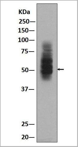
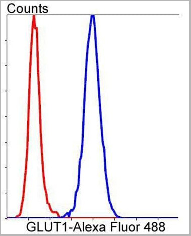 FCM (Flow Cytometry)
(Flow cytometric analysis of Hela cells with GLUT1 antibody at 1/50 dilution (blue) compared with an unlabelled control (cells without incubation with primary antibody.)
FCM (Flow Cytometry)
(Flow cytometric analysis of Hela cells with GLUT1 antibody at 1/50 dilution (blue) compared with an unlabelled control (cells without incubation with primary antibody.)
GLUT1, Monoclonal Antibody (Cat# AAA11692)
Full Name
Anti-GLUT1 Rabbit Monoclonal Antibody
Gene Names
SLC2A1; CSE; PED; DYT9; GLUT; DYT17; DYT18; EIG12; GLUT1; HTLVR; GLUT-1; SDCHCN; GLUT1DS
Reactivity
Human, Mouse, Rat
Applications
Flow Cytometry, Immunofluorescence, Immunohistochemistry, Immunocytochemistry, Western Blot
Purity
Affinity-chromatography
Pricing