Filters
Clonality
Type
Reactivity
Gene Name
Isotype
Host
Application
Clone
391 results for " Isotype" - showing 1-50

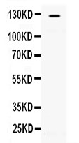
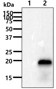 Western Blot (WB)
(The cell lysates(40ug) were resolved by SDS-PAGE, transferred to PVDF membrane and probed with anti-human TREM2 antibody (1:1000). Proteins were visualized using a goat anti-mouse secondary antibody conjugated to HRP and an ECL detection system.Lane 1 : 293T cell lysateLane 2 : TREM2 19-161aa Transfected 293T cell lysate)
Western Blot (WB)
(The cell lysates(40ug) were resolved by SDS-PAGE, transferred to PVDF membrane and probed with anti-human TREM2 antibody (1:1000). Proteins were visualized using a goat anti-mouse secondary antibody conjugated to HRP and an ECL detection system.Lane 1 : 293T cell lysateLane 2 : TREM2 19-161aa Transfected 293T cell lysate)
TREM2, Monoclonal Antibody (Cat# AAA332953)
Full Name
Human TREM2 antibody
Reactivity
Human
Applications
Western Blot, Immunocytochemistry, Flow Cytometry
Purity
By protein-A affinity chromatography
Pricing


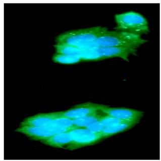
 ICC (Immunocytochemistry)
(Immunostaining analysis in HeLa cells. HeLa cells were fixed with 4% paraformaldehyde and permeabilized with 0.1% Triton X-100 in PBS. The cells were immunostained with anti-MARS mAb. [Lot No. MARSD10B4-2])
ICC (Immunocytochemistry)
(Immunostaining analysis in HeLa cells. HeLa cells were fixed with 4% paraformaldehyde and permeabilized with 0.1% Triton X-100 in PBS. The cells were immunostained with anti-MARS mAb. [Lot No. MARSD10B4-2])
MARS, Monoclonal Antibody (Cat# AAA10600)
Full Name
Mouse monoclonal antibody Anti-Human MARS
Gene Names
MARS; MRS; METRS; MTRNS; SPG70
Reactivity
Human
Applications
Dot Blot, Immunocytochemistry, Immunoprecipitation, Western Blot, Flow Cytometry
Pricing


 ICC (Immunocytochemistry)
(Immunostaining analysis in HeLa cells. HeLa cells were fixed with 4% paraformaldehyde and permeabilized with 0.1% Triton X-100 in PBS. The cells were immunostained with anti-STAT3 mAb. [Lot No. STAAD22A-1])
ICC (Immunocytochemistry)
(Immunostaining analysis in HeLa cells. HeLa cells were fixed with 4% paraformaldehyde and permeabilized with 0.1% Triton X-100 in PBS. The cells were immunostained with anti-STAT3 mAb. [Lot No. STAAD22A-1])
STAT3, Monoclonal Antibody (Cat# AAA10599)
Full Name
Mouse monoclonal antibody Anti-Human STAT3
Gene Names
STAT3; APRF; HIES; ADMIO
Reactivity
Human
Applications
Immunocytochemistry, Immunoprecipitation, Western Blot, Flow Cytometry
Pricing


 ICC (Immunocytochemistry)
(Immunostaining analysis in HeLa cells. HeLa cells were fixed with 4% paraformaldehyde and permeabilized with 0.1% Triton X-100 in PBS. The cells were immunostained with anti-BTAF1 mAb. [Lot No. BTA3D61-1])
ICC (Immunocytochemistry)
(Immunostaining analysis in HeLa cells. HeLa cells were fixed with 4% paraformaldehyde and permeabilized with 0.1% Triton X-100 in PBS. The cells were immunostained with anti-BTAF1 mAb. [Lot No. BTA3D61-1])
BTAF1, Monoclonal Antibody (Cat# AAA10597)
Full Name
Mouse monoclonal antibody Anti-Human BTAF1
Gene Names
BTAF1; MOT1; TAF172; TAFII170; TAF(II)170
Reactivity
Human
Applications
Dot Blot, Immunocytochemistry, Immunoprecipitation, Western Blot, Flow Cytometry
Pricing


 IF (Immunofluorescence)
(Figure 6. IF analysis of CD72 using anti- CD72 antibody (AAA19329).CD72 was detected in immunocytochemical section of HEPA1-6 cells. Enzyme antigen retrieval was performed using IHC enzyme antigen retrieval reagent for 15 mins. The cells were blocked with 10% goat serum. And then incubated with 5μg/mL rabbit anti- CD72 Antibody (AAA19329) overnight at 4 degree C. DyLight®488 Conjugated Goat Anti-Rabbit IgG was used as secondary antibody at 1:100 dilution and incubated for 30 minutes at 37 degree C. The section was counterstained with DAPI. Visualize using a fluorescence microscope and filter sets appropriate for the label used.)
IF (Immunofluorescence)
(Figure 6. IF analysis of CD72 using anti- CD72 antibody (AAA19329).CD72 was detected in immunocytochemical section of HEPA1-6 cells. Enzyme antigen retrieval was performed using IHC enzyme antigen retrieval reagent for 15 mins. The cells were blocked with 10% goat serum. And then incubated with 5μg/mL rabbit anti- CD72 Antibody (AAA19329) overnight at 4 degree C. DyLight®488 Conjugated Goat Anti-Rabbit IgG was used as secondary antibody at 1:100 dilution and incubated for 30 minutes at 37 degree C. The section was counterstained with DAPI. Visualize using a fluorescence microscope and filter sets appropriate for the label used.)
CD72, Polyclonal Antibody (Cat# AAA19329)
Full Name
Anti-CD72 Antibody
Gene Names
Cd72; CD72c; Ly-19; Ly-32; Lyb-2; Ly-m19
Reactivity
Mouse, Rat
Applications
WB, IHC-P, ICC, IF, FC/FACS/FCM, EIA
Purity
Immunogen affinity purified.
Pricing


 Application Data
(Published customer image:RPE conjugated Mouse anti Canine CD4 antibody, clone YKIC302.9used for the assessment of CD4 levels on canine cells by flow cytometry.Image caption:Immunophenotypic profile of tumor infiltrating lymphocyte in canine mammary carcinomas. Analysis of tumor infiltrating T-cells, B-lymphocytes and T-cell subsets from MC-BMT or MC (A), further subcategorized according to the absence (-) or presence (+) of lymph node metastasis (-) (B). Lymphocyte populations and subsets were identified by flow cytometric immunostaining as described in Material and Methods. Data were expressed as percentage of positive cells within gated lymphocytes and CD4+/CD8+ T-cell ratio. Significant differences at p < 0.05 are highlighted by asterisk.)
Application Data
(Published customer image:RPE conjugated Mouse anti Canine CD4 antibody, clone YKIC302.9used for the assessment of CD4 levels on canine cells by flow cytometry.Image caption:Immunophenotypic profile of tumor infiltrating lymphocyte in canine mammary carcinomas. Analysis of tumor infiltrating T-cells, B-lymphocytes and T-cell subsets from MC-BMT or MC (A), further subcategorized according to the absence (-) or presence (+) of lymph node metastasis (-) (B). Lymphocyte populations and subsets were identified by flow cytometric immunostaining as described in Material and Methods. Data were expressed as percentage of positive cells within gated lymphocytes and CD4+/CD8+ T-cell ratio. Significant differences at p < 0.05 are highlighted by asterisk.)
CD4, Monoclonal Antibody (Cat# AAA12278)
Full Name
RAT ANTI DOG CD4: APC
Reactivity
Dog
Applications
Flow Cytometry
Pricing


 FCM (Flow Cytometry)
(Figure 10. Flow Cytometry analysis of THP-1 cells using anti-HDAC5 antibody (AAA19236).Overlay histogram showing THP-1 cells stained with AAA19236 (Blue line). The cells were blocked with 10% normal goat serum. And then incubated with rabbit anti-HDAC5 Antibody (AAA19236, 1μg/1x106 cells) for 30 min at 20 degree C. DyLight®488 conjugated goat anti-rabbit IgG (5-10μg/1x106 cells) was used as secondary antibody for 30 minutes at 20 degree C. Isotype control antibody (Green line) was rabbit IgG (1μg/1x106) used under the same conditions. Unlabelled sample (Red line) was also used as a control.)
FCM (Flow Cytometry)
(Figure 10. Flow Cytometry analysis of THP-1 cells using anti-HDAC5 antibody (AAA19236).Overlay histogram showing THP-1 cells stained with AAA19236 (Blue line). The cells were blocked with 10% normal goat serum. And then incubated with rabbit anti-HDAC5 Antibody (AAA19236, 1μg/1x106 cells) for 30 min at 20 degree C. DyLight®488 conjugated goat anti-rabbit IgG (5-10μg/1x106 cells) was used as secondary antibody for 30 minutes at 20 degree C. Isotype control antibody (Green line) was rabbit IgG (1μg/1x106) used under the same conditions. Unlabelled sample (Red line) was also used as a control.)
HDAC5, Polyclonal Antibody (Cat# AAA19236)
Full Name
Anti-HDAC5 Antibody
Gene Names
HDAC5; HD5; NY-CO-9
Reactivity
Human, Mouse, Rat
Applications
Western Blot, Immunohistochemistry, Immunocytochemistry, Immunofluorescence, Flow Cytometry, Direct ELISA
Purity
Immunogen affinity purified.
Pricing


 FCM (Flow Cytometry)
(Overlay histogram showing Hela cells stained with CSB-MA878942A1m (red line) at 1:300. The cells were incubated in 1x PBS /10% normal goat serum to block non-specific protein-protein interactions followed by primary antibody for 1 h at 4 degree C. The secondary antibody used was FITC goat anti-mouse IgG(H+L) at 1/200 dilution for 1 h at 4 degree C. Isotype control antibody (green line) was used under the same conditions. Acquisition of >10,000 events was performed.)
FCM (Flow Cytometry)
(Overlay histogram showing Hela cells stained with CSB-MA878942A1m (red line) at 1:300. The cells were incubated in 1x PBS /10% normal goat serum to block non-specific protein-protein interactions followed by primary antibody for 1 h at 4 degree C. The secondary antibody used was FITC goat anti-mouse IgG(H+L) at 1/200 dilution for 1 h at 4 degree C. Isotype control antibody (green line) was used under the same conditions. Acquisition of >10,000 events was performed.)
PD-L1, Monoclonal Antibody (Cat# AAA27017)
Full Name
PD-L1 Monoclonal Antibody
Gene Names
CD274; B7-H; B7H1; PDL1; PD-L1; PDCD1L1; PDCD1LG1
Reactivity
Human
Applications
Western Blot, Immunohistochemistry, Immunofluorescence, Flow Cytometry
Purity
>95%
Protein G Purified
Protein G Purified
Pricing


 WB (Western Blot)
(Western blot analysis of lysates from Hela, HepG2, HL-60 cell line (from left to right), using HIST1H2AG Antibody (Center). AAA28746 was diluted at 1:1000 at each lane. A goat anti-rabbit IgG H&L(HRP) at 1:5000 dilution was used as the secondary antibody. Lysates at 35ug per lane.)
WB (Western Blot)
(Western blot analysis of lysates from Hela, HepG2, HL-60 cell line (from left to right), using HIST1H2AG Antibody (Center). AAA28746 was diluted at 1:1000 at each lane. A goat anti-rabbit IgG H&L(HRP) at 1:5000 dilution was used as the secondary antibody. Lysates at 35ug per lane.)
HIST1H2AG, Polyclonal Antibody (Cat# AAA28746)
Full Name
HIST1H2AG Antibody (Center)
Gene Names
HIST1H2AI; H2A/c; H2AFC
Reactivity
Human (Predicted Reactivity: Rat, Mouse, Bovine, Xenopus, Yeast, Chicken, Monkey, Zebrafish, Drosophila)
Applications
Immunohistochemistry, Western Blot, Flow Cytometry
Purity
Purified Rabbit Polyclonal Antibody (Pab)
Pricing


 Application Data
(Staining of human peripheral blood monocytes with MOUSE ANTI HUMAN CD274:RPE)
Application Data
(Staining of human peripheral blood monocytes with MOUSE ANTI HUMAN CD274:RPE)
CD274, Monoclonal Antibody (Cat# AAA26778)
Full Name
CD274 (CD274 Antigen, B7 Homolog 1, B7H1, B7-H1, B7-H, MGC142294, MGC142296, OTTHUMP00000021029, Programmed Cell Death 1 Ligand 1, PDCD1 Ligand 1, PDCD1L1, PDCD1LG1, Programmed Death Ligand 1, PDL1, PD-L1, RGD1566211) (MaxLight 405)
Gene Names
CD274; B7-H; B7H1; PDL1; PD-L1; PDCD1L1; PDCD1LG1
Reactivity
Human
Applications
Flow Cytometry
Purity
Purified by protein G affinity chromatography from tissue culture supernatant.
Pricing


 FCM (Flow Cytometry)
(Figure 7. Flow Cytometry analysis of U251 cells using anti-CGKI/PRKG1 antibody (AAA19246).Overlay histogram showing U251 cells stained with AAA19246 (Blue line). The cells were blocked with 10% normal goat serum. And then incubated with rabbit anti-CGKI/PRKG1 Antibody (AAA19246, 1μg/1x106 cells) for 30 min at 20 degree C. DyLight®488 conjugated goat anti-rabbit IgG (5-10μg/1x106 cells) was used as secondary antibody for 30 minutes at 20 degree C. Isotype control antibody (Green line) was rabbit IgG (1μg/1x106) used under the same conditions. Unlabelled sample (Red line) was also used as a control.)
FCM (Flow Cytometry)
(Figure 7. Flow Cytometry analysis of U251 cells using anti-CGKI/PRKG1 antibody (AAA19246).Overlay histogram showing U251 cells stained with AAA19246 (Blue line). The cells were blocked with 10% normal goat serum. And then incubated with rabbit anti-CGKI/PRKG1 Antibody (AAA19246, 1μg/1x106 cells) for 30 min at 20 degree C. DyLight®488 conjugated goat anti-rabbit IgG (5-10μg/1x106 cells) was used as secondary antibody for 30 minutes at 20 degree C. Isotype control antibody (Green line) was rabbit IgG (1μg/1x106) used under the same conditions. Unlabelled sample (Red line) was also used as a control.)
cGKI/PRKG1, Polyclonal Antibody (Cat# AAA19246)
Full Name
Anti-cGKI/PRKG1 Antibody
Gene Names
PRKG1; 1; PKG; cGK; AAT8; cGK1; cGKI; cGK 1; PRKG1B; PRKGR1B; cGKI-BETA; cGKI-alpha
Reactivity
Human, Mouse, Rat
Applications
Western Blot, Immunohistochemistry, Immunocytochemistry, Immunofluorescence, Flow Cytometry, Direct ELISA
Purity
Immunogen affinity purified.
Pricing


 FCM (Flow Cytometry)
(Overlay histogram showing HepG2 cells stained with AAA28061 (red line) at 1:100. The cells were fixed in 4% formaldehyde and permeated by 0.2% TritonX-100. Then 10% normal goat serum was Incubated to block non-specific protein-protein interactions followed by the antibody (1ug/1*106cells) for 1 h at 4 degree C. The secondary antibody used was FITC-conjugated Goat Anti-Mouse IgG(H+L) at 1/100 dilution for 30min at 4 degree C. Isotype control antibody (green line) was mouse IgG2b (1ug/1*106cells) used under the same conditions. Acquisition of >10,000 events was performed.)
FCM (Flow Cytometry)
(Overlay histogram showing HepG2 cells stained with AAA28061 (red line) at 1:100. The cells were fixed in 4% formaldehyde and permeated by 0.2% TritonX-100. Then 10% normal goat serum was Incubated to block non-specific protein-protein interactions followed by the antibody (1ug/1*106cells) for 1 h at 4 degree C. The secondary antibody used was FITC-conjugated Goat Anti-Mouse IgG(H+L) at 1/100 dilution for 30min at 4 degree C. Isotype control antibody (green line) was mouse IgG2b (1ug/1*106cells) used under the same conditions. Acquisition of >10,000 events was performed.)
CD63, Monoclonal Antibody (Cat# AAA28061)
Full Name
CD63 Monoclonal Antibody
Gene Names
Cd63; ME491; C75951; Tspan30
Reactivity
Human
Applications
Western Blot, Immunohistochemistry, Immunofluorescence, Flow Cytometry
Purity
>95%, Protein A purified
Pricing


 Application Data
(Published Customer Image:Rat anti Mouse Gr-1 antibody, clone RB6-8C5 used for the identification of neutrophils by immunofluorescence.Image caption:S. typhimurium-Infected Macrophages Containing Phagocytosed Neutrophils and T Cells Confocal fluorescence microscopy of 50-mum-thick liver sections from 1-wk-infected Slc11a1 wild-type mice. (A-C) S.Typhimurium (O-antigen, arrows) are red, macrophages (F4-80 and MOMA-2) are blue, DNA (DAPI) is gray, phalloidin is green, and neutrophils (Gr-1/Ly-6G/RB6-8C5) are pink (arrowheads). (A) Collapsed image from a 40-mum Z-stack. Scale bar is 20 mum. (B and C) Sections from (A) that are 4 mum apart. The video from which (A-C) were derived (Video S2) is available online. (D-G) T cells within multinucleate macrophages.Macrophages (F4-80 and MOMA-2) are blue (D, G, and H), T cells (CD3zeta) are red (D, G, arrowheads), DAPI is gray (E, G), actin-bound phalloidin is green (F, G). (G) Is a composite of (D, E, and F). Scale bars are 16 mum. (H) An image from a different mouse stained and labeled as described for (D-G). Scale bar is 8 mum. A video showing a T cell inside of a macrophage is available online (Video S3).From: Nix RN, Altschuler SE, Henson PM, Detweiler CS (2007) Hemophagocytic Macrophages Harbor Salmonella enterica during Persistent Infection.PLoS Pathog 3(12): e193.)
Application Data
(Published Customer Image:Rat anti Mouse Gr-1 antibody, clone RB6-8C5 used for the identification of neutrophils by immunofluorescence.Image caption:S. typhimurium-Infected Macrophages Containing Phagocytosed Neutrophils and T Cells Confocal fluorescence microscopy of 50-mum-thick liver sections from 1-wk-infected Slc11a1 wild-type mice. (A-C) S.Typhimurium (O-antigen, arrows) are red, macrophages (F4-80 and MOMA-2) are blue, DNA (DAPI) is gray, phalloidin is green, and neutrophils (Gr-1/Ly-6G/RB6-8C5) are pink (arrowheads). (A) Collapsed image from a 40-mum Z-stack. Scale bar is 20 mum. (B and C) Sections from (A) that are 4 mum apart. The video from which (A-C) were derived (Video S2) is available online. (D-G) T cells within multinucleate macrophages.Macrophages (F4-80 and MOMA-2) are blue (D, G, and H), T cells (CD3zeta) are red (D, G, arrowheads), DAPI is gray (E, G), actin-bound phalloidin is green (F, G). (G) Is a composite of (D, E, and F). Scale bars are 16 mum. (H) An image from a different mouse stained and labeled as described for (D-G). Scale bar is 8 mum. A video showing a T cell inside of a macrophage is available online (Video S3).From: Nix RN, Altschuler SE, Henson PM, Detweiler CS (2007) Hemophagocytic Macrophages Harbor Salmonella enterica during Persistent Infection.PLoS Pathog 3(12): e193.)
Gr-1, Monoclonal Antibody (Cat# AAA12257)
Full Name
Rat Anti Mouse Gr-1: FITC
Reactivity
Mouse
Applications
Flow Cytometry
Purity
Purified IgG prepared by affinity chromatography on Protein G from tissue culture supernatant
Pricing

 WB (Western Blot)
(Western blot analysis of lysates from Hela, HepG2, MCF-7, SH-SY5Y, mouse NIH/3T3 cell line (from left to right), using FGFR1 Antibody (Center). AAA28687 was diluted at 1:1000 at each lane. A goat anti-rabbit IgG H&L(HRP) at 1:10000 dilution was used as the secondary antibody. Lysates at 20ug per lane.)
WB (Western Blot)
(Western blot analysis of lysates from Hela, HepG2, MCF-7, SH-SY5Y, mouse NIH/3T3 cell line (from left to right), using FGFR1 Antibody (Center). AAA28687 was diluted at 1:1000 at each lane. A goat anti-rabbit IgG H&L(HRP) at 1:10000 dilution was used as the secondary antibody. Lysates at 20ug per lane.)
FGFR1, Polyclonal Antibody (Cat# AAA28687)
Full Name
FGFR1 Antibody (Center)
Gene Names
FGFR1; CEK; FLG; HH2; OGD; FLT2; KAL2; BFGFR; CD331; FGFBR; FLT-2; HBGFR; N-SAM; FGFR-1; HRTFDS; bFGF-R-1
Reactivity
Human, mouse (Predicted Reactivity: Chicken, Rat)
Applications
Flow Cytometry, Western Blot
Purity
Purified Rabbit Polyclonal Antibody (Pab)
Pricing
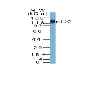


 WB (Western Blot)
(Western Blot Analysis of human Jurkat cell lysate using CD31 Mouse Monoclonal Antibody (C31.7).)
WB (Western Blot)
(Western Blot Analysis of human Jurkat cell lysate using CD31 Mouse Monoclonal Antibody (C31.7).)
CD31 / PECAM-1, Monoclonal Antibody (Cat# AAA13801)
Full Name
CD31 / PECAM-1 (Endothelial Cell Marker) Mouse Monoclonal Antibody
Gene Names
PECAM1; CD31; PECA1; GPIIA'; PECAM-1; endoCAM; CD31/EndoCAM
Reactivity
Human, Cynomolgus Monkey, Rabbit.
Weakly reacts with Rat
Weakly reacts with Rat
Applications
Flow Cytometry, Immunofluorescence, Western Blot, Immunohistochemistry
Pricing


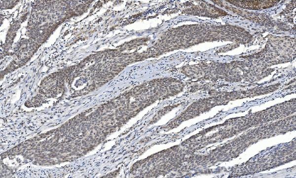
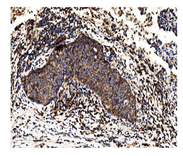
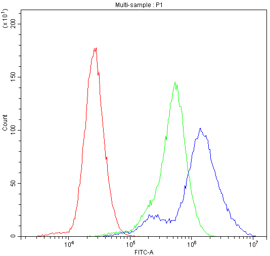
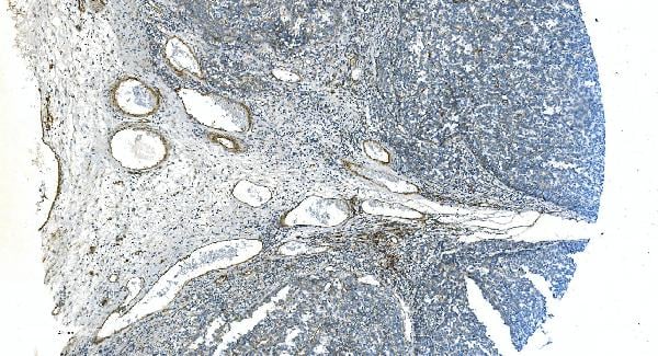
 IHC (Immunohistochemistry)
(Figure 8. IHC analysis of MEK2/MAP2K2 using anti-MEK2/MAP2K2 antibody (AAA19230).MEK2/MAP2K2 was detected in paraffin-embedded section of mouse brain tissue. Heat mediated antigen retrieval was performed in EDTA buffer (pH8. 0, epitope retrieval solution). The tissue section was blocked with 10% goat serum. The tissue section was then incubated with 2μg/ml rabbit anti-MEK2/MAP2K2 Antibody (AAA19230) overnight at 4 degree C. Biotinylated goat anti-rabbit IgG was used as secondary antibody and incubated for 30 minutes at 37 degree C. The tissue section was developed using Strepavidin-Biotin-Complex (SABC) (Catalog # with DAB as the chromogen.)
IHC (Immunohistochemistry)
(Figure 8. IHC analysis of MEK2/MAP2K2 using anti-MEK2/MAP2K2 antibody (AAA19230).MEK2/MAP2K2 was detected in paraffin-embedded section of mouse brain tissue. Heat mediated antigen retrieval was performed in EDTA buffer (pH8. 0, epitope retrieval solution). The tissue section was blocked with 10% goat serum. The tissue section was then incubated with 2μg/ml rabbit anti-MEK2/MAP2K2 Antibody (AAA19230) overnight at 4 degree C. Biotinylated goat anti-rabbit IgG was used as secondary antibody and incubated for 30 minutes at 37 degree C. The tissue section was developed using Strepavidin-Biotin-Complex (SABC) (Catalog # with DAB as the chromogen.)
MEK2/MAP2K2, Polyclonal Antibody (Cat# AAA19230)
Full Name
Anti-MEK2/MAP2K2 Antibody
Gene Names
MAP2K2; CFC4; MEK2; MKK2; MAPKK2; PRKMK2
Reactivity
Human, Mouse, Rat
Applications
Western Blot, Immunohistochemistry, Immunocytochemistry, Immunofluorescence, Flow Cytometry, Direct ELISA
Purity
Immunogen affinity purified.
Pricing


 FCM (Flow Cytometry)
(Figure 6. Flow Cytometry analysis of HEPG2 cells using anti-Alkaline phosphatase/ALPL antibody (AAA19231).Overlay histogram showing HEPG2 cells stained with AAA19231 (Blue line). The cells were blocked with 10% normal goat serum. And then incubated with rabbit anti-Alkaline phosphatase/ALPL Antibody (AAA19231, 1μg/1x106 cells) for 30 min at 20 degree C. DyLight®488 conjugated goat anti-rabbit IgG (5-10μg/1x106 cells) was used as secondary antibody for 30 minutes at 20 degree C. Isotype control antibody (Green line) was rabbit IgG (1μg/1x106) used under the same conditions. Unlabelled sample (Red line) was also used as a control.)
FCM (Flow Cytometry)
(Figure 6. Flow Cytometry analysis of HEPG2 cells using anti-Alkaline phosphatase/ALPL antibody (AAA19231).Overlay histogram showing HEPG2 cells stained with AAA19231 (Blue line). The cells were blocked with 10% normal goat serum. And then incubated with rabbit anti-Alkaline phosphatase/ALPL Antibody (AAA19231, 1μg/1x106 cells) for 30 min at 20 degree C. DyLight®488 conjugated goat anti-rabbit IgG (5-10μg/1x106 cells) was used as secondary antibody for 30 minutes at 20 degree C. Isotype control antibody (Green line) was rabbit IgG (1μg/1x106) used under the same conditions. Unlabelled sample (Red line) was also used as a control.)
Alkaline phosphatase/ALPL, Polyclonal Antibody (Cat# AAA19231)
Full Name
Anti-Alkaline phosphatase/ALPL Antibody
Gene Names
ALPL; HOPS; TNAP; APTNAP; TNSALP; AP-TNAP
Reactivity
Human, Mouse, Rat
Applications
Western Blot, Immunohistochemistry, Immunocytochemistry, Immunofluorescence, Flow Cytometry
Purity
Immunogen affinity purified.
Pricing


 FCM (Flow Cytometry)
(Figure 8. Flow Cytometry analysis of A549 cells using anti-MCM4 antibody (AAA19257).Overlay histogram showing A549 cells stained with AAA19257 (Blue line). The cells were blocked with 10% normal goat serum. And then incubated with rabbit anti-MCM4 Antibody (AAA19257, 1μg/1x106 cells) for 30 min at 20 degree C. DyLight®488 conjugated goat anti-rabbit IgG (5-10μg/1x106 cells) was used as secondary antibody for 30 minutes at 20 degree C. Isotype control antibody (Green line) was rabbit IgG (1μg/1x106) used under the same conditions. Unlabelled sample (Red line) was also used as a control.)
FCM (Flow Cytometry)
(Figure 8. Flow Cytometry analysis of A549 cells using anti-MCM4 antibody (AAA19257).Overlay histogram showing A549 cells stained with AAA19257 (Blue line). The cells were blocked with 10% normal goat serum. And then incubated with rabbit anti-MCM4 Antibody (AAA19257, 1μg/1x106 cells) for 30 min at 20 degree C. DyLight®488 conjugated goat anti-rabbit IgG (5-10μg/1x106 cells) was used as secondary antibody for 30 minutes at 20 degree C. Isotype control antibody (Green line) was rabbit IgG (1μg/1x106) used under the same conditions. Unlabelled sample (Red line) was also used as a control.)
MCM4, Polyclonal Antibody (Cat# AAA19257)
Full Name
Anti-MCM4 Antibody
Gene Names
MCM4; NKCD; CDC21; CDC54; NKGCD; hCdc21; P1-CDC21
Reactivity
Human, Mouse, Rat
Applications
Western Blot, Immunohistochemistry, Immunocytochemistry, Immunofluorescence, Flow Cytometry, Direct ELISA
Purity
Immunogen affinity purified.
Pricing
 WB (Western Blot)
IgG, Antibody (Cat# AAA13798)
Full Name
Goat Anti-Rabbit IgG
Reactivity
Rabbit
Applications
Western Blot
Pricing


 Application Data
(Staining of human peripheral blood monocytes with MOUSE ANTI HUMAN CD274:RPE)
Application Data
(Staining of human peripheral blood monocytes with MOUSE ANTI HUMAN CD274:RPE)
CD274, Monoclonal Antibody (Cat# AAA26743)
Full Name
CD274 (CD274 Antigen, B7 Homolog 1, B7H1, B7-H1, B7-H, MGC142294, MGC142296, OTTHUMP00000021029, Programmed Cell Death 1 Ligand 1, PDCD1 Ligand 1, PDCD1L1, PDCD1LG1, Programmed Death Ligand 1, PDL1, PD-L1, RGD1566211) (FITC)
Gene Names
CD274; B7-H; B7H1; PDL1; PD-L1; PDCD1L1; PDCD1LG1
Reactivity
Human
Applications
Flow Cytometry
Purity
Purified by protein G affinity chromatography from tissue culture supernatant.
Pricing


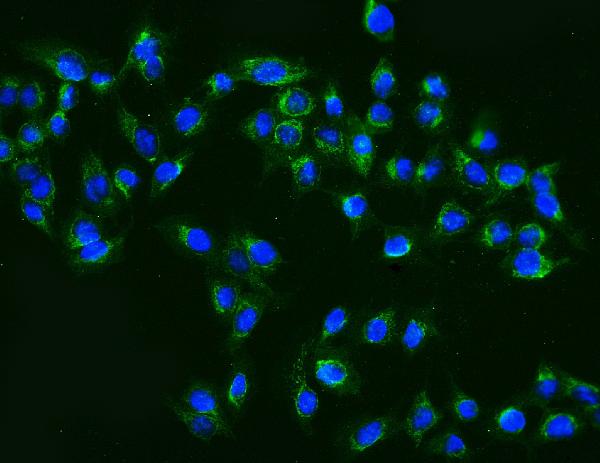
 IF (Immunofluorescence)
(Immunofluorescence of CX3CR1 in rat heart tissue with CX3CR1 antibody at 20 μg/ml.Green: CX3CR1 Antibody (2093)Blue: DAPI staining)
IF (Immunofluorescence)
(Immunofluorescence of CX3CR1 in rat heart tissue with CX3CR1 antibody at 20 μg/ml.Green: CX3CR1 Antibody (2093)Blue: DAPI staining)
CX3CR1, Polyclonal Antibody (Cat# AAA10919)
Full Name
CX3CR1 Antibody
Gene Names
CX3CR1; V28; CCRL1; GPR13; CMKDR1; GPRV28; CMKBRL1
Reactivity
Human, Mouse, Rat
Applications
Western Blot, Immunohistochemistry, Immunofluorescence, Flow Cytometry
Purity
CX3CR1 Antibody is affinity chromatography purified via peptide column.
Pricing


 FCM (Flow Cytometry)
(Figure 8. Flow Cytometry analysis of A549 cells using anti-Caveolin-2/CAV2 antibody (AAA19243).Overlay histogram showing A549 cells stained with AAA19243 (Blue line). The cells were blocked with 10% normal goat serum. And then incubated with rabbit anti-Caveolin-2/CAV2 Antibody (AAA19243,1μg/1x106 cells) for 30 min at 20 degree C. DyLight®488 conjugated goat anti-rabbit IgG (5-10μg/1x106 cells) was used as secondary antibody for 30 minutes at 20 degree C. Isotype control antibody (Green line) was rabbit IgG (1μg/1x106) used under the same conditions. Unlabelled sample (Red line) was also used as a control.)
FCM (Flow Cytometry)
(Figure 8. Flow Cytometry analysis of A549 cells using anti-Caveolin-2/CAV2 antibody (AAA19243).Overlay histogram showing A549 cells stained with AAA19243 (Blue line). The cells were blocked with 10% normal goat serum. And then incubated with rabbit anti-Caveolin-2/CAV2 Antibody (AAA19243,1μg/1x106 cells) for 30 min at 20 degree C. DyLight®488 conjugated goat anti-rabbit IgG (5-10μg/1x106 cells) was used as secondary antibody for 30 minutes at 20 degree C. Isotype control antibody (Green line) was rabbit IgG (1μg/1x106) used under the same conditions. Unlabelled sample (Red line) was also used as a control.)
Caveolin-2/CAV2, Polyclonal Antibody (Cat# AAA19243)
Full Name
Anti-Caveolin-2/CAV2 Antibody
Gene Names
CAV2; CAV
Reactivity
Human, Mouse, Rat, Monkey
Applications
Western Blot, Immunohistochemistry, Immunocytochemistry, Immunofluorescence, Flow Cytometry, Direct ELISA
Purity
Immunogen affinity purified.
Pricing


 ICC (Immunocytochemistry)
(Immunostaining analysis in HeLa cells. HeLa cells were fixed with 4% paraformaldehyde and permeabilized with 0.1% Triton X-100 in PBS. The cells were immunostained with anti-TWIST mAb. [Lot No. Twist2C1a-1])
ICC (Immunocytochemistry)
(Immunostaining analysis in HeLa cells. HeLa cells were fixed with 4% paraformaldehyde and permeabilized with 0.1% Triton X-100 in PBS. The cells were immunostained with anti-TWIST mAb. [Lot No. Twist2C1a-1])
TWIST, Monoclonal Antibody (Cat# AAA10606)
Full Name
Mouse monoclonal antibody Anti-Human TWIST
Gene Names
TWIST1; CRS; CSO; SCS; ACS3; CRS1; BPES2; BPES3; TWIST; bHLHa38
Reactivity
Human
Applications
Dot Blot, Immunocytochemistry, Western Blot, Flow Cytometry
Pricing


 FCM (Flow Cytometry)
(Figure 8. Flow Cytometry analysis of C6 cells using anti-non-muscle Myosin IIB/MYH10 antibody (AAA19262).Overlay histogram showing C6 cells stained with AAA19262 (Blue line). The cells were blocked with 10% normal goat serum. And then incubated with rabbit anti-non-muscle Myosin IIB/MYH10 Antibody (AAA19262,1μg/1x106 cells) for 30 min at 20 degree C. DyLight®488 conjugated goat anti-rabbit IgG (5-10μg/1x106 cells) was used as secondary antibody for 30 minutes at 20 degree C. Isotype control antibody (Green line) was rabbit IgG (1μg/1x106) used under the same conditions. Unlabelled sample (Red line) was also used as a control.)
FCM (Flow Cytometry)
(Figure 8. Flow Cytometry analysis of C6 cells using anti-non-muscle Myosin IIB/MYH10 antibody (AAA19262).Overlay histogram showing C6 cells stained with AAA19262 (Blue line). The cells were blocked with 10% normal goat serum. And then incubated with rabbit anti-non-muscle Myosin IIB/MYH10 Antibody (AAA19262,1μg/1x106 cells) for 30 min at 20 degree C. DyLight®488 conjugated goat anti-rabbit IgG (5-10μg/1x106 cells) was used as secondary antibody for 30 minutes at 20 degree C. Isotype control antibody (Green line) was rabbit IgG (1μg/1x106) used under the same conditions. Unlabelled sample (Red line) was also used as a control.)
non-muscle Myosin IIB/MYH10, Polyclonal Antibody (Cat# AAA19262)
Full Name
Anti-non-muscle Myosin IIB/MYH10 Antibody
Gene Names
MYH10; NMMHCB; NMMHC-IIB
Reactivity
Human, Mouse, Rat
Applications
Western Blot, Immunohistochemistry, Immunocytochemistry, Immunofluorescence, Flow Cytometry
Purity
Immunogen affinity purified.
Pricing


 Application Data
(Staining of human peripheral blood monocytes with MOUSE ANTI HUMAN CD274:RPE)
Application Data
(Staining of human peripheral blood monocytes with MOUSE ANTI HUMAN CD274:RPE)
CD274, Monoclonal Antibody (Cat# AAA26806)
Full Name
CD274 (CD274 Antigen, B7 Homolog 1, B7H1, B7-H1, B7-H, MGC142294, MGC142296, OTTHUMP00000021029, Programmed Cell Death 1 Ligand 1, PDCD1 Ligand 1, PDCD1L1, PDCD1LG1, Programmed Death Ligand 1, PDL1, PD-L1, RGD1566211) (MaxLight 550)
Gene Names
CD274; B7-H; B7H1; PDL1; PD-L1; PDCD1L1; PDCD1LG1
Reactivity
Human
Applications
Flow Cytometry
Purity
Purified by protein G affinity chromatography from tissue culture supernatant.
Pricing


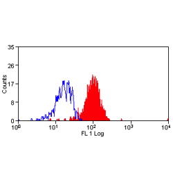
 FCM (Flow Cytometry)
(HAS2 Antibody (Center) (AAA28714) flow cytometric analysis of K562 cells (bottom histogram) compared to a Rabbit IgG Isotype control (negative control-top histogram).FITC-conjugated goat-anti-rabbit secondary antibodies were used for the analysis.)
FCM (Flow Cytometry)
(HAS2 Antibody (Center) (AAA28714) flow cytometric analysis of K562 cells (bottom histogram) compared to a Rabbit IgG Isotype control (negative control-top histogram).FITC-conjugated goat-anti-rabbit secondary antibodies were used for the analysis.)
HAS2, Polyclonal Antibody (Cat# AAA28714)
Full Name
HAS2 Antibody (Center)
Reactivity
Human, mouse
Applications
Western Blot, Immunohistochemistry, Flow Cytometry
Purity
Peptide Affinity Purified Rabbit Polyclonal Antibody (Pab)
Pricing


 FCM (Flow Cytometry)
(Figure 6. Flow Cytometry analysis of U-87 cells using anti-ATP citrate lyase antibody (AAA11689).Overlay histogram showing U-87 cells stained with AAA11689 (Blue line).The cells were blocked with 10% normal goat serum. And then incubated with rabbit anti-ATP citrate lyase Antibody (AAA11689,1ug/1x10^6 cells) for 30 min at 20 degree C. DyLight®488 conjugated goat anti-rabbit IgG (5-10ug/1x10^6 cells) was used as secondary antibody for 30 minutes at 20 degree C. Isotype control antibody (Green line) was rabbit IgG (1ug/1x106) used under the same conditions. Unlabelled sample (Red line) was also used as a control.)
FCM (Flow Cytometry)
(Figure 6. Flow Cytometry analysis of U-87 cells using anti-ATP citrate lyase antibody (AAA11689).Overlay histogram showing U-87 cells stained with AAA11689 (Blue line).The cells were blocked with 10% normal goat serum. And then incubated with rabbit anti-ATP citrate lyase Antibody (AAA11689,1ug/1x10^6 cells) for 30 min at 20 degree C. DyLight®488 conjugated goat anti-rabbit IgG (5-10ug/1x10^6 cells) was used as secondary antibody for 30 minutes at 20 degree C. Isotype control antibody (Green line) was rabbit IgG (1ug/1x106) used under the same conditions. Unlabelled sample (Red line) was also used as a control.)
ATP citrate lyase, Polyclonal Antibody (Cat# AAA11689)
Full Name
Anti-ATP citrate lyase Antibody
Gene Names
ACLY; ACL; ATPCL; CLATP
Reactivity
Human, Rat
Applications
Western Blot, Immunohistochemistry
Purity
Immunogen affinity purified.
Pricing


 Application Data
(Published Customer Image:Mouse CD31 antibody, clone ER-MP12 used for the demonstration of vasculature in mouse brain by immunofluorescence.Image caption:Inhibition of 2-AG hydrolysis reduces LPS-induced BBB permeability. a, b Fibrinogen levels in b plasma and the a ratio of brain to plasma fibrinogen were assessed by ELISA. n?=?5/7 mice per group. c, d Fluorescent immunostaining in the striatum for fibrinogen (red) and vascular marker (CD31; green) demonstrated leakage of fibrinogen into the brain with vehicle treatment, whereas vascular integrity was preserved when (e, f) MAGL was inhibited. g Extravascular fibrinogen was semi-quantitated in fluorescently labeled sections of the striatum. Bar graphs were plotted with mean?+/-SEM and data analyzed using one-way analysis of variance (ANOVA) with Tukey post-hoc comparisons. n = 5/7 mice per group. Significance is shown as *p?)
Application Data
(Published Customer Image:Mouse CD31 antibody, clone ER-MP12 used for the demonstration of vasculature in mouse brain by immunofluorescence.Image caption:Inhibition of 2-AG hydrolysis reduces LPS-induced BBB permeability. a, b Fibrinogen levels in b plasma and the a ratio of brain to plasma fibrinogen were assessed by ELISA. n?=?5/7 mice per group. c, d Fluorescent immunostaining in the striatum for fibrinogen (red) and vascular marker (CD31; green) demonstrated leakage of fibrinogen into the brain with vehicle treatment, whereas vascular integrity was preserved when (e, f) MAGL was inhibited. g Extravascular fibrinogen was semi-quantitated in fluorescently labeled sections of the striatum. Bar graphs were plotted with mean?+/-SEM and data analyzed using one-way analysis of variance (ANOVA) with Tukey post-hoc comparisons. n = 5/7 mice per group. Significance is shown as *p?)
CD31, Monoclonal Antibody (Cat# AAA12258)
Full Name
Rat Anti Mouse CD31: FITC
Gene Names
Pecam1; Cd31; Pecam; C85791; PECAM-1
Reactivity
Mouse
Applications
Flow Cytometry
Purity
Purified IgG prepared by affinity chromatography on Protein G from tissue culture supernatant
Pricing

 Application Data
MPO, Monoclonal Antibody (Cat# AAA14608)
Full Name
MPO, Rat, mAb 2D4, Biotin
Applications
Flow Cytometry, Immunoassay
Purity
Protein G
Pricing


 SDS-PAGE
(SDS Page analysis of purified S100B Mouse Monoclonal Antibody (S100B/1012).)
SDS-PAGE
(SDS Page analysis of purified S100B Mouse Monoclonal Antibody (S100B/1012).)
S100B, Monoclonal Antibody (Cat# AAA23893)
Full Name
S100B (Astrocyte and Melanoma Marker)
Gene Names
S100B; NEF; S100; S100-B; S100beta
Reactivity
Human, Mouse, Rat, Cow. Others not known.
Applications
Flow Cytometry, Immunofluorescence, Western Blot, Immunohistochemistry
Pricing


 SDS-PAGE
(Purified ER-beta Mouse Monoclonal Antibody (ESR2/686). Confirmation of Integrity and Purity of Antibody.)
SDS-PAGE
(Purified ER-beta Mouse Monoclonal Antibody (ESR2/686). Confirmation of Integrity and Purity of Antibody.)
ER-beta1 (Estrogen Receptor beta-1), Monoclonal Antibody (Cat# AAA13846)
Full Name
ER-beta1 (Estrogen Receptor beta-1) Mouse Monoclonal Antibody
Gene Names
ESR2; Erb; ESRB; ESTRB; NR3A2; ER-BETA; ESR-BETA
Reactivity
Human
Applications
Flow Cytometry, Immunofluorescence, Western Blot, Immunohistochemistry
Pricing


 Application Data
(Publised customer image:Mouse anti Human CD163 antibody, clone EDHu-1 used for the identification of perivascular macrophages in human brain by immunofluorescence.Image caption:Images demonstrating immunohistological stainings of amylin and double immunofluorescence staining against NG2/amylin, laminin/amylin and CD163/amylin in the hippocampus of the patient with AD and T2D.Amylin cell inclusions are indicated with arrows in (a) and shown in a higher magnification in (b). Pericytes with round cell bodies and NG2-positive coverage of the microvessel surface (green in c), without amylin cell inclusions (red in d), displayed round DAPI-positive cell nuclei (blue in e). The images in (c), (d) and (e) are merged in (f). Cells with more diffuse and weak NG2 staining (indicated by the arrowhead, green in g) and cytosolic amylin cell inclusions (red in h) showed altered cell nuclei (indicated with arrow, blue in i).The adjacent unaffected NG2-positive cell is indicated with an arrowhead in (i). The images in (g), (h) and (i) are merged in (j). Cells enclosed by laminin (green in K) contained amylin grains (red in l) and fragmented DAPI-positive cell nuclei (indicated with an arrow blue in m). The images in (k), (l) and (m) are merged in (n).Loss of NG2 coverage (green in o) was associated with polarized amylin cell inclusion (red in p) and fragmented DAPI-positive cell nuclei (indicated with an arrow, blue in q). The images in (o), (p) and (q) are merged in (r). Staining against macrophage marker CD163 (green in s) did not co-localize with amylin cell inclusions (red in t). The cell nucleus was stained with DAPI (blue in U). The images in (s), (t) and (u) are merged in (v). Scale bars: (a) 50 mum, (b), (c) to (v) 5 mum.From: Schultz, N. et al. (2016).Amylin alters human brain pericyte viability and NG2 expression.J Cereb Blood Flow & Metab. Jun 28 [Epub ahead of print]This is from an open access article distributed under the terms of the Creative Commons Attribution License.)
Application Data
(Publised customer image:Mouse anti Human CD163 antibody, clone EDHu-1 used for the identification of perivascular macrophages in human brain by immunofluorescence.Image caption:Images demonstrating immunohistological stainings of amylin and double immunofluorescence staining against NG2/amylin, laminin/amylin and CD163/amylin in the hippocampus of the patient with AD and T2D.Amylin cell inclusions are indicated with arrows in (a) and shown in a higher magnification in (b). Pericytes with round cell bodies and NG2-positive coverage of the microvessel surface (green in c), without amylin cell inclusions (red in d), displayed round DAPI-positive cell nuclei (blue in e). The images in (c), (d) and (e) are merged in (f). Cells with more diffuse and weak NG2 staining (indicated by the arrowhead, green in g) and cytosolic amylin cell inclusions (red in h) showed altered cell nuclei (indicated with arrow, blue in i).The adjacent unaffected NG2-positive cell is indicated with an arrowhead in (i). The images in (g), (h) and (i) are merged in (j). Cells enclosed by laminin (green in K) contained amylin grains (red in l) and fragmented DAPI-positive cell nuclei (indicated with an arrow blue in m). The images in (k), (l) and (m) are merged in (n).Loss of NG2 coverage (green in o) was associated with polarized amylin cell inclusion (red in p) and fragmented DAPI-positive cell nuclei (indicated with an arrow, blue in q). The images in (o), (p) and (q) are merged in (r). Staining against macrophage marker CD163 (green in s) did not co-localize with amylin cell inclusions (red in t). The cell nucleus was stained with DAPI (blue in U). The images in (s), (t) and (u) are merged in (v). Scale bars: (a) 50 mum, (b), (c) to (v) 5 mum.From: Schultz, N. et al. (2016).Amylin alters human brain pericyte viability and NG2 expression.J Cereb Blood Flow & Metab. Jun 28 [Epub ahead of print]This is from an open access article distributed under the terms of the Creative Commons Attribution License.)
CD163, Monoclonal Antibody (Cat# AAA12256)
Full Name
Mouse Anti Human CD163: RPE
Gene Names
CD163; M130; MM130; SCARI1
Reactivity
Human
Applications
Flow Cytometry
Pricing


 IF (Immunofluorescence)
(Figure 11. IF analysis of H Cadherin/CDH13 using anti- H Cadherin/CDH13 antibody (AAA19252).H Cadherin/CDH13 was detected in immunocytochemical section of SIHA cells. Enzyme antigen retrieval was performed using IHC enzyme antigen retrieval reagent for 15 mins. The cells were blocked with 10% goat serum. And then incubated with 5μg/mL rabbit anti- H Cadherin/CDH13 Antibody (AAA19252) overnight at 4 degree C. DyLight®488 Conjugated Goat Anti-Rabbit IgG was used as secondary antibody at 1:100 dilution and incubated for 30 minutes at 37 degree C. The section was counterstained with DAPI. Visualize using a fluorescence microscope and filter sets appropriate for the label used.)
IF (Immunofluorescence)
(Figure 11. IF analysis of H Cadherin/CDH13 using anti- H Cadherin/CDH13 antibody (AAA19252).H Cadherin/CDH13 was detected in immunocytochemical section of SIHA cells. Enzyme antigen retrieval was performed using IHC enzyme antigen retrieval reagent for 15 mins. The cells were blocked with 10% goat serum. And then incubated with 5μg/mL rabbit anti- H Cadherin/CDH13 Antibody (AAA19252) overnight at 4 degree C. DyLight®488 Conjugated Goat Anti-Rabbit IgG was used as secondary antibody at 1:100 dilution and incubated for 30 minutes at 37 degree C. The section was counterstained with DAPI. Visualize using a fluorescence microscope and filter sets appropriate for the label used.)
H Cadherin/CDH13, Polyclonal Antibody (Cat# AAA19252)
Full Name
Anti-H Cadherin/CDH13 Antibody
Gene Names
CDH13; CDHH; P105
Reactivity
Human, Rat
Applications
Western Blot, Immunohistochemistry, Immunocytochemistry, Immunofluorescence, Flow Cytometry, Direct ELISA
Purity
Immunogen affinity purified.
Pricing


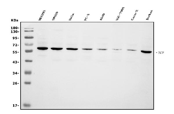
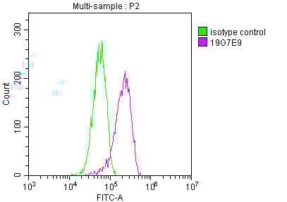
 FCM (Flow Cytometry)
(Figure 6. Flow Cytometry analysis of CACO-2 cells using anti-PLTP antibody (AAA19255).Overlay histogram showing CACO-2 cells stained with AAA19255 (Blue line). The cells were blocked with 10% normal goat serum. And then incubated with rabbit anti-PLTP Antibody (AAA19255, 1μg/1x106 cells) for 30 min at 20 degree C. DyLight®488 conjugated goat anti-rabbit IgG (5-10μg/1x106 cells) was used as secondary antibody for 30 minutes at 20 degree C. Isotype control antibody (Green line) was rabbit IgG (1μg/1x106) used under the same conditions. Unlabelled sample (Red line) was also used as a control.)
FCM (Flow Cytometry)
(Figure 6. Flow Cytometry analysis of CACO-2 cells using anti-PLTP antibody (AAA19255).Overlay histogram showing CACO-2 cells stained with AAA19255 (Blue line). The cells were blocked with 10% normal goat serum. And then incubated with rabbit anti-PLTP Antibody (AAA19255, 1μg/1x106 cells) for 30 min at 20 degree C. DyLight®488 conjugated goat anti-rabbit IgG (5-10μg/1x106 cells) was used as secondary antibody for 30 minutes at 20 degree C. Isotype control antibody (Green line) was rabbit IgG (1μg/1x106) used under the same conditions. Unlabelled sample (Red line) was also used as a control.)
PLTP, Polyclonal Antibody (Cat# AAA19255)
Full Name
Anti-PLTP Antibody
Gene Names
PLTP; BPIFE; HDLCQ9
Reactivity
Human
Applications
Western Blot, Immunohistochemistry, Immunocytochemistry, Immunofluorescence, Flow Cytometry, Direct ELISA
Purity
Immunogen affinity purified.
Pricing


 FCM (Flow Cytometry)
(Figure 8. Flow Cytometry analysis of A431 cells using anti-EGFR antibody (AAA19214).Overlay histogram showing A431 cells stained with AAA19214 (Blue line). The cells were blocked with 10% normal goat serum. And then incubated with rabbit anti-EGFR Antibody (AAA19214,1μg/1x106 cells) for 30 min at 20 degree C. DyLight®488 conjugated goat anti-rabbit IgG (5-10μg/1x106 cells) was used as secondary antibody for 30 minutes at 20 degree C. Isotype control antibody (Green line) was rabbit IgG (1μg/1x106) used under the same conditions. Unlabelled sample (Red line) was also used as a control.)
FCM (Flow Cytometry)
(Figure 8. Flow Cytometry analysis of A431 cells using anti-EGFR antibody (AAA19214).Overlay histogram showing A431 cells stained with AAA19214 (Blue line). The cells were blocked with 10% normal goat serum. And then incubated with rabbit anti-EGFR Antibody (AAA19214,1μg/1x106 cells) for 30 min at 20 degree C. DyLight®488 conjugated goat anti-rabbit IgG (5-10μg/1x106 cells) was used as secondary antibody for 30 minutes at 20 degree C. Isotype control antibody (Green line) was rabbit IgG (1μg/1x106) used under the same conditions. Unlabelled sample (Red line) was also used as a control.)
EGFR, Polyclonal Antibody (Cat# AAA19214)
Full Name
Anti-EGFR Antibody
Gene Names
EGFR; ERBB; HER1; mENA; ERBB1; PIG61
Reactivity
Human, Mouse, Rat
Applications
Western Blot, Immunohistochemistry, Immunocytochemistry, Immunofluorescence, Flow Cytometry, Direct ELISA
Purity
Immunogen affinity purified.
Pricing


 ICC (Immunocytochemistry)
(Immunostaining analysis in HeLa cells. HeLa cells were fixed with 4% paraformaldehyde and permeabilized with 0.1% Triton X-100 in PBS. The cells were immunostained with anti-TPX2 mAb. Nuclear was stained with Hoechst (blue fluorescence). [Lot No. 3164C6a-1])
ICC (Immunocytochemistry)
(Immunostaining analysis in HeLa cells. HeLa cells were fixed with 4% paraformaldehyde and permeabilized with 0.1% Triton X-100 in PBS. The cells were immunostained with anti-TPX2 mAb. Nuclear was stained with Hoechst (blue fluorescence). [Lot No. 3164C6a-1])
TPX2, Monoclonal Antibody (Cat# AAA10610)
Full Name
Mouse monoclonal antibody Anti-Human TPX2
Gene Names
TPX2; DIL2; p100; DIL-2; HCTP4; FLS353; HCA519; REPP86; C20orf1; C20orf2; GD:C20orf1
Reactivity
Human
Applications
Dot Blot, Immunocytochemistry, Immunoprecipitation, Western Blot, Flow Cytometry
Pricing


 FCM (Flow Cytometry)
(Overlay histogram showing MCF-7 cells stained with CSB-MA013563A0m (red line) at 1:400. The cells were incubated in 1x PBS /10% normal goat serum to block non-specific protein-protein interactions followed by primary antibody for 1 h at 4 degree C. The secondary antibody used was FITC goat anti-mouse IgG(H+L) at 1/200 dilution for 1 h at 4 degree C. Isotype control antibody (green line) was used under the same conditions. Acquisition of >10,000 events was performed.)
FCM (Flow Cytometry)
(Overlay histogram showing MCF-7 cells stained with CSB-MA013563A0m (red line) at 1:400. The cells were incubated in 1x PBS /10% normal goat serum to block non-specific protein-protein interactions followed by primary antibody for 1 h at 4 degree C. The secondary antibody used was FITC goat anti-mouse IgG(H+L) at 1/200 dilution for 1 h at 4 degree C. Isotype control antibody (green line) was used under the same conditions. Acquisition of >10,000 events was performed.)
CD146, Monoclonal Antibody (Cat# AAA27014)
Full Name
CD146 Monoclonal Antibody
Gene Names
MCAM; CD146; MUC18; HEMCAM; METCAM; MelCAM
Reactivity
Human
Applications
Western Blot, Immunofluorescence, Flow Cytometry
Purity
>95%
Protein G Purified
Protein G Purified
Pricing


 WB (Western Blot)
(Flow cytometric analysis of SK-BR-3 cells using MICA Antibody (Center) (green) compared to an isotype control of rabbit IgG(blue). AAA28710 was diluted at 1:25 dilution. An Alexa Fluor 488 goat anti-rabbit lgG at 1:400 dilution was used as the secondary antibody.)
WB (Western Blot)
(Flow cytometric analysis of SK-BR-3 cells using MICA Antibody (Center) (green) compared to an isotype control of rabbit IgG(blue). AAA28710 was diluted at 1:25 dilution. An Alexa Fluor 488 goat anti-rabbit lgG at 1:400 dilution was used as the secondary antibody.)
MICA, Polyclonal Antibody (Cat# AAA28710)
Full Name
MICA Antibody (Center)
Gene Names
MICA; MIC-A; PERB11.1
Reactivity
Human
Applications
Western Blot, Immunohistochemistry, Immunofluorescence, Flow Cytometry
Purity
Peptide Affinity Purified Rabbit Polyclonal Antibody (Pab)
Pricing


 WB (Western Blot)
(Western blot analysis of HLA-B (arrow) using rabbit polyclonal HLA-B Antibody (N-term). 293 cell lysates (2 ug/lane) either nontransfected (Lane 1) or transiently transfected (Lane 2) with the HLA-B gene.)
WB (Western Blot)
(Western blot analysis of HLA-B (arrow) using rabbit polyclonal HLA-B Antibody (N-term). 293 cell lysates (2 ug/lane) either nontransfected (Lane 1) or transiently transfected (Lane 2) with the HLA-B gene.)
HLA-B, Polyclonal Antibody (Cat# AAA28743)
Full Name
HLA-B Antibody (N-term)
Reactivity
Human
Applications
Western Blot, Immunohistochemistry, Flow Cytometry
Purity
Peptide Affinity Purified Rabbit Polyclonal Antibody (Pab)
Pricing


 Application Data
(Overlay histogram showing HL-60 cells stained with AAA27009 (red line) at 1:500. The cells were incubated in 1x PBS /10% normal goat serum to block non-specific protein-protein interactions followed by primary antibody for 1 h at 4 degree C.The secondary antibody used was FITC goat anti-mouse IgG (H+L) at 1/200 dilution for 1 h at 4 degree C. Isotype control antibody (green line) was used under the same conditions. Acquisition of >10, 000 events was performed.)
Application Data
(Overlay histogram showing HL-60 cells stained with AAA27009 (red line) at 1:500. The cells were incubated in 1x PBS /10% normal goat serum to block non-specific protein-protein interactions followed by primary antibody for 1 h at 4 degree C.The secondary antibody used was FITC goat anti-mouse IgG (H+L) at 1/200 dilution for 1 h at 4 degree C. Isotype control antibody (green line) was used under the same conditions. Acquisition of >10, 000 events was performed.)
CD31, Monoclonal Antibody (Cat# AAA27009)
Full Name
CD31 Monoclonal Antibody
Gene Names
PECAM1; CD31; PECA1; GPIIA'; PECAM-1; endoCAM; CD31/EndoCAM
Reactivity
Human
Applications
Western Blot, Immunohistochemistry, Immunofluorescence, Flow Cytometry
Purity
>95%, Protein G purified
Pricing


 FCM (Flow Cytometry)
(Overlay histogram showing Hela cells stained with (red line) at 1:100. The cells were fixed in 4% formaldehyde and permeated by 0.2% TritonX-100. Then 10% normal goat serum was Incubated to block non-specific protein-protein interactions followed by the antibody (1ug/1*106cells) for 1 h at 4 degree C. The secondary antibody used was FITC-conjugated Goat Anti-Mouse IgG(H+L) at 1/100 dilution for 30min at 4 degree C. Isotype control antibody (green line) was mouse IgG2b (1ug/1*106cells) used under the same conditions. Acquisition of >10,000 events was performed.)
FCM (Flow Cytometry)
(Overlay histogram showing Hela cells stained with (red line) at 1:100. The cells were fixed in 4% formaldehyde and permeated by 0.2% TritonX-100. Then 10% normal goat serum was Incubated to block non-specific protein-protein interactions followed by the antibody (1ug/1*106cells) for 1 h at 4 degree C. The secondary antibody used was FITC-conjugated Goat Anti-Mouse IgG(H+L) at 1/100 dilution for 30min at 4 degree C. Isotype control antibody (green line) was mouse IgG2b (1ug/1*106cells) used under the same conditions. Acquisition of >10,000 events was performed.)
YWHAZ, Monoclonal Antibody (Cat# AAA27050)
Full Name
YWHAZ Monoclonal Antibody
Gene Names
YWHAZ; YWHAD; KCIP-1; 14-3-3-zeta
Reactivity
Human, Mouse, Rat
Applications
Western Blot, Immunohistochemistry, Immunofluorescence, Flow Cytometry
Purity
>95%, Protein A purified
Pricing


 FCM (Flow Cytometry)
(Figure 8. Flow Cytometry analysis of THP-1 cells using anti-NOX2/gp91phox/CYBB antibody (AAA19218).Overlay histogram showing THP-1 cells stained with AAA19218 (Blue line). The cells were blocked with 10% normal goat serum. And then incubated with rabbit anti-NOX2/gp91phox/CYBB Antibody (AAA19218, 1μg/1x106 cells) for 30 min at 20 degree C. DyLight®488 conjugated goat anti-rabbit IgG (5-10μg/1x106 cells) was used as secondary antibody for 30 minutes at 20 degree C. Isotype control antibody (Green line) was rabbit IgG (1μg/1x106) used under the same conditions. Unlabelled sample (Red line) was also used as a control.)
FCM (Flow Cytometry)
(Figure 8. Flow Cytometry analysis of THP-1 cells using anti-NOX2/gp91phox/CYBB antibody (AAA19218).Overlay histogram showing THP-1 cells stained with AAA19218 (Blue line). The cells were blocked with 10% normal goat serum. And then incubated with rabbit anti-NOX2/gp91phox/CYBB Antibody (AAA19218, 1μg/1x106 cells) for 30 min at 20 degree C. DyLight®488 conjugated goat anti-rabbit IgG (5-10μg/1x106 cells) was used as secondary antibody for 30 minutes at 20 degree C. Isotype control antibody (Green line) was rabbit IgG (1μg/1x106) used under the same conditions. Unlabelled sample (Red line) was also used as a control.)
NOX2/gp91phox/CYBB, Polyclonal Antibody (Cat# AAA19218)
Full Name
Anti-NOX2/gp91phox/CYBB Antibody
Gene Names
CYBB; CGD; NOX2; AMCBX2; GP91-1; GP91PHOX; p91-PHOX; GP91-PHOX
Reactivity
Human
Applications
Western Blot, Immunohistochemistry, Immunocytochemistry, Immunofluorescence, Flow Cytometry, Direct ELISA
Purity
Immunogen affinity purified.
Pricing


 FCM (Flow Cytometry)
(Figure 9. Flow Cytometry analysis of K562 cells using anti-CYP1A1 antibody (AAA11667).Overlay histogram showing K562 cells stained with AAA11667 (Blue line).The cells were blocked with 10% normal goat serum. And then incubated with rabbit anti-CYP1A1 Antibody (AAA11667,1ug/1x10^6 cells) for 30 min at 20 degree C. DyLight®488 conjugated goat anti-rabbit IgG (5-10ug/1x10^6 cells) was used as secondary antibody for 30 minutes at 20 degree C. Isotype control antibody (Green line) was rabbit IgG (1ug/1x106) used under the same conditions. Unlabelled sample (Red line) was also used as a control.)
FCM (Flow Cytometry)
(Figure 9. Flow Cytometry analysis of K562 cells using anti-CYP1A1 antibody (AAA11667).Overlay histogram showing K562 cells stained with AAA11667 (Blue line).The cells were blocked with 10% normal goat serum. And then incubated with rabbit anti-CYP1A1 Antibody (AAA11667,1ug/1x10^6 cells) for 30 min at 20 degree C. DyLight®488 conjugated goat anti-rabbit IgG (5-10ug/1x10^6 cells) was used as secondary antibody for 30 minutes at 20 degree C. Isotype control antibody (Green line) was rabbit IgG (1ug/1x106) used under the same conditions. Unlabelled sample (Red line) was also used as a control.)
CYP1A1, Polyclonal Antibody (Cat# AAA11667)
Full Name
Anti-CYP1A1 Antibody
Gene Names
CYP1A1; AHH; AHRR; CP11; CYP1; CYPIA1; P1-450; P450-C; P450DX
Reactivity
Human, Mouse, Rat
Applications
Western Blot, Immunohistochemistry, Immunohistochemistry, Immunocytochemistry, Flow Cytometry
Purity
Immunogen Affinity Purified
Pricing


 ICC (Immunocytochemistry)
(Immunostaining analysis in HeLa cells. HeLa cells were fixed with 4% paraformaldehyde and permeabilized with 0.1% Triton X-100 in PBS. The cells were immunostained with anti-BRD3 mAb. [Lot No. 2088C3a-1])
ICC (Immunocytochemistry)
(Immunostaining analysis in HeLa cells. HeLa cells were fixed with 4% paraformaldehyde and permeabilized with 0.1% Triton X-100 in PBS. The cells were immunostained with anti-BRD3 mAb. [Lot No. 2088C3a-1])
BRD3, Monoclonal Antibody (Cat# AAA10603)
Full Name
Mouse monoclonal antibody Anti-Human BRD3
Gene Names
BRD3; ORFX; RING3L
Reactivity
Human
Applications
Dot Blot, Immunocytochemistry, Immunoprecipitation, Western Blot, Flow Cytometry
Pricing


 IHC (Immunohistchemistry)
(Formalin-fixed, paraffin-embedded Rat Pancreas stained with ODC1 Monoclonal Antibody (ODC1/485))
IHC (Immunohistchemistry)
(Formalin-fixed, paraffin-embedded Rat Pancreas stained with ODC1 Monoclonal Antibody (ODC1/485))
Ornithine Decarboxylase-1 (ODC-1), Monoclonal Antibody (Cat# AAA13817)
Full Name
Ornithine Decarboxylase-1 (ODC-1) Mouse Monoclonal Antibody
Gene Names
ODC1; ODC
Reactivity
Human, Rat
Applications
Flow Cytometry, Immunofluorescence, Western Blot, Immunohistochemistry
Pricing


 FCM (Flow Cytometry)
(Figure 7. Flow Cytometry analysis of THP-1 cells using anti-S100A4 antibody (AAA19235).Overlay histogram showing THP-1 cells stained with AAA19235 (Blue line). The cells were blocked with 10% normal goat serum. And then incubated with rabbit anti-S100A4 Antibody (AAA19235, 1μg/1x106 cells) for 30 min at 20 degree C. DyLight®488 conjugated goat anti-rabbit IgG (5-10μg/1x106 cells) was used as secondary antibody for 30 minutes at 20 degree C. Isotype control antibody (Green line) was rabbit IgG (1μg/1x106) used under the same conditions. Unlabelled sample (Red line) was also used as a control.)
FCM (Flow Cytometry)
(Figure 7. Flow Cytometry analysis of THP-1 cells using anti-S100A4 antibody (AAA19235).Overlay histogram showing THP-1 cells stained with AAA19235 (Blue line). The cells were blocked with 10% normal goat serum. And then incubated with rabbit anti-S100A4 Antibody (AAA19235, 1μg/1x106 cells) for 30 min at 20 degree C. DyLight®488 conjugated goat anti-rabbit IgG (5-10μg/1x106 cells) was used as secondary antibody for 30 minutes at 20 degree C. Isotype control antibody (Green line) was rabbit IgG (1μg/1x106) used under the same conditions. Unlabelled sample (Red line) was also used as a control.)
S100A4, Polyclonal Antibody (Cat# AAA19235)
Full Name
Anti-S100A4 Antibody
Gene Names
S100A4; 42A; 18A2; CAPL; FSP1; MTS1; P9KA; PEL98
Reactivity
Human
Applications
Western Blot, Immunohistochemistry, Immunocytochemistry, Immunofluorescence, Flow Cytometry, Direct ELISA
Purity
Immunogen affinity purified.
Pricing


 Application Data
(Staining of human peripheral blood monocytes with MOUSE ANTI HUMAN CD274:RPE)
Application Data
(Staining of human peripheral blood monocytes with MOUSE ANTI HUMAN CD274:RPE)
CD274, Monoclonal Antibody (Cat# AAA26767)
Full Name
CD274 (CD274 Antigen, B7 Homolog 1, B7H1, B7-H1, B7-H, MGC142294, MGC142296, OTTHUMP00000021029, Programmed Cell Death 1 Ligand 1, PDCD1 Ligand 1, PDCD1L1, PDCD1LG1, Programmed Death Ligand 1, PDL1, PD-L1, RGD1566211) (PE)
Gene Names
CD274; B7-H; B7H1; PDL1; PD-L1; PDCD1L1; PDCD1LG1
Reactivity
Human
Applications
Flow Cytometry
Purity
Purified by protein G affinity chromatography from tissue culture supernatant.
Pricing


 ICC (Immunocytochemistry)
(Immunostaining analysis in HeLa cells. HeLa cells were fixed with 4% paraformaldehyde and permeabilized with 0.1% Triton X-100 in PBS. The cells were immunostained with anti-PAX8 mAb. [Lot No. PAX8R1-3])
ICC (Immunocytochemistry)
(Immunostaining analysis in HeLa cells. HeLa cells were fixed with 4% paraformaldehyde and permeabilized with 0.1% Triton X-100 in PBS. The cells were immunostained with anti-PAX8 mAb. [Lot No. PAX8R1-3])
PAX8, Monoclonal Antibody (Cat# AAA10602)
Full Name
Mouse monoclonal antibody Anti-Human PAX8
Reactivity
Human
Applications
Dot Blot, Immunocytochemistry, Immunoprecipitation, Western Blot, Flow Cytometry
Pricing


 FCM (Flow Cytometry)
(Figure 10. Flow Cytometry analysis of RH35 cells using anti-Prohibitin/PHB antibody (AAA19234).Overlay histogram showing RH35 cells stained with AAA19234 (Blue line). The cells were blocked with 10% normal goat serum. And then incubated with rabbit anti-Prohibitin/PHB Antibody (AAA19234, 1μg/1x106 cells) for 30 min at 20 degree C. DyLight®488 conjugated goat anti-rabbit IgG (5-10μg/1x106 cells) was used as secondary antibody for 30 minutes at 20 degree C. Isotype control antibody (Green line) was rabbit IgG (1μg/1x106) used under the same conditions. Unlabelled sample (Red line) was also used as a control.)
FCM (Flow Cytometry)
(Figure 10. Flow Cytometry analysis of RH35 cells using anti-Prohibitin/PHB antibody (AAA19234).Overlay histogram showing RH35 cells stained with AAA19234 (Blue line). The cells were blocked with 10% normal goat serum. And then incubated with rabbit anti-Prohibitin/PHB Antibody (AAA19234, 1μg/1x106 cells) for 30 min at 20 degree C. DyLight®488 conjugated goat anti-rabbit IgG (5-10μg/1x106 cells) was used as secondary antibody for 30 minutes at 20 degree C. Isotype control antibody (Green line) was rabbit IgG (1μg/1x106) used under the same conditions. Unlabelled sample (Red line) was also used as a control.)
Prohibitin/PHB, Polyclonal Antibody (Cat# AAA19234)
Full Name
Anti-Prohibitin/PHB Antibody
Gene Names
PHB; PHB1
Reactivity
Human, Mouse, Rat
Applications
Western Blot, Immunohistochemistry, Flow Cytometry, Direct ELISA
Purity
Immunogen affinity purified.
Pricing


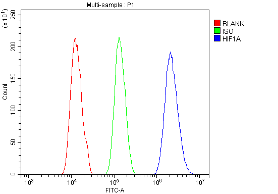 FCM (Flow Cytometry)
(Figure 6. Flow Cytometry analysis of A431 cells using anti-HIF-1 alpha/HIF1A antibody (AAA19213).Overlay histogram showing A431 cells stained with AAA19213 (Blue line). The cells were blocked with 10% normal goat serum. And then incubated with rabbit anti-HIF-1 alpha/HIF1A Antibody (AAA19213, 1μg/1x106 cells) for 30 min at 20 degree C. DyLight®488 conjugated goat anti-rabbit IgG (5-10μg/1x106 cells) was used as secondary antibody for 30 minutes at 20 degree C. Isotype control antibody (Green line) was rabbit IgG (1μg/1x106) used under the same conditions. Unlabelled sample (Red line) was also used as a control.)
FCM (Flow Cytometry)
(Figure 6. Flow Cytometry analysis of A431 cells using anti-HIF-1 alpha/HIF1A antibody (AAA19213).Overlay histogram showing A431 cells stained with AAA19213 (Blue line). The cells were blocked with 10% normal goat serum. And then incubated with rabbit anti-HIF-1 alpha/HIF1A Antibody (AAA19213, 1μg/1x106 cells) for 30 min at 20 degree C. DyLight®488 conjugated goat anti-rabbit IgG (5-10μg/1x106 cells) was used as secondary antibody for 30 minutes at 20 degree C. Isotype control antibody (Green line) was rabbit IgG (1μg/1x106) used under the same conditions. Unlabelled sample (Red line) was also used as a control.)
HIF-1 alpha/HIF1A, Polyclonal Antibody (Cat# AAA19213)
Full Name
Anti-HIF-1 alpha/HIF1A Antibody
Gene Names
HIF1A; HIF1; MOP1; PASD8; HIF-1A; bHLHe78; HIF-1alpha; HIF1-ALPHA
Reactivity
Human, Mouse, Rat
Applications
Western Blot, Immunohistochemistry, Flow Cytometry, Direct ELISA
Purity
Immunogen affinity purified.
Pricing