Filters
Clonality
Type
Reactivity
Gene Name
Isotype
Host
Application
Clone
20 results for " Growth Factor Receptors" - showing 1-20


 Application Data
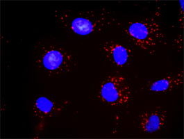
(Proximity Ligation Analysis of protein-protein interactions between STAT1 and PDGFRB Mahlavu cells were stained with anti-STAT1 rabbit purified polyclonal (1:1200) and 131071 (1:50). Each red dot represents the detection of protein-protein interaction complex, and nuclei were counterstained with DAPI (blue).)
Application Data
(Proximity Ligation Analysis of protein-protein interactions between STAT1 and PDGFRB Mahlavu cells were stained with anti-STAT1 rabbit purified polyclonal (1:1200) and 131071 (1:50). Each red dot represents the detection of protein-protein interaction complex, and nuclei were counterstained with DAPI (blue).)
PDGFRB, Monoclonal Antibody (Cat# AAA25488)
Full Name
PDGFRB (PDGFR, PDGFR1, Platelet-derived Growth Factor Receptor beta, Beta Platelet-derived Growth Factor Receptor, Beta-type Platelet-derived Growth Factor Receptor, CD140 Antigen-like Family Member B, Platelet-derived Growth Factor Receptor 1, CD140b) (H
Gene Names
PDGFRB; IMF1; KOGS; IBGC4; JTK12; PDGFR; PENTT; CD140B; PDGFR1; PDGFR-1
Reactivity
Human
Applications
Immunoprecipitation, Western Blot
Purity
Purified by Protein A Affinity Chromatography.
Pricing


 Application Data
(Proximity Ligation Analysis of protein-protein interactions between STAT1 and PDGFRB Mahlavu cells were stained with anti-STAT1 rabbit purified polyclonal (1:1200) and (1:50). Each red dot represents the detection of protein-protein interaction complex, and nuclei were counterstained with DAPI (blue).)
Application Data
(Proximity Ligation Analysis of protein-protein interactions between STAT1 and PDGFRB Mahlavu cells were stained with anti-STAT1 rabbit purified polyclonal (1:1200) and (1:50). Each red dot represents the detection of protein-protein interaction complex, and nuclei were counterstained with DAPI (blue).)
PDGFRB, Monoclonal Antibody (Cat# AAA26690)
Full Name
PDGFRB (PDGFR, PDGFR1, Platelet-derived Growth Factor Receptor beta, Beta Platelet-derived Growth Factor Receptor, Beta-type Platelet-derived Growth Factor Receptor, CD140 Antigen-like Family Member B, Platelet-derived Growth Factor Receptor 1, CD140b) (M
Gene Names
PDGFRB; IMF1; KOGS; IBGC4; JTK12; PDGFR; PENTT; CD140B; PDGFR1; PDGFR-1
Reactivity
Human
Applications
Immunoprecipitation, Western Blot
Purity
Purified by Protein A Affinity Chromatography.
Pricing


 Application Data
(Proximity Ligation Analysis of protein-protein interactions between STAT1 and PDGFRB Mahlavu cells were stained with anti-STAT1 rabbit purified polyclonal (1:1200) and 131071 (1:50). Each red dot represents the detection of protein-protein interaction complex, and nuclei were counterstained with DAPI (blue).)
Application Data
(Proximity Ligation Analysis of protein-protein interactions between STAT1 and PDGFRB Mahlavu cells were stained with anti-STAT1 rabbit purified polyclonal (1:1200) and 131071 (1:50). Each red dot represents the detection of protein-protein interaction complex, and nuclei were counterstained with DAPI (blue).)
PDGFRB, Monoclonal Antibody (Cat# AAA26698)
Full Name
PDGFRB (PDGFR, PDGFR1, Platelet-derived Growth Factor Receptor beta, Beta Platelet-derived Growth Factor Receptor, Beta-type Platelet-derived Growth Factor Receptor, CD140 Antigen-like Family Member B, Platelet-derived Growth Factor Receptor 1, CD140b) (M
Gene Names
PDGFRB; IMF1; KOGS; IBGC4; JTK12; PDGFR; PENTT; CD140B; PDGFR1; PDGFR-1
Reactivity
Human
Applications
Immunoprecipitation, Western Blot
Purity
Purified by Protein A Affinity Chromatography.
Pricing


 Application Data
(Proximity Ligation Analysis of protein-protein interactions between STAT1 and PDGFRB Mahlavu cells were stained with anti-STAT1 rabbit purified polyclonal (1:1200) and 131071 (1:50). Each red dot represents the detection of protein-protein interaction complex, and nuclei were counterstained with DAPI (blue).)
Application Data
(Proximity Ligation Analysis of protein-protein interactions between STAT1 and PDGFRB Mahlavu cells were stained with anti-STAT1 rabbit purified polyclonal (1:1200) and 131071 (1:50). Each red dot represents the detection of protein-protein interaction complex, and nuclei were counterstained with DAPI (blue).)
PDGFRB, Monoclonal Antibody (Cat# AAA24602)
Full Name
PDGFRB (PDGFR, PDGFR1, Platelet-derived Growth Factor Receptor beta, Beta Platelet-derived Growth Factor Receptor, Beta-type Platelet-derived Growth Factor Receptor, CD140 Antigen-like Family Member B, Platelet-derived Growth Factor Receptor 1, CD140b) AP
Gene Names
PDGFRB; IMF1; KOGS; IBGC4; JTK12; PDGFR; PENTT; CD140B; PDGFR1; PDGFR-1
Reactivity
Human
Applications
Immunoprecipitation, Western Blot
Purity
Purified by Protein A Affinity Chromatography.
Pricing


 Application Data
(Published customer image Infiltration of GFP+ BM-cells in infarct and peri-infarct regions. (A-B) Dot plots of viable macrophages/granulocytes (CD11b+CD45high, top right quadrants) and microglia (CD11b+CD45dim, bottom right quadrants) in cortex from BM-chimeric unmanipulated mice and mice exposed to pMCAO. (C) Bar graph showing mean numbers of CD11b+CD45dim microglia and CD11b+CD45high macrophages/granulocytes in BM-chimeric mice 24 hours after pMCAO, subdivided based on expression of GFP (n = 5). Approximately 92% of of the CD45high population were GFP+. (D) Estimation and comparison of mean numbers of CD11b+CD45dim microglia in non-chimeric (n = 10) versus BM-chimeric mice (n = 5) 24 hours after of pMCAO shows significantly fewer CD11b+CD45dim microglial cells in irradiated mice. (E) Overview, showing distribution of infiltrating GFP+ BM-derived cells into infarct (IF) and peri-infarct (P-IF) regions 24 hours after pMCAO. (E-G) By 24 hours, GFP+ single cells (F) and vessel-associated aggregates of GFP+ cells (arrows in G) were observed in infarct and peri-infarct regions. Some of the vessel-associated cells were round, leukocyte-like cells (arrows) while others were elongated cells lining the vasculature (arrow heads in G and in insert). (H) Bar graph showing mean numbers of single GFP+ cells and vessel-associated aggregates of GFP+ cells in ipsi- and contralateral cortex 24 hours after surgery (n = 10). (I-P) Immunohistochemical staining of CD45.1 (I, K), CD45.2 (J, L), IgG2a (M, O) and CD45 (N, P) in ischemic tissue in BM-chimeric (I, J, M, N) and non-chimeric mice (K, L, O, P) 24 hours after pMCAO. N.D, none detected. Scale bars: 200 um (A), 10 um (B, C). 50 um (I-P) *P < 0.05, **P < 0.01, and ***P < 0.001.From: Clausen BH, Lambertsen KL, Babcock AA, Holm TH, Dagnaes-Hansen F, Finsen B. Interleukin-1beta and tumor necrosis factor-alpha are expressed by different subsets of microglia and macrophages after ischemic stroke in mice. J Neuroinflammation. 2008 Oct 23;5:46.)
Application Data
(Published customer image Infiltration of GFP+ BM-cells in infarct and peri-infarct regions. (A-B) Dot plots of viable macrophages/granulocytes (CD11b+CD45high, top right quadrants) and microglia (CD11b+CD45dim, bottom right quadrants) in cortex from BM-chimeric unmanipulated mice and mice exposed to pMCAO. (C) Bar graph showing mean numbers of CD11b+CD45dim microglia and CD11b+CD45high macrophages/granulocytes in BM-chimeric mice 24 hours after pMCAO, subdivided based on expression of GFP (n = 5). Approximately 92% of of the CD45high population were GFP+. (D) Estimation and comparison of mean numbers of CD11b+CD45dim microglia in non-chimeric (n = 10) versus BM-chimeric mice (n = 5) 24 hours after of pMCAO shows significantly fewer CD11b+CD45dim microglial cells in irradiated mice. (E) Overview, showing distribution of infiltrating GFP+ BM-derived cells into infarct (IF) and peri-infarct (P-IF) regions 24 hours after pMCAO. (E-G) By 24 hours, GFP+ single cells (F) and vessel-associated aggregates of GFP+ cells (arrows in G) were observed in infarct and peri-infarct regions. Some of the vessel-associated cells were round, leukocyte-like cells (arrows) while others were elongated cells lining the vasculature (arrow heads in G and in insert). (H) Bar graph showing mean numbers of single GFP+ cells and vessel-associated aggregates of GFP+ cells in ipsi- and contralateral cortex 24 hours after surgery (n = 10). (I-P) Immunohistochemical staining of CD45.1 (I, K), CD45.2 (J, L), IgG2a (M, O) and CD45 (N, P) in ischemic tissue in BM-chimeric (I, J, M, N) and non-chimeric mice (K, L, O, P) 24 hours after pMCAO. N.D, none detected. Scale bars: 200 um (A), 10 um (B, C). 50 um (I-P) *P < 0.05, **P < 0.01, and ***P < 0.001.From: Clausen BH, Lambertsen KL, Babcock AA, Holm TH, Dagnaes-Hansen F, Finsen B. Interleukin-1beta and tumor necrosis factor-alpha are expressed by different subsets of microglia and macrophages after ischemic stroke in mice. J Neuroinflammation. 2008 Oct 23;5:46.)
CD11b, Monoclonal Antibody (Cat# AAA12182)
Full Name
RAT ANTI MOUSE CD11b:FITC
Gene Names
Itgam; CR3; CR3A; MAC1; Cd11b; Ly-40; Mac-1; Mac-1a; CD11b/CD18; F730045J24Rik
Applications
Flow Cytometry
Pricing


 Application Data
(Proximity Ligation Analysis of protein-protein interactions between STAT1 and PDGFRB Mahlavu cells were stained with anti-STAT1 rabbit purified polyclonal (1:1200) and (1:50). Each red dot represents the detection of protein-protein interaction complex, and nuclei were counterstained with DAPI (blue).)
Application Data
(Proximity Ligation Analysis of protein-protein interactions between STAT1 and PDGFRB Mahlavu cells were stained with anti-STAT1 rabbit purified polyclonal (1:1200) and (1:50). Each red dot represents the detection of protein-protein interaction complex, and nuclei were counterstained with DAPI (blue).)
PDGFRB, Monoclonal Antibody (Cat# AAA26694)
Full Name
PDGFRB (PDGFR, PDGFR1, Platelet-derived Growth Factor Receptor beta, Beta Platelet-derived Growth Factor Receptor, Beta-type Platelet-derived Growth Factor Receptor, CD140 Antigen-like Family Member B, Platelet-derived Growth Factor Receptor 1, CD140b) (M
Gene Names
PDGFRB; IMF1; KOGS; IBGC4; JTK12; PDGFR; PENTT; CD140B; PDGFR1; PDGFR-1
Reactivity
Human
Applications
Immunoprecipitation, Western Blot
Purity
Purified by Protein A Affinity Chromatography.
Pricing


 Application Data
(Published customer image Infiltration of GFP+ BM-cells in infarct and peri-infarct regions. (A-B) Dot plots of viable macrophages/granulocytes (CD11b+CD45high, top right quadrants) and microglia (CD11b+CD45dim, bottom right quadrants) in cortex from BM-chimeric unmanipulated mice and mice exposed to pMCAO. (C) Bar graph showing mean numbers of CD11b+CD45dim microglia and CD11b+CD45high macrophages/granulocytes in BM-chimeric mice 24 hours after pMCAO, subdivided based on expression of GFP (n = 5). Approximately 92% of of the CD45high population were GFP+. (D) Estimation and comparison of mean numbers of CD11b+CD45dim microglia in non-chimeric (n = 10) versus BM-chimeric mice (n = 5) 24 hours after of pMCAO shows significantly fewer CD11b+CD45dim microglial cells in irradiated mice. (E) Overview, showing distribution of infiltrating GFP+ BM-derived cells into infarct (IF) and peri-infarct (P-IF) regions 24 hours after pMCAO. (E-G) By 24 hours, GFP+ single cells (F) and vessel-associated aggregates of GFP+ cells (arrows in G) were observed in infarct and peri-infarct regions. Some of the vessel-associated cells were round, leukocyte-like cells (arrows) while others were elongated cells lining the vasculature (arrow heads in G and in insert). (H) Bar graph showing mean numbers of single GFP+ cells and vessel-associated aggregates of GFP+ cells in ipsi- and contralateral cortex 24 hours after surgery (n = 10). (I-P) Immunohistochemical staining of CD45.1 (I, K), CD45.2 (J, L), IgG2a (M, O) and CD45 (N, P) in ischemic tissue in BM-chimeric (I, J, M, N) and non-chimeric mice (K, L, O, P) 24 hours after pMCAO. N.D, none detected. Scale bars: 200 um (A), 10 um (B, C). 50 um (I-P) *P < 0.05, **P < 0.01, and ***P < 0.001.From: Clausen BH, Lambertsen KL, Babcock AA, Holm TH, Dagnaes-Hansen F, Finsen B. Interleukin-1beta and tumor necrosis factor-alpha are expressed by different subsets of microglia and macrophages after ischemic stroke in mice. J Neuroinflammation. 2008 Oct 23;5:46.)
Application Data
(Published customer image Infiltration of GFP+ BM-cells in infarct and peri-infarct regions. (A-B) Dot plots of viable macrophages/granulocytes (CD11b+CD45high, top right quadrants) and microglia (CD11b+CD45dim, bottom right quadrants) in cortex from BM-chimeric unmanipulated mice and mice exposed to pMCAO. (C) Bar graph showing mean numbers of CD11b+CD45dim microglia and CD11b+CD45high macrophages/granulocytes in BM-chimeric mice 24 hours after pMCAO, subdivided based on expression of GFP (n = 5). Approximately 92% of of the CD45high population were GFP+. (D) Estimation and comparison of mean numbers of CD11b+CD45dim microglia in non-chimeric (n = 10) versus BM-chimeric mice (n = 5) 24 hours after of pMCAO shows significantly fewer CD11b+CD45dim microglial cells in irradiated mice. (E) Overview, showing distribution of infiltrating GFP+ BM-derived cells into infarct (IF) and peri-infarct (P-IF) regions 24 hours after pMCAO. (E-G) By 24 hours, GFP+ single cells (F) and vessel-associated aggregates of GFP+ cells (arrows in G) were observed in infarct and peri-infarct regions. Some of the vessel-associated cells were round, leukocyte-like cells (arrows) while others were elongated cells lining the vasculature (arrow heads in G and in insert). (H) Bar graph showing mean numbers of single GFP+ cells and vessel-associated aggregates of GFP+ cells in ipsi- and contralateral cortex 24 hours after surgery (n = 10). (I-P) Immunohistochemical staining of CD45.1 (I, K), CD45.2 (J, L), IgG2a (M, O) and CD45 (N, P) in ischemic tissue in BM-chimeric (I, J, M, N) and non-chimeric mice (K, L, O, P) 24 hours after pMCAO. N.D, none detected. Scale bars: 200 um (A), 10 um (B, C). 50 um (I-P) *P < 0.05, **P < 0.01, and ***P < 0.001.From: Clausen BH, Lambertsen KL, Babcock AA, Holm TH, Dagnaes-Hansen F, Finsen B. Interleukin-1beta and tumor necrosis factor-alpha are expressed by different subsets of microglia and macrophages after ischemic stroke in mice. J Neuroinflammation. 2008 Oct 23;5:46.)
CD11b, Monoclonal Antibody (Cat# AAA12183)
Full Name
RAT ANTI MOUSE CD11b:FITC
Gene Names
Itgam; CR3; CR3A; MAC1; Cd11b; Ly-40; Mac-1; Mac-1a; CD11b/CD18; F730045J24Rik
Applications
Flow Cytometry
Pricing


 Application Data
(Proximity Ligation Analysis of protein-protein interactions between STAT1 and PDGFRB Mahlavu cells were stained with anti-STAT1 rabbit purified polyclonal (1:1200) and 131071 (1:50). Each red dot represents the detection of protein-protein interaction complex, and nuclei were counterstained with DAPI (blue).)
Application Data
(Proximity Ligation Analysis of protein-protein interactions between STAT1 and PDGFRB Mahlavu cells were stained with anti-STAT1 rabbit purified polyclonal (1:1200) and 131071 (1:50). Each red dot represents the detection of protein-protein interaction complex, and nuclei were counterstained with DAPI (blue).)
PDGFRB, Monoclonal Antibody (Cat# AAA25194)
Full Name
PDGFRB (PDGFR, PDGFR1, Platelet-derived Growth Factor Receptor beta, Beta Platelet-derived Growth Factor Receptor, Beta-type Platelet-derived Growth Factor Receptor, CD140 Antigen-like Family Member B, Platelet-derived Growth Factor Receptor 1, CD140b) (F
Gene Names
PDGFRB; IMF1; KOGS; IBGC4; JTK12; PDGFR; PENTT; CD140B; PDGFR1; PDGFR-1
Reactivity
Human
Applications
Immunoprecipitation, Western Blot
Purity
Purified by Protein A Affinity Chromatography.
Pricing


 Application Data
(Proximity Ligation Analysis of protein-protein interactions between STAT1 and PDGFRB Mahlavu cells were stained with anti-STAT1 rabbit purified polyclonal (1:1200) and 131071 (1:50). Each red dot represents the detection of protein-protein interaction complex, and nuclei were counterstained with DAPI (blue).)
Application Data
(Proximity Ligation Analysis of protein-protein interactions between STAT1 and PDGFRB Mahlavu cells were stained with anti-STAT1 rabbit purified polyclonal (1:1200) and 131071 (1:50). Each red dot represents the detection of protein-protein interaction complex, and nuclei were counterstained with DAPI (blue).)
PDGFRB, Monoclonal Antibody (Cat# AAA26701)
Full Name
PDGFRB (PDGFR, PDGFR1, Platelet-derived Growth Factor Receptor beta, Beta Platelet-derived Growth Factor Receptor, Beta-type Platelet-derived Growth Factor Receptor, CD140 Antigen-like Family Member B, Platelet-derived Growth Factor Receptor 1, CD140b) (M
Gene Names
PDGFRB; IMF1; KOGS; IBGC4; JTK12; PDGFR; PENTT; CD140B; PDGFR1; PDGFR-1
Reactivity
Human
Applications
Immunoprecipitation, Western Blot
Purity
Purified by Protein A Affinity Chromatography.
Pricing


 Application Data
(Published customer image Infiltration of GFP+ BM-cells in infarct and peri-infarct regions. (A-B) Dot plots of viable macrophages/granulocytes (CD11b+CD45high, top right quadrants) and microglia (CD11b+CD45dim, bottom right quadrants) in cortex from BM-chimeric unmanipulated mice and mice exposed to pMCAO. (C) Bar graph showing mean numbers of CD11b+CD45dim microglia and CD11b+CD45high macrophages/granulocytes in BM-chimeric mice 24 hours after pMCAO, subdivided based on expression of GFP (n = 5). Approximately 92% of of the CD45high population were GFP+. (D) Estimation and comparison of mean numbers of CD11b+CD45dim microglia in non-chimeric (n = 10) versus BM-chimeric mice (n = 5) 24 hours after of pMCAO shows significantly fewer CD11b+CD45dim microglial cells in irradiated mice. (E) Overview, showing distribution of infiltrating GFP+ BM-derived cells into infarct (IF) and peri-infarct (P-IF) regions 24 hours after pMCAO. (E-G) By 24 hours, GFP+ single cells (F) and vessel-associated aggregates of GFP+ cells (arrows in G) were observed in infarct and peri-infarct regions. Some of the vessel-associated cells were round, leukocyte-like cells (arrows) while others were elongated cells lining the vasculature (arrow heads in G and in insert). (H) Bar graph showing mean numbers of single GFP+ cells and vessel-associated aggregates of GFP+ cells in ipsi- and contralateral cortex 24 hours after surgery (n = 10). (I-P) Immunohistochemical staining of CD45.1 (I, K), CD45.2 (J, L), IgG2a (M, O) and CD45 (N, P) in ischemic tissue in BM-chimeric (I, J, M, N) and non-chimeric mice (K, L, O, P) 24 hours after pMCAO. N.D, none detected. Scale bars: 200 um (A), 10 um (B, C). 50 um (I-P) *P < 0.05, **P < 0.01, and ***P < 0.001.From: Clausen BH, Lambertsen KL, Babcock AA, Holm TH, Dagnaes-Hansen F, Finsen B. Interleukin-1beta and tumor necrosis factor-alpha are expressed by different subsets of microglia and macrophages after ischemic stroke in mice. J Neuroinflammation. 2008 Oct 23;5:46.)
Application Data
(Published customer image Infiltration of GFP+ BM-cells in infarct and peri-infarct regions. (A-B) Dot plots of viable macrophages/granulocytes (CD11b+CD45high, top right quadrants) and microglia (CD11b+CD45dim, bottom right quadrants) in cortex from BM-chimeric unmanipulated mice and mice exposed to pMCAO. (C) Bar graph showing mean numbers of CD11b+CD45dim microglia and CD11b+CD45high macrophages/granulocytes in BM-chimeric mice 24 hours after pMCAO, subdivided based on expression of GFP (n = 5). Approximately 92% of of the CD45high population were GFP+. (D) Estimation and comparison of mean numbers of CD11b+CD45dim microglia in non-chimeric (n = 10) versus BM-chimeric mice (n = 5) 24 hours after of pMCAO shows significantly fewer CD11b+CD45dim microglial cells in irradiated mice. (E) Overview, showing distribution of infiltrating GFP+ BM-derived cells into infarct (IF) and peri-infarct (P-IF) regions 24 hours after pMCAO. (E-G) By 24 hours, GFP+ single cells (F) and vessel-associated aggregates of GFP+ cells (arrows in G) were observed in infarct and peri-infarct regions. Some of the vessel-associated cells were round, leukocyte-like cells (arrows) while others were elongated cells lining the vasculature (arrow heads in G and in insert). (H) Bar graph showing mean numbers of single GFP+ cells and vessel-associated aggregates of GFP+ cells in ipsi- and contralateral cortex 24 hours after surgery (n = 10). (I-P) Immunohistochemical staining of CD45.1 (I, K), CD45.2 (J, L), IgG2a (M, O) and CD45 (N, P) in ischemic tissue in BM-chimeric (I, J, M, N) and non-chimeric mice (K, L, O, P) 24 hours after pMCAO. N.D, none detected. Scale bars: 200 um (A), 10 um (B, C). 50 um (I-P) *P < 0.05, **P < 0.01, and ***P < 0.001.From: Clausen BH, Lambertsen KL, Babcock AA, Holm TH, Dagnaes-Hansen F, Finsen B. Interleukin-1beta and tumor necrosis factor-alpha are expressed by different subsets of microglia and macrophages after ischemic stroke in mice. J Neuroinflammation. 2008 Oct 23;5:46.)
CD11b, Monoclonal Antibody (Cat# AAA12186)
Full Name
RAT ANTI MOUSE CD11b:RPE
Gene Names
Itgam; CR3; CR3A; MAC1; Cd11b; Ly-40; Mac-1; Mac-1a; CD11b/CD18; F730045J24Rik
Applications
Flow Cytometry
Pricing


 Application Data
(Proximity Ligation Analysis of protein-protein interactions between STAT1 and PDGFRB Mahlavu cells were stained with anti-STAT1 rabbit purified polyclonal (1:1200) and 131071 (1:50). Each red dot represents the detection of protein-protein interaction complex, and nuclei were counterstained with DAPI (blue).)
Application Data
(Proximity Ligation Analysis of protein-protein interactions between STAT1 and PDGFRB Mahlavu cells were stained with anti-STAT1 rabbit purified polyclonal (1:1200) and 131071 (1:50). Each red dot represents the detection of protein-protein interaction complex, and nuclei were counterstained with DAPI (blue).)
PDGFRB, Monoclonal Antibody (Cat# AAA24897)
Full Name
PDGFRB (PDGFR, PDGFR1, Platelet-derived Growth Factor Receptor beta, Beta Platelet-derived Growth Factor Receptor, Beta-type Platelet-derived Growth Factor Receptor, CD140 Antigen-like Family Member B, Platelet-derived Growth Factor Receptor 1, CD140b) (B
Gene Names
PDGFRB; IMF1; KOGS; IBGC4; JTK12; PDGFR; PENTT; CD140B; PDGFR1; PDGFR-1
Reactivity
Human
Applications
Immunoprecipitation, Western Blot
Purity
Purified by Protein A Affinity Chromatography.
Pricing


 Application Data
(Proximity Ligation Analysis of protein-protein interactions between STAT1 and PDGFRB Mahlavu cells were stained with anti-STAT1 rabbit purified polyclonal (1:1200) and (1:50). Each red dot represents the detection of protein-protein interaction complex, and nuclei were counterstained with DAPI (blue).)
Application Data
(Proximity Ligation Analysis of protein-protein interactions between STAT1 and PDGFRB Mahlavu cells were stained with anti-STAT1 rabbit purified polyclonal (1:1200) and (1:50). Each red dot represents the detection of protein-protein interaction complex, and nuclei were counterstained with DAPI (blue).)
PDGFRB, Monoclonal Antibody (Cat# AAA26687)
Full Name
PDGFRB (PDGFR, PDGFR1, Platelet-derived Growth Factor Receptor beta, Beta Platelet-derived Growth Factor Receptor, Beta-type Platelet-derived Growth Factor Receptor, CD140 Antigen-like Family Member B, Platelet-derived Growth Factor Receptor 1, CD140b) (M
Gene Names
PDGFRB; IMF1; KOGS; IBGC4; JTK12; PDGFR; PENTT; CD140B; PDGFR1; PDGFR-1
Reactivity
Human
Applications
Immunoprecipitation, Western Blot
Purity
Purified by Protein A Affinity Chromatography.
Pricing


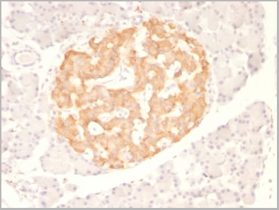
 WB (Western Blot)
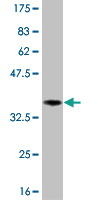
(WB Suggested Anti-NTRK3 Antibody Titration: 0.2-1 ug/mlELISA Titer: 1:312500Positive Control: Human Liver)
WB (Western Blot)
(WB Suggested Anti-NTRK3 Antibody Titration: 0.2-1 ug/mlELISA Titer: 1:312500Positive Control: Human Liver)
NTRK3, Polyclonal Antibody (Cat# AAA23541)
Full Name
NTRK3 antibody - C-terminal region
Gene Names
NTRK3; TRKC; GP145-TrkC; gp145(trkC)
Reactivity
Cow, Guinea Pig, Horse, Human, Mouse, Pig, Rabbit, Rat, Zebrafish
Applications
WB
Purity
Affinity Purified
Pricing
 Standard Curve (Sample)
Interleukin-2, ELISA Kit (Cat# AAA14616)
Full Name
Interleukin-2 (IL-2)
Gene Names
IL2; IL-2; TCGF; lymphokine
Pricing


 Application Data
(Proximity Ligation Analysis of protein-protein interactions between STAT1 and PDGFRB Mahlavu cells were stained with anti-STAT1 rabbit purified polyclonal (1:1200) and 131071 (1:50). Each red dot represents the detection of protein-protein interaction complex, and nuclei were counterstained with DAPI (blue).)
Application Data
(Proximity Ligation Analysis of protein-protein interactions between STAT1 and PDGFRB Mahlavu cells were stained with anti-STAT1 rabbit purified polyclonal (1:1200) and 131071 (1:50). Each red dot represents the detection of protein-protein interaction complex, and nuclei were counterstained with DAPI (blue).)
PDGFRB, Monoclonal Antibody (Cat# AAA25784)
Full Name
PDGFRB (PDGFR, PDGFR1, Platelet-derived Growth Factor Receptor beta, Beta Platelet-derived Growth Factor Receptor, Beta-type Platelet-derived Growth Factor Receptor, CD140 Antigen-like Family Member B, Platelet-derived Growth Factor Receptor 1, CD140b) (P
Gene Names
PDGFRB; IMF1; KOGS; IBGC4; JTK12; PDGFR; PENTT; CD140B; PDGFR1; PDGFR-1
Reactivity
Human
Applications
EIA, IP, WB
Purity
Purified by Protein A Affinity Chromatography.
Pricing


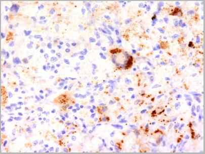
 WB (Western Blot)
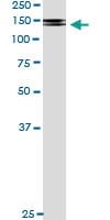
(WB Suggested Anti-TGFB1 Antibody Titration: 0.2-1 ug/ml or 1:5000 to 1:1000 dilution.SP2/0 cell lysate)
WB (Western Blot)
(WB Suggested Anti-TGFB1 Antibody Titration: 0.2-1 ug/ml or 1:5000 to 1:1000 dilution.SP2/0 cell lysate)
TGFB1, Polyclonal Antibody (Cat# AAA23459)
Full Name
TGFB1 antibody - middle region
Gene Names
Tgfb1; Tgfb; Tgfb-1; TGFbeta1; TGF-beta1
Reactivity
Predicted Reactivity: Cow, Dog, Goat, Guinea Pig, Horse, Mouse, Pig, Rat, Sheep (Tested Reactivity: Human, Mouse)
Applications
IHC, WB, IF
Purity
Affinity Purified
Pricing


 WB (Western Blot)
(FG Pancreatic Carcinoma Cell Lines stably expressing vector along (FG-V) the b3 integrin subunit (FG-b3) or a b3 truncation mutant (FG-759x). Src Mab (AAA28639) was diluted 1:500 in 1% BSA/TBST and incubated Overnight at 4 degree C. After washing 3x 5 min. with TBST the blots were incubated with 1:5000 Goat anti-mouse or Goat anti-rabbit secondary antibody for 1 hr at Room temperature. The blots were again washed 3x 5 min. with TBST and developed using ECL reagent.Data and protocol kindly provided by Dr. Weis of Cheresh Lab, UCSD.)
WB (Western Blot)
(FG Pancreatic Carcinoma Cell Lines stably expressing vector along (FG-V) the b3 integrin subunit (FG-b3) or a b3 truncation mutant (FG-759x). Src Mab (AAA28639) was diluted 1:500 in 1% BSA/TBST and incubated Overnight at 4 degree C. After washing 3x 5 min. with TBST the blots were incubated with 1:5000 Goat anti-mouse or Goat anti-rabbit secondary antibody for 1 hr at Room temperature. The blots were again washed 3x 5 min. with TBST and developed using ECL reagent.Data and protocol kindly provided by Dr. Weis of Cheresh Lab, UCSD.)
SRC, Monoclonal Antibody (Cat# AAA28639)
Full Name
SRC Antibody
Gene Names
SRC; ASV; SRC1; c-SRC; p60-Src
Reactivity
Human, mouse
Applications
WB, EIA, IF
Purity
This antibody is purified through a protein G column, followed by dialysis against PBS.
Pricing

 FCM (Flow Cytometry)
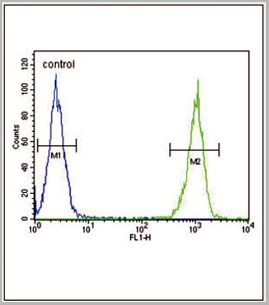
(LTF Antibody flow cytometric analysis of MDA-MB231 cells (right histogram) compared to a negative control cell (lefthistogram).FITC-conjugated goat-anti-rabbit secondary antibodies were used for theanalysis.)
FCM (Flow Cytometry)
(LTF Antibody flow cytometric analysis of MDA-MB231 cells (right histogram) compared to a negative control cell (lefthistogram).FITC-conjugated goat-anti-rabbit secondary antibodies were used for theanalysis.)
LTF, Polyclonal Antibody (Cat# AAA28680)
Full Name
LTF Antibody
Gene Names
LTF; LF; HLF2; GIG12; HEL110
Reactivity
Human, mouse
Applications
Western Blot, Flow Cytometry
Purity
Peptide Affinity Purified Rabbit Polyclonal Antibody (Pab)
Pricing
 IF (Immunofluorescence)
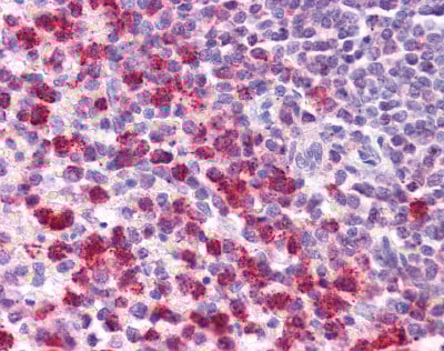
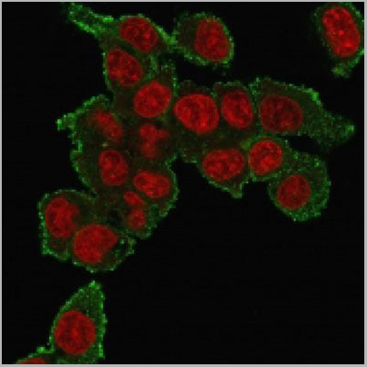
(Immunofluorescence staining of PFA-fixed HePG2 cells using Tumor Necrosis Factor (TNF alpha) Mouse Monoclonal Antibody (TNF706) followed by goat anti-mouse IgG-CF488 (green). Nuclei stained with RedDot.)
IF (Immunofluorescence)
(Immunofluorescence staining of PFA-fixed HePG2 cells using Tumor Necrosis Factor (TNF alpha) Mouse Monoclonal Antibody (TNF706) followed by goat anti-mouse IgG-CF488 (green). Nuclei stained with RedDot.)
TNF-alpha (Tumor Necrosis Factor alpha), Monoclonal Antibody (Cat# AAA13806)
Full Name
TNF-alpha (Tumor Necrosis Factor alpha) Mouse Monoclonal Antibody
Gene Names
TNF; DIF; TNFA; TNFSF2; TNLG1F; TNF-alpha
Reactivity
Human, Mouse, Rat, Rabbit, Cat, Dog, Zebrafish
Applications
Flow Cytometry, Immunofluorescence, Immunohistochemistry
Pricing
PDGFRB, Monoclonal Antibody (Cat# AAA24306)
Full Name
PDGFRB (PDGFR, PDGFR1, Platelet-derived Growth Factor Receptor beta, Beta Platelet-derived Growth Factor Receptor, Beta-type Platelet-derived Growth Factor Receptor, CD140 Antigen-like Family Member B, Platelet-derived Growth Factor Receptor 1, CD140b) (A
Gene Names
PDGFRB; IMF1; KOGS; IBGC4; JTK12; PDGFR; PENTT; CD140B; PDGFR1; PDGFR-1
Reactivity
Human
Applications
EIA, IP, WB
Purity
Purified by Protein A Affinity Chromatography.
Pricing