Filters
Clonality
Type
Reactivity
Gene Name
Isotype
Host
Application
Clone
94 results for " Cell Tissue Markers" - showing 1-50


 Application Data
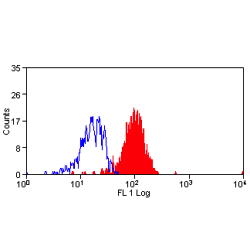
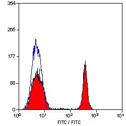
(Staining of CD209 transfected K562 cells with MOUSE ANTI HUMAN CD209: FITC)
Application Data
(Staining of CD209 transfected K562 cells with MOUSE ANTI HUMAN CD209: FITC)
DC-SIGN, Monoclonal Antibody (Cat# AAA26785)
Full Name
DC-SIGN (Dendritic Cell-specific ICAM3 grabbing Non-integrin, C Type Lectin Domain Family 4 Member L, CLEC4L, CD209, CD209 Antigen, CDSIGN, Dendritic Cell-specific ICAM-3-grabbing Non-integrin 1, DC SIGN1, DC-SIGN1, HIV GP120 Binding Protein, MGC129965) (
Reactivity
Human
Applications
Flow Cytometry, Immunofluorescence
Purity
Purified by protein G affinity chromatography from tissue culture supernatant.
Pricing


 Application Data
(Staining of human peripheral blood monocytes with MOUSE ANTI HUMAN CD274:RPE)
Application Data
(Staining of human peripheral blood monocytes with MOUSE ANTI HUMAN CD274:RPE)
CD274, Monoclonal Antibody (Cat# AAA26778)
Full Name
CD274 (CD274 Antigen, B7 Homolog 1, B7H1, B7-H1, B7-H, MGC142294, MGC142296, OTTHUMP00000021029, Programmed Cell Death 1 Ligand 1, PDCD1 Ligand 1, PDCD1L1, PDCD1LG1, Programmed Death Ligand 1, PDL1, PD-L1, RGD1566211) (MaxLight 405)
Gene Names
CD274; B7-H; B7H1; PDL1; PD-L1; PDCD1L1; PDCD1LG1
Reactivity
Human
Applications
Flow Cytometry
Purity
Purified by protein G affinity chromatography from tissue culture supernatant.
Pricing


 Application Data
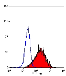
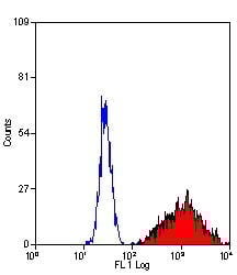
(Staining of NALM6 cells with MOUSE ANTI HUMAN CD10:ALEXA 488)
Application Data
(Staining of NALM6 cells with MOUSE ANTI HUMAN CD10:ALEXA 488)
CD10, Monoclonal Antibody (Cat# AAA26775)
Full Name
CD10 (CALLA, CD 10, Common Acute Lymphocytic Leukemia Antigen, Enkephalinase, gp100, Membrane Metalloendopeptidase, MME, Neprilysin, Neutral Endopeptidase, Pmel17) (MaxLight 405)
Gene Names
MME; NEP; SFE; CD10; CALLA
Reactivity
Human
Applications
Flow Cytometry, Immunohistochemistry, Immunoprecipitation, Western Blot
Purity
Purified by protein G affinity chromatography from tissue culture supernatant.
Pricing


 Application Data
(Staining of CD209 transfected K562 cells with MOUSE ANTI HUMAN CD209: FITC)
Application Data
(Staining of CD209 transfected K562 cells with MOUSE ANTI HUMAN CD209: FITC)
DC-SIGN, Monoclonal Antibody (Cat# AAA26761)
Full Name
DC-SIGN (Dendritic Cell-specific ICAM3 grabbing Non-integrin, C Type Lectin Domain Family 4 Member L, CLEC4L, CD209, CD209 Antigen, CDSIGN, Dendritic Cell-specific ICAM-3-grabbing Non-integrin 1, DC SIGN1, DC-SIGN1, HIV GP120 Binding Protein, MGC129965) (
Reactivity
Human
Applications
Flow Cytometry, Immunofluorescence
Purity
Purified by protein G affinity chromatography from tissue culture supernatant.
Pricing


 Application Data
(Staining of CD209 transfected K562 cells with MOUSE ANTI HUMAN CD209: FITC)
Application Data
(Staining of CD209 transfected K562 cells with MOUSE ANTI HUMAN CD209: FITC)
DC-SIGN, Monoclonal Antibody (Cat# AAA26799)
Full Name
DC-SIGN (Dendritic Cell-specific ICAM3 grabbing Non-integrin, C Type Lectin Domain Family 4 Member L, CLEC4L, CD209, CD209 Antigen, CDSIGN, Dendritic Cell-specific ICAM-3-grabbing Non-integrin 1, DC SIGN1, DC-SIGN1, HIV GP120 Binding Protein, MGC129965) (
Reactivity
Human
Applications
Flow Cytometry, Immunofluorescence
Purity
Purified by protein G affinity chromatography from tissue culture supernatant.
Pricing


 Application Data
(Staining of CD209 transfected K562 cells with MOUSE ANTI HUMAN CD209: FITC)
Application Data
(Staining of CD209 transfected K562 cells with MOUSE ANTI HUMAN CD209: FITC)
DC-SIGN, Monoclonal Antibody (Cat# AAA26736)
Full Name
DC-SIGN (Dendritic Cell-specific ICAM3 grabbing Non-integrin, C Type Lectin Domain Family 4 Member L, CLEC4L, CD209, CD209 Antigen, CDSIGN, Dendritic Cell-specific ICAM-3-grabbing Non-integrin 1, DC SIGN1, DC-SIGN1, HIV GP120 Binding Protein, MGC129965) (
Reactivity
Human
Applications
Flow Cytometry, Immunofluorescence
Purity
Purified by protein G affinity chromatography from tissue culture supernatant.
Pricing


 Application Data
(Staining of human peripheral blood monocytes with MOUSE ANTI HUMAN CD274:RPE)
Application Data
(Staining of human peripheral blood monocytes with MOUSE ANTI HUMAN CD274:RPE)
CD274, Monoclonal Antibody (Cat# AAA26743)
Full Name
CD274 (CD274 Antigen, B7 Homolog 1, B7H1, B7-H1, B7-H, MGC142294, MGC142296, OTTHUMP00000021029, Programmed Cell Death 1 Ligand 1, PDCD1 Ligand 1, PDCD1L1, PDCD1LG1, Programmed Death Ligand 1, PDL1, PD-L1, RGD1566211) (FITC)
Gene Names
CD274; B7-H; B7H1; PDL1; PD-L1; PDCD1L1; PDCD1LG1
Reactivity
Human
Applications
Flow Cytometry
Purity
Purified by protein G affinity chromatography from tissue culture supernatant.
Pricing


 Application Data
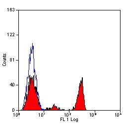
(Staining of human peripheral blood lymphocytes with MOUSE ANTI HUMAN CD45RA:FITC (MCA88F))
Application Data
(Staining of human peripheral blood lymphocytes with MOUSE ANTI HUMAN CD45RA:FITC (MCA88F))
CD45RA, Monoclonal Antibody (Cat# AAA26770)
Full Name
CD45RA (CD45 Antigen, B220, GP180, Leukocyte Common Antigen, LCA, L-CA, LY5, LY-5, Protein Tyrosine Phosphatase Receptor Type C Polypeptide, PTPRC, T200, T200 Glycoprotein) (PE)
Reactivity
Human, Monkey
Applications
Flow Cytometry, Immunohistochemistry
Purity
Purified by protein G affinity chromatography from tissue culture supernatant.
Pricing


 Application Data
(Staining of human peripheral blood monocytes with MOUSE ANTI HUMAN CD274:RPE)
Application Data
(Staining of human peripheral blood monocytes with MOUSE ANTI HUMAN CD274:RPE)
CD274, Monoclonal Antibody (Cat# AAA26806)
Full Name
CD274 (CD274 Antigen, B7 Homolog 1, B7H1, B7-H1, B7-H, MGC142294, MGC142296, OTTHUMP00000021029, Programmed Cell Death 1 Ligand 1, PDCD1 Ligand 1, PDCD1L1, PDCD1LG1, Programmed Death Ligand 1, PDL1, PD-L1, RGD1566211) (MaxLight 550)
Gene Names
CD274; B7-H; B7H1; PDL1; PD-L1; PDCD1L1; PDCD1LG1
Reactivity
Human
Applications
Flow Cytometry
Purity
Purified by protein G affinity chromatography from tissue culture supernatant.
Pricing


 Application Data
(Published customer image Infiltration of GFP+ BM-cells in infarct and peri-infarct regions. (A-B) Dot plots of viable macrophages/granulocytes (CD11b+CD45high, top right quadrants) and microglia (CD11b+CD45dim, bottom right quadrants) in cortex from BM-chimeric unmanipulated mice and mice exposed to pMCAO. (C) Bar graph showing mean numbers of CD11b+CD45dim microglia and CD11b+CD45high macrophages/granulocytes in BM-chimeric mice 24 hours after pMCAO, subdivided based on expression of GFP (n = 5). Approximately 92% of of the CD45high population were GFP+. (D) Estimation and comparison of mean numbers of CD11b+CD45dim microglia in non-chimeric (n = 10) versus BM-chimeric mice (n = 5) 24 hours after of pMCAO shows significantly fewer CD11b+CD45dim microglial cells in irradiated mice. (E) Overview, showing distribution of infiltrating GFP+ BM-derived cells into infarct (IF) and peri-infarct (P-IF) regions 24 hours after pMCAO. (E-G) By 24 hours, GFP+ single cells (F) and vessel-associated aggregates of GFP+ cells (arrows in G) were observed in infarct and peri-infarct regions. Some of the vessel-associated cells were round, leukocyte-like cells (arrows) while others were elongated cells lining the vasculature (arrow heads in G and in insert). (H) Bar graph showing mean numbers of single GFP+ cells and vessel-associated aggregates of GFP+ cells in ipsi- and contralateral cortex 24 hours after surgery (n = 10). (I-P) Immunohistochemical staining of CD45.1 (I, K), CD45.2 (J, L), IgG2a (M, O) and CD45 (N, P) in ischemic tissue in BM-chimeric (I, J, M, N) and non-chimeric mice (K, L, O, P) 24 hours after pMCAO. N.D, none detected. Scale bars: 200 um (A), 10 um (B, C). 50 um (I-P) *P < 0.05, **P < 0.01, and ***P < 0.001.From: Clausen BH, Lambertsen KL, Babcock AA, Holm TH, Dagnaes-Hansen F, Finsen B. Interleukin-1beta and tumor necrosis factor-alpha are expressed by different subsets of microglia and macrophages after ischemic stroke in mice. J Neuroinflammation. 2008 Oct 23;5:46.)
Application Data
(Published customer image Infiltration of GFP+ BM-cells in infarct and peri-infarct regions. (A-B) Dot plots of viable macrophages/granulocytes (CD11b+CD45high, top right quadrants) and microglia (CD11b+CD45dim, bottom right quadrants) in cortex from BM-chimeric unmanipulated mice and mice exposed to pMCAO. (C) Bar graph showing mean numbers of CD11b+CD45dim microglia and CD11b+CD45high macrophages/granulocytes in BM-chimeric mice 24 hours after pMCAO, subdivided based on expression of GFP (n = 5). Approximately 92% of of the CD45high population were GFP+. (D) Estimation and comparison of mean numbers of CD11b+CD45dim microglia in non-chimeric (n = 10) versus BM-chimeric mice (n = 5) 24 hours after of pMCAO shows significantly fewer CD11b+CD45dim microglial cells in irradiated mice. (E) Overview, showing distribution of infiltrating GFP+ BM-derived cells into infarct (IF) and peri-infarct (P-IF) regions 24 hours after pMCAO. (E-G) By 24 hours, GFP+ single cells (F) and vessel-associated aggregates of GFP+ cells (arrows in G) were observed in infarct and peri-infarct regions. Some of the vessel-associated cells were round, leukocyte-like cells (arrows) while others were elongated cells lining the vasculature (arrow heads in G and in insert). (H) Bar graph showing mean numbers of single GFP+ cells and vessel-associated aggregates of GFP+ cells in ipsi- and contralateral cortex 24 hours after surgery (n = 10). (I-P) Immunohistochemical staining of CD45.1 (I, K), CD45.2 (J, L), IgG2a (M, O) and CD45 (N, P) in ischemic tissue in BM-chimeric (I, J, M, N) and non-chimeric mice (K, L, O, P) 24 hours after pMCAO. N.D, none detected. Scale bars: 200 um (A), 10 um (B, C). 50 um (I-P) *P < 0.05, **P < 0.01, and ***P < 0.001.From: Clausen BH, Lambertsen KL, Babcock AA, Holm TH, Dagnaes-Hansen F, Finsen B. Interleukin-1beta and tumor necrosis factor-alpha are expressed by different subsets of microglia and macrophages after ischemic stroke in mice. J Neuroinflammation. 2008 Oct 23;5:46.)
CD11b, Monoclonal Antibody (Cat# AAA12182)
Full Name
RAT ANTI MOUSE CD11b:FITC
Gene Names
Itgam; CR3; CR3A; MAC1; Cd11b; Ly-40; Mac-1; Mac-1a; CD11b/CD18; F730045J24Rik
Applications
Flow Cytometry
Pricing


 Application Data
(Staining of CD209 transfected K562 cells with MOUSE ANTI HUMAN CD209: FITC)
Application Data
(Staining of CD209 transfected K562 cells with MOUSE ANTI HUMAN CD209: FITC)
DC-SIGN, Monoclonal Antibody (Cat# AAA26722)
Full Name
DC-SIGN (Dendritic Cell-specific ICAM3 grabbing Non-integrin, C Type Lectin Domain Family 4 Member L, CLEC4L, CD209, CD209 Antigen, CDSIGN, Dendritic Cell-specific ICAM-3-grabbing Non-integrin 1, DC SIGN1, DC-SIGN1, HIV GP120 Binding Protein, MGC129965) (
Reactivity
Human
Applications
Immunofluorescence
Purity
Purified by protein G affinity chromatography from tissue culture supernatant.
Pricing


 Application Data
(Published customer image Infiltration of GFP+ BM-cells in infarct and peri-infarct regions. (A-B) Dot plots of viable macrophages/granulocytes (CD11b+CD45high, top right quadrants) and microglia (CD11b+CD45dim, bottom right quadrants) in cortex from BM-chimeric unmanipulated mice and mice exposed to pMCAO. (C) Bar graph showing mean numbers of CD11b+CD45dim microglia and CD11b+CD45high macrophages/granulocytes in BM-chimeric mice 24 hours after pMCAO, subdivided based on expression of GFP (n = 5). Approximately 92% of of the CD45high population were GFP+. (D) Estimation and comparison of mean numbers of CD11b+CD45dim microglia in non-chimeric (n = 10) versus BM-chimeric mice (n = 5) 24 hours after of pMCAO shows significantly fewer CD11b+CD45dim microglial cells in irradiated mice. (E) Overview, showing distribution of infiltrating GFP+ BM-derived cells into infarct (IF) and peri-infarct (P-IF) regions 24 hours after pMCAO. (E-G) By 24 hours, GFP+ single cells (F) and vessel-associated aggregates of GFP+ cells (arrows in G) were observed in infarct and peri-infarct regions. Some of the vessel-associated cells were round, leukocyte-like cells (arrows) while others were elongated cells lining the vasculature (arrow heads in G and in insert). (H) Bar graph showing mean numbers of single GFP+ cells and vessel-associated aggregates of GFP+ cells in ipsi- and contralateral cortex 24 hours after surgery (n = 10). (I-P) Immunohistochemical staining of CD45.1 (I, K), CD45.2 (J, L), IgG2a (M, O) and CD45 (N, P) in ischemic tissue in BM-chimeric (I, J, M, N) and non-chimeric mice (K, L, O, P) 24 hours after pMCAO. N.D, none detected. Scale bars: 200 um (A), 10 um (B, C). 50 um (I-P) *P < 0.05, **P < 0.01, and ***P < 0.001.From: Clausen BH, Lambertsen KL, Babcock AA, Holm TH, Dagnaes-Hansen F, Finsen B. Interleukin-1beta and tumor necrosis factor-alpha are expressed by different subsets of microglia and macrophages after ischemic stroke in mice. J Neuroinflammation. 2008 Oct 23;5:46.)
Application Data
(Published customer image Infiltration of GFP+ BM-cells in infarct and peri-infarct regions. (A-B) Dot plots of viable macrophages/granulocytes (CD11b+CD45high, top right quadrants) and microglia (CD11b+CD45dim, bottom right quadrants) in cortex from BM-chimeric unmanipulated mice and mice exposed to pMCAO. (C) Bar graph showing mean numbers of CD11b+CD45dim microglia and CD11b+CD45high macrophages/granulocytes in BM-chimeric mice 24 hours after pMCAO, subdivided based on expression of GFP (n = 5). Approximately 92% of of the CD45high population were GFP+. (D) Estimation and comparison of mean numbers of CD11b+CD45dim microglia in non-chimeric (n = 10) versus BM-chimeric mice (n = 5) 24 hours after of pMCAO shows significantly fewer CD11b+CD45dim microglial cells in irradiated mice. (E) Overview, showing distribution of infiltrating GFP+ BM-derived cells into infarct (IF) and peri-infarct (P-IF) regions 24 hours after pMCAO. (E-G) By 24 hours, GFP+ single cells (F) and vessel-associated aggregates of GFP+ cells (arrows in G) were observed in infarct and peri-infarct regions. Some of the vessel-associated cells were round, leukocyte-like cells (arrows) while others were elongated cells lining the vasculature (arrow heads in G and in insert). (H) Bar graph showing mean numbers of single GFP+ cells and vessel-associated aggregates of GFP+ cells in ipsi- and contralateral cortex 24 hours after surgery (n = 10). (I-P) Immunohistochemical staining of CD45.1 (I, K), CD45.2 (J, L), IgG2a (M, O) and CD45 (N, P) in ischemic tissue in BM-chimeric (I, J, M, N) and non-chimeric mice (K, L, O, P) 24 hours after pMCAO. N.D, none detected. Scale bars: 200 um (A), 10 um (B, C). 50 um (I-P) *P < 0.05, **P < 0.01, and ***P < 0.001.From: Clausen BH, Lambertsen KL, Babcock AA, Holm TH, Dagnaes-Hansen F, Finsen B. Interleukin-1beta and tumor necrosis factor-alpha are expressed by different subsets of microglia and macrophages after ischemic stroke in mice. J Neuroinflammation. 2008 Oct 23;5:46.)
CD11b, Monoclonal Antibody (Cat# AAA12184)
Full Name
RAT ANTI MOUSE CD11b
Gene Names
Itgam; CR3; CR3A; MAC1; Cd11b; Ly-40; Mac-1; Mac-1a; CD11b/CD18; F730045J24Rik
Applications
Immunohistochemistry, Flow Cytometry, Immunofluorescence, Immunoprecipitation
Pricing


 Application Data
(Staining of NALM6 cells with MOUSE ANTI HUMAN CD10:ALEXA 488)
Application Data
(Staining of NALM6 cells with MOUSE ANTI HUMAN CD10:ALEXA 488)
CD10, Monoclonal Antibody (Cat# AAA26713)
Full Name
CD10 (CALLA, CD 10, Common Acute Lymphocytic Leukemia Antigen, Enkephalinase, gp100, Membrane Metalloendopeptidase, MME, Neprilysin, Neutral Endopeptidase, Pmel17) (AP)
Gene Names
MME; NEP; SFE; CD10; CALLA
Reactivity
Human
Applications
Immunohistochemistry, Immunoprecipitation, Western Blot
Purity
Purified by protein G affinity chromatography from tissue culture supernatant.
Pricing


 Application Data
(Staining of human peripheral blood monocytes with MOUSE ANTI HUMAN CD274:RPE)
Application Data
(Staining of human peripheral blood monocytes with MOUSE ANTI HUMAN CD274:RPE)
CD274, Monoclonal Antibody (Cat# AAA26767)
Full Name
CD274 (CD274 Antigen, B7 Homolog 1, B7H1, B7-H1, B7-H, MGC142294, MGC142296, OTTHUMP00000021029, Programmed Cell Death 1 Ligand 1, PDCD1 Ligand 1, PDCD1L1, PDCD1LG1, Programmed Death Ligand 1, PDL1, PD-L1, RGD1566211) (PE)
Gene Names
CD274; B7-H; B7H1; PDL1; PD-L1; PDCD1L1; PDCD1LG1
Reactivity
Human
Applications
Flow Cytometry
Purity
Purified by protein G affinity chromatography from tissue culture supernatant.
Pricing


 IF (Immunofluorescence)
(Immunofluorescentstaining of PFA-fixedHeLa cells with Pan-Nuclear Antigen Monoclonal Antibody (AAA13844) followed by goat anti-mouse IgG-CF488 (green). Membranes labeled with phalloidin (red).)
IF (Immunofluorescence)
(Immunofluorescentstaining of PFA-fixedHeLa cells with Pan-Nuclear Antigen Monoclonal Antibody (AAA13844) followed by goat anti-mouse IgG-CF488 (green). Membranes labeled with phalloidin (red).)
Nuclear Antigen, Monoclonal Antibody (Cat# AAA13844)
Full Name
Nuclear Antigen (Pan-Nuclear Marker) Mouse Monoclonal Antibody
Reactivity
Human, Mouse, Rat.
Does not react with Pig
Does not react with Pig
Applications
Flow Cytometry, Immunofluorescence, Immunocytochemistry, Immunohistochemistry
Pricing


 Application Data
(Staining of NALM6 cells with MOUSE ANTI HUMAN CD10:ALEXA 488)
Application Data
(Staining of NALM6 cells with MOUSE ANTI HUMAN CD10:ALEXA 488)
CD10, Monoclonal Antibody (Cat# AAA26751)
Full Name
CD10 (CALLA, CD 10, Common Acute Lymphocytic Leukemia Antigen, Enkephalinase, gp100, Membrane Metalloendopeptidase, MME, Neprilysin, Neutral Endopeptidase, Pmel17) (HRP)
Gene Names
MME; NEP; SFE; CD10; CALLA
Reactivity
Human
Applications
Flow Cytometry, Immunohistochemistry, Immunoprecipitation, Western Blot
Purity
Purified by protein G affinity chromatography from tissue culture supernatant.
Pricing


 Application Data
CD274, Monoclonal Antibody (Cat# AAA26716)
Full Name
CD274 (CD274 Antigen, B7 Homolog 1, B7H1, B7-H1, B7-H, MGC142294, MGC142296, OTTHUMP00000021029, Programmed Cell Death 1 Ligand 1, PDCD1 Ligand 1, PDCD1L1, PDCD1LG1, Programmed Death Ligand 1, PDL1, PD-L1, RGD1566211) (AP)
Gene Names
CD274; B7-H; B7H1; PDL1; PD-L1; PDCD1L1; PDCD1LG1
Reactivity
Human
Applications
ELISA
Purity
Purified by protein G affinity chromatography from tissue culture supernatant.
Pricing


 Application Data
(Staining of human peripheral blood lymphocytes with MOUSE ANTI HUMAN CD4:ALEXA 405)
Application Data
(Staining of human peripheral blood lymphocytes with MOUSE ANTI HUMAN CD4:ALEXA 405)
CD4, Monoclonal Antibody (Cat# AAA26732)
Full Name
CD4 (CD4 Antigen, CD4 Molecule, CD4 Receptor, CD4mut, T Cell Surface Antigen T4/Leu3, T Cell Antigen T4, T Cell Surface Glycoprotein CD4) (Biotin)
Gene Names
CD4; CD4mut
Reactivity
Human
Applications
Flow Cytometry, Immunohistochemistry
Purity
Purified by protein A affinity chromatography from tissue culture supernatant.
Pricing


 Application Data
(Staining of human peripheral blood lymphocytes with MOUSE ANTI HUMAN CD4:ALEXA 405)
Application Data
(Staining of human peripheral blood lymphocytes with MOUSE ANTI HUMAN CD4:ALEXA 405)
CD4, Monoclonal Antibody (Cat# AAA26795)
Full Name
CD4 (CD4 Antigen, CD4 Molecule, CD4 Receptor, CD4mut, T Cell Surface Antigen T4/Leu3, T Cell Antigen T4, T Cell Surface Glycoprotein CD4) (MaxLight 490)
Gene Names
CD4; CD4mut
Reactivity
Human
Applications
Flow Cytometry, Immunohistochemistry
Purity
Purified by protein A affinity chromatography from tissue culture supernatant.
Pricing


 Application Data
(Staining of human peripheral blood lymphocytes with MOUSE ANTI HUMAN CD4:ALEXA 405)
Application Data
(Staining of human peripheral blood lymphocytes with MOUSE ANTI HUMAN CD4:ALEXA 405)
CD4, Monoclonal Antibody (Cat# AAA26757)
Full Name
CD4 (CD4 Antigen, CD4 Molecule, CD4 Receptor, CD4mut, T Cell Surface Antigen T4/Leu3, T Cell Antigen T4, T Cell Surface Glycoprotein CD4) (HRP)
Gene Names
CD4; CD4mut
Reactivity
Human
Applications
Flow Cytometry, Immunohistochemistry
Purity
Purified by protein A affinity chromatography from tissue culture supernatant.
Pricing


 Application Data
(Staining of human peripheral blood lymphocytes with MOUSE ANTI HUMAN CD45RA:FITC (MCA88F))
Application Data
(Staining of human peripheral blood lymphocytes with MOUSE ANTI HUMAN CD45RA:FITC (MCA88F))
CD45RA, Monoclonal Antibody (Cat# AAA26783)
Full Name
CD45RA (CD45 Antigen, B220, GP180, Leukocyte Common Antigen, LCA, L-CA, LY5, LY-5, Protein Tyrosine Phosphatase Receptor Type C Polypeptide, PTPRC, T200, T200 Glycoprotein) (MaxLight 405)
Reactivity
Human, Monkey
Applications
Flow Cytometry, Immunohistochemistry
Purity
Purified by protein G affinity chromatography from tissue culture supernatant.
Pricing


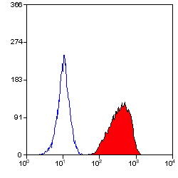
 Application Data
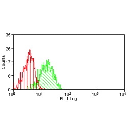
(Overlay histogram showing SY5Y cells stained with AAA27008 (red line). The cells were fixed with 70% Ethylalcohol (18h) and then permeabilized with 0.3% Triton X-100 for 2 min. The cells were then incubated in 1x PBS /10% normal goat serum to block non-specific protein-protein interactions followed by the antibody (10ug/1x10^6cells) for 1 h at 4 degree C. The secondary antibody used was FITC goat anti-mouse IgG (H+L) at 1/200 dilution for 1 h at 4 degree C. Isotype control antibody (green line) was mouse IgG2b (10ug/1x10^6cells) used under the same conditions. Acquisition of >10, 000 events was performed.)
Application Data
(Overlay histogram showing SY5Y cells stained with AAA27008 (red line). The cells were fixed with 70% Ethylalcohol (18h) and then permeabilized with 0.3% Triton X-100 for 2 min. The cells were then incubated in 1x PBS /10% normal goat serum to block non-specific protein-protein interactions followed by the antibody (10ug/1x10^6cells) for 1 h at 4 degree C. The secondary antibody used was FITC goat anti-mouse IgG (H+L) at 1/200 dilution for 1 h at 4 degree C. Isotype control antibody (green line) was mouse IgG2b (10ug/1x10^6cells) used under the same conditions. Acquisition of >10, 000 events was performed.)
GFAP, Monoclonal Antibody (Cat# AAA27008)
Full Name
GFAP Monoclonal Antibody
Gene Names
GFAP; ALXDRD
Reactivity
Human, Mouse, Rat
Applications
Western Blot, Immunohistochemistry, Immunofluorescence, Flow Cytometry
Purity
>95%, Protein G purified
Pricing


 Application Data
(Staining of human peripheral blood lymphocytes with MOUSE ANTI HUMAN CD4:ALEXA 405)
Application Data
(Staining of human peripheral blood lymphocytes with MOUSE ANTI HUMAN CD4:ALEXA 405)
CD4, Monoclonal Antibody (Cat# AAA26719)
Full Name
CD4 (CD4 Antigen, CD4 Molecule, CD4 Receptor, CD4mut, T Cell Surface Antigen T4/Leu3, T Cell Antigen T4, T Cell Surface Glycoprotein CD4) (AP)
Gene Names
CD4; CD4mut
Reactivity
Human
Applications
Immunohistochemistry
Purity
Purified by protein A affinity chromatography from tissue culture supernatant.
Pricing


 Application Data
(Staining of human peripheral blood monocytes with MOUSE ANTI HUMAN CD274:RPE)
Application Data
(Staining of human peripheral blood monocytes with MOUSE ANTI HUMAN CD274:RPE)
CD274, Monoclonal Antibody (Cat# AAA26729)
Full Name
CD274 (CD274 Antigen, B7 Homolog 1, B7H1, B7-H1, B7-H, MGC142294, MGC142296, OTTHUMP00000021029, Programmed Cell Death 1 Ligand 1, PDCD1 Ligand 1, PDCD1L1, PDCD1LG1, Programmed Death Ligand 1, PDL1, PD-L1, RGD1566211) (Biotin)
Gene Names
CD274; B7-H; B7H1; PDL1; PD-L1; PDCD1L1; PDCD1LG1
Reactivity
Human
Applications
Flow Cytometry
Purity
Purified by protein G affinity chromatography from tissue culture supernatant.
Pricing


 Application Data
(Staining of NALM6 cells with MOUSE ANTI HUMAN CD10:ALEXA 488)
Application Data
(Staining of NALM6 cells with MOUSE ANTI HUMAN CD10:ALEXA 488)
CD10, Monoclonal Antibody (Cat# AAA26726)
Full Name
CD10 (CALLA, CD 10, Common Acute Lymphocytic Leukemia Antigen, Enkephalinase, gp100, Membrane Metalloendopeptidase, MME, Neprilysin, Neutral Endopeptidase, Pmel17) (Biotin)
Gene Names
MME; NEP; SFE; CD10; CALLA
Reactivity
Human
Applications
Flow Cytometry, Immunohistochemistry, Immunoprecipitation, Western Blot
Purity
Purified by protein G affinity chromatography from tissue culture supernatant.
Pricing


 Application Data
(Published customer image Infiltration of GFP+ BM-cells in infarct and peri-infarct regions. (A-B) Dot plots of viable macrophages/granulocytes (CD11b+CD45high, top right quadrants) and microglia (CD11b+CD45dim, bottom right quadrants) in cortex from BM-chimeric unmanipulated mice and mice exposed to pMCAO. (C) Bar graph showing mean numbers of CD11b+CD45dim microglia and CD11b+CD45high macrophages/granulocytes in BM-chimeric mice 24 hours after pMCAO, subdivided based on expression of GFP (n = 5). Approximately 92% of of the CD45high population were GFP+. (D) Estimation and comparison of mean numbers of CD11b+CD45dim microglia in non-chimeric (n = 10) versus BM-chimeric mice (n = 5) 24 hours after of pMCAO shows significantly fewer CD11b+CD45dim microglial cells in irradiated mice. (E) Overview, showing distribution of infiltrating GFP+ BM-derived cells into infarct (IF) and peri-infarct (P-IF) regions 24 hours after pMCAO. (E-G) By 24 hours, GFP+ single cells (F) and vessel-associated aggregates of GFP+ cells (arrows in G) were observed in infarct and peri-infarct regions. Some of the vessel-associated cells were round, leukocyte-like cells (arrows) while others were elongated cells lining the vasculature (arrow heads in G and in insert). (H) Bar graph showing mean numbers of single GFP+ cells and vessel-associated aggregates of GFP+ cells in ipsi- and contralateral cortex 24 hours after surgery (n = 10). (I-P) Immunohistochemical staining of CD45.1 (I, K), CD45.2 (J, L), IgG2a (M, O) and CD45 (N, P) in ischemic tissue in BM-chimeric (I, J, M, N) and non-chimeric mice (K, L, O, P) 24 hours after pMCAO. N.D, none detected. Scale bars: 200 um (A), 10 um (B, C). 50 um (I-P) *P < 0.05, **P < 0.01, and ***P < 0.001.From: Clausen BH, Lambertsen KL, Babcock AA, Holm TH, Dagnaes-Hansen F, Finsen B. Interleukin-1beta and tumor necrosis factor-alpha are expressed by different subsets of microglia and macrophages after ischemic stroke in mice. J Neuroinflammation. 2008 Oct 23;5:46.)
Application Data
(Published customer image Infiltration of GFP+ BM-cells in infarct and peri-infarct regions. (A-B) Dot plots of viable macrophages/granulocytes (CD11b+CD45high, top right quadrants) and microglia (CD11b+CD45dim, bottom right quadrants) in cortex from BM-chimeric unmanipulated mice and mice exposed to pMCAO. (C) Bar graph showing mean numbers of CD11b+CD45dim microglia and CD11b+CD45high macrophages/granulocytes in BM-chimeric mice 24 hours after pMCAO, subdivided based on expression of GFP (n = 5). Approximately 92% of of the CD45high population were GFP+. (D) Estimation and comparison of mean numbers of CD11b+CD45dim microglia in non-chimeric (n = 10) versus BM-chimeric mice (n = 5) 24 hours after of pMCAO shows significantly fewer CD11b+CD45dim microglial cells in irradiated mice. (E) Overview, showing distribution of infiltrating GFP+ BM-derived cells into infarct (IF) and peri-infarct (P-IF) regions 24 hours after pMCAO. (E-G) By 24 hours, GFP+ single cells (F) and vessel-associated aggregates of GFP+ cells (arrows in G) were observed in infarct and peri-infarct regions. Some of the vessel-associated cells were round, leukocyte-like cells (arrows) while others were elongated cells lining the vasculature (arrow heads in G and in insert). (H) Bar graph showing mean numbers of single GFP+ cells and vessel-associated aggregates of GFP+ cells in ipsi- and contralateral cortex 24 hours after surgery (n = 10). (I-P) Immunohistochemical staining of CD45.1 (I, K), CD45.2 (J, L), IgG2a (M, O) and CD45 (N, P) in ischemic tissue in BM-chimeric (I, J, M, N) and non-chimeric mice (K, L, O, P) 24 hours after pMCAO. N.D, none detected. Scale bars: 200 um (A), 10 um (B, C). 50 um (I-P) *P < 0.05, **P < 0.01, and ***P < 0.001.From: Clausen BH, Lambertsen KL, Babcock AA, Holm TH, Dagnaes-Hansen F, Finsen B. Interleukin-1beta and tumor necrosis factor-alpha are expressed by different subsets of microglia and macrophages after ischemic stroke in mice. J Neuroinflammation. 2008 Oct 23;5:46.)
CD11b, Monoclonal Antibody (Cat# AAA12185)
Full Name
RAT ANTI MOUSE CD11b
Gene Names
Itgam; CR3; CR3A; MAC1; Cd11b; Ly-40; Mac-1; Mac-1a; CD11b/CD18; F730045J24Rik
Applications
Immunohistochemistry, Flow Cytometry, Immunofluorescence, Immunoprecipitation
Pricing


 Application Data
(Published customer image Infiltration of GFP+ BM-cells in infarct and peri-infarct regions. (A-B) Dot plots of viable macrophages/granulocytes (CD11b+CD45high, top right quadrants) and microglia (CD11b+CD45dim, bottom right quadrants) in cortex from BM-chimeric unmanipulated mice and mice exposed to pMCAO. (C) Bar graph showing mean numbers of CD11b+CD45dim microglia and CD11b+CD45high macrophages/granulocytes in BM-chimeric mice 24 hours after pMCAO, subdivided based on expression of GFP (n = 5). Approximately 92% of of the CD45high population were GFP+. (D) Estimation and comparison of mean numbers of CD11b+CD45dim microglia in non-chimeric (n = 10) versus BM-chimeric mice (n = 5) 24 hours after of pMCAO shows significantly fewer CD11b+CD45dim microglial cells in irradiated mice. (E) Overview, showing distribution of infiltrating GFP+ BM-derived cells into infarct (IF) and peri-infarct (P-IF) regions 24 hours after pMCAO. (E-G) By 24 hours, GFP+ single cells (F) and vessel-associated aggregates of GFP+ cells (arrows in G) were observed in infarct and peri-infarct regions. Some of the vessel-associated cells were round, leukocyte-like cells (arrows) while others were elongated cells lining the vasculature (arrow heads in G and in insert). (H) Bar graph showing mean numbers of single GFP+ cells and vessel-associated aggregates of GFP+ cells in ipsi- and contralateral cortex 24 hours after surgery (n = 10). (I-P) Immunohistochemical staining of CD45.1 (I, K), CD45.2 (J, L), IgG2a (M, O) and CD45 (N, P) in ischemic tissue in BM-chimeric (I, J, M, N) and non-chimeric mice (K, L, O, P) 24 hours after pMCAO. N.D, none detected. Scale bars: 200 um (A), 10 um (B, C). 50 um (I-P) *P < 0.05, **P < 0.01, and ***P < 0.001.From: Clausen BH, Lambertsen KL, Babcock AA, Holm TH, Dagnaes-Hansen F, Finsen B. Interleukin-1beta and tumor necrosis factor-alpha are expressed by different subsets of microglia and macrophages after ischemic stroke in mice. J Neuroinflammation. 2008 Oct 23;5:46.)
Application Data
(Published customer image Infiltration of GFP+ BM-cells in infarct and peri-infarct regions. (A-B) Dot plots of viable macrophages/granulocytes (CD11b+CD45high, top right quadrants) and microglia (CD11b+CD45dim, bottom right quadrants) in cortex from BM-chimeric unmanipulated mice and mice exposed to pMCAO. (C) Bar graph showing mean numbers of CD11b+CD45dim microglia and CD11b+CD45high macrophages/granulocytes in BM-chimeric mice 24 hours after pMCAO, subdivided based on expression of GFP (n = 5). Approximately 92% of of the CD45high population were GFP+. (D) Estimation and comparison of mean numbers of CD11b+CD45dim microglia in non-chimeric (n = 10) versus BM-chimeric mice (n = 5) 24 hours after of pMCAO shows significantly fewer CD11b+CD45dim microglial cells in irradiated mice. (E) Overview, showing distribution of infiltrating GFP+ BM-derived cells into infarct (IF) and peri-infarct (P-IF) regions 24 hours after pMCAO. (E-G) By 24 hours, GFP+ single cells (F) and vessel-associated aggregates of GFP+ cells (arrows in G) were observed in infarct and peri-infarct regions. Some of the vessel-associated cells were round, leukocyte-like cells (arrows) while others were elongated cells lining the vasculature (arrow heads in G and in insert). (H) Bar graph showing mean numbers of single GFP+ cells and vessel-associated aggregates of GFP+ cells in ipsi- and contralateral cortex 24 hours after surgery (n = 10). (I-P) Immunohistochemical staining of CD45.1 (I, K), CD45.2 (J, L), IgG2a (M, O) and CD45 (N, P) in ischemic tissue in BM-chimeric (I, J, M, N) and non-chimeric mice (K, L, O, P) 24 hours after pMCAO. N.D, none detected. Scale bars: 200 um (A), 10 um (B, C). 50 um (I-P) *P < 0.05, **P < 0.01, and ***P < 0.001.From: Clausen BH, Lambertsen KL, Babcock AA, Holm TH, Dagnaes-Hansen F, Finsen B. Interleukin-1beta and tumor necrosis factor-alpha are expressed by different subsets of microglia and macrophages after ischemic stroke in mice. J Neuroinflammation. 2008 Oct 23;5:46.)
CD11b, Monoclonal Antibody (Cat# AAA12183)
Full Name
RAT ANTI MOUSE CD11b:FITC
Gene Names
Itgam; CR3; CR3A; MAC1; Cd11b; Ly-40; Mac-1; Mac-1a; CD11b/CD18; F730045J24Rik
Applications
Flow Cytometry
Pricing


 Application Data
(Immunoperoxidase staining of a human tonsil cryosection with Mouse anti Human CD163 antibody, clone EDHu-1 followed by the Histar detection system . Low power)
Application Data
(Immunoperoxidase staining of a human tonsil cryosection with Mouse anti Human CD163 antibody, clone EDHu-1 followed by the Histar detection system . Low power)
CD163, Monoclonal Antibody (Cat# AAA12100)
Full Name
MOUSE ANTI HUMAN CD163
Gene Names
CD163; M130; MM130
Applications
Immunohistochemistry, Flow Cytometry, Immunofluorescence, Immunoassay, Immunohistochemistry, Western Blot
Pricing


 Application Data
(Staining of human peripheral blood lymphocytes with MOUSE ANTI HUMAN CD45RA:FITC (MCA88F))
Application Data
(Staining of human peripheral blood lymphocytes with MOUSE ANTI HUMAN CD45RA:FITC (MCA88F))
CD45RA, Monoclonal Antibody (Cat# AAA26734)
Full Name
CD45RA (CD45 Antigen, B220, GP180, Leukocyte Common Antigen, LCA, L-CA, LY5, LY-5, Protein Tyrosine Phosphatase Receptor Type C Polypeptide, PTPRC, T200, T200 Glycoprotein) (Biotin)
Reactivity
Human, Monkey
Applications
Flow Cytometry, Immunohistochemistry
Purity
Purified by protein G affinity chromatography from tissue culture supernatant.
Pricing


 Application Data
(Staining of human peripheral blood lymphocytes with MOUSE ANTI HUMAN CD45RA:FITC (MCA88F))
Application Data
(Staining of human peripheral blood lymphocytes with MOUSE ANTI HUMAN CD45RA:FITC (MCA88F))
CD45RA, Monoclonal Antibody (Cat# AAA26747)
Full Name
CD45RA (CD45 Antigen, B220, GP180, Leukocyte Common Antigen, LCA, L-CA, LY5, LY-5, Protein Tyrosine Phosphatase Receptor Type C Polypeptide, PTPRC, T200, T200 Glycoprotein) (FITC)
Reactivity
Human, Monkey
Applications
Flow Cytometry, Immunohistochemistry
Purity
Purified by protein G affinity chromatography from tissue culture supernatant.
Pricing


 Application Data
(Staining of human peripheral blood lymphocytes with MOUSE ANTI HUMAN CD4:ALEXA 405)
Application Data
(Staining of human peripheral blood lymphocytes with MOUSE ANTI HUMAN CD4:ALEXA 405)
CD4, Monoclonal Antibody (Cat# AAA26781)
Full Name
CD4 (CD4 Antigen, CD4 Molecule, CD4 Receptor, CD4mut, T Cell Surface Antigen T4/Leu3, T Cell Antigen T4, T Cell Surface Glycoprotein CD4) (MaxLight 405)
Gene Names
CD4; CD4mut
Reactivity
Human
Applications
Flow Cytometry, Immunohistochemistry
Purity
Purified by protein A affinity chromatography from tissue culture supernatant.
Pricing


 Application Data
(Staining of human peripheral blood monocytes with MOUSE ANTI HUMAN CD274:RPE)
Application Data
(Staining of human peripheral blood monocytes with MOUSE ANTI HUMAN CD274:RPE)
CD274, Monoclonal Antibody (Cat# AAA26792)
Full Name
CD274 (CD274 Antigen, B7 Homolog 1, B7H1, B7-H1, B7-H, MGC142294, MGC142296, OTTHUMP00000021029, Programmed Cell Death 1 Ligand 1, PDCD1 Ligand 1, PDCD1L1, PDCD1LG1, Programmed Death Ligand 1, PDL1, PD-L1, RGD1566211) (MaxLight 490)
Gene Names
CD274; B7-H; B7H1; PDL1; PD-L1; PDCD1L1; PDCD1LG1
Reactivity
Human
Applications
Flow Cytometry
Purity
Purified by protein G affinity chromatography from tissue culture supernatant.
Pricing


 Application Data
(Staining of mouse spleen with Hamster anti Mouse CD81: Alexa Fluor 488)
Application Data
(Staining of mouse spleen with Hamster anti Mouse CD81: Alexa Fluor 488)
CD81, Monoclonal Antibody (Cat# AAA12097)
Full Name
HAMSTER ANTI MOUSE CD81
Gene Names
Cd81; Tapa1; Tapa-1; Tspan28
Applications
Immunohistochemistry, Flow Cytometry, Immunoprecipitation, Western Blot
Pricing


 Application Data
(Staining of NALM6 cells with MOUSE ANTI HUMAN CD10:ALEXA 488)
Application Data
(Staining of NALM6 cells with MOUSE ANTI HUMAN CD10:ALEXA 488)
CD10, Monoclonal Antibody (Cat# AAA26740)
Full Name
CD10 (CALLA, CD 10, Common Acute Lymphocytic Leukemia Antigen, Enkephalinase, gp100, Membrane Metalloendopeptidase, MME, Neprilysin, Neutral Endopeptidase, Pmel17) (FITC)
Gene Names
MME; NEP; SFE; CD10; CALLA
Reactivity
Human
Applications
Flow Cytometry, Immunohistochemistry, Immunoprecipitation, Western Blot
Purity
Purified by protein G affinity chromatography from tissue culture supernatant.
Pricing


 Application Data
(Staining of CD209 transfected K562 cells with MOUSE ANTI HUMAN CD209: FITC)
Application Data
(Staining of CD209 transfected K562 cells with MOUSE ANTI HUMAN CD209: FITC)
DC-SIGN, Monoclonal Antibody (Cat# AAA26841)
Full Name
DC-SIGN (Dendritic Cell-specific ICAM3 grabbing Non-integrin, C Type Lectin Domain Family 4 Member L, CLEC4L, CD209, CD209 Antigen, CDSIGN, Dendritic Cell-specific ICAM-3-grabbing Non-integrin 1, DC SIGN1, DC-SIGN1, HIV GP120 Binding Protein, MGC129965) (
Reactivity
Human
Applications
FC/FACS, IF
Purity
Purified by protein G affinity chromatography from tissue culture supernatant.
Pricing


 Application Data
(Staining of human peripheral blood lymphocytes with MOUSE ANTI HUMAN CD45RA:FITC (MCA88F))
Application Data
(Staining of human peripheral blood lymphocytes with MOUSE ANTI HUMAN CD45RA:FITC (MCA88F))
CD45RA, Monoclonal Antibody (Cat# AAA26720)
Full Name
CD45RA (CD45 Antigen, B220, GP180, Leukocyte Common Antigen, LCA, L-CA, LY5, LY-5, Protein Tyrosine Phosphatase Receptor Type C Polypeptide, PTPRC, T200, T200 Glycoprotein) (AP)
Reactivity
Human, Monkey
Applications
Immunohistochemistry
Purity
Purified by protein G affinity chromatography from tissue culture supernatant.
Pricing


 Application Data
(Staining of human peripheral blood lymphocytes with MOUSE ANTI HUMAN CD45RA:FITC (MCA88F))
Application Data
(Staining of human peripheral blood lymphocytes with MOUSE ANTI HUMAN CD45RA:FITC (MCA88F))
CD45RA, Monoclonal Antibody (Cat# AAA26759)
Full Name
CD45RA (CD45 Antigen, B220, GP180, Leukocyte Common Antigen, LCA, L-CA, LY5, LY-5, Protein Tyrosine Phosphatase Receptor Type C Polypeptide, PTPRC, T200, T200 Glycoprotein) (HRP)
Reactivity
Human, Monkey
Applications
Flow Cytometry, Immunohistochemistry
Purity
Purified by protein G affinity chromatography from tissue culture supernatant.
Pricing


 Application Data
(Staining of CD209 transfected K562 cells with MOUSE ANTI HUMAN CD209: FITC)
Application Data
(Staining of CD209 transfected K562 cells with MOUSE ANTI HUMAN CD209: FITC)
DC-SIGN, Monoclonal Antibody (Cat# AAA26813)
Full Name
DC-SIGN (Dendritic Cell-specific ICAM3 grabbing Non-integrin, C Type Lectin Domain Family 4 Member L, CLEC4L, CD209, CD209 Antigen, CDSIGN, Dendritic Cell-specific ICAM-3-grabbing Non-integrin 1, DC SIGN1, DC-SIGN1, HIV GP120 Binding Protein, MGC129965) (
Reactivity
Human
Applications
FC/FACS, IF
Purity
Purified by protein G affinity chromatography from tissue culture supernatant.
Pricing


 Application Data
(Staining of NALM6 cells with MOUSE ANTI HUMAN CD10:ALEXA 488)
Application Data
(Staining of NALM6 cells with MOUSE ANTI HUMAN CD10:ALEXA 488)
CD10, Monoclonal Antibody (Cat# AAA26765)
Full Name
CD10 (CALLA, CD 10, Common Acute Lymphocytic Leukemia Antigen, Enkephalinase, gp100, Membrane Metalloendopeptidase, MME, Neprilysin, Neutral Endopeptidase, Pmel17) (PE)
Gene Names
MME; NEP; SFE; CD10; CALLA
Reactivity
Human
Applications
Flow Cytometry, Immunohistochemistry, Immunoprecipitation, Western Blot
Purity
Purified by protein G affinity chromatography from tissue culture supernatant.
Pricing


 Application Data
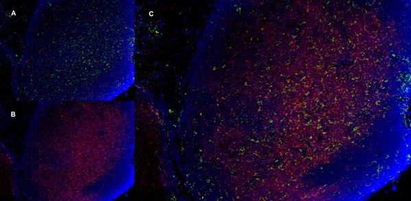
(C:FGFR2/isolectinB4 (C) and FGFR1/isolectinB4 (D) staining of apparent mesenchymal cells and the subpopulation of endothelial cells. Virtually all other dispersed apparent mesenchymal cells express FGFR1 and FGFR2 (merged image in E). F: FGFR2 (F) and FGFR1 (G) staining in clustered cells of epithelial origin (inferred by morphology here) demonstrating that epithelial cells express both FGFR1 and FGFR2 (merged image with DAPI staining in H).)
Application Data
(C:FGFR2/isolectinB4 (C) and FGFR1/isolectinB4 (D) staining of apparent mesenchymal cells and the subpopulation of endothelial cells. Virtually all other dispersed apparent mesenchymal cells express FGFR1 and FGFR2 (merged image in E). F: FGFR2 (F) and FGFR1 (G) staining in clustered cells of epithelial origin (inferred by morphology here) demonstrating that epithelial cells express both FGFR1 and FGFR2 (merged image with DAPI staining in H).)
FGFR2, Polyclonal Antibody (Cat# AAA26853)
Full Name
FGFR2, NT (FGFR2, BEK, KGFR, KSAM, Fibroblast growth factor receptor 2, K-sam, Keratinocyte growth factor receptor, CD332) (Biotin)
Gene Names
FGFR2; BEK; JWS; BBDS; CEK3; CFD1; ECT1; KGFR; TK14; TK25; BFR-1; CD332; K-SAM
Reactivity
Human, Monkey, Mouse, Rat
Applications
FC/FACS, EIA, IF, IHC, WB
Purity
Purified by Protein G Affinity Chromatography.
Pricing


 Application Data
CD274, Monoclonal Antibody (Cat# AAA26754)
Full Name
CD274 (CD274 Antigen, B7 Homolog 1, B7H1, B7-H1, B7-H, MGC142294, MGC142296, OTTHUMP00000021029, Programmed Cell Death 1 Ligand 1, PDCD1 Ligand 1, PDCD1L1, PDCD1LG1, Programmed Death Ligand 1, PDL1, PD-L1, RGD1566211) (HRP)
Gene Names
CD274; B7-H; B7H1; PDL1; PD-L1; PDCD1L1; PDCD1LG1
Reactivity
Human
Applications
ELISA
Purity
Purified by protein G affinity chromatography from tissue culture supernatant.
Pricing


 Application Data
(Staining of NALM6 cells with MOUSE ANTI HUMAN CD10:ALEXA 488)
Application Data
(Staining of NALM6 cells with MOUSE ANTI HUMAN CD10:ALEXA 488)
CD10, Monoclonal Antibody (Cat# AAA26789)
Full Name
CD10 (CALLA, CD 10, Common Acute Lymphocytic Leukemia Antigen, Enkephalinase, gp100, Membrane Metalloendopeptidase, MME, Neprilysin, Neutral Endopeptidase, Pmel17) (MaxLight 490)
Gene Names
MME; NEP; SFE; CD10; CALLA
Reactivity
Human
Applications
Flow Cytometry, Immunohistochemistry, Immunoprecipitation, Western Blot
Purity
Purified by protein G affinity chromatography from tissue culture supernatant.
Pricing


 Application Data
(Staining of mouse spleen with Hamster anti Mouse CD81: Alexa Fluor 488 (AAA11941A488))
Application Data
(Staining of mouse spleen with Hamster anti Mouse CD81: Alexa Fluor 488 (AAA11941A488))
CD81, Monoclonal Antibody (Cat# AAA11941)
Full Name
HAMSTER ANTI MOUSE CD81
Gene Names
Cd81; Tapa1; Tapa-1; Tspan28
Reactivity
Rat
Applications
Immunohistochemistry, Flow Cytometry, Immunoprecipitation, Western Blot
Pricing


 Application Data
(C:FGFR2/isolectinB4 (C) and FGFR1/isolectinB4 (D) staining of apparent mesenchymal cells and the subpopulation of endothelial cells. Virtually all other dispersed apparent mesenchymal cells express FGFR1 and FGFR2 (merged image in E). F: FGFR2 (F) and FGFR1 (G) staining in clustered cells of epithelial origin (inferred by morphology here) demonstrating that epithelial cells express both FGFR1 and FGFR2 (merged image with DAPI staining in H).)
Application Data
(C:FGFR2/isolectinB4 (C) and FGFR1/isolectinB4 (D) staining of apparent mesenchymal cells and the subpopulation of endothelial cells. Virtually all other dispersed apparent mesenchymal cells express FGFR1 and FGFR2 (merged image in E). F: FGFR2 (F) and FGFR1 (G) staining in clustered cells of epithelial origin (inferred by morphology here) demonstrating that epithelial cells express both FGFR1 and FGFR2 (merged image with DAPI staining in H).)
FGFR2, Polyclonal Antibody (Cat# AAA26855)
Full Name
FGFR2, NT (FGFR2, BEK, KGFR, KSAM, Fibroblast growth factor receptor 2, K-sam, Keratinocyte growth factor receptor, CD332) (Azide free) (HRP)
Gene Names
FGFR2; BEK; JWS; BBDS; CEK3; CFD1; ECT1; KGFR; TK14; TK25; BFR-1; CD332; K-SAM
Reactivity
Human, Monkey, Mouse, Rat
Applications
IHC, EIA, WB
Purity
Purified by Protein G Affinity Chromatography.
Pricing


 Application Data
(Published customer image Infiltration of GFP+ BM-cells in infarct and peri-infarct regions. (A-B) Dot plots of viable macrophages/granulocytes (CD11b+CD45high, top right quadrants) and microglia (CD11b+CD45dim, bottom right quadrants) in cortex from BM-chimeric unmanipulated mice and mice exposed to pMCAO. (C) Bar graph showing mean numbers of CD11b+CD45dim microglia and CD11b+CD45high macrophages/granulocytes in BM-chimeric mice 24 hours after pMCAO, subdivided based on expression of GFP (n = 5). Approximately 92% of of the CD45high population were GFP+. (D) Estimation and comparison of mean numbers of CD11b+CD45dim microglia in non-chimeric (n = 10) versus BM-chimeric mice (n = 5) 24 hours after of pMCAO shows significantly fewer CD11b+CD45dim microglial cells in irradiated mice. (E) Overview, showing distribution of infiltrating GFP+ BM-derived cells into infarct (IF) and peri-infarct (P-IF) regions 24 hours after pMCAO. (E-G) By 24 hours, GFP+ single cells (F) and vessel-associated aggregates of GFP+ cells (arrows in G) were observed in infarct and peri-infarct regions. Some of the vessel-associated cells were round, leukocyte-like cells (arrows) while others were elongated cells lining the vasculature (arrow heads in G and in insert). (H) Bar graph showing mean numbers of single GFP+ cells and vessel-associated aggregates of GFP+ cells in ipsi- and contralateral cortex 24 hours after surgery (n = 10). (I-P) Immunohistochemical staining of CD45.1 (I, K), CD45.2 (J, L), IgG2a (M, O) and CD45 (N, P) in ischemic tissue in BM-chimeric (I, J, M, N) and non-chimeric mice (K, L, O, P) 24 hours after pMCAO. N.D, none detected. Scale bars: 200 um (A), 10 um (B, C). 50 um (I-P) *P < 0.05, **P < 0.01, and ***P < 0.001.From: Clausen BH, Lambertsen KL, Babcock AA, Holm TH, Dagnaes-Hansen F, Finsen B. Interleukin-1beta and tumor necrosis factor-alpha are expressed by different subsets of microglia and macrophages after ischemic stroke in mice. J Neuroinflammation. 2008 Oct 23;5:46.)
Application Data
(Published customer image Infiltration of GFP+ BM-cells in infarct and peri-infarct regions. (A-B) Dot plots of viable macrophages/granulocytes (CD11b+CD45high, top right quadrants) and microglia (CD11b+CD45dim, bottom right quadrants) in cortex from BM-chimeric unmanipulated mice and mice exposed to pMCAO. (C) Bar graph showing mean numbers of CD11b+CD45dim microglia and CD11b+CD45high macrophages/granulocytes in BM-chimeric mice 24 hours after pMCAO, subdivided based on expression of GFP (n = 5). Approximately 92% of of the CD45high population were GFP+. (D) Estimation and comparison of mean numbers of CD11b+CD45dim microglia in non-chimeric (n = 10) versus BM-chimeric mice (n = 5) 24 hours after of pMCAO shows significantly fewer CD11b+CD45dim microglial cells in irradiated mice. (E) Overview, showing distribution of infiltrating GFP+ BM-derived cells into infarct (IF) and peri-infarct (P-IF) regions 24 hours after pMCAO. (E-G) By 24 hours, GFP+ single cells (F) and vessel-associated aggregates of GFP+ cells (arrows in G) were observed in infarct and peri-infarct regions. Some of the vessel-associated cells were round, leukocyte-like cells (arrows) while others were elongated cells lining the vasculature (arrow heads in G and in insert). (H) Bar graph showing mean numbers of single GFP+ cells and vessel-associated aggregates of GFP+ cells in ipsi- and contralateral cortex 24 hours after surgery (n = 10). (I-P) Immunohistochemical staining of CD45.1 (I, K), CD45.2 (J, L), IgG2a (M, O) and CD45 (N, P) in ischemic tissue in BM-chimeric (I, J, M, N) and non-chimeric mice (K, L, O, P) 24 hours after pMCAO. N.D, none detected. Scale bars: 200 um (A), 10 um (B, C). 50 um (I-P) *P < 0.05, **P < 0.01, and ***P < 0.001.From: Clausen BH, Lambertsen KL, Babcock AA, Holm TH, Dagnaes-Hansen F, Finsen B. Interleukin-1beta and tumor necrosis factor-alpha are expressed by different subsets of microglia and macrophages after ischemic stroke in mice. J Neuroinflammation. 2008 Oct 23;5:46.)
CD11b, Monoclonal Antibody (Cat# AAA12186)
Full Name
RAT ANTI MOUSE CD11b:RPE
Gene Names
Itgam; CR3; CR3A; MAC1; Cd11b; Ly-40; Mac-1; Mac-1a; CD11b/CD18; F730045J24Rik
Applications
Flow Cytometry
Pricing


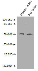
 WB (Western Blot)
(CD58 monoclonal antibody, Western Blot analysis of CD58 expression in Jurkat.)
WB (Western Blot)
(CD58 monoclonal antibody, Western Blot analysis of CD58 expression in Jurkat.)
CD58, Monoclonal Antibody (Cat# AAA25637)
Full Name
CD58 (Lymphocyte Function-associated Antigen 3, Ag3, Surface Glycoprotein LFA-3, LFA3) (PE)
Gene Names
CD58; ag3; LFA3; LFA-3
Reactivity
Human
Applications
Immunofluorescence, Immunohistochemistry, Western Blot
Purity
Purified by Protein A Affinity Chromatography.
Pricing


 Application Data
(Staining of human peripheral blood lymphocytes with MOUSE ANTI HUMAN CD45RA:FITC (MCA88F))
Application Data
(Staining of human peripheral blood lymphocytes with MOUSE ANTI HUMAN CD45RA:FITC (MCA88F))
CD45RA, Monoclonal Antibody (Cat# AAA26797)
Full Name
CD45RA (CD45 Antigen, B220, GP180, Leukocyte Common Antigen, LCA, L-CA, LY5, LY-5, Protein Tyrosine Phosphatase Receptor Type C Polypeptide, PTPRC, T200, T200 Glycoprotein) (MaxLight 490)
Reactivity
Human, Monkey
Applications
Flow Cytometry, Immunohistochemistry
Purity
Purified by protein G affinity chromatography from tissue culture supernatant.
Pricing


 Application Data
(Staining of CD209 transfected K562 cells with MOUSE ANTI HUMAN CD209: FITC)
Application Data
(Staining of CD209 transfected K562 cells with MOUSE ANTI HUMAN CD209: FITC)
DC-SIGN, Monoclonal Antibody (Cat# AAA26827)
Full Name
DC-SIGN (Dendritic Cell-specific ICAM3 grabbing Non-integrin, C Type Lectin Domain Family 4 Member L, CLEC4L, CD209, CD209 Antigen, CDSIGN, Dendritic Cell-specific ICAM-3-grabbing Non-integrin 1, DC SIGN1, DC-SIGN1, HIV GP120 Binding Protein, MGC129965) (
Reactivity
Human
Applications
FC/FACS, IF
Purity
Purified by protein G affinity chromatography from tissue culture supernatant.
Pricing


 FCM (Flow Cytometry)
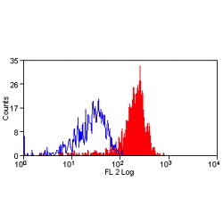
(Dual staining of pig peripheral blood lymphocytes with Mouse anti Pig CD335 detected with Goat anti Mouse IgG (H/L):FITC (STAR117F), and Mouse anti Pig wCD8a:RPE)
FCM (Flow Cytometry)
(Dual staining of pig peripheral blood lymphocytes with Mouse anti Pig CD335 detected with Goat anti Mouse IgG (H/L):FITC (STAR117F), and Mouse anti Pig wCD8a:RPE)
CD335, Monoclonal Antibody (Cat# AAA12253)
Full Name
MOUSE ANTI PIG CD335
Reactivity
Pig
Applications
Flow Cytometry, Immunofluorescence
Pricing


 Application Data
(Published customer image Infiltration of GFP+ BM-cells in infarct and peri-infarct regions. (A-B) Dot plots of viable macrophages/granulocytes (CD11b+CD45high, top right quadrants) and microglia (CD11b+CD45dim, bottom right quadrants) in cortex from BM-chimeric unmanipulated mice and mice exposed to pMCAO. (C) Bar graph showing mean numbers of CD11b+CD45dim microglia and CD11b+CD45high macrophages/granulocytes in BM-chimeric mice 24 hours after pMCAO, subdivided based on expression of GFP (n = 5). Approximately 92% of of the CD45high population were GFP+. (D) Estimation and comparison of mean numbers of CD11b+CD45dim microglia in non-chimeric (n = 10) versus BM-chimeric mice (n = 5) 24 hours after of pMCAO shows significantly fewer CD11b+CD45dim microglial cells in irradiated mice. (E) Overview, showing distribution of infiltrating GFP+ BM-derived cells into infarct (IF) and peri-infarct (P-IF) regions 24 hours after pMCAO. (E-G) By 24 hours, GFP+ single cells (F) and vessel-associated aggregates of GFP+ cells (arrows in G) were observed in infarct and peri-infarct regions. Some of the vessel-associated cells were round, leukocyte-like cells (arrows) while others were elongated cells lining the vasculature (arrow heads in G and in insert). (H) Bar graph showing mean numbers of single GFP+ cells and vessel-associated aggregates of GFP+ cells in ipsi- and contralateral cortex 24 hours after surgery (n = 10). (I-P) Immunohistochemical staining of CD45.1 (I, K), CD45.2 (J, L), IgG2a (M, O) and CD45 (N, P) in ischemic tissue in BM-chimeric (I, J, M, N) and non-chimeric mice (K, L, O, P) 24 hours after pMCAO. N.D, none detected. Scale bars: 200 um (A), 10 um (B, C). 50 um (I-P) *P < 0.05, **P < 0.01, and ***P < 0.001.From: Clausen BH, Lambertsen KL, Babcock AA, Holm TH, Dagnaes-Hansen F, Finsen B. Interleukin-1beta and tumor necrosis factor-alpha are expressed by different subsets of microglia and macrophages after ischemic stroke in mice. J Neuroinflammation. 2008 Oct 23;5:46.)
Application Data
(Published customer image Infiltration of GFP+ BM-cells in infarct and peri-infarct regions. (A-B) Dot plots of viable macrophages/granulocytes (CD11b+CD45high, top right quadrants) and microglia (CD11b+CD45dim, bottom right quadrants) in cortex from BM-chimeric unmanipulated mice and mice exposed to pMCAO. (C) Bar graph showing mean numbers of CD11b+CD45dim microglia and CD11b+CD45high macrophages/granulocytes in BM-chimeric mice 24 hours after pMCAO, subdivided based on expression of GFP (n = 5). Approximately 92% of of the CD45high population were GFP+. (D) Estimation and comparison of mean numbers of CD11b+CD45dim microglia in non-chimeric (n = 10) versus BM-chimeric mice (n = 5) 24 hours after of pMCAO shows significantly fewer CD11b+CD45dim microglial cells in irradiated mice. (E) Overview, showing distribution of infiltrating GFP+ BM-derived cells into infarct (IF) and peri-infarct (P-IF) regions 24 hours after pMCAO. (E-G) By 24 hours, GFP+ single cells (F) and vessel-associated aggregates of GFP+ cells (arrows in G) were observed in infarct and peri-infarct regions. Some of the vessel-associated cells were round, leukocyte-like cells (arrows) while others were elongated cells lining the vasculature (arrow heads in G and in insert). (H) Bar graph showing mean numbers of single GFP+ cells and vessel-associated aggregates of GFP+ cells in ipsi- and contralateral cortex 24 hours after surgery (n = 10). (I-P) Immunohistochemical staining of CD45.1 (I, K), CD45.2 (J, L), IgG2a (M, O) and CD45 (N, P) in ischemic tissue in BM-chimeric (I, J, M, N) and non-chimeric mice (K, L, O, P) 24 hours after pMCAO. N.D, none detected. Scale bars: 200 um (A), 10 um (B, C). 50 um (I-P) *P < 0.05, **P < 0.01, and ***P < 0.001.From: Clausen BH, Lambertsen KL, Babcock AA, Holm TH, Dagnaes-Hansen F, Finsen B. Interleukin-1beta and tumor necrosis factor-alpha are expressed by different subsets of microglia and macrophages after ischemic stroke in mice. J Neuroinflammation. 2008 Oct 23;5:46.)
CD11b, Monoclonal Antibody (Cat# AAA12231)
Full Name
RAT ANTI MOUSE CD11b:Low Endotoxin
Gene Names
Itgam; CR3; CR3A; MAC1; Cd11b; Ly-40; Mac-1; Mac-1a; CD11b/CD18; F730045J24Rik
Applications
Immunohistochemistry, Flow Cytometry, Functional Assay, Immunofluorescence, Immunoprecipitation
Pricing