Filters
Clonality
Type
Reactivity
Gene Name
Isotype
Host
Application
Clone
232 results for " Bone" - showing 1-50
 Standard Curve (Sample)
Neutrophil Elastase (NE), ELISA Kit (Cat# AAA13136)
Full Name
Porcine Neutrophil Elastase (NE) ELISA Kit
Gene Names
ELANE; GE; NE; HLE; HNE; ELA2; SCN1; PMN-E
Reactivity
Porcine
Pricing


 Application Data
(At 25 degree C. Samples were then incubated with primary Ab(At 37 degree C. An AlexaFluor594 conjugated goat anti-rabbit IgG(H+L) Ab(Red) and an AlexaFluor488 conjugated goat anti-mouse IgG(H+L) Ab(Green) were used as the secondary antibody.The nuclear counter stain is DAPI (blue).)
Application Data
(At 25 degree C. Samples were then incubated with primary Ab(At 37 degree C. An AlexaFluor594 conjugated goat anti-rabbit IgG(H+L) Ab(Red) and an AlexaFluor488 conjugated goat anti-mouse IgG(H+L) Ab(Green) were used as the secondary antibody.The nuclear counter stain is DAPI (blue).)
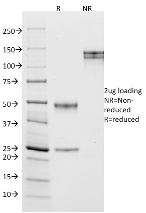
Bub1, Polyclonal Antibody (Cat# AAA31312)
Full Name
Phospho-Bub1 (Ser459) Antibody
Gene Names
BUB1; BUB1A; BUB1L; hBUB1
Reactivity
Human, Rat
Applications
Immunohistochemistry, Immunofluorescence, Immunocytochemistry, Peptide ELISA
Purity
The antibody is from purified rabbit serum by affinity purification via sequential chromatography on phospho-peptide and non-phospho-peptide affinity columns.
Pricing


 IF (Immunofluorescence)
(AAA30931 staining NIH/3T3 cells by IF/ICC. The sample were fixed with PFA and permeabilized in 0.1% Triton X-100, then blocked in 10% serum for 45 minutes at 25 degree C. The primary antibody was diluted at 1/200 and incubated with the sample for 1 hour at 37 degree C. An Alexa Fluor 594 conjugated goat anti-rabbit IgG (H+L) antibody, diluted at 1/600, was used as secondary antibody.)
IF (Immunofluorescence)
(AAA30931 staining NIH/3T3 cells by IF/ICC. The sample were fixed with PFA and permeabilized in 0.1% Triton X-100, then blocked in 10% serum for 45 minutes at 25 degree C. The primary antibody was diluted at 1/200 and incubated with the sample for 1 hour at 37 degree C. An Alexa Fluor 594 conjugated goat anti-rabbit IgG (H+L) antibody, diluted at 1/600, was used as secondary antibody.)
Acetyl-p53, Polyclonal Antibody (Cat# AAA30931)
Full Name
Acetyl-p53 (Lys317) Antibody
Gene Names
TP53; P53; BCC7; LFS1; TRP53
Reactivity
Human, Mouse, Rat
Applications
Western Blot, Immunohistochemistry, Immunofluorescence, Immunocytochemistry
Purity
Affinity Purification
Pricing


 FCM (Flow Cytometry)
(Flow cytometric analysis of HepG2 cells with Neutrophil Elastase antibody at 1/50 dilution (red) compared with an unlabelled control (cells without incubation with primary antibody; black). Alexa Fluor 488-conjugated goat anti rabbit IgG was used as the secondary antibody)
FCM (Flow Cytometry)
(Flow cytometric analysis of HepG2 cells with Neutrophil Elastase antibody at 1/50 dilution (red) compared with an unlabelled control (cells without incubation with primary antibody; black). Alexa Fluor 488-conjugated goat anti rabbit IgG was used as the secondary antibody)
Neutrophil Elastase, Monoclonal Antibody (Cat# AAA30292)
Full Name
Neutrophil Elastase Antibody
Gene Names
ELANE; GE; NE; HLE; HNE; ELA2; SCN1; PMN-E
Reactivity
Human
Applications
Western Blot, Immunocytochemistry, Immunofluorescence, Immunohistochemistry, Flow Cytometry
Purity
ProA affinity purified
Pricing


 IHC (Immunohistochemistry)
(At 1/100 staining Human esophageal cancer by IHC-P. The sample was formaldehyde fixed and a heat mediated antigen retrieval step in citrate buffer was performed. The sample was then blocked and incubated with the primary antibody at 4 degree C overnight. An HRP conjugated anti-Rabbit antibody was used as the secondary antibody.)
IHC (Immunohistochemistry)
(At 1/100 staining Human esophageal cancer by IHC-P. The sample was formaldehyde fixed and a heat mediated antigen retrieval step in citrate buffer was performed. The sample was then blocked and incubated with the primary antibody at 4 degree C overnight. An HRP conjugated anti-Rabbit antibody was used as the secondary antibody.)
p53, Polyclonal Antibody (Cat# AAA31354)
Full Name
Acetyl-p53 (Lys373) Antibody
Gene Names
TP53; P53; BCC7; LFS1; TRP53
Reactivity
Human, Mouse, Rat
Predicted Reactivity: Pig (91%), Sheep (80%), Rabbit (91%), Dog (91%)
Predicted Reactivity: Pig (91%), Sheep (80%), Rabbit (91%), Dog (91%)
Applications
Western Blot, Immunohistochemistry, Peptide ELISA
Purity
The antiserum was purified by peptide affinity chromatography using SulfoLink Coupling Resin
Pricing

 SDS-PAGE
Major Basic Protein (MBP), Recombinant Protein (Cat# AAA20242)
Full Name
Recombinant Major Basic Protein (MBP)
Gene Names
PRG2; MBP; BMPG; MBP1
Reactivity
Homo sapiens (Human)
Applications
Western Blot
Purity
> 90%
Pricing


 WB (Western Blot)
(BST2 monoclonal antibody (M15), clone 2E6. Western Blot analysis of BST2 expression in human placenta.)
WB (Western Blot)
(BST2 monoclonal antibody (M15), clone 2E6. Western Blot analysis of BST2 expression in human placenta.)
BST2, Monoclonal Antibody (Cat# AAA26271)
Full Name
BST2 (Bone Marrow Stromal Cell antigen 2, CD317) (Biotin)
Gene Names
BST2; CD317; TETHERIN
Applications
Immunofluorescence, Immunoprecipitation, Western Blot
Purity
Purified
Pricing
 IF (Immunofluorescence)
(Immunofluorescence Analysis of HeLa cells labeling IGF-1. IGF-1 Mouse Monoclonal Antibody (IGF/1020) conjugated to CF647R (green). DAPI (blue) nuclear counterstain.)
IF (Immunofluorescence)
(Immunofluorescence Analysis of HeLa cells labeling IGF-1. IGF-1 Mouse Monoclonal Antibody (IGF/1020) conjugated to CF647R (green). DAPI (blue) nuclear counterstain.)
Insulin-like Growth Factor-1 (IGF-1), Monoclonal Antibody (Cat# AAA13834)
Full Name
Insulin-like Growth Factor-1 (IGF-1) Mouse Monoclonal Antibody
Gene Names
IGF1; MGF; IGFI; IGF-I
Reactivity
Human, Mouse, Rat, Rabbit
Applications
Flow Cytometry, Immunofluorescence
Pricing


 Application Data
(Published Customer Image:Rat anti Mouse Gr-1 antibody, clone RB6-8C5 used for the identification of neutrophils by immunofluorescence.Image caption:S. typhimurium-Infected Macrophages Containing Phagocytosed Neutrophils and T Cells Confocal fluorescence microscopy of 50-mum-thick liver sections from 1-wk-infected Slc11a1 wild-type mice. (A-C) S.Typhimurium (O-antigen, arrows) are red, macrophages (F4-80 and MOMA-2) are blue, DNA (DAPI) is gray, phalloidin is green, and neutrophils (Gr-1/Ly-6G/RB6-8C5) are pink (arrowheads). (A) Collapsed image from a 40-mum Z-stack. Scale bar is 20 mum. (B and C) Sections from (A) that are 4 mum apart. The video from which (A-C) were derived (Video S2) is available online. (D-G) T cells within multinucleate macrophages.Macrophages (F4-80 and MOMA-2) are blue (D, G, and H), T cells (CD3zeta) are red (D, G, arrowheads), DAPI is gray (E, G), actin-bound phalloidin is green (F, G). (G) Is a composite of (D, E, and F). Scale bars are 16 mum. (H) An image from a different mouse stained and labeled as described for (D-G). Scale bar is 8 mum. A video showing a T cell inside of a macrophage is available online (Video S3).From: Nix RN, Altschuler SE, Henson PM, Detweiler CS (2007) Hemophagocytic Macrophages Harbor Salmonella enterica during Persistent Infection.PLoS Pathog 3(12): e193.)
Application Data
(Published Customer Image:Rat anti Mouse Gr-1 antibody, clone RB6-8C5 used for the identification of neutrophils by immunofluorescence.Image caption:S. typhimurium-Infected Macrophages Containing Phagocytosed Neutrophils and T Cells Confocal fluorescence microscopy of 50-mum-thick liver sections from 1-wk-infected Slc11a1 wild-type mice. (A-C) S.Typhimurium (O-antigen, arrows) are red, macrophages (F4-80 and MOMA-2) are blue, DNA (DAPI) is gray, phalloidin is green, and neutrophils (Gr-1/Ly-6G/RB6-8C5) are pink (arrowheads). (A) Collapsed image from a 40-mum Z-stack. Scale bar is 20 mum. (B and C) Sections from (A) that are 4 mum apart. The video from which (A-C) were derived (Video S2) is available online. (D-G) T cells within multinucleate macrophages.Macrophages (F4-80 and MOMA-2) are blue (D, G, and H), T cells (CD3zeta) are red (D, G, arrowheads), DAPI is gray (E, G), actin-bound phalloidin is green (F, G). (G) Is a composite of (D, E, and F). Scale bars are 16 mum. (H) An image from a different mouse stained and labeled as described for (D-G). Scale bar is 8 mum. A video showing a T cell inside of a macrophage is available online (Video S3).From: Nix RN, Altschuler SE, Henson PM, Detweiler CS (2007) Hemophagocytic Macrophages Harbor Salmonella enterica during Persistent Infection.PLoS Pathog 3(12): e193.)
Gr-1, Monoclonal Antibody (Cat# AAA12257)
Full Name
Rat Anti Mouse Gr-1: FITC
Reactivity
Mouse
Applications
Flow Cytometry
Purity
Purified IgG prepared by affinity chromatography on Protein G from tissue culture supernatant
Pricing


 Application Data
(Staining of mouse peripheral blood granulocytes with Rat anti Mouse CD13)
Application Data
(Staining of mouse peripheral blood granulocytes with Rat anti Mouse CD13)
CD13, Monoclonal Antibody (Cat# AAA12144)
Full Name
RAT ANTI MOUSE CD13
Gene Names
Anpep; Apn; AP-M; AP-N; Cd13; P150
Applications
Immunohistochemistry, Flow Cytometry
Purity
Affinity chromatography on Protein G from tissue culture supernatant
Pricing
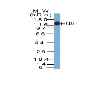


 WB (Western Blot)
(Western Blot Analysis of human Jurkat cell lysate using CD31 Mouse Monoclonal Antibody (C31.7).)
WB (Western Blot)
(Western Blot Analysis of human Jurkat cell lysate using CD31 Mouse Monoclonal Antibody (C31.7).)
CD31 / PECAM-1, Monoclonal Antibody (Cat# AAA13801)
Full Name
CD31 / PECAM-1 (Endothelial Cell Marker) Mouse Monoclonal Antibody
Gene Names
PECAM1; CD31; PECA1; GPIIA'; PECAM-1; endoCAM; CD31/EndoCAM
Reactivity
Human, Cynomolgus Monkey, Rabbit.
Weakly reacts with Rat
Weakly reacts with Rat
Applications
Flow Cytometry, Immunofluorescence, Western Blot, Immunohistochemistry
Pricing


 FCM (Flow Cytometry)
(Figure 6. Flow Cytometry analysis of HEPG2 cells using anti-Alkaline phosphatase/ALPL antibody (AAA19231).Overlay histogram showing HEPG2 cells stained with AAA19231 (Blue line). The cells were blocked with 10% normal goat serum. And then incubated with rabbit anti-Alkaline phosphatase/ALPL Antibody (AAA19231, 1μg/1x106 cells) for 30 min at 20 degree C. DyLight®488 conjugated goat anti-rabbit IgG (5-10μg/1x106 cells) was used as secondary antibody for 30 minutes at 20 degree C. Isotype control antibody (Green line) was rabbit IgG (1μg/1x106) used under the same conditions. Unlabelled sample (Red line) was also used as a control.)
FCM (Flow Cytometry)
(Figure 6. Flow Cytometry analysis of HEPG2 cells using anti-Alkaline phosphatase/ALPL antibody (AAA19231).Overlay histogram showing HEPG2 cells stained with AAA19231 (Blue line). The cells were blocked with 10% normal goat serum. And then incubated with rabbit anti-Alkaline phosphatase/ALPL Antibody (AAA19231, 1μg/1x106 cells) for 30 min at 20 degree C. DyLight®488 conjugated goat anti-rabbit IgG (5-10μg/1x106 cells) was used as secondary antibody for 30 minutes at 20 degree C. Isotype control antibody (Green line) was rabbit IgG (1μg/1x106) used under the same conditions. Unlabelled sample (Red line) was also used as a control.)
Alkaline phosphatase/ALPL, Polyclonal Antibody (Cat# AAA19231)
Full Name
Anti-Alkaline phosphatase/ALPL Antibody
Gene Names
ALPL; HOPS; TNAP; APTNAP; TNSALP; AP-TNAP
Reactivity
Human, Mouse, Rat
Applications
Western Blot, Immunohistochemistry, Immunocytochemistry, Immunofluorescence, Flow Cytometry
Purity
Immunogen affinity purified.
Pricing


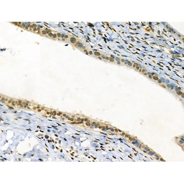
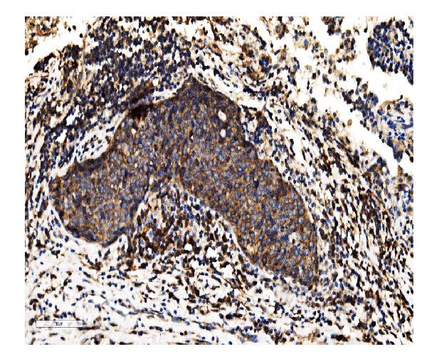
 IHC (Immunohistochemistry)
(At 1/100 staining Human esophageal cancer by IHC-P. The sample was formaldehyde fixed and a heat mediated antigen retrieval step in citrate buffer was performed. The sample was then blocked and incubated with the primary antibody at 4 degree C overnight. An HRP conjugated anti-Rabbit antibody was used as the secondary antibody.)
IHC (Immunohistochemistry)
(At 1/100 staining Human esophageal cancer by IHC-P. The sample was formaldehyde fixed and a heat mediated antigen retrieval step in citrate buffer was performed. The sample was then blocked and incubated with the primary antibody at 4 degree C overnight. An HRP conjugated anti-Rabbit antibody was used as the secondary antibody.)
p53, Polyclonal Antibody (Cat# AAA31377)
Full Name
p53 Antibody
Gene Names
TP53; P53; BCC7; LFS1; TRP53
Reactivity
Human, Mouse, Rat
Predicted Reactivity: Pig (91%), Sheep (80%), Rabbit (91%), Dog (91%)
Predicted Reactivity: Pig (91%), Sheep (80%), Rabbit (91%), Dog (91%)
Applications
Western Blot, Immunohistochemistry, Peptide ELISA
Purity
The antiserum was purified by peptide affinity chromatography using SulfoLink Coupling Resin
Pricing


 IHC (Immunohistchemistry)
(Formalin-fixed, paraffin-embedded human Breast Carcinoma stained with MUC-1 / EMA Monoclonal Antibody (MUC1/520).)
IHC (Immunohistchemistry)
(Formalin-fixed, paraffin-embedded human Breast Carcinoma stained with MUC-1 / EMA Monoclonal Antibody (MUC1/520).)
MUC1 / EMA /CD227, Monoclonal Antibody (Cat# AAA13843)
Full Name
MUC1 / EMA /CD227 (Epithelial Marker) Mouse Monoclonal Antibody
Gene Names
MUC1; EMA; MCD; PEM; PUM; KL-6; MAM6; MCKD; PEMT; CD227; H23AG; MCKD1; MUC-1; ADMCKD; ADMCKD1; CA 15-3; MUC-1/X; MUC1/ZD; MUC-1/SEC
Reactivity
Human
Applications
Flow Cytometry, Immunofluorescence, Immunohistochemistry
Pricing


 Application Data
(Detection limit for recombinant GST tagged BMP7 is 10 ng/ml as a capture antibody.)
Application Data
(Detection limit for recombinant GST tagged BMP7 is 10 ng/ml as a capture antibody.)
BMP7, Monoclonal Antibody (Cat# AAA25919)
Full Name
BMP7 (Bone Morphogenetic Protein 7, OP-1) (AP)
Gene Names
BMP7; OP-1
Applications
Western Blot
Purity
Purified
Pricing


 Application Data
(At 25 degree C. Samples were then incubated with primary Ab(At 37 degree C. An AlexaFluor594 conjugated goat anti-rabbit IgG(H+L) Ab(Red) and an AlexaFluor488 conjugated goat anti-mouse IgG(H+L) Ab(Green) were used as the secondary antibody.The nuclear counter stain is DAPI(blue).)
Application Data
(At 25 degree C. Samples were then incubated with primary Ab(At 37 degree C. An AlexaFluor594 conjugated goat anti-rabbit IgG(H+L) Ab(Red) and an AlexaFluor488 conjugated goat anti-mouse IgG(H+L) Ab(Green) were used as the secondary antibody.The nuclear counter stain is DAPI(blue).)
Rac1/cdc42, Polyclonal Antibody (Cat# AAA31342)
Full Name
Rac1/cdc42 Antibody
Gene Names
RAC1; MIG5; Rac-1; TC-25; p21-Rac1
Reactivity
Human, Mouse, Rat
Predicted Reactivity: Pig (100%), Bovine (100%), Sheep (100%), Dog (100%), Xenopus (100%)
Predicted Reactivity: Pig (100%), Bovine (100%), Sheep (100%), Dog (100%), Xenopus (100%)
Applications
Western Blot, Immunohistochemistry, Immunofluorescence, Immunocytochemistry, Peptide ELISA
Purity
The antiserum was purified by peptide affinity chromatography using SulfoLink Coupling Resin
Pricing


 Application Data
(Published customer image Infiltration of GFP+ BM-cells in infarct and peri-infarct regions. (A-B) Dot plots of viable macrophages/granulocytes (CD11b+CD45high, top right quadrants) and microglia (CD11b+CD45dim, bottom right quadrants) in cortex from BM-chimeric unmanipulated mice and mice exposed to pMCAO. (C) Bar graph showing mean numbers of CD11b+CD45dim microglia and CD11b+CD45high macrophages/granulocytes in BM-chimeric mice 24 hours after pMCAO, subdivided based on expression of GFP (n = 5). Approximately 92% of of the CD45high population were GFP+. (D) Estimation and comparison of mean numbers of CD11b+CD45dim microglia in non-chimeric (n = 10) versus BM-chimeric mice (n = 5) 24 hours after of pMCAO shows significantly fewer CD11b+CD45dim microglial cells in irradiated mice. (E) Overview, showing distribution of infiltrating GFP+ BM-derived cells into infarct (IF) and peri-infarct (P-IF) regions 24 hours after pMCAO. (E-G) By 24 hours, GFP+ single cells (F) and vessel-associated aggregates of GFP+ cells (arrows in G) were observed in infarct and peri-infarct regions. Some of the vessel-associated cells were round, leukocyte-like cells (arrows) while others were elongated cells lining the vasculature (arrow heads in G and in insert). (H) Bar graph showing mean numbers of single GFP+ cells and vessel-associated aggregates of GFP+ cells in ipsi- and contralateral cortex 24 hours after surgery (n = 10). (I-P) Immunohistochemical staining of CD45.1 (I, K), CD45.2 (J, L), IgG2a (M, O) and CD45 (N, P) in ischemic tissue in BM-chimeric (I, J, M, N) and non-chimeric mice (K, L, O, P) 24 hours after pMCAO. N.D, none detected. Scale bars: 200 um (A), 10 um (B, C). 50 um (I-P) *P < 0.05, **P < 0.01, and ***P < 0.001.From: Clausen BH, Lambertsen KL, Babcock AA, Holm TH, Dagnaes-Hansen F, Finsen B. Interleukin-1beta and tumor necrosis factor-alpha are expressed by different subsets of microglia and macrophages after ischemic stroke in mice. J Neuroinflammation. 2008 Oct 23;5:46.)
Application Data
(Published customer image Infiltration of GFP+ BM-cells in infarct and peri-infarct regions. (A-B) Dot plots of viable macrophages/granulocytes (CD11b+CD45high, top right quadrants) and microglia (CD11b+CD45dim, bottom right quadrants) in cortex from BM-chimeric unmanipulated mice and mice exposed to pMCAO. (C) Bar graph showing mean numbers of CD11b+CD45dim microglia and CD11b+CD45high macrophages/granulocytes in BM-chimeric mice 24 hours after pMCAO, subdivided based on expression of GFP (n = 5). Approximately 92% of of the CD45high population were GFP+. (D) Estimation and comparison of mean numbers of CD11b+CD45dim microglia in non-chimeric (n = 10) versus BM-chimeric mice (n = 5) 24 hours after of pMCAO shows significantly fewer CD11b+CD45dim microglial cells in irradiated mice. (E) Overview, showing distribution of infiltrating GFP+ BM-derived cells into infarct (IF) and peri-infarct (P-IF) regions 24 hours after pMCAO. (E-G) By 24 hours, GFP+ single cells (F) and vessel-associated aggregates of GFP+ cells (arrows in G) were observed in infarct and peri-infarct regions. Some of the vessel-associated cells were round, leukocyte-like cells (arrows) while others were elongated cells lining the vasculature (arrow heads in G and in insert). (H) Bar graph showing mean numbers of single GFP+ cells and vessel-associated aggregates of GFP+ cells in ipsi- and contralateral cortex 24 hours after surgery (n = 10). (I-P) Immunohistochemical staining of CD45.1 (I, K), CD45.2 (J, L), IgG2a (M, O) and CD45 (N, P) in ischemic tissue in BM-chimeric (I, J, M, N) and non-chimeric mice (K, L, O, P) 24 hours after pMCAO. N.D, none detected. Scale bars: 200 um (A), 10 um (B, C). 50 um (I-P) *P < 0.05, **P < 0.01, and ***P < 0.001.From: Clausen BH, Lambertsen KL, Babcock AA, Holm TH, Dagnaes-Hansen F, Finsen B. Interleukin-1beta and tumor necrosis factor-alpha are expressed by different subsets of microglia and macrophages after ischemic stroke in mice. J Neuroinflammation. 2008 Oct 23;5:46.)
CD11b, Monoclonal Antibody (Cat# AAA12182)
Full Name
RAT ANTI MOUSE CD11b:FITC
Gene Names
Itgam; CR3; CR3A; MAC1; Cd11b; Ly-40; Mac-1; Mac-1a; CD11b/CD18; F730045J24Rik
Applications
Flow Cytometry
Pricing


 Application Data
(Detection limit for recombinant GST tagged BMP7 is 10 ng/ml as a capture antibody.)
Application Data
(Detection limit for recombinant GST tagged BMP7 is 10 ng/ml as a capture antibody.)
BMP7, Monoclonal Antibody (Cat# AAA26451)
Full Name
BMP7 (Bone Morphogenetic Protein 7, OP-1) (HRP)
Gene Names
BMP7; OP-1
Applications
Western Blot
Purity
Purified
Pricing


 WB (Western Blot)
(BST2 monoclonal antibody (M15), clone 2E6. Western Blot analysis of BST2 expression in human placenta.)
WB (Western Blot)
(BST2 monoclonal antibody (M15), clone 2E6. Western Blot analysis of BST2 expression in human placenta.)
BST2, Monoclonal Antibody (Cat# AAA26404)
Full Name
BST2 (Bone Marrow Stromal Cell antigen 2, CD317) (FITC)
Gene Names
BST2; CD317; TETHERIN
Applications
Immunofluorescence, Immunoprecipitation, Western Blot
Purity
Purified
Pricing


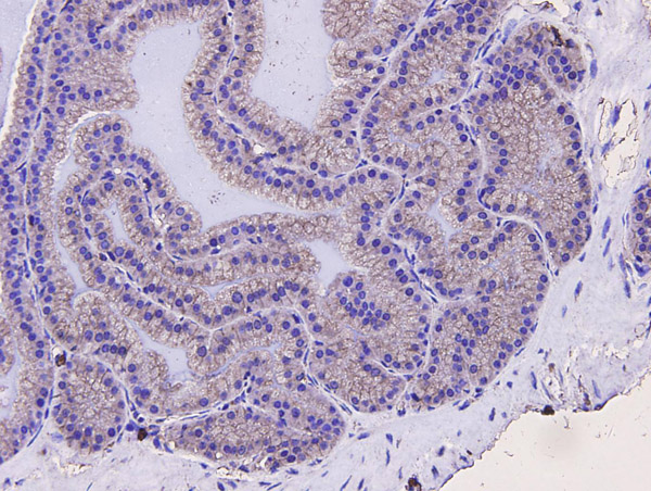
 IHC (Immunohistochemistry)
(At 1/100 staining Human pancreatic cancer by IHC-P. The sample was formaldehyde fixed and a heat mediated antigen retrieval step in citrate buffer was performed. The sample was then blocked and incubated with the primary antibody at 4 degree C overnight. An HRP conjugated anti-Rabbit antibody was used as the secondary antibody.)
IHC (Immunohistochemistry)
(At 1/100 staining Human pancreatic cancer by IHC-P. The sample was formaldehyde fixed and a heat mediated antigen retrieval step in citrate buffer was performed. The sample was then blocked and incubated with the primary antibody at 4 degree C overnight. An HRP conjugated anti-Rabbit antibody was used as the secondary antibody.)
p53, Polyclonal Antibody (Cat# AAA31355)
Full Name
Acetyl-p53 (Lys381) Antibody
Gene Names
TP53; P53; BCC7; LFS1; TRP53
Reactivity
Human, Mouse, Rat
Predicted Reactivity: Rabbit (90%), Dog (100%)
Predicted Reactivity: Rabbit (90%), Dog (100%)
Applications
Western Blot, Immunohistochemistry, Peptide ELISA
Purity
The antiserum was purified by peptide affinity chromatography using SulfoLink Coupling Resin
Pricing


 Application Data
(At 25 degree C. The primary antibody was diluted at 1/200 and incubated with the sample for 1 hour at 37 degree C. An Alexa Fluor 594 conjugated goat anti-rabbit IgG (H+L) Ab, diluted at 1/600, was used as the secondary antibody.)
Application Data
(At 25 degree C. The primary antibody was diluted at 1/200 and incubated with the sample for 1 hour at 37 degree C. An Alexa Fluor 594 conjugated goat anti-rabbit IgG (H+L) Ab, diluted at 1/600, was used as the secondary antibody.)
p53, Polyclonal Antibody (Cat# AAA31429)
Full Name
Phospho-p53 (Ser392) Antibody
Gene Names
TP53; P53; BCC7; LFS1; TRP53
Reactivity
Human, Mouse
Predicted Reactivity: Pig (88%), Bovine (88%), Sheep (88%), Rabbit (88%)
Predicted Reactivity: Pig (88%), Bovine (88%), Sheep (88%), Rabbit (88%)
Applications
Western Blot, Immunohistochemistry, Immunofluorescence, Immunocytochemistry, Peptide ELISA
Purity
The antibody is from purified rabbit serum by affinity purification via sequential chromatography on phospho-peptide and non-phospho-peptide affinity columns.
Pricing


 WB (Western Blot)
(Western BlotPositive WB detected in:Mouse brain tissue,Mouse skeletal muscle tissue,ZebrafishAll lanes: Bmp4 antibody at 3ug/mlSecondaryGoat polyclonal to rabbit IgG at 1/50000 dilutionPredicted band size: 47 kDaObserved band size: 47 kDa)
WB (Western Blot)
(Western BlotPositive WB detected in:Mouse brain tissue,Mouse skeletal muscle tissue,ZebrafishAll lanes: Bmp4 antibody at 3ug/mlSecondaryGoat polyclonal to rabbit IgG at 1/50000 dilutionPredicted band size: 47 kDaObserved band size: 47 kDa)
Bone morphogenetic protein 4, Polyclonal Antibody (Cat# AAA26924)
Full Name
Rabbit anti-human Bone morphogenetic protein 4 polyclonal Antibody
Gene Names
BMP4; ZYME; BMP2B; OFC11; BMP2B1; MCOPS6
Reactivity
Human
Applications
Western Blot, Immunohistochemistry
Purity
Caprylic Acid Ammonium Sulfate Precipitation purified
Pricing


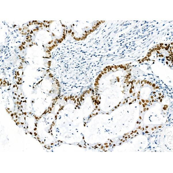
 IHC (Immunohistochemistry)
(At 1/100 staining Human gastric cancer by IHC-P. The sample was formaldehyde fixed and a heat mediated antigen retrieval step in citrate buffer was performed. The sample was then blocked and incubated with the primary antibody at 4 degree C overnight. An HRP conjugated anti-Rabbit antibody was used as the secondary antibody.)
IHC (Immunohistochemistry)
(At 1/100 staining Human gastric cancer by IHC-P. The sample was formaldehyde fixed and a heat mediated antigen retrieval step in citrate buffer was performed. The sample was then blocked and incubated with the primary antibody at 4 degree C overnight. An HRP conjugated anti-Rabbit antibody was used as the secondary antibody.)
Smad1/9, Polyclonal Antibody (Cat# AAA31453)
Full Name
Phospho-Smad1/9 (Ser463/Ser465) Antibody
Gene Names
SMAD1; BSP1; JV41; BSP-1; JV4-1; MADH1; MADR1
Reactivity
Human, Mouse, Rat
Predicted Reactivity: Pig (100%), Zebrafish (100%), Bovine (100%), Sheep (100%), Rabbit (100%), Dog (100%), Chicken (100%), Xenopus (100%)
Predicted Reactivity: Pig (100%), Zebrafish (100%), Bovine (100%), Sheep (100%), Rabbit (100%), Dog (100%), Chicken (100%), Xenopus (100%)
Applications
Western Blot, Immunohistochemistry, Peptide ELISA
Purity
The antibody is from purified rabbit serum by affinity purification via sequential chromatography on phospho-peptide and non-phospho-peptide affinity columns.
Pricing


 IHC (Immunohistchemistry)
(Immunohistochemistry of paraffin-embedded mouse lung using BMP5 antibody at dilution of 1:100 (40x lens).)
IHC (Immunohistchemistry)
(Immunohistochemistry of paraffin-embedded mouse lung using BMP5 antibody at dilution of 1:100 (40x lens).)
BMP5, Polyclonal Antibody (Cat# AAA28209)
Full Name
BMP5 Polyclonal Antibody
Reactivity
Human, Mouse, Rat
Applications
Western Blot
Purity
Affinity Purification
Pricing


 Application Data
(Published customer image Infiltration of GFP+ BM-cells in infarct and peri-infarct regions. (A-B) Dot plots of viable macrophages/granulocytes (CD11b+CD45high, top right quadrants) and microglia (CD11b+CD45dim, bottom right quadrants) in cortex from BM-chimeric unmanipulated mice and mice exposed to pMCAO. (C) Bar graph showing mean numbers of CD11b+CD45dim microglia and CD11b+CD45high macrophages/granulocytes in BM-chimeric mice 24 hours after pMCAO, subdivided based on expression of GFP (n = 5). Approximately 92% of of the CD45high population were GFP+. (D) Estimation and comparison of mean numbers of CD11b+CD45dim microglia in non-chimeric (n = 10) versus BM-chimeric mice (n = 5) 24 hours after of pMCAO shows significantly fewer CD11b+CD45dim microglial cells in irradiated mice. (E) Overview, showing distribution of infiltrating GFP+ BM-derived cells into infarct (IF) and peri-infarct (P-IF) regions 24 hours after pMCAO. (E-G) By 24 hours, GFP+ single cells (F) and vessel-associated aggregates of GFP+ cells (arrows in G) were observed in infarct and peri-infarct regions. Some of the vessel-associated cells were round, leukocyte-like cells (arrows) while others were elongated cells lining the vasculature (arrow heads in G and in insert). (H) Bar graph showing mean numbers of single GFP+ cells and vessel-associated aggregates of GFP+ cells in ipsi- and contralateral cortex 24 hours after surgery (n = 10). (I-P) Immunohistochemical staining of CD45.1 (I, K), CD45.2 (J, L), IgG2a (M, O) and CD45 (N, P) in ischemic tissue in BM-chimeric (I, J, M, N) and non-chimeric mice (K, L, O, P) 24 hours after pMCAO. N.D, none detected. Scale bars: 200 um (A), 10 um (B, C). 50 um (I-P) *P < 0.05, **P < 0.01, and ***P < 0.001.From: Clausen BH, Lambertsen KL, Babcock AA, Holm TH, Dagnaes-Hansen F, Finsen B. Interleukin-1beta and tumor necrosis factor-alpha are expressed by different subsets of microglia and macrophages after ischemic stroke in mice. J Neuroinflammation. 2008 Oct 23;5:46.)
Application Data
(Published customer image Infiltration of GFP+ BM-cells in infarct and peri-infarct regions. (A-B) Dot plots of viable macrophages/granulocytes (CD11b+CD45high, top right quadrants) and microglia (CD11b+CD45dim, bottom right quadrants) in cortex from BM-chimeric unmanipulated mice and mice exposed to pMCAO. (C) Bar graph showing mean numbers of CD11b+CD45dim microglia and CD11b+CD45high macrophages/granulocytes in BM-chimeric mice 24 hours after pMCAO, subdivided based on expression of GFP (n = 5). Approximately 92% of of the CD45high population were GFP+. (D) Estimation and comparison of mean numbers of CD11b+CD45dim microglia in non-chimeric (n = 10) versus BM-chimeric mice (n = 5) 24 hours after of pMCAO shows significantly fewer CD11b+CD45dim microglial cells in irradiated mice. (E) Overview, showing distribution of infiltrating GFP+ BM-derived cells into infarct (IF) and peri-infarct (P-IF) regions 24 hours after pMCAO. (E-G) By 24 hours, GFP+ single cells (F) and vessel-associated aggregates of GFP+ cells (arrows in G) were observed in infarct and peri-infarct regions. Some of the vessel-associated cells were round, leukocyte-like cells (arrows) while others were elongated cells lining the vasculature (arrow heads in G and in insert). (H) Bar graph showing mean numbers of single GFP+ cells and vessel-associated aggregates of GFP+ cells in ipsi- and contralateral cortex 24 hours after surgery (n = 10). (I-P) Immunohistochemical staining of CD45.1 (I, K), CD45.2 (J, L), IgG2a (M, O) and CD45 (N, P) in ischemic tissue in BM-chimeric (I, J, M, N) and non-chimeric mice (K, L, O, P) 24 hours after pMCAO. N.D, none detected. Scale bars: 200 um (A), 10 um (B, C). 50 um (I-P) *P < 0.05, **P < 0.01, and ***P < 0.001.From: Clausen BH, Lambertsen KL, Babcock AA, Holm TH, Dagnaes-Hansen F, Finsen B. Interleukin-1beta and tumor necrosis factor-alpha are expressed by different subsets of microglia and macrophages after ischemic stroke in mice. J Neuroinflammation. 2008 Oct 23;5:46.)
CD11b, Monoclonal Antibody (Cat# AAA12184)
Full Name
RAT ANTI MOUSE CD11b
Gene Names
Itgam; CR3; CR3A; MAC1; Cd11b; Ly-40; Mac-1; Mac-1a; CD11b/CD18; F730045J24Rik
Applications
Immunohistochemistry, Flow Cytometry, Immunofluorescence, Immunoprecipitation
Pricing
 Application Data
Collagen 1, alpha 1 telopeptide, Polyclonal Antibody (Cat# AAA14236)
Full Name
Anti-Collagen I alpha1 Telopeptide Sequence
Gene Names
COL1A1; OI4
Reactivity
Human, mouse, sheep
Applications
Western Blot, Immunohistochemistry
Purity
Affinity Purified
Pricing


 Application Data
(Staining of mouse peripheral blood granulocytes with Rat anti Mouse CD13)
Application Data
(Staining of mouse peripheral blood granulocytes with Rat anti Mouse CD13)
CD13, Monoclonal Antibody (Cat# AAA12145)
Full Name
RAT ANTI MOUSE CD13
Gene Names
Anpep; Apn; AP-M; AP-N; Cd13; P150
Applications
Immunohistochemistry, Flow Cytometry
Pricing


 Application Data
(Detection limit for recombinant GST tagged SPP1 is ~1ng/ml as a capture antibody.)
Application Data
(Detection limit for recombinant GST tagged SPP1 is ~1ng/ml as a capture antibody.)
Osteopontin, Monoclonal Antibody (Cat# AAA25480)
Full Name
Osteopontin (Bone Sialoprotein 1, Nephropontin, Secreted Phosphoprotein 1, SPP-1, Urinary Stone Protein, Uropontin, SPP1, BNSP, OPN, PSEC0156, MGC110940) (HRP)
Gene Names
SPP1; OPN; BNSP; BSPI; ETA-1
Reactivity
Human, Mouse
Applications
Western Blot
Purity
Purified by Protein A Affinity Chromatography.
Pricing


 Application Data
(Detection limit for recombinant GST tagged BMP7 is 10 ng/ml as a capture antibody.)
Application Data
(Detection limit for recombinant GST tagged BMP7 is 10 ng/ml as a capture antibody.)
BMP7, Monoclonal Antibody (Cat# AAA26185)
Full Name
BMP7 (Bone Morphogenetic Protein 7, OP-1) (Biotin)
Gene Names
BMP7; OP-1
Applications
Western Blot
Purity
Purified
Pricing


 Application Data
(Detection limit for recombinant GST tagged SPP1 is ~1ng/ml as a capture antibody.)
Application Data
(Detection limit for recombinant GST tagged SPP1 is ~1ng/ml as a capture antibody.)
Osteopontin, Monoclonal Antibody (Cat# AAA24889)
Full Name
Osteopontin (Bone Sialoprotein 1, Nephropontin, Secreted Phosphoprotein 1, SPP-1, Urinary Stone Protein, Uropontin, SPP1, BNSP, OPN, PSEC0156, MGC110940) (Biotin)
Gene Names
SPP1; OPN; BNSP; BSPI; ETA-1
Reactivity
Human, Mouse
Applications
Western Blot
Purity
Purified by Protein A Affinity Chromatography.
Pricing


 Application Data
(Analysis of Protein Array containing more than 19, 000 full-length human proteins using CD10 Mouse Monoclonal Antibody (MME/1870)Z- and S- Score: The Z-score represents the strength of a signal that a monoclonal antibody (MAb) (in combination with a fluorescently-tagged anti-IgG secondary antibody) produces when binding to a particular protein on the HuProtTM array. Z-scores are described in units of standard deviations (SD's) above the mean value of all signals generated on that array. If targets on HuProtTM are arranged in descending order of the Z-score, the S-score is the difference (also in units of SD's) between the Z-score. S-score therefore represents the relative target specificity of a MAb to its intended target. A MAb is considered to specific to its intended target, if the MAb has an S-score of at least 2.5. For example, if a MAb binds to protein X with a Z-score of 43 and to protein Y with a Z-score of 14, then the S-score for the binding of that MAb to protein X is equal to 29.)
Application Data
(Analysis of Protein Array containing more than 19, 000 full-length human proteins using CD10 Mouse Monoclonal Antibody (MME/1870)Z- and S- Score: The Z-score represents the strength of a signal that a monoclonal antibody (MAb) (in combination with a fluorescently-tagged anti-IgG secondary antibody) produces when binding to a particular protein on the HuProtTM array. Z-scores are described in units of standard deviations (SD's) above the mean value of all signals generated on that array. If targets on HuProtTM are arranged in descending order of the Z-score, the S-score is the difference (also in units of SD's) between the Z-score. S-score therefore represents the relative target specificity of a MAb to its intended target. A MAb is considered to specific to its intended target, if the MAb has an S-score of at least 2.5. For example, if a MAb binds to protein X with a Z-score of 43 and to protein Y with a Z-score of 14, then the S-score for the binding of that MAb to protein X is equal to 29.)
CD10, Monoclonal Antibody (Cat# AAA23901)
Full Name
CD10 (Membrane Metalloendopeptidase)
Gene Names
MME; NEP; SFE; CD10; CALLA; CMT2T; SCA43
Reactivity
Human. Others not tested.
Applications
Immunohistochemistry
Pricing


 Application Data
(Published customer image Infiltration of GFP+ BM-cells in infarct and peri-infarct regions. (A-B) Dot plots of viable macrophages/granulocytes (CD11b+CD45high, top right quadrants) and microglia (CD11b+CD45dim, bottom right quadrants) in cortex from BM-chimeric unmanipulated mice and mice exposed to pMCAO. (C) Bar graph showing mean numbers of CD11b+CD45dim microglia and CD11b+CD45high macrophages/granulocytes in BM-chimeric mice 24 hours after pMCAO, subdivided based on expression of GFP (n = 5). Approximately 92% of of the CD45high population were GFP+. (D) Estimation and comparison of mean numbers of CD11b+CD45dim microglia in non-chimeric (n = 10) versus BM-chimeric mice (n = 5) 24 hours after of pMCAO shows significantly fewer CD11b+CD45dim microglial cells in irradiated mice. (E) Overview, showing distribution of infiltrating GFP+ BM-derived cells into infarct (IF) and peri-infarct (P-IF) regions 24 hours after pMCAO. (E-G) By 24 hours, GFP+ single cells (F) and vessel-associated aggregates of GFP+ cells (arrows in G) were observed in infarct and peri-infarct regions. Some of the vessel-associated cells were round, leukocyte-like cells (arrows) while others were elongated cells lining the vasculature (arrow heads in G and in insert). (H) Bar graph showing mean numbers of single GFP+ cells and vessel-associated aggregates of GFP+ cells in ipsi- and contralateral cortex 24 hours after surgery (n = 10). (I-P) Immunohistochemical staining of CD45.1 (I, K), CD45.2 (J, L), IgG2a (M, O) and CD45 (N, P) in ischemic tissue in BM-chimeric (I, J, M, N) and non-chimeric mice (K, L, O, P) 24 hours after pMCAO. N.D, none detected. Scale bars: 200 um (A), 10 um (B, C). 50 um (I-P) *P < 0.05, **P < 0.01, and ***P < 0.001.From: Clausen BH, Lambertsen KL, Babcock AA, Holm TH, Dagnaes-Hansen F, Finsen B. Interleukin-1beta and tumor necrosis factor-alpha are expressed by different subsets of microglia and macrophages after ischemic stroke in mice. J Neuroinflammation. 2008 Oct 23;5:46.)
Application Data
(Published customer image Infiltration of GFP+ BM-cells in infarct and peri-infarct regions. (A-B) Dot plots of viable macrophages/granulocytes (CD11b+CD45high, top right quadrants) and microglia (CD11b+CD45dim, bottom right quadrants) in cortex from BM-chimeric unmanipulated mice and mice exposed to pMCAO. (C) Bar graph showing mean numbers of CD11b+CD45dim microglia and CD11b+CD45high macrophages/granulocytes in BM-chimeric mice 24 hours after pMCAO, subdivided based on expression of GFP (n = 5). Approximately 92% of of the CD45high population were GFP+. (D) Estimation and comparison of mean numbers of CD11b+CD45dim microglia in non-chimeric (n = 10) versus BM-chimeric mice (n = 5) 24 hours after of pMCAO shows significantly fewer CD11b+CD45dim microglial cells in irradiated mice. (E) Overview, showing distribution of infiltrating GFP+ BM-derived cells into infarct (IF) and peri-infarct (P-IF) regions 24 hours after pMCAO. (E-G) By 24 hours, GFP+ single cells (F) and vessel-associated aggregates of GFP+ cells (arrows in G) were observed in infarct and peri-infarct regions. Some of the vessel-associated cells were round, leukocyte-like cells (arrows) while others were elongated cells lining the vasculature (arrow heads in G and in insert). (H) Bar graph showing mean numbers of single GFP+ cells and vessel-associated aggregates of GFP+ cells in ipsi- and contralateral cortex 24 hours after surgery (n = 10). (I-P) Immunohistochemical staining of CD45.1 (I, K), CD45.2 (J, L), IgG2a (M, O) and CD45 (N, P) in ischemic tissue in BM-chimeric (I, J, M, N) and non-chimeric mice (K, L, O, P) 24 hours after pMCAO. N.D, none detected. Scale bars: 200 um (A), 10 um (B, C). 50 um (I-P) *P < 0.05, **P < 0.01, and ***P < 0.001.From: Clausen BH, Lambertsen KL, Babcock AA, Holm TH, Dagnaes-Hansen F, Finsen B. Interleukin-1beta and tumor necrosis factor-alpha are expressed by different subsets of microglia and macrophages after ischemic stroke in mice. J Neuroinflammation. 2008 Oct 23;5:46.)
CD11b, Monoclonal Antibody (Cat# AAA12185)
Full Name
RAT ANTI MOUSE CD11b
Gene Names
Itgam; CR3; CR3A; MAC1; Cd11b; Ly-40; Mac-1; Mac-1a; CD11b/CD18; F730045J24Rik
Applications
Immunohistochemistry, Flow Cytometry, Immunofluorescence, Immunoprecipitation
Pricing


 Application Data
(Published customer image Infiltration of GFP+ BM-cells in infarct and peri-infarct regions. (A-B) Dot plots of viable macrophages/granulocytes (CD11b+CD45high, top right quadrants) and microglia (CD11b+CD45dim, bottom right quadrants) in cortex from BM-chimeric unmanipulated mice and mice exposed to pMCAO. (C) Bar graph showing mean numbers of CD11b+CD45dim microglia and CD11b+CD45high macrophages/granulocytes in BM-chimeric mice 24 hours after pMCAO, subdivided based on expression of GFP (n = 5). Approximately 92% of of the CD45high population were GFP+. (D) Estimation and comparison of mean numbers of CD11b+CD45dim microglia in non-chimeric (n = 10) versus BM-chimeric mice (n = 5) 24 hours after of pMCAO shows significantly fewer CD11b+CD45dim microglial cells in irradiated mice. (E) Overview, showing distribution of infiltrating GFP+ BM-derived cells into infarct (IF) and peri-infarct (P-IF) regions 24 hours after pMCAO. (E-G) By 24 hours, GFP+ single cells (F) and vessel-associated aggregates of GFP+ cells (arrows in G) were observed in infarct and peri-infarct regions. Some of the vessel-associated cells were round, leukocyte-like cells (arrows) while others were elongated cells lining the vasculature (arrow heads in G and in insert). (H) Bar graph showing mean numbers of single GFP+ cells and vessel-associated aggregates of GFP+ cells in ipsi- and contralateral cortex 24 hours after surgery (n = 10). (I-P) Immunohistochemical staining of CD45.1 (I, K), CD45.2 (J, L), IgG2a (M, O) and CD45 (N, P) in ischemic tissue in BM-chimeric (I, J, M, N) and non-chimeric mice (K, L, O, P) 24 hours after pMCAO. N.D, none detected. Scale bars: 200 um (A), 10 um (B, C). 50 um (I-P) *P < 0.05, **P < 0.01, and ***P < 0.001.From: Clausen BH, Lambertsen KL, Babcock AA, Holm TH, Dagnaes-Hansen F, Finsen B. Interleukin-1beta and tumor necrosis factor-alpha are expressed by different subsets of microglia and macrophages after ischemic stroke in mice. J Neuroinflammation. 2008 Oct 23;5:46.)
Application Data
(Published customer image Infiltration of GFP+ BM-cells in infarct and peri-infarct regions. (A-B) Dot plots of viable macrophages/granulocytes (CD11b+CD45high, top right quadrants) and microglia (CD11b+CD45dim, bottom right quadrants) in cortex from BM-chimeric unmanipulated mice and mice exposed to pMCAO. (C) Bar graph showing mean numbers of CD11b+CD45dim microglia and CD11b+CD45high macrophages/granulocytes in BM-chimeric mice 24 hours after pMCAO, subdivided based on expression of GFP (n = 5). Approximately 92% of of the CD45high population were GFP+. (D) Estimation and comparison of mean numbers of CD11b+CD45dim microglia in non-chimeric (n = 10) versus BM-chimeric mice (n = 5) 24 hours after of pMCAO shows significantly fewer CD11b+CD45dim microglial cells in irradiated mice. (E) Overview, showing distribution of infiltrating GFP+ BM-derived cells into infarct (IF) and peri-infarct (P-IF) regions 24 hours after pMCAO. (E-G) By 24 hours, GFP+ single cells (F) and vessel-associated aggregates of GFP+ cells (arrows in G) were observed in infarct and peri-infarct regions. Some of the vessel-associated cells were round, leukocyte-like cells (arrows) while others were elongated cells lining the vasculature (arrow heads in G and in insert). (H) Bar graph showing mean numbers of single GFP+ cells and vessel-associated aggregates of GFP+ cells in ipsi- and contralateral cortex 24 hours after surgery (n = 10). (I-P) Immunohistochemical staining of CD45.1 (I, K), CD45.2 (J, L), IgG2a (M, O) and CD45 (N, P) in ischemic tissue in BM-chimeric (I, J, M, N) and non-chimeric mice (K, L, O, P) 24 hours after pMCAO. N.D, none detected. Scale bars: 200 um (A), 10 um (B, C). 50 um (I-P) *P < 0.05, **P < 0.01, and ***P < 0.001.From: Clausen BH, Lambertsen KL, Babcock AA, Holm TH, Dagnaes-Hansen F, Finsen B. Interleukin-1beta and tumor necrosis factor-alpha are expressed by different subsets of microglia and macrophages after ischemic stroke in mice. J Neuroinflammation. 2008 Oct 23;5:46.)
CD11b, Monoclonal Antibody (Cat# AAA12183)
Full Name
RAT ANTI MOUSE CD11b:FITC
Gene Names
Itgam; CR3; CR3A; MAC1; Cd11b; Ly-40; Mac-1; Mac-1a; CD11b/CD18; F730045J24Rik
Applications
Flow Cytometry
Pricing


 IF (Immunofluorescence)
(Immunofluorescence analysis of human breast cancer using COL1A1 antibody.)
IF (Immunofluorescence)
(Immunofluorescence analysis of human breast cancer using COL1A1 antibody.)
COL1A1, Polyclonal Antibody (Cat# AAA29806)
Full Name
COL1A1 Polyclonal Antibody
Gene Names
COL1A1; OI4
Reactivity
Human, Mouse, Rat
Applications
Western Blot, Immunohistochemistry, Immunofluorescence
Purity
Affinity purification
Pricing
 Standard Curve (Sample)
Bone Sialoprotein (BSP), ELISA Kit (Cat# AAA12836)
Full Name
Rat Bone Sialoprotein (BSP) ELISA Kit
Gene Names
IBSP; BSP; BNSP; SP-II; BSP-II
Reactivity
Rat
Pricing
 Application Data
(ATH mouse bone marrow stained with anti-CD44 (clone: KM81) (filled histogram) or Rat IgG2a isotype control (open histogram).N.B. Appropriate control samples should always be included in any labelling studies. * For optimal results in various applications, it is recommended that each investigator determine dilutions appropriate for individual use.)
Application Data
(ATH mouse bone marrow stained with anti-CD44 (clone: KM81) (filled histogram) or Rat IgG2a isotype control (open histogram).N.B. Appropriate control samples should always be included in any labelling studies. * For optimal results in various applications, it is recommended that each investigator determine dilutions appropriate for individual use.)
CD44, Monoclonal Antibody (Cat# AAA14269)
Full Name
Anti-Mouse CD44, Purified (Clone KM81) (rat IgG2a)
Gene Names
CD44; IN; LHR; MC56; MDU2; MDU3; MIC4; Pgp1; CDW44; CSPG8; HCELL; HUTCH-I; ECMR-III
Reactivity
Mouse
Applications
Immunohistochemistry, Flow Cytometry
Pricing


 FCM (Flow Cytometry)
(Flow cytometric analysis of Hela cells with SQSTM1 antibody at 1/50 dilution (blue) compared with an unlabelled control (cells without incubation with primary antibody; red). Goat anti mouse IgG (FITC) was used as the secondary antibody.)
FCM (Flow Cytometry)
(Flow cytometric analysis of Hela cells with SQSTM1 antibody at 1/50 dilution (blue) compared with an unlabelled control (cells without incubation with primary antibody; red). Goat anti mouse IgG (FITC) was used as the secondary antibody.)
Sequestosome-1, Monoclonal Antibody (Cat# AAA29890)
Full Name
Sequestosome-1 Antibody
Gene Names
SQSTM1; p60; p62; A170; OSIL; PDB3; ZIP3; p62B
Reactivity
Human, Mouse, Rat
Applications
Western Blot, Immunocytochemistry, Immunohistochemistry, Flow Cytometry
Purity
Peptide affinity purified.
Pricing


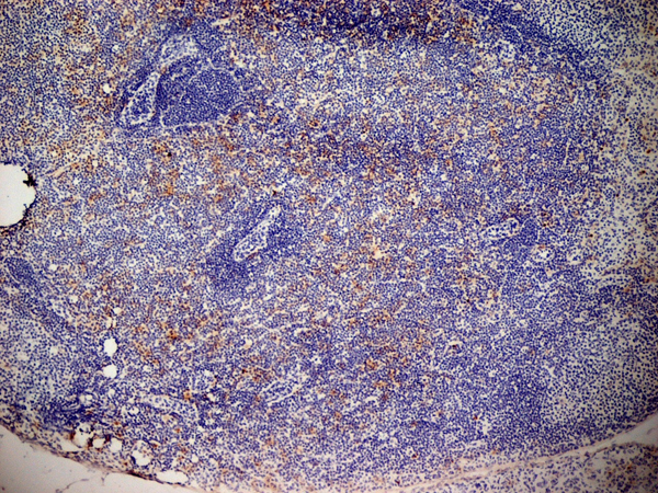
 IHC (Immunohistochemistry)
(At 1/100 staining Human gastric cancer and adjacent normal tissues by IHC-P. The sample was formaldehyde fixed and a heat mediated antigen retrieval step in citrate buffer was performed. The sample was then blocked and incubated with the primary antibody at 4 degree C overnight. An HRP conjugated anti-Rabbit antibody was used as the secondary antibody.)
IHC (Immunohistochemistry)
(At 1/100 staining Human gastric cancer and adjacent normal tissues by IHC-P. The sample was formaldehyde fixed and a heat mediated antigen retrieval step in citrate buffer was performed. The sample was then blocked and incubated with the primary antibody at 4 degree C overnight. An HRP conjugated anti-Rabbit antibody was used as the secondary antibody.)
AIRE, Polyclonal Antibody (Cat# AAA31324)
Full Name
Phospho-AIRE (Ser156) Antibody
Gene Names
AIRE; APS1; APSI; PGA1; AIRE1; APECED
Reactivity
Human, Mouse, Rat
Applications
Immunohistochemistry, Peptide ELISA
Purity
The antibody is from purified rabbit serum by affinity purification via sequential chromatography on phospho-peptide and non-phospho-peptide affinity columns.
Pricing


 Application Data
(Detection limit for recombinant GST tagged SPP1 is ~1ng/ml as a capture antibody.)
Application Data
(Detection limit for recombinant GST tagged SPP1 is ~1ng/ml as a capture antibody.)
Osteopontin, Monoclonal Antibody (Cat# AAA24594)
Full Name
Osteopontin (Bone Sialoprotein 1, Nephropontin, Secreted Phosphoprotein 1, SPP-1, Urinary Stone Protein, Uropontin, SPP1, BNSP, OPN, PSEC0156, MGC110940) APC
Gene Names
SPP1; OPN; BNSP; BSPI; ETA-1
Reactivity
Human, Mouse
Applications
Western Blot
Purity
Purified by Protein A Affinity Chromatography.
Pricing


 IP (Immunoprecipitation)
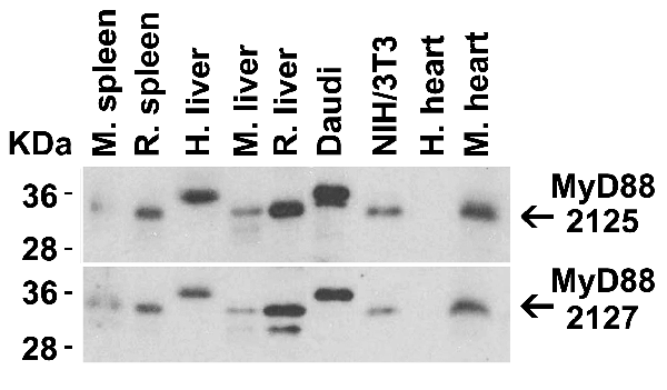
(Figure 13 Immunoprecipitation Validation in HEK293 cells (Kawai et al., 2004)HEK293 cells were transiently transfected with DYKDDDDK-IRF7. Ccell lysates were immunoprecipitated with control rabbit anti-mouse immunoglobulin serum (IgG) or anti-MyD88 (Ab1 and Ab2), followed by immunoblotting with anti-DYKDDDDK.)
IP (Immunoprecipitation)
(Figure 13 Immunoprecipitation Validation in HEK293 cells (Kawai et al., 2004)HEK293 cells were transiently transfected with DYKDDDDK-IRF7. Ccell lysates were immunoprecipitated with control rabbit anti-mouse immunoglobulin serum (IgG) or anti-MyD88 (Ab1 and Ab2), followed by immunoblotting with anti-DYKDDDDK.)
MYD88, Polyclonal Antibody (Cat# AAA10930)
Full Name
MYD88 Antibody
Gene Names
MYD88; MYD88D
Reactivity
Human, Mouse
Applications
Western Blot, Immunohistochemistry, Immunofluorescence
Purity
MYD88 Antibody is affinity chromatography purified via peptide column.
Pricing


 Application Data
(Immunoperoxidase staining of rat lymph node cryosection with Mouse anti Rat CD25 followed by horseradish peroxidase conjugated Goat anti Mouse IgG1 as a detection reagent. High power)
Application Data
(Immunoperoxidase staining of rat lymph node cryosection with Mouse anti Rat CD25 followed by horseradish peroxidase conjugated Goat anti Mouse IgG1 as a detection reagent. High power)
CD25, Monoclonal Antibody (Cat# AAA11965)
Full Name
MOUSE ANTI RAT CD25
Gene Names
Il2ra; IL2RAC
Applications
Immunohistochemistry, Flow Cytometry, Immunoprecipitation, Immunohistochemistry
Pricing


 Application Data
(Immunoperoxidase staining of rat lymph node cryosection with Mouse anti Rat CD25 followed by horseradish peroxidase conjugated Goat anti Mouse IgG1 as a detection reagent. High power)
Application Data
(Immunoperoxidase staining of rat lymph node cryosection with Mouse anti Rat CD25 followed by horseradish peroxidase conjugated Goat anti Mouse IgG1 as a detection reagent. High power)
CD25, Monoclonal Antibody (Cat# AAA11966)
Full Name
MOUSE ANTI RAT CD25
Gene Names
Il2ra; IL2RAC
Applications
Immunohistochemistry, Flow Cytometry, Immunoprecipitation, Immunohistochemistry
Pricing


 Application Data
(At 25 degree C. Samples were then incubated with primary Ab(At 37 degree C. An AlexaFluor594 conjugated goat anti-rabbit IgG(H+L) Ab(Red) and an AlexaFluor488 conjugated goat anti-mouse IgG(H+L) Ab(Green) were used as the secondary antibody.The nuclear counter stain is DAPI(blue).)
Application Data
(At 25 degree C. Samples were then incubated with primary Ab(At 37 degree C. An AlexaFluor594 conjugated goat anti-rabbit IgG(H+L) Ab(Red) and an AlexaFluor488 conjugated goat anti-mouse IgG(H+L) Ab(Green) were used as the secondary antibody.The nuclear counter stain is DAPI(blue).)
CDK5, Polyclonal Antibody (Cat# AAA31445)
Full Name
Phospho-CDK5 (Ser159) Antibody
Gene Names
CDK5; PSSALRE
Reactivity
Human, Mouse, Rat, Monkey
Predicted Reactivity: Zebrafish (100%), Bovine (100%), Horse (100%), Sheep (100%), Rabbit (100%), Dog (100%), Chicken (100%), Xenopus (100%)
Predicted Reactivity: Zebrafish (100%), Bovine (100%), Horse (100%), Sheep (100%), Rabbit (100%), Dog (100%), Chicken (100%), Xenopus (100%)
Applications
Western Blot, Immunohistochemistry, Immunofluorescence, Immunocytochemistry, Peptide ELISA
Purity
The antibody is from purified rabbit serum by affinity purification via sequential chromatography on phospho-peptide and non-phospho-peptide affinity columns.
Pricing
 Standard Curve (Sample)
Procollagen I N-Terminal Propeptide (PINP), ELISA Kit (Cat# AAA21071)
Full Name
Mouse Procollagen I N-Terminal Propeptide (PINP) Mini Samples ELISA Kit
Reactivity
Mouse
Pricing


 IHC (Immunohistochemistry)
(AAA31076 at 1/200 staining human liver cancer tissue sections by IHC-P. The tissue was formaldehyde fixed and a heat mediated antigen retrieval step in citrate buffer was performed. The tissue was then blocked and incubated with the antibody for 1.5 hours at 22 degree C. An HRP conjugated goat anti-rabbit antibody was used as the secondary.)
IHC (Immunohistochemistry)
(AAA31076 at 1/200 staining human liver cancer tissue sections by IHC-P. The tissue was formaldehyde fixed and a heat mediated antigen retrieval step in citrate buffer was performed. The tissue was then blocked and incubated with the antibody for 1.5 hours at 22 degree C. An HRP conjugated goat anti-rabbit antibody was used as the secondary.)
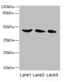
BMP3, Polyclonal Antibody (Cat# AAA31076)
Full Name
BMP3 Antibody
Gene Names
BMP3; BMP-3A
Reactivity
Human, Mouse, Rat
Applications
Western Blot, Immunohistochemistry, Immunofluorescence, Immunocytochemistry
Purity
The antiserum was purified by peptide affinity chromatography using SulfoLink Coupling Resin.
Pricing
 Standard Curve (Sample)
bone morphogenetic protein receptor, type II (serine/threonine kinase), ELISA Kit (Cat# AAA15514)
Full Name
Human Bone morphogenetic protein receptor II, BMPR-II ELISA Kit
Gene Names
BMPR2; BMR2; PPH1; BMPR3; BRK-3; POVD1; T-ALK; BMPR-II
Reactivity
Human
Pricing


 WB (Western Blot)
(Western blot analysis of Phospho-MOB1A (Tyr26) in lysates of 293 IFN 2500U/ml 30', using Phospho-MOB1A (Tyr26) Antibody.)
WB (Western Blot)
(Western blot analysis of Phospho-MOB1A (Tyr26) in lysates of 293 IFN 2500U/ml 30', using Phospho-MOB1A (Tyr26) Antibody.)
MOB1A, Polyclonal Antibody (Cat# AAA31161)
Full Name
Phospho-MOB1A (Tyr26) Antibody
Gene Names
MOB1B; MATS2; MOB4A; MOBKL1A
Reactivity
Human, Mouse, Rat
Predicted: Bovine, Horse, Sheep, Rabbit, Dog, Xenopus
Predicted: Bovine, Horse, Sheep, Rabbit, Dog, Xenopus
Applications
Western Blot, Immunohistochemistry, Peptide ELISA
Purity
Purified rabbit serum by affinity purification via sequential chromatography on phospho- and non-phospho-peptide affinity columns.
Pricing


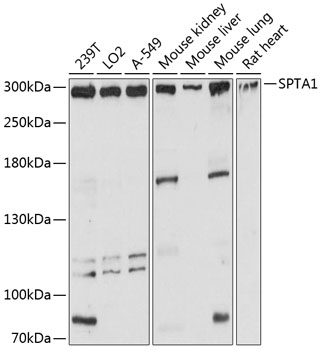
 IHC (Immunohistchemistry)
(Immunohistochemistry of paraffin-embedded mouse liver using SPTA1 antibody at dilution of 1:100 .)
IHC (Immunohistchemistry)
(Immunohistochemistry of paraffin-embedded mouse liver using SPTA1 antibody at dilution of 1:100 .)
SPTA1, Polyclonal Antibody (Cat# AAA30554)
Full Name
SPTA1 Rabbit Polyclonal Antibody
Gene Names
SPTA1; EL2; HPP; HS3; SPH3; SPTA
Reactivity
Human, Mouse, Rat
Applications
Western Blot, Immunohistochemistry, Immunofluorescence
Purity
Affinity purification
Pricing


 WB (Western Blot)
(BST2 monoclonal antibody (M15), clone 2E6. Western Blot analysis of BST2 expression in human placenta.)
WB (Western Blot)
(BST2 monoclonal antibody (M15), clone 2E6. Western Blot analysis of BST2 expression in human placenta.)
BST2, Monoclonal Antibody (Cat# AAA26005)
Full Name
BST2 (Bone Marrow Stromal Cell antigen 2, CD317) (AP)
Gene Names
BST2; CD317; TETHERIN
Applications
Immunoprecipitation, Western Blot
Purity
Purified
Pricing
 Standard Curve (Sample)
Procollagen I N-Terminal Propeptide (PINP), ELISA Kit (Cat# AAA20796)
Full Name
Rat Procollagen I N-Terminal Propeptide (PINP) ELISA Kit
Reactivity
Rat
Pricing