Filters
▼Clonality
▼Type
▼Reactivity
▼Gene Name
▼Isotype
▼Host
▼Application
▼Clone
▼Phospho Antibodies
Phospho-specific antibodies’ typical purpose is to enable researchers to detect changes in proteins. They will exclusively bind to the amino acid sequence on a protein that has been phosphorylated (which is both a physical & chemical change) and do not bind to the same amino acid sequence on said protein if it lacks said phosphorylation. This aids in being able to clearly see and understand the data produced from this particular protein modification.
Viewing 1-50 of 5298 product results
 WB (Western Blot)
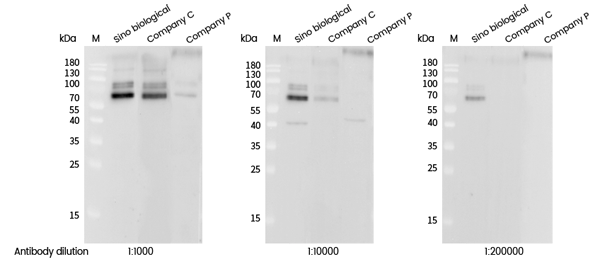
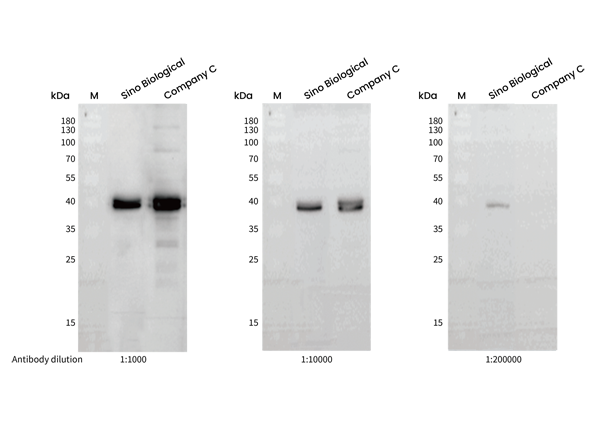
(Western blot analysis of extracts from serum-starved MCF7, treated with IGF-1 (100 ng/mL, 30 min), using Phospho-S6K1 (Thr389) rabbit monoclonal Antibody (#AAA258873) and other brands’ antibodies (company C, company P) at dilution of 1:1000, 1:10000 and 1:200000.)
WB (Western Blot)
(Western blot analysis of extracts from serum-starved MCF7, treated with IGF-1 (100 ng/mL, 30 min), using Phospho-S6K1 (Thr389) rabbit monoclonal Antibody (#AAA258873) and other brands’ antibodies (company C, company P) at dilution of 1:1000, 1:10000 and 1:200000.)
p70 S6 Kinase 1, Monoclonal Recombinant Antibody (Cat# AAA258873)
 IF (Immunofluorescence)
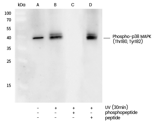
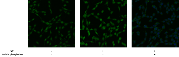
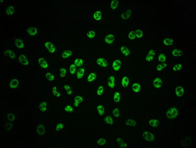
(Immunofluorescence staining of Phospho-p38 MAPK (Thr180, Tyr182) in serum-starved NIH-3T3 cells, untreated (left), treated with UV (30mins) (middle) or treated with UV (30mins) and lambda phosphatase (right). Cells were fixed with 4% PFA, permeabilzed with 0.1% Triton X-100 in PBS, blocked with 10% serum, and incubated with rabbit anti-Mouse Phospho-p38 MAPK (Thr180, Tyr182) monoclonal antibody (dilution ratio 1:60) at 4? overnight. Then cells were stained with the Alexa Fluor488-conjugated Goat Anti-rabbit IgG secondary antibody (green). Positive staining was mainly localized to Nucleus.)
IF (Immunofluorescence)
(Immunofluorescence staining of Phospho-p38 MAPK (Thr180, Tyr182) in serum-starved NIH-3T3 cells, untreated (left), treated with UV (30mins) (middle) or treated with UV (30mins) and lambda phosphatase (right). Cells were fixed with 4% PFA, permeabilzed with 0.1% Triton X-100 in PBS, blocked with 10% serum, and incubated with rabbit anti-Mouse Phospho-p38 MAPK (Thr180, Tyr182) monoclonal antibody (dilution ratio 1:60) at 4? overnight. Then cells were stained with the Alexa Fluor488-conjugated Goat Anti-rabbit IgG secondary antibody (green). Positive staining was mainly localized to Nucleus.)
p38 MAPK, Monoclonal Recombinant Antibody (Cat# AAA258875)
 IHC (Immunohiostchemistry)
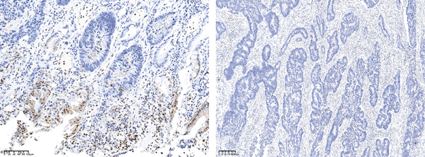
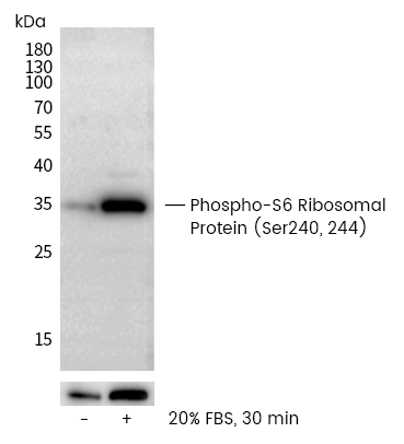
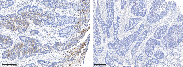
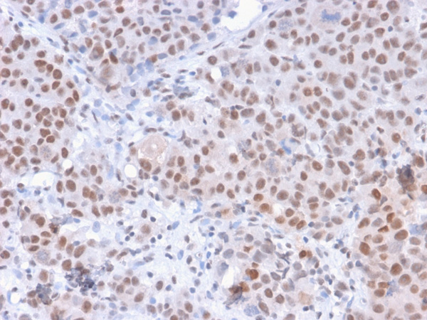
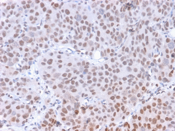
(Immunohistochemical analysis of paraffin-embedded human carcinoma of sigmoid tissue,untreated (left) or lambda phosphatase-treated (right), using Recombinant Phospho- S6 Ribosomal Protein (Ser240/244) Antibody, Rabbit Monoclonal (#AAA258880) at 1:2000 dilution.)
IHC (Immunohiostchemistry)
(Immunohistochemical analysis of paraffin-embedded human carcinoma of sigmoid tissue,untreated (left) or lambda phosphatase-treated (right), using Recombinant Phospho- S6 Ribosomal Protein (Ser240/244) Antibody, Rabbit Monoclonal (#AAA258880) at 1:2000 dilution.)
S6 Ribosomal, Monoclonal Antibody (Cat# AAA258880)
 WB (Western Blot)
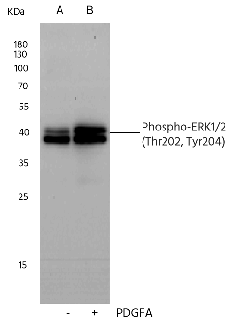
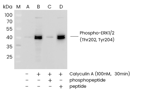
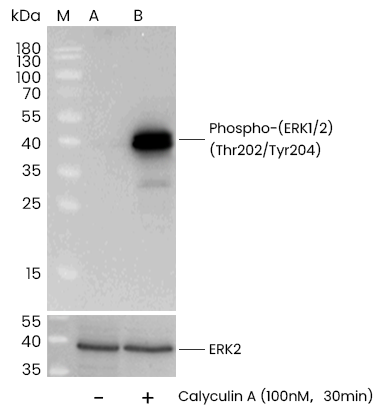
(Western blot analysis of extracts from serum-starved Hela, treated with Calyculin A (100 nM, 30 min), using Phospho-p44/42 MAPK (ERK1/2) (Thr202, Tyr204) rabbit monoclonal Antibody (#AAA258876) and other brands’ antibodies (company C) at dilution of 1:1000, 1:10000 and 1:200000.)
WB (Western Blot)
(Western blot analysis of extracts from serum-starved Hela, treated with Calyculin A (100 nM, 30 min), using Phospho-p44/42 MAPK (ERK1/2) (Thr202, Tyr204) rabbit monoclonal Antibody (#AAA258876) and other brands’ antibodies (company C) at dilution of 1:1000, 1:10000 and 1:200000.)
ERK1/2, Monoclonal Antibody (Cat# AAA258876)
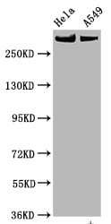
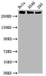
 WB (Western Blot)
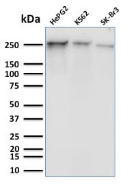
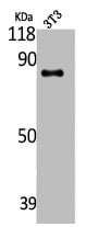
(Western Blot Analysis of Human HepG2, K562 and SK-Br3 cell lysates using RNA Poll II Mouse Monoclonal Antibody (8A7).)
WB (Western Blot)
(Western Blot Analysis of Human HepG2, K562 and SK-Br3 cell lysates using RNA Poll II Mouse Monoclonal Antibody (8A7).)
RNA Polymerase II CTD Repeat YSPTSPS, Monoclonal Antibody (Cat# AAA215151)
 WB (Western Blot)
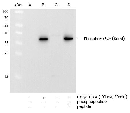
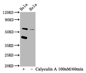
(Western blot analysis of extracts from serum-starved Hela, treated with Calyculin A (100 nM, 30 min), using Phospho-eIF2? (Ser51) Antibody (#AAA258877) and other brands’ antibodies (company C) at dilution of 1:1000, 1:10000 and 1:200000.)
WB (Western Blot)
(Western blot analysis of extracts from serum-starved Hela, treated with Calyculin A (100 nM, 30 min), using Phospho-eIF2? (Ser51) Antibody (#AAA258877) and other brands’ antibodies (company C) at dilution of 1:1000, 1:10000 and 1:200000.)
eIF2alpha, Monoclonal Recombinant Antibody (Cat# AAA258877)
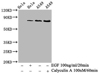
 WB (Western Blot)
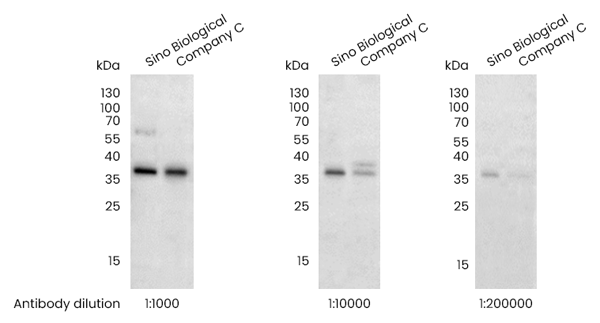
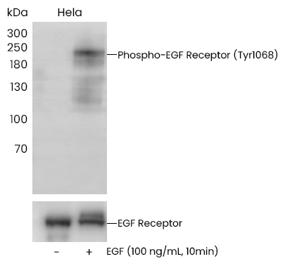
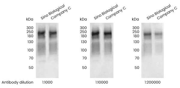
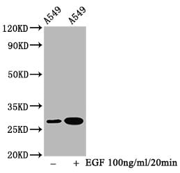
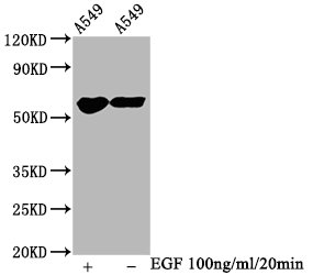
(Western blot analysis of extracts from serum-starved Hela, treated with EGF (100 ng/mL, 10 min), using Phospho-EGF Receptor (Tyr1068) Antibody (#AAA258878) and other brands’ antibodies (company C) at dilution of 1:1000, 1:10000 and 1:200000.)
WB (Western Blot)
(Western blot analysis of extracts from serum-starved Hela, treated with EGF (100 ng/mL, 10 min), using Phospho-EGF Receptor (Tyr1068) Antibody (#AAA258878) and other brands’ antibodies (company C) at dilution of 1:1000, 1:10000 and 1:200000.)
EGF Receptor, Monoclonal Antibody (Cat# AAA258878)
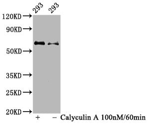
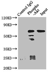 IP (Immunoprecipitation)
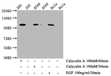
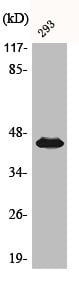
(Immunoprecipitating Phospho-AKT1 in 293 whole cell lysate treated with Calyculin ALane 1: Rabbit control IgG(1ug)instead of CSB-RA001553A473phHU in 293 whole cell lysate treated with Calyculin A.For western blotting,a HRP-conjugated Protein G antibody was used as the secondary antibody (1/2000)Lane 2: CSB-RA001553A473phHU(3ug)+ 293 whole cell lysate treated with Calyculin A(1mg)Lane 3: 293 whole cell lysate treated with Calyculin A (20ug))
IP (Immunoprecipitation)
(Immunoprecipitating Phospho-AKT1 in 293 whole cell lysate treated with Calyculin ALane 1: Rabbit control IgG(1ug)instead of CSB-RA001553A473phHU in 293 whole cell lysate treated with Calyculin A.For western blotting,a HRP-conjugated Protein G antibody was used as the secondary antibody (1/2000)Lane 2: CSB-RA001553A473phHU(3ug)+ 293 whole cell lysate treated with Calyculin A(1mg)Lane 3: 293 whole cell lysate treated with Calyculin A (20ug))
AKT1, Monoclonal Recombinant Antibody (Cat# AAA235519)

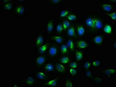
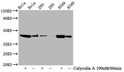
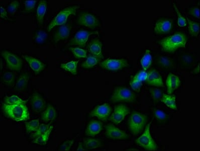
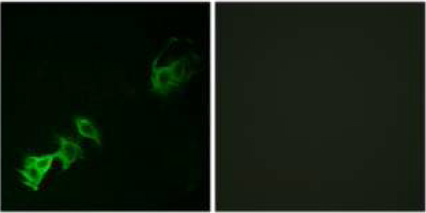
 IF (Immunofluorescence)
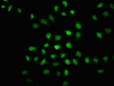
(Immunofluorescence staining of Hela cells(treated with 50mM Calyculin A for 30min) with CSB-RA007511A446phHU at 1:100,counter-stained with DAPI. The cells were fixed in 4% formaldehyde, permeabilized using 0.2% Triton X-100 and blocked in 10% normal Goat Serum. The cells were then incubated with the antibody overnight at 4 degree C. The secondary antibody was Alexa Fluor 488-congugated AffiniPure Goat Anti-Rabbit IgG (H+L).)
IF (Immunofluorescence)
(Immunofluorescence staining of Hela cells(treated with 50mM Calyculin A for 30min) with CSB-RA007511A446phHU at 1:100,counter-stained with DAPI. The cells were fixed in 4% formaldehyde, permeabilized using 0.2% Triton X-100 and blocked in 10% normal Goat Serum. The cells were then incubated with the antibody overnight at 4 degree C. The secondary antibody was Alexa Fluor 488-congugated AffiniPure Goat Anti-Rabbit IgG (H+L).)
EIF2AK2, Monoclonal Recombinant Antibody (Cat# AAA235536)

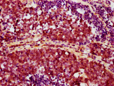
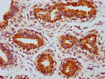
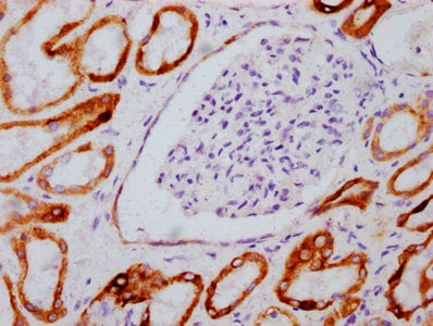
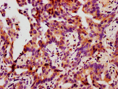
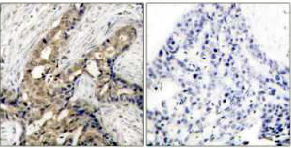
 IHC (Immunohiostchemistry)
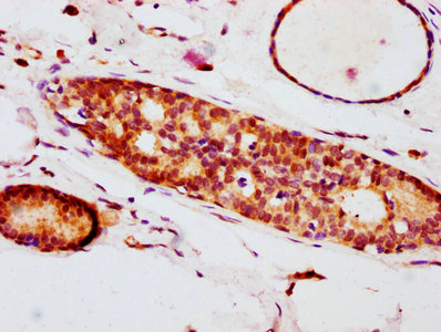
(IHC image of CSB-RA007556A209phHU diluted at 1:100 and staining in paraffin-embedded human breast cancer performed on a Leica BondTM system. After dewaxing and hydration, antigen retrieval was mediated by high pressure in a citrate buffer (pH 6.0). Section was blocked with 10% normal goat serum 30min at RT. Then primary antibody (1% BSA) was incubated at 4 degree C overnight. The primary is detected by a biotinylated secondary antibody and visualized using an HRP conjugated SP system.)
IHC (Immunohiostchemistry)
(IHC image of CSB-RA007556A209phHU diluted at 1:100 and staining in paraffin-embedded human breast cancer performed on a Leica BondTM system. After dewaxing and hydration, antigen retrieval was mediated by high pressure in a citrate buffer (pH 6.0). Section was blocked with 10% normal goat serum 30min at RT. Then primary antibody (1% BSA) was incubated at 4 degree C overnight. The primary is detected by a biotinylated secondary antibody and visualized using an HRP conjugated SP system.)
EIF4E, Monoclonal Recombinant Antibody (Cat# AAA235538)
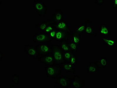
 IF (Immunofluorescence)
(Immunofluorescence staining of Hela cells with CSB-RA007795A724phHU at 1:100,counter-stained with DAPI. The cells were fixed in 4% formaldehyde, permeabilized using 0.2% Triton X-100 and blocked in 10% normal Goat Serum. The cells were then incubated with the antibody overnight at 4 degree C. The secondary antibody was Alexa Fluor 488-congugated AffiniPure Goat Anti-Rabbit IgG (H+L).)
IF (Immunofluorescence)
(Immunofluorescence staining of Hela cells with CSB-RA007795A724phHU at 1:100,counter-stained with DAPI. The cells were fixed in 4% formaldehyde, permeabilized using 0.2% Triton X-100 and blocked in 10% normal Goat Serum. The cells were then incubated with the antibody overnight at 4 degree C. The secondary antibody was Alexa Fluor 488-congugated AffiniPure Goat Anti-Rabbit IgG (H+L).)
ERN1, Monoclonal Recombinant Antibody (Cat# AAA235539)
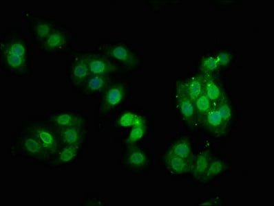
 IF (Immunofluorescence)
(Immunofluorescence staining of Hela cells with CSB-RA008968A2448phHU at 1:100,counter-stained with DAPI. The cells were fixed in 4% formaldehyde, permeabilized using 0.2% Triton X-100 and blocked in 10% normal Goat Serum. The cells were then incubated with the antibody overnight at 4 degree C. The secondary antibody was Alexa Fluor 488-congugated AffiniPure Goat Anti-Rabbit IgG (H+L).)
IF (Immunofluorescence)
(Immunofluorescence staining of Hela cells with CSB-RA008968A2448phHU at 1:100,counter-stained with DAPI. The cells were fixed in 4% formaldehyde, permeabilized using 0.2% Triton X-100 and blocked in 10% normal Goat Serum. The cells were then incubated with the antibody overnight at 4 degree C. The secondary antibody was Alexa Fluor 488-congugated AffiniPure Goat Anti-Rabbit IgG (H+L).)
MTOR, Monoclonal Recombinant Antibody (Cat# AAA235544)
 IF (Immunofluorescence)
(Immunofluorescence staining of Hela cells(treated with 50mM Calyculin A for 30min) with CSB-RA009963A09phHU at 1:100,counter-stained with DAPI. The cells were fixed in 4% formaldehyde, permeabilized using 0.2% Triton X-100 and blocked in 10% normal Goat Serum. The cells were then incubated with the antibody overnight at 4 degree C. The secondary antibody was Alexa Fluor 488-congugated AffiniPure Goat Anti-Rabbit IgG (H+L).)
IF (Immunofluorescence)
(Immunofluorescence staining of Hela cells(treated with 50mM Calyculin A for 30min) with CSB-RA009963A09phHU at 1:100,counter-stained with DAPI. The cells were fixed in 4% formaldehyde, permeabilized using 0.2% Triton X-100 and blocked in 10% normal Goat Serum. The cells were then incubated with the antibody overnight at 4 degree C. The secondary antibody was Alexa Fluor 488-congugated AffiniPure Goat Anti-Rabbit IgG (H+L).)
GSK3B, Monoclonal Recombinant Antibody (Cat# AAA235550)
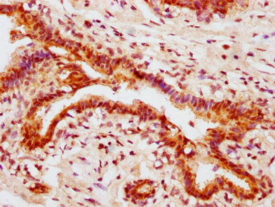
 IHC (Immunohistochemisry)
(IHC image of CSB-RA010833A78phHU diluted at 1:100 and staining in paraffin-embedded human breast cancer performed on a Leica BondTM system. After dewaxing and hydration, antigen retrieval was mediated by high pressure in a citrate buffer (pH 6.0). Section was blocked with 10% normal goat serum 30min at RT. Then primary antibody (1% BSA) was incubated at 4 degree C overnight. The primary is detected by a biotinylated secondary antibody and visualized using an HRP conjugated SP system.)
IHC (Immunohistochemisry)
(IHC image of CSB-RA010833A78phHU diluted at 1:100 and staining in paraffin-embedded human breast cancer performed on a Leica BondTM system. After dewaxing and hydration, antigen retrieval was mediated by high pressure in a citrate buffer (pH 6.0). Section was blocked with 10% normal goat serum 30min at RT. Then primary antibody (1% BSA) was incubated at 4 degree C overnight. The primary is detected by a biotinylated secondary antibody and visualized using an HRP conjugated SP system.)
HSPB1, Monoclonal Recombinant Antibody (Cat# AAA235561)

 IF (Immunofluorescence)
(Immunofluorescence staining of Hela cells with CSB-RA015270A62phHU at 1:100,counter-stained with DAPI. The cells were fixed in 4% formaldehyde, permeabilized using 0.2% Triton X-100 and blocked in 10% normal Goat Serum. The cells were then incubated with the antibody overnight at 4 degree C. The secondary antibody was Alexa Fluor 488-congugated AffiniPure Goat Anti-Rabbit IgG (H+L).)
IF (Immunofluorescence)
(Immunofluorescence staining of Hela cells with CSB-RA015270A62phHU at 1:100,counter-stained with DAPI. The cells were fixed in 4% formaldehyde, permeabilized using 0.2% Triton X-100 and blocked in 10% normal Goat Serum. The cells were then incubated with the antibody overnight at 4 degree C. The secondary antibody was Alexa Fluor 488-congugated AffiniPure Goat Anti-Rabbit IgG (H+L).)
MYC, Monoclonal Recombinant Antibody (Cat# AAA235574)
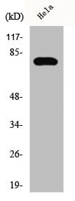
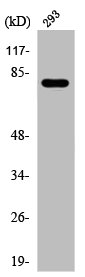
 WB (Western Blot)
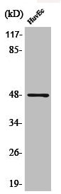
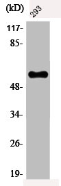
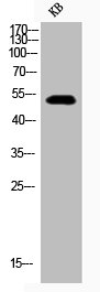
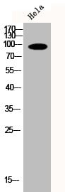
(Western Blot analysis of KB using Phospho-Akt (S473) Polyclonal Antibody)
WB (Western Blot)
(Western Blot analysis of KB using Phospho-Akt (S473) Polyclonal Antibody)
AKT1/AKT2/AKT3, Polyclonal Antibody (Cat# AAA235762)
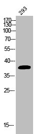
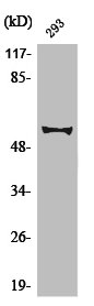
 WB (Western Blot)
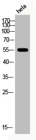
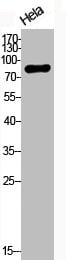
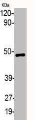
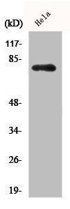
(Western Blot analysis of HELA cells using Phospho-Akt (T308) Polyclonal Antibody)
WB (Western Blot)
(Western Blot analysis of HELA cells using Phospho-Akt (T308) Polyclonal Antibody)
AKT1/AKT2/AKT3, Polyclonal Antibody (Cat# AAA235763)
 WB (Western Blot)
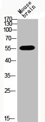
(Western Blot analysis of MOUSE-BRAIN cells using Phospho-Akt2 (S474) Polyclonal Antibody)
WB (Western Blot)
(Western Blot analysis of MOUSE-BRAIN cells using Phospho-Akt2 (S474) Polyclonal Antibody)
AKT2, Polyclonal Antibody (Cat# AAA235764)
 WB (Western Blot)
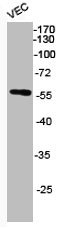
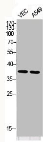
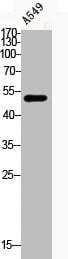
(Western Blot analysis of VEC A549 cells using Phospho-AP-1/Jun D (S73/100) Polyclonal Antibody)
WB (Western Blot)
(Western Blot analysis of VEC A549 cells using Phospho-AP-1/Jun D (S73/100) Polyclonal Antibody)
JUN/JUND, Polyclonal Antibody (Cat# AAA235765)
 WB (Western Blot)
(Western Blot analysis of HELA cells using Phospho-Catenin-beta (T41/S45) Polyclonal Antibody)
WB (Western Blot)
(Western Blot analysis of HELA cells using Phospho-Catenin-beta (T41/S45) Polyclonal Antibody)
CTNNB1, Polyclonal Antibody (Cat# AAA235773)
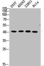
 WB (Western Blot)
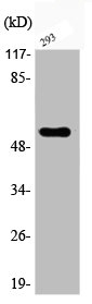
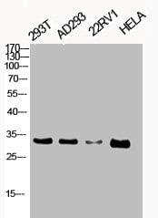
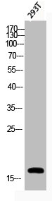
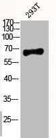
(Western Blot analysis of 293T AD293 22RV1 HELA cells using Phospho-Cdk1/2/3 (T14) Polyclonal Antibody)
WB (Western Blot)
(Western Blot analysis of 293T AD293 22RV1 HELA cells using Phospho-Cdk1/2/3 (T14) Polyclonal Antibody)
CDK1/CDK2/CDK3, Polyclonal Antibody (Cat# AAA235778)
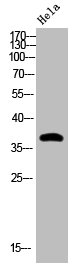
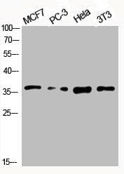
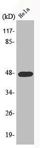
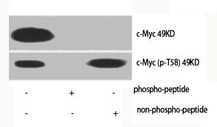 WB (Western Blot)
(Western Blot analysis of various cells using Phospho-c-Myc (T58) Polyclonal Antibody)
WB (Western Blot)
(Western Blot analysis of various cells using Phospho-c-Myc (T58) Polyclonal Antibody)
MYC, Polyclonal Antibody (Cat# AAA235780)
 WB (Western Blot)
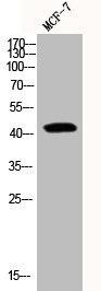
(Western Blot analysis of MCF7 PC-3 HELA NIH-3T3 cells using Phospho-CREB-1 (S133) Polyclonal Antibody)
WB (Western Blot)
(Western Blot analysis of MCF7 PC-3 HELA NIH-3T3 cells using Phospho-CREB-1 (S133) Polyclonal Antibody)
CREB1, Polyclonal Antibody (Cat# AAA235782)
 WB (Western Blot)
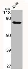
(Western Blot analysis of A549 cells using Phospho-Cyclin E1 (T395) Polyclonal Antibody)
WB (Western Blot)
(Western Blot analysis of A549 cells using Phospho-Cyclin E1 (T395) Polyclonal Antibody)
CCNE1, Polyclonal Antibody (Cat# AAA235784)
 WB (Western Blot)
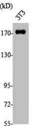
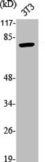
(Western Blot analysis of NIH-3T3 cells using Phospho-EGFR (Y1197) Polyclonal Antibody)
WB (Western Blot)
(Western Blot analysis of NIH-3T3 cells using Phospho-EGFR (Y1197) Polyclonal Antibody)
EGFR, Polyclonal Antibody (Cat# AAA235786)
 WB (Western Blot)
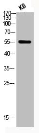
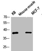
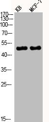
(Western Blot analysis of KB MCF7 MOUSE-MUSCLE cells using Phospho-eIF2alpha (S51) Polyclonal Antibody)
WB (Western Blot)
(Western Blot analysis of KB MCF7 MOUSE-MUSCLE cells using Phospho-eIF2alpha (S51) Polyclonal Antibody)
EIF2S1, Polyclonal Antibody (Cat# AAA235787)
 WB (Western Blot)
(Western Blot analysis of various cells using Phospho-GSK3alpha/beta (Y279/216) Polyclonal Antibody)
WB (Western Blot)
(Western Blot analysis of various cells using Phospho-GSK3alpha/beta (Y279/216) Polyclonal Antibody)
GSK3A/GSK3B, Polyclonal Antibody (Cat# AAA235793)
 WB (Western Blot)
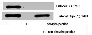
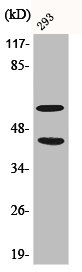
(Western Blot analysis of various cells using Phospho-Histone H3 (S28) Polyclonal Antibody)
WB (Western Blot)
(Western Blot analysis of various cells using Phospho-Histone H3 (S28) Polyclonal Antibody)
Histone H3, Polyclonal Antibody (Cat# AAA235799)
 WB (Western Blot)
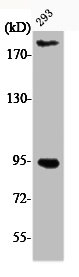
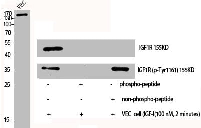
(Western Blot analysis of VEC cells using Phospho-IGF-IR (Y1161) Polyclonal Antibody)
WB (Western Blot)
(Western Blot analysis of VEC cells using Phospho-IGF-IR (Y1161) Polyclonal Antibody)
IGF1R/INSR, Polyclonal Antibody (Cat# AAA235800)
 WB (Western Blot)
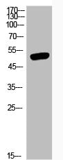
(Western Blot analysis of KB MCF7 cells using Phospho-JNK1/2/3 (T183/Y185) Polyclonal Antibody)
WB (Western Blot)
(Western Blot analysis of KB MCF7 cells using Phospho-JNK1/2/3 (T183/Y185) Polyclonal Antibody)
MAPK8/MAPK9/MAPK10, Polyclonal Antibody (Cat# AAA235808)
 WB (Western Blot)
(Western Blot analysis of various cells using Phospho-MEK-1/2 (S218/222) Polyclonal Antibody)
WB (Western Blot)
(Western Blot analysis of various cells using Phospho-MEK-1/2 (S218/222) Polyclonal Antibody)
MAP2K1/MAP2K2, Polyclonal Antibody (Cat# AAA235810)
 WB (Western Blot)
(Western Blot analysis of NIH-3T3 cells using Phospho-p73 (Y99) Polyclonal Antibody)
WB (Western Blot)
(Western Blot analysis of NIH-3T3 cells using Phospho-p73 (Y99) Polyclonal Antibody)
TP73, Polyclonal Antibody (Cat# AAA235818)
 WB (Western Blot)
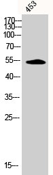
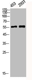
(Western Blot analysis of 453 293T cells using Phospho-CaMKIIalpha/beta/delta (T305) Polyclonal Antibody)
WB (Western Blot)
(Western Blot analysis of 453 293T cells using Phospho-CaMKIIalpha/beta/delta (T305) Polyclonal Antibody)
CAMK2A/CAMK2B/CAMK2D, Polyclonal Antibody (Cat# AAA235825)
 WB (Western Blot)
(Western Blot analysis of JK cells using Phospho-DRP1 (S637) Polyclonal Antibody)
WB (Western Blot)
(Western Blot analysis of JK cells using Phospho-DRP1 (S637) Polyclonal Antibody)
DNM1L, Polyclonal Antibody (Cat# AAA235675)
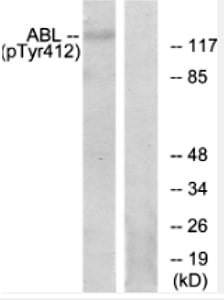 WB (Western Blot)
(Western blot analysis of lysates from RAW264.7 cells, using Abl (Phospho-Tyr393/412) Antibody. The lane on the right is blocked with the phospho peptide.)
WB (Western Blot)
(Western blot analysis of lysates from RAW264.7 cells, using Abl (Phospho-Tyr393/412) Antibody. The lane on the right is blocked with the phospho peptide.)
ABL1/ABL2, Polyclonal Antibody (Cat# AAA235691)
VAV1, Polyclonal Antibody (Cat# AAA235701)
 WB (Western Blot)
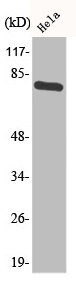
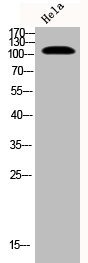
(Western Blot analysis of HELA cells using Phospho-Btk (Y551) Polyclonal Antibody)
WB (Western Blot)
(Western Blot analysis of HELA cells using Phospho-Btk (Y551) Polyclonal Antibody)
BTK, Polyclonal Antibody (Cat# AAA235828)
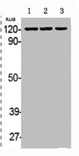
 WB (Western Blot)
(Western Blot analysis of HELA cells using Phospho-PKD2 (S876) Polyclonal Antibody)
WB (Western Blot)
(Western Blot analysis of HELA cells using Phospho-PKD2 (S876) Polyclonal Antibody)
PRKD2, Polyclonal Antibody (Cat# AAA235833)
 WB (Western Blot)
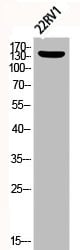
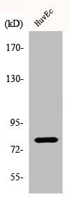
(Western Blot analysis of 22RV1 cells using Phospho-NOS3 (S1177) Polyclonal Antibody)
WB (Western Blot)
(Western Blot analysis of 22RV1 cells using Phospho-NOS3 (S1177) Polyclonal Antibody)
NOS3, Polyclonal Antibody (Cat# AAA235839)
 WB (Western Blot)
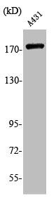
(Western Blot analysis of HELA A431 cells using Phospho-EphB1/2 (Y594/604) Polyclonal Antibody)
WB (Western Blot)
(Western Blot analysis of HELA A431 cells using Phospho-EphB1/2 (Y594/604) Polyclonal Antibody)
EPHB1/EPHB2, Polyclonal Antibody (Cat# AAA235840)
 WB (Western Blot)
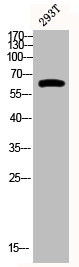
(Western Blot analysis of 293T cells using Phospho-p63 (S455) Polyclonal Antibody)
WB (Western Blot)
(Western Blot analysis of 293T cells using Phospho-p63 (S455) Polyclonal Antibody)
TP63, Polyclonal Antibody (Cat# AAA235849)
 WB (Western Blot)
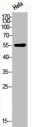
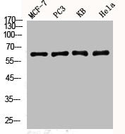
(Western Blot analysis of MCF7 PC-3 HELA KB cells using Phospho-YAP (S127) Polyclonal Antibody)
WB (Western Blot)
(Western Blot analysis of MCF7 PC-3 HELA KB cells using Phospho-YAP (S127) Polyclonal Antibody)
YAP1, Polyclonal Antibody (Cat# AAA235851)
 WB (Western Blot)
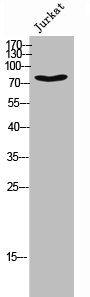
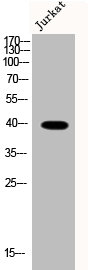
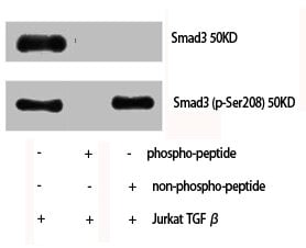
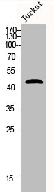
(Western Blot analysis of Jurkat cells using Phospho-Smad3 (S208) Polyclonal Antibody)
WB (Western Blot)
(Western Blot analysis of Jurkat cells using Phospho-Smad3 (S208) Polyclonal Antibody)
SMAD3, Polyclonal Antibody (Cat# AAA235854)
 WB (Western Blot)
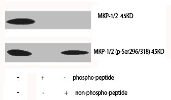
(Western Blot analysis of various cells using Phospho-MKP-1/2 (S296/318) Polyclonal Antibody)
WB (Western Blot)
(Western Blot analysis of various cells using Phospho-MKP-1/2 (S296/318) Polyclonal Antibody)
DUSP1/DUSP4, Polyclonal Antibody (Cat# AAA235858)
 WB (Western Blot)
(Western Blot analysis of A549 cells using Phospho-PKC delta (S645) Polyclonal Antibody)
WB (Western Blot)
(Western Blot analysis of A549 cells using Phospho-PKC delta (S645) Polyclonal Antibody)
PRKCD, Polyclonal Antibody (Cat# AAA235861)
 WB (Western Blot)
(Western Blot analysis of 293T AD293 22RV1 HELA cells using Phospho-c-Myc (S62) Polyclonal Antibody)
WB (Western Blot)
(Western Blot analysis of 293T AD293 22RV1 HELA cells using Phospho-c-Myc (S62) Polyclonal Antibody)
MYC, Polyclonal Antibody (Cat# AAA235865)
 WB (Western Blot)
(Western Blot analysis of NIH-3T3 cells using Phospho-Caldesmon (S789) Polyclonal Antibody)
WB (Western Blot)
(Western Blot analysis of NIH-3T3 cells using Phospho-Caldesmon (S789) Polyclonal Antibody)
CALD1, Polyclonal Antibody (Cat# AAA235868)
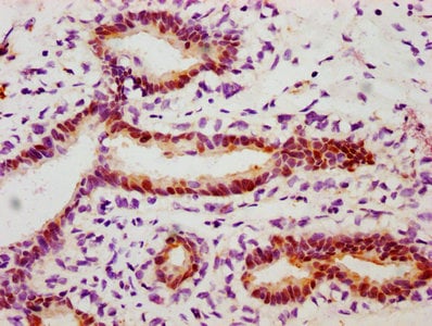
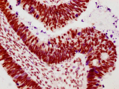
 IHC (Immunohiostchemistry)
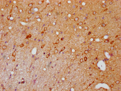
(IHC image of CSB-RA017407A144phHU diluted at 1:100 and staining in paraffin-embedded rat brain tissue performed on a Leica BondTM system. After dewaxing and hydration, antigen retrieval was mediated by high pressure in a citrate buffer (pH 6.0). Section was blocked with 10% normal goat serum 30min at RT. Then primary antibody (1% BSA) was incubated at 4 degree C overnight. The primary is detected by a biotinylated secondary antibody and visualized using an HRP conjugated SP system.)
IHC (Immunohiostchemistry)
(IHC image of CSB-RA017407A144phHU diluted at 1:100 and staining in paraffin-embedded rat brain tissue performed on a Leica BondTM system. After dewaxing and hydration, antigen retrieval was mediated by high pressure in a citrate buffer (pH 6.0). Section was blocked with 10% normal goat serum 30min at RT. Then primary antibody (1% BSA) was incubated at 4 degree C overnight. The primary is detected by a biotinylated secondary antibody and visualized using an HRP conjugated SP system.)
PAK1/PAK2/PAK3, Monoclonal Recombinant Antibody (Cat# AAA235577)
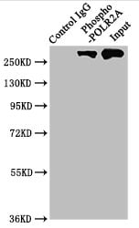 IP (Immunoprecipitation)
(Immunoprecipitating Phospho-POLR2A in Hela whole cell lysateLane 1: Rabbit control IgG(1ug)instead of CSB-RA018327A02phHU in Hela whole cell lysate.For western blotting,a HRP-conjugated Protein G antibody was used as the secondary antibody (1/2000)Lane 2: CSB-RA018327A02phHU(3ug)+ Hela whole cell lysate(1mg)Lane 3: Hela whole cell lysate (20ug))
IP (Immunoprecipitation)
(Immunoprecipitating Phospho-POLR2A in Hela whole cell lysateLane 1: Rabbit control IgG(1ug)instead of CSB-RA018327A02phHU in Hela whole cell lysate.For western blotting,a HRP-conjugated Protein G antibody was used as the secondary antibody (1/2000)Lane 2: CSB-RA018327A02phHU(3ug)+ Hela whole cell lysate(1mg)Lane 3: Hela whole cell lysate (20ug))
POLR2A, Monoclonal Recombinant Antibody (Cat# AAA235579)
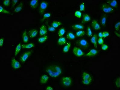
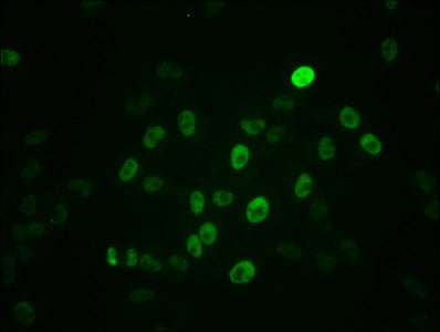
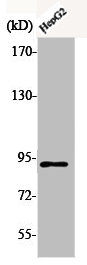
 IF (Immunofluorescence)
(Immunofluorescence staining of HepG2 cells with CSB-RA018699A638phHU at 1:100,counter-stained with DAPI. The cells were fixed in 4% formaldehyde, permeabilized using 0.2% Triton X-100 and blocked in 10% normal Goat Serum. The cells were then incubated with the antibody overnight at 4 degree C. The secondary antibody was Alexa Fluor 488-congugated AffiniPure Goat Anti-Rabbit IgG (H+L).)
IF (Immunofluorescence)
(Immunofluorescence staining of HepG2 cells with CSB-RA018699A638phHU at 1:100,counter-stained with DAPI. The cells were fixed in 4% formaldehyde, permeabilized using 0.2% Triton X-100 and blocked in 10% normal Goat Serum. The cells were then incubated with the antibody overnight at 4 degree C. The secondary antibody was Alexa Fluor 488-congugated AffiniPure Goat Anti-Rabbit IgG (H+L).)
PRKCA, Monoclonal Recombinant Antibody (Cat# AAA235582)
What Are Phospho Antibodies?
Protein phosphorylation is a process where a phosphate group is added to certain amino acid residues of a protein – usually serine (S), threonine (T), or tyrosine (Y) - by enzymes called kinases. This process is integral in controlling cellular signaling, cellular growth, and other biological functions.
Our catalog includes a wide range of phospho-specific antibodies that can accurately detect this important marker. They perform strongly in widely-used laboratory applications such as Western blot, flow cytometry, immunohistochemistry, and immunofluorescence microscopy. We value your trust in us and are committed to providing top-quality products and services. All of our antibodies are guaranteed to work for the applications and species indicated on our website & associated product pages.
What Are The Key Applications of Phospho Antibodies?
1. Western Blotting
One of the first steps a researcher can take in utilizing these phospho-specific antibodies, is to check if the antibody works using a technique referred to as “Western blot”. For those unfamiliar, Western Blot aids in showing whether the protein that the antibody recognizes is appearing at the correct/expected size. These phospho-specific antibodies should also be able to detect changes in the target protein’s phosphorylation (on/off state) when cells are stimulated in certain ways.
2. Staining of Fixed Cells (Immunocytochemistry)
Another routine use of these phospho-specific antibodies, is to test if the antibody is able to demonstrate similar performance when used on fixed cells (intact cells that have been preserved) as it did in the Western blot tests. It is an important aspect in many cases to confirm that the antibody works in actual intact cell samples. Ideally, the method used for cellular fixation should be the same as what is used in pathology labs (like using 10% formalin). To check if the antibody works well in tissue sections (FFPE), researchers will often test it on fixed cells that are processed similar to tissue samples.
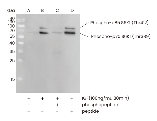
3. Specificity Tests Using Peptides
In order to make sure that the antibody is only binding to the right target:
- Laboratory technicians will mix the antibody with phospho-peptides (short segments of the protein containing the phosphate group modification).
- If the antibody signal disappears, it is confirmation that it is binding to the correct phosphorylated location.
- A more robust test is to use both the phosphorylated and non-phosphorylated (dephosphorylated) versions of the protein. The antibody should react only with the phosphorylated one.
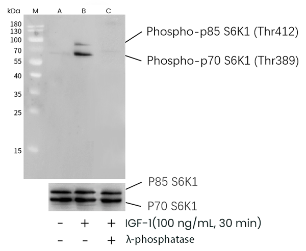
- Another method sometimes utilized is to treat the sample with an enzyme, such as alkaline phosphatase, that specifically removes phosphate groups. If the antibody signal disappears after this, it also confirms specificity.
4. Genetic Confirmation
As a final step, scientists can genetically manipulate the nucleotide sequence and alter the target protein by removing the exact site where phosphorylation happens. If the antibody no longer appears to detect the modified protein, it is strong evidence supporting the antibody being specific for that phosphorylated site.
Why Buy Phospho Antibodies Through Us?
- The production laboratory adheres to strict and consistent protocols prior to releasing any of these phospho-specific antibodies:
- Standard methods and proper controls in all tests to ensure high quality.
- These antibodies are tested and validated in different cell types and species.
- High quality control criterion to ensure each batch is consistent, so you will obtain reliable results every time.
FAQ
1. What Are Phospho-Specific Antibodies?
Phospho-specific antibodies are made to detect proteins only when they have a phosphate group linked to a specific amino acid residue. This empowers scientists understand if a protein is "turned on" or active, based on its phosphorylation state.
2. How to Detect Phosphorylated Proteins in a Western Blot?
To find out if a protein is phosphorylated using Western blot:
- Use a phospho-specific antibody that binds only to the phosphorylated form of the protein.
- You can also use a “regular” antibody for the same amino acid sequence of the protein that the phospho-specific antibody is binding to (but in this case, this antibody will not bind if there is a phosphate group present) in order to compare how much of it is phosphorylated versus how much is non-phosphorylated (or “total” protein, if the “normal” antibody’s epitopes are non-phospho-site-specific).
3. How to Choose the Best Antibody?
Here are some simple tips to help you pick the right antibody:
- Know your target
- Match your sample characteristics
- Confirm the intended use is appropriate
- Check “host” and “type”
- Check the “quality” of the presented data/images
- Appraise whether the available validation meets your needs